MiR-210 disturbs mitotic progression through regulating a group of mitosis-related genes Jie He 1,2 , Jiangbin Wu 1,2 , Naihan Xu 2 , Weidong Xie 2 , Mengnan Li 1,2 , Jianna Li 2 , Yuyang Jiang 3 , Burton B. Yang 4, * and Yaou Zhang 2, * 5 1 School of Life Sciences, Tsinghua University, Beijing 100084, 2 Key Laboratory in Healthy Science and Technology, Division of Life Science, Graduate School at Shenzhen, Tsinghua University, Shenzhen 518055, PR China, 3 The State Key Laboratory Breeding Base-Shenzhen Key Laboratory of Chemical Biology, the Graduate School at Shenzhen, Tsinghua University, Shenzhen 518055, P. R. China and 4 Sunnybrook Research Institute, and Department of Laboratory Medicine and Pathobiology, University of Toronto, Toronto, Canada 10 Received March 28, 2012; Revised September 26, 2012; Accepted September 30, 2012 ABSTRACT MiR-210 is up-regulated in multiple cancer types but its function is disputable and further investigation is necessary. Using a bioinformatics approach, we 15 identified the putative target genes of miR-210 in hypoxia-induced CNE cells from genome-wide scale. Two functional gene groups related to cell cycle and RNA processing were recognized as the major targets of miR-210. Here, we investigated the 20 molecular mechanism and biological consequence of miR-210 in cell cycle regulation, particularly mitosis. Hypoxia-induced up-regulation of miR-210 was highly correlated with the down-regulation of a group of mitosis-related genes, including Plk1, 25 Cdc25B, Cyclin F, Bub1B and Fam83D. MiR-210 sup- pressed the expression of these genes by directly targeting their 3 0 -UTRs. Over-expression of exogen- ous miR-210 disturbed mitotic progression and caused aberrant mitosis. Furthermore, miR-210 30 mimic with pharmacological doses reduced tumor formation in a mouse metastatic tumor model. Taken together, these results implicate that miR-210 disturbs mitosis through targeting multi- genes involved in mitotic progression, which may 35 contribute to its inhibitory role on tumor formation. INTRODUCTION MicroRNAs (miRNAs) are a class of small non-coding RNA of 19–24 nucleotides in length, which play pivotal roles in various biological and pathologic processes, 40 including cell proliferation, differentiation, apoptosis, me- tabolism, organ morphology, angiogenesis and cancer (1–8). There is mounting evidence that miRNA expression is distinct between normal and tumor tissues. In some in- stances, these differences are associated with the initiation 45 and progression of cancer (9–13). By modulating onco- genic and tumor suppressor pathways, miRNAs can act as oncogenes or tumor suppressors, or oncomiRs or tumor-suppressors miRs, respectively (6,13–15). Some miRNAs, such as miR-210, are up-regulated in 50 multiple cancer types (16–19). However, they cannot simply be described as oncomiRs or tumor-suppressor miRs because of the wide array of targets involved in regulating different cellular functions. MiR-210 is a hypoxia-induced gene regulated by (hypoxia-inducible 55 factor 1) HIF-1a and plays various roles in the cells (7,17,19–21). In a murine model of myocardial infarction, miR-210 inhibited apoptosis and improved angiogenesis and cardiac function through potential targets Efna3 and Ptp1b (22). MiR-210 also down-regulates mitochon- 60 drial function by repressing the expression of iron–sulfur cluster scaffold homolog and COX10 (cytochrome c oxidase assembly protein) (23). It has recently been reported that the expression of miR-210 is induced during erythroid differentiation in a time- and 65 dosage-dependent manner. Consistent with increased ex- pression of the fetal gamma-globin genes, miR-210 might be involved in erythropoiesis (24). The role of miR-210 in cancer is complex and disputable. Data from clinical cancer samples show that miR-210 expression is inversely 70 correlated with disease-free states and overall survival rates (16,18–19). However, other studies have reported that a remarkably high frequency of miR-210 gene dele- tions was found in ovarian cancer patients (25). *To whom correspondence should be addressed. Tel/Fax: +86 0755 26036884; Email: [email protected] Correspondence may also be addressed to Burton B. Yang. Tel: +1 416 480 5874; Fax: +1 416 480 5737; Email: [email protected] The authors wish it to be known that, in their opinion, the first three authors should be regarded as joint First Authors. Nucleic Acids Research, 2012, 1–11 doi:10.1093/nar/gks995 ß The Author(s) 2012. Published by Oxford University Press. This is an Open Access article distributed under the terms of the Creative Commons Attribution License (http://creativecommons.org/licenses/by-nc/3.0/), which permits non-commercial reuse, distribution, and reproduction in any medium, provided the original work is properly cited. For commercial re-use, please contact [email protected].

Welcome message from author
This document is posted to help you gain knowledge. Please leave a comment to let me know what you think about it! Share it to your friends and learn new things together.
Transcript

MiR-210 disturbs mitotic progression throughregulating a group of mitosis-related genesJie He1,2, Jiangbin Wu1,2, Naihan Xu2, Weidong Xie2, Mengnan Li1,2, Jianna Li2,
Yuyang Jiang3, Burton B. Yang4,* and Yaou Zhang2,*
51School of Life Sciences, Tsinghua University, Beijing 100084, 2Key Laboratory in Healthy Science andTechnology, Division of Life Science, Graduate School at Shenzhen, Tsinghua University, Shenzhen 518055, PRChina, 3The State Key Laboratory Breeding Base-Shenzhen Key Laboratory of Chemical Biology, the GraduateSchool at Shenzhen, Tsinghua University, Shenzhen 518055, P. R. China and 4Sunnybrook Research Institute,and Department of Laboratory Medicine and Pathobiology, University of Toronto, Toronto, Canada
10 Received March 28, 2012; Revised September 26, 2012; Accepted September 30, 2012
ABSTRACT
MiR-210 is up-regulated in multiple cancer types butits function is disputable and further investigation isnecessary. Using a bioinformatics approach, we
15 identified the putative target genes of miR-210 inhypoxia-induced CNE cells from genome-widescale. Two functional gene groups related to cellcycle and RNA processing were recognized as themajor targets of miR-210. Here, we investigated the
20 molecular mechanism and biological consequenceof miR-210 in cell cycle regulation, particularlymitosis. Hypoxia-induced up-regulation of miR-210was highly correlated with the down-regulation of agroup of mitosis-related genes, including Plk1,
25 Cdc25B, Cyclin F, Bub1B and Fam83D. MiR-210 sup-pressed the expression of these genes by directlytargeting their 30-UTRs. Over-expression of exogen-ous miR-210 disturbed mitotic progression andcaused aberrant mitosis. Furthermore, miR-210
30 mimic with pharmacological doses reduced tumorformation in a mouse metastatic tumor model.Taken together, these results implicate thatmiR-210 disturbs mitosis through targeting multi-genes involved in mitotic progression, which may
35 contribute to its inhibitory role on tumor formation.
INTRODUCTION
MicroRNAs (miRNAs) are a class of small non-codingRNA of 19–24 nucleotides in length, which play pivotalroles in various biological and pathologic processes,
40including cell proliferation, differentiation, apoptosis, me-tabolism, organ morphology, angiogenesis and cancer(1–8). There is mounting evidence that miRNA expressionis distinct between normal and tumor tissues. In some in-stances, these differences are associated with the initiation
45and progression of cancer (9–13). By modulating onco-genic and tumor suppressor pathways, miRNAs can actas oncogenes or tumor suppressors, or oncomiRs ortumor-suppressors miRs, respectively (6,13–15).Some miRNAs, such as miR-210, are up-regulated in
50multiple cancer types (16–19). However, they cannotsimply be described as oncomiRs or tumor-suppressormiRs because of the wide array of targets involvedin regulating different cellular functions. MiR-210is a hypoxia-induced gene regulated by (hypoxia-inducible
55factor 1) HIF-1a and plays various roles in the cells(7,17,19–21). In a murine model of myocardial infarction,miR-210 inhibited apoptosis and improved angiogenesisand cardiac function through potential targets Efna3and Ptp1b (22). MiR-210 also down-regulates mitochon-
60drial function by repressing the expression of iron–sulfurcluster scaffold homolog and COX10 (cytochrome coxidase assembly protein) (23). It has recently beenreported that the expression of miR-210 is inducedduring erythroid differentiation in a time- and
65dosage-dependent manner. Consistent with increased ex-pression of the fetal gamma-globin genes, miR-210 mightbe involved in erythropoiesis (24). The role of miR-210 incancer is complex and disputable. Data from clinicalcancer samples show that miR-210 expression is inversely
70correlated with disease-free states and overall survivalrates (16,18–19). However, other studies have reportedthat a remarkably high frequency of miR-210 gene dele-tions was found in ovarian cancer patients (25).
*To whom correspondence should be addressed. Tel/Fax: +86 0755 26036884; Email: [email protected] may also be addressed to Burton B. Yang. Tel: +1 416 480 5874; Fax: +1 416 480 5737; Email: [email protected]
The authors wish it to be known that, in their opinion, the first three authors should be regarded as joint First Authors.
Nucleic Acids Research, 2012, 1–11doi:10.1093/nar/gks995
� The Author(s) 2012. Published by Oxford University Press.This is an Open Access article distributed under the terms of the Creative Commons Attribution License (http://creativecommons.org/licenses/by-nc/3.0/), whichpermits non-commercial reuse, distribution, and reproduction in any medium, provided the original work is properly cited. For commercial re-use, please [email protected].

Over-expression of ectopic miR-210 could function asa tumor suppressor because it attenuates cancer cell pro-liferation through down-regulating E2F3, fibroblastgrowth factor receptor-like 1, Homeobox protein Hox-
5 A1 (HOXA1), Homeobox protein Hox-A9 (HOXA9)and Max-binding protein (MNT) (17,26,27).Through a combination of genome-wide scale analysis
and bioinformatics approach, we identified two functionalgene groups as the potential targets of miR-210. One is
10 involved in cell cycle regulation and the other is related toRNA processing. Here, we investigated the molecularmechanism and biological consequences of miR-210 incell cycle regulation. We found that hypoxia-inducedup-regulation of miR-210 was highly correlated with the
15 down-regulation of Plk1, Cdc25B, Cyclin F, Bub1B andFam83D genes involved in mitotic regulation. MiR-210suppressed the expression of these genes by directly tar-geting their 30-untranslated regions (30-UTRs). Over-expression of exogenous miR-210 also disturbed mitotic
20 progression and caused aberrant mitosis. Moreover,miR-210 mimic with pharmacological doses reducedtumor formation in a mouse metastatic tumor modelduring the initial stage. These results indicate that the in-hibitory role of miR-210 on tumor formation may be par-
25 tially due to mitotic disturbances in cancer cells.
MATERIALS AND METHODS
Cell culture and hypoxia induction
HeLa or CNE cells (Kunming Cell Bank, China) werecultured in Dulbecco’s modified Eagle’s medium contain-
30 ing 10% fetal bovine serum at 37�C with 5% carbondioxide (CO2). Hypoxia was induced by treating cellswith deferoxamine mesylate (DFOM, Sigma-AldrichCo., MO, USA) at a final concentration of 130 mM.
mRNA expression profile
35 Total RNA was isolated using Trizol reagent (Invitrogen)and the samples were analysed by mRNA microarray(Capital Bio Corp., Beijing, China). Procedures were per-formed as described in detail on the website of CapitalBio(http://www.capitalbio.com) (28). For each test and
40 control samples, two hybridizations were performed byusing a reversal Fuorescent strategy. Genes showing agreater than 2-fold induction or repression in both micro-array were selected as differentially expressed genes.
MiRNAs, siRNAs and transfection
45 MiRNA mimics were designed according to the miRBasesequence database (http://microrna.sanger.ac.uk).miRNA mimics, miRNA inhibitors and small interferingRNA (siRNA) duplexes were synthesized and purified byShanghai GenePharma Co. (Shanghai, China). The se-
50 quences of these inhibitors are the exact antisense copyof the mature miRNAs, and all the nucleotides in the in-hibitors contain 20-O-Methyl modification (20-OMe) modi-fications at each base. SiRNA duplexes with randomsequences were used as a negative control (NC). The
55 sequence of these miRNA and siRNAs were included in
the Supplementary Table S1. Cells were transfected withsiRNA or miRNAs duplexes using Lipofectaime 2000(Invitrogen Corp., Carlsbad, CA, USA) according tomanufacturer’s instructions.
60Lentiviral preparation and transfection
The recombinant lentivirus for miR-210 (Lv-miR210),which expresses mature human miR-210, and negativecontrol (Lv-NC), which expresses a scrambled RNA,were purchased from GenePharma (Shanghai, China).
65Protocol of lentivirus infection is according to theGenePharma Recombinant Lentivirus OperationManual (http://www.genepharma.com).
Immunocytochemistry
CNEcells transfectedwithmiR-210 orNCwereExed in 4%70paraformaldehyde in phosphate buffered saline (PBS) for
15 min, followed by permeabilization with 0.25% TritonX-100 in PBS for 10 min at room temperature. Fixedsamples were blocked with 3% bovine serum albumin inPBS for 1 h at room temperature before incubation with
75primary and secondary antibodies. Primary antibodiesused were a-tubulin and g-tubulin (Sigma-Aldrich Co.,MO, USA). Secondary antibodies used were Alexa Fluor488 or 555 conjugates (Invitrogen, USA). Images werecaptured using an Olympus confocal microscope.
80Cell synchronization and flow cytometry analysis
CNE cells transfected with miR-210, miR-9, Plk1-siRNAor NC were synchronized at mitosis by nocodazole(100 ng/ml) treatment for 12 h. The mitotic cells werereleased from nocodazole block by washing the cells
85three times with PBS. At 1 h after nocodazole release,the cells were fixed in 70% ethanol in PBS overnight.Cells were then counterstained with propidium iodide(PI) and analysed for DNA content by use of a BDInfluxTM flow cytometer.
90Luciferase activity assay
Reporter vectors were generated by inserting the 30-UTRsof Plk1 (nt 267–285), Bub1B (nt 87–108), Cyclin F (nt537–557), Cdc5B (nt 870–887), Fam83D (nt 216–236)and their corresponding mutated 30-UTR fragments into
95[pRL-TK Vector(Promega Corporation) (pRL-TK)]plasmid (Promega, USA). Then, 30-UTRs were amplifiedby PCR using the primers shown in Supplementary TableS1. Luciferase activities were assayed according to a pre-viously published protocol (29). Briefly, CNE cells
100cultured in 24-well plate were transfected with 100 ng ofplasmid and 20 nM of each miRNA using Lipofectamine2000. Cells were collected at 30 h after transfection toperform Renilla luciferase activity assay.
Mice and tumor formation assays
105Athymic nude mice aged 4 weeks were purchased from theExperimental Animal Center of Guang Zhou Universityof Chinese Medicine (Guang Zhou, China) and kept inpathogen-free animal facilities for 2 weeks at Tsinghua
2 Nucleic Acids Research, 2012

University Shenzhen Graduate School. The mice weredivided into three groups for the treatment of miR-210,Plk1-siRNA or NC respectively, and each group had 8mice. CNE cells were prepared for mouse tail vein injec-
5 tions. Briefly, the cells were suspended in PBS, and theirviability assessed by trypan blue staining was typically>95%. Six-week-old mice were injected with approxi-mately 5� 105 CNE cells in the tail vein. Transferrin-polyethylenimine (TF-PEI)/modified-miRNA-mimics
10 complex were delivered through tail vein injection every3 days. After 6 injections, mice were euthanized and theirlungs were removed and rinsed. The left lung lobes werecollected to generate histological tablets to calculate thepulmonary metastases. The right lung lobes were fixed
15 with formalin, embedded with paraffin for lung tissue sec-tions, which were prepared at 5mm on a transverse plain.The sections were examined using H&E staining. The pul-monary metastases were counted and photographed. Allexperiments on animals were conducted according to the
20 US Public Health Service’s Policy on Human Care andUse of Laboratory Animals.
RESULTS
Identification of miR-210 target genes inhypoxia-induced CNE cells
25 Results from mRNA microarray analysis revealed that351 genes were down-regulated in hypoxia-induced CNEnasopharyngeal carcinoma cells. To clarify the potentialtargets of miR-210, we used FindTar, a prediction algo-rithm designed by our laboratory, to screen miR-210
30 targets from those down-regulated genes under hypoxiacondition (30). Among them, 107 genes were predicted asthe putative targets of miR-210 (Supplementary Table S2).
The putative targets of miR-210 were then uploadedinto DAVID bioinformatics resources (http://david.abcc.
35 ncifcrf.gov/) for functional annotation clustering and en-richment scoring. A higher enrichment score for a clusterindicated that the gene members within the cluster wereinvolved in more important (enriched) terms in a givenstudy (31). The results showed seven clusters with enrich-
40 ment scores higher than 1.3, representing four biologicalfunctions, including structure molecules of organelle,RNA processing, cell cycle and a few methyltransferases(Supplementary Table S3). Search tool for the retrievalof interacting genes/proteins (http://string-db.org/)
45 (STRING), a database of protein interactions, was thenused to investigate functional associations among the can-didate target genes of miR-210 and networks involved inthe biological function of miR-210 (32). The 107 putativemiR-210 genes were uploaded into STRING 9 (http://
50 string-db.org/) and two areas where the genes cohered toform networks were identified. As shown in Figure 1A,one network consisted of cell cycle–related genes, andthe other one related to RNA process. This resultindicated that the role of miR-210 might be related to
55 cell cycle regulation and RNA process.In this investigation, we studied the function of
miR-210 on cell cycle regulation. We analysed the func-tional cluster 4 from DAVID bioinformatics resources: 27
genes associated with the cell cycle were divided into 2860enriched functional annotations (Supplementary Table
S3). After removing the overlapping genes, eight geneswere identified from over 10 enriched functional annota-tions, including Plk1 [polo-like kinase 1 (PLK1)], Bub1B[budding uninhibited by benzimidazoles 1 homolog
65beta(yeast) or Mitotic checkpoint serine/threonine kinaseBUB1 beta (BUB1B)], Cyclin F (CCNF), Pds5B [sisterchromatid cohesion protein PDS5 homolog B orandrogen induced inhibitor of proliferation (PDS5B)],Fam83D [family with sequence similarity 83, member D
70(FAM83D)], Cdc25B [cell division cycle 25 homolog B(S.pombe) (CDC25B)], g-tubulin [tubulin, gamma 1(TUBG1)] and Cyclin D1 (CCND1) (Figure 1B). All ofthe genes, except Cyclin D1, were related to mitosis. Weexamined their mRNA expression under normoxia or
75hypoxia condition. Results from quantitative reverse tran-scriptase polymerase chain reaction (qRT-PCR) con-firmed that hypoxic stress significantly suppressed theexpression of these genes (Figure 1C). In contrast, inhib-ition of endogenous miR-210 with specific miRNA inhibi-
80tor could reverse the down-regulation of Plk1, Bub1B,Cyclin F, Pds5B and Cdc25B by hypoxic stress, suggestingthat these genes might be miR-210 targets (Figure 1D).
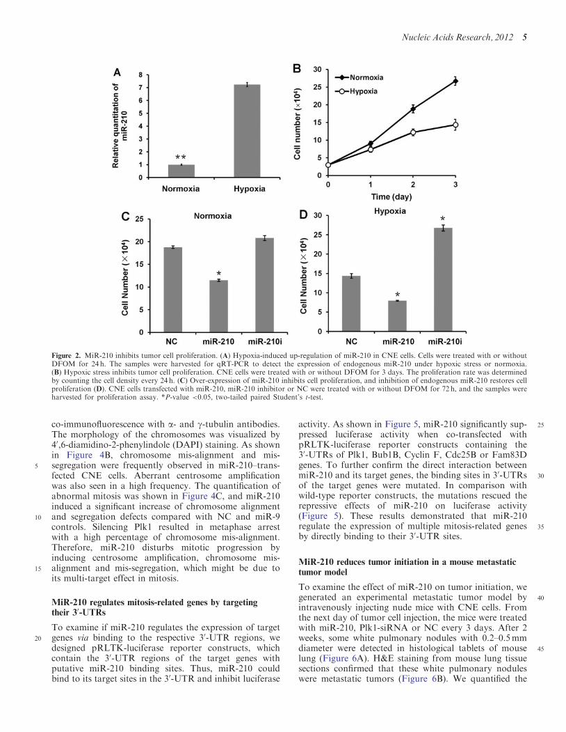
MiR-210 inhibits tumor cell proliferation
To investigate the correlation between miR-210 expression85and cell proliferation, we examined the expression level of
miR-210 under hypoxic stress. CNE cells were treatedwith the hypoxia mimetic agent DFOM for 24 h. Thelevel of endogenous miR-210 was determined by qRT-PCR. As shown in Figure 2A, hypoxic stress induced a
907-fold increase of endogenous miR-210 expression. Thecell proliferation assay also showed a retardation of cellgrowth in hypoxia-treated cells (Figure 2B). Then, weexamined the effect of miR-210 over-expression on cellproliferation under normoxia or hypoxia condition. As
95shown in Figure 2C and D, over-expression of exogenousmiR-210 significantly reduced the rate of cell proliferation.Conversely, inhibition of endogenous miR-210 restoredcell proliferation rate, particularly in hypoxia-treatedcells, when miR-210 is highly up-regulated (Figure 2D).
100MiR-210 down-regulates the expression ofmitosis-related genes
Bioinformatics analysis indicates that eight mitosis regu-latory genes are putative targets of miR-210 in hypoxia-induced CNE cells. To validate, we over-expressed
105miR-210 in hypoxia-induced CNE cells and harvestedcells for qRT-PCR and Western blotting. As shown inFigure 3A, miR-210 over-expression resulted in 40–80%reduction in transcript abundance for Plk1, Cdc25B,Bub1B, Cyclin F and Fam83D. Similarly, the protein ex-
110pression of these genes was also down-regulated to variousextents by exogenous miR-210. Among them, the proteinlevel of Plk1, a key protein involved in both mitotic entryand spindle checkpoint, decreased almost 80% comparedwith control (Figure 3B). There was little, if any, reduction
115of Cyclin D1, Pds5B and g-tubulin from both qRT-PCRand Western blotting (Data not shown).
Nucleic Acids Research, 2012 3

We also generated CNE and HeLa cells that stablyexpressed miR-210 and NC by lentiviral infection. Forvisualization, the GenePharma Supersilencing vector wasdesigned to carry a green fluorescent protein (GFP) gene
5 in its backbone (Figure 3C). We harvested the cellsinfected with either miR-210 or NC for Westernblotting. It was shown that over-expression of miR-210also down-regulated the protein expression of Plk1,Cyclin F, Bub1B, Cdc25B and Fam83D in both HeLa
10 and CNE cells in normal culture condition (Figure 3D).
MiR-210 disturbs mitotic progression and causes defectsof chromosome alignment and segregation
Having shown that miR-210 down-regulated the expres-sion of some genes related to mitosis, we next investigated
15 the function of miR-210 on mitotic progression. CNE cellstransfected with NC, miR-210, Plk1-siRNA and miR-9were synchronized at mitosis by nocodazole treatment,and the mitotic cells were released from nocodazoleblock for 1 h. The cells were fixed and stained with PI
20for flow cytometry analysis. As shown in Figure 4A,>25% of the NC-transfected cells exited mitosis andentered G1 phase at 1 h after nocodazole release. Bycontrast, there was a significant delay of mitotic exit inmiR-210–transfected cells, as <10% of the cells entered
25G1. For comparison, we used miR-9 as a controlmiRNA because it had no predicted target genes relatedto mitosis. Similar to NC, 22.7% of G1 phase cells wereobserved in miR-9–transfected cells at 1 h afternocodazole release. Plk1, a major target of miR-210,
30plays an important role in mitotic spindle checkpoint.We found that depletion of Plk1 by siRNA almost com-pletely blocked the mitotic exit and the majority of cellswere arrested in mitosis after nocodazole release. Theseresults indicate that miR-210 is involved in regulating
35mitotic progression.It is well known that some of the miR-210 targets, such
as Plk1, Bub1B and cyclin F, are important regulators ofmitotic spindle checkpoint. To investigate if miR-210affected spindle checkpoint and chromosome segregation,
40we examined the mitotic spindle formation by
Figure 1. Identification of putative miR-210 target genes. (A) The functional association of miR-210 target genes. Totally 107 candidate target geneswere uploaded into STRING 9 (http://string-db.org/), and two networks related to cell cycle and RNA process were identified. (B) Eight genesrelated to cell cycle were identified as putative miR-210 targets. (C) Hypoxia-induced down-regulation of PLK1, BUB1B, CCNF, PDS5B, FAM83D,CDC25B, CCND1 and TUBG1 mRNAs. CNE cells were treated with either Dimethyl Sulfoxide (DMSO) or DFOM at a final concentration of130mM. After 20 h of DFOM treatment, cells were harvested for qRT-PCR. Data are representative of three independent experiments. Cathepsin L(CTS-L) and Nuclear factor I/B (NFIB) were used as negative controls. (D) CNE cells were transfected with miR-210 inhibitor and treated withDFOM for 24 h. The down-regulation of PLK1, BUB1B, CCNF, FAM83D and CDC25B were reversed by miR-210 inhibitor. NC (small RNA withrandom sequence) was used as negative control.
4 Nucleic Acids Research, 2012
co-immunofluorescence with a- and g-tubulin antibodies.The morphology of the chromosomes was visualized by40,6-diamidino-2-phenylindole (DAPI) staining. As shownin Figure 4B, chromosome mis-alignment and mis-
5 segregation were frequently observed in miR-210–trans-fected CNE cells. Aberrant centrosome amplificationwas also seen in a high frequency. The quantification ofabnormal mitosis was shown in Figure 4C, and miR-210induced a significant increase of chromosome alignment
10 and segregation defects compared with NC and miR-9controls. Silencing Plk1 resulted in metaphase arrestwith a high percentage of chromosome mis-alignment.Therefore, miR-210 disturbs mitotic progression byinducing centrosome amplification, chromosome mis-
15 alignment and mis-segregation, which might be due toits multi-target effect in mitosis.
MiR-210 regulates mitosis-related genes by targetingtheir 30-UTRs
To examine if miR-210 regulates the expression of target20 genes via binding to the respective 30-UTR regions, we
designed pRLTK-luciferase reporter constructs, whichcontain the 30-UTR regions of the target genes withputative miR-210 binding sites. Thus, miR-210 couldbind to its target sites in the 30-UTR and inhibit luciferase
25activity. As shown in Figure 5, miR-210 significantly sup-pressed luciferase activity when co-transfected withpRLTK-luciferase reporter constructs containing the30-UTRs of Plk1, Bub1B, Cyclin F, Cdc25B or Fam83Dgenes. To further confirm the direct interaction between
30miR-210 and its target genes, the binding sites in 30-UTRsof the target genes were mutated. In comparison withwild-type reporter constructs, the mutations rescued therepressive effects of miR-210 on luciferase activity(Figure 5). These results demonstrated that miR-210
35regulate the expression of multiple mitosis-related genesby directly binding to their 30-UTR sites.
MiR-210 reduces tumor initiation in a mouse metastatictumor model
To examine the effect of miR-210 on tumor initiation, we40generated an experimental metastatic tumor model by
intravenously injecting nude mice with CNE cells. Fromthe next day of tumor cell injection, the mice were treatedwith miR-210, Plk1-siRNA or NC every 3 days. After 2weeks, some white pulmonary nodules with 0.2–0.5mm
45diameter were detected in histological tablets of mouselung (Figure 6A). H&E staining from mouse lung tissuesections confirmed that these white pulmonary noduleswere metastatic tumors (Figure 6B). We quantified the
Figure 2. MiR-210 inhibits tumor cell proliferation. (A) Hypoxia-induced up-regulation of miR-210 in CNE cells. Cells were treated with or withoutDFOM for 24 h. The samples were harvested for qRT-PCR to detect the expression of endogenous miR-210 under hypoxic stress or normoxia.(B) Hypoxic stress inhibits tumor cell proliferation. CNE cells were treated with or without DFOM for 3 days. The proliferation rate was determinedby counting the cell density every 24 h. (C) Over-expression of miR-210 inhibits cell proliferation, and inhibition of endogenous miR-210 restores cellproliferation (D). CNE cells transfected with miR-210, miR-210 inhibitor or NC were treated with or without DFOM for 72 h, and the samples wereharvested for proliferation assay. *P-value <0.05, two-tailed paired Student’s t-test.
Nucleic Acids Research, 2012 5

pulmonary nodules by microscopy and found that thenumber of pulmonary nodules in miR-210–treated micewas much less than that in NC groups. Plk1-siRNAshowed a similar effect with miR-210 on tumor initiation
5 (Figure 6C).
DISCUSSION
In patients with poor prognosis, the tumor growth rate isusually higher and the tumor size is also larger than that inpatients with better prognosis (33). Oxygen homeostasis is
10 frequently disrupted in tumor, and hypoxia appears to bestrongly associated with tumor propagation and malig-nant progression. As a hypoxia-induced miRNA,miR-210 up-regulation is frequently observed in patientswith poor prognosis (18,19). However, it does not mean
15 that miR-210 is the cause of the poorer prognosis incancer patients.Multi-targets might be the most important biologic
characteristic of miRNAs. Data from computational pre-diction and biological investigations of genome-wide scale
20 suggest that one miRNA may have tens to hundreds targetgenes (34–36). Co-regulation of a group of functionally
related genes to provoke detectable functional changes isa common mode of miRNA-mediated gene regulation(36). However, the identification of functionally related
25target gene groups of a miRNA is a challenge. Here, wecombined the biologic approach of genome-wide scale andbioinformatics approach to identify the functionallyrelated target genes of miR-210. We found that miR-210target a group of mitosis-related genes. Through down-
30regulating several important mitotic regulators (Plk1,Cdc25B, Bub1B, cyclin F and Fam83D), miR-210disturbs mitotic progression, exerts subtle effects on thekey events of mitosis, and also inhibits tumor cells prolif-eration and tumor formation.
35Numerous studies demonstrate that both Plk1 andCdc25B control the onset of mitosis though a positiveand negative feedback loop. The regulation of mitosis iscomplex. The Cdc25 phosphatase family plays an import-ant role in G2-M transitions during unperturbed cell cycle
40(37,38). Among them, Cdc25B and Cdc25C are primarilyrequired for entry into mitosis (39). An incipient activa-tion of Cdk1/cyclin B is initiated by cdc25B during theG2/M transition, whereas the full activation of Cdk1/cyclin B is governed by Cdc25C at the onset of mitosis
Figure 3. MiR-210 down-regulates the expression of mitosis-related genes. CNE cells transfected with miR-210, miR-210 inhibitor or NC weretreated with DFOM for 24 h. Cells were harvested for qRT-PCR (A) or Western blotting (B) to detect the expression of PLK1, BUB1B, CCNF,FAM83D and CDC25B. (C) Lentivirus transfection of miR-210 or NC in CNE or HeLa cells; GFP was used as an indicator for transfectionefficiency. (D) CNE or HeLa cells stably expressing miR-210 or NC were collected for Western blotting to detect the expression of PLK1, BUB1B,CCNF, FAM83D and CDC25B. *P-value <0.05, **P-value <0.01.
6 Nucleic Acids Research, 2012

(39,40). The active Cdk1/cyclin B then phosphorylatesCdc25B and C, leading to an irreversible auto-amplification loop that drives cells into mitosis (41,42).On the other hand, Plk1 promotes the activation of
5 Cdk1/cyclin B by activating Cdc25C (43). Plk1 alsoleads to a further increase in Cdk1 activity by additionallyinhibiting Myt1 and inducing the degradation of Wee1 byphosphorylation (44–46). In addition to mitotic entry,Plk1 controls multiple steps during mitotic progression.
10 Plk1 phosphoryaltes ninein-like protein and kizuna topromote the recruitment of g-tubulin ring complexes andmaintain the integrity of the centrosomes (45,47,48). Plk1regulates sister chromatid resolution by promoting theremoval of cohesins in prophase and facilitating their
15 cleavage by separase (44,45). Plk1 is also essential forcytokinesis by activating the Rho GTPase, an activatorof the actomyosin ring to stimulate the contraction ofthe contractile ring and promote cytokinesis (44,45). Ithas been demonstrated recently that over-expression of
20miR-210 induces growth suppression and G2/M arrest(49). Therefore, this investigation provides a potentialmechanism that miR-210 may regulate G2/M transitionby targeting Plk1 and Cdc25B (Figure 7).MiR-210 also suppresses the expression of Bub1B,
25Cyclin F and Fam83D. The checkpoint serine/threonine-protein kinase Bub1B localizes to the kinetochores andplays a role in the inhibition of the anaphase-promotingcomplex (50). Cyclin F physically associates with CP110, aprotein essential for centrosome duplication. Knocking
30down cyclin F expression with siRNA induces multipolarspindles and asymmetric bipolar spindles with laggingchromosomes (51). Fam83D is also named as CHICAbecause it interacts with kinesin-like protein (KIF22)/chromokinesin Kid (KID) to the spindle microtubules
35and is required for proper chromosome congression andalignment during mitosis (52). In this study, we demon-strate that over-expression of miR-210 induces subtleeffects on mitosis, including chromosome mis-alignment
Figure 4. MiR-210 disturbs mitotic progression and causes aberrant mitosis. (A) CNE cells transfected with miR-210, miR-9, Plk1-siRNA or NCwere synchronized at mitosis by nocodazole treatment. The mitotic cells were released from nocodazole block for 1 h. The cells were fixed and stainedwith PI for flow cytometry analysis. MiR-9, which has no predicted target genes related to mitosis, was used as a control miRNA. (B) CNE cellstransfected with miR-210 or NC were fixed and subjected to co-immunofluorescence with a- and g-tubulin antibodies, and the chromosomes werestained with DAPI. Scale bar: 10 mm. (C) CNE cells transfected with miR-210, miR-9, Plk1-siRNA or NC were partially synchronized by nocodazoletreatment for 4 h, and then released for 1 h to quantify the number of aberrant mitosis. Data are representative of three independent experiments.
Nucleic Acids Research, 2012 7

and mis-segregation, centrosome amplification. This effectcould be due to the direct down-regulation of Bub1B,cyclin F and Fam83D. Recent study also reported thatmiR-210 can regulate centrosome duplication cycle by tar-
5 geting the expression of E2F3 in renal carcinoma cells(49). Take together, the biological function of miR-210on cell cycle progression, particularly mitosis, is deter-mined by its multiple target effects. MiR-210 controlsthe onset of mitosis by regulating the expression of
10 Cdc25B and Plk1, and it also governs the mitotic progres-sion by targeting Plk1, Cyclin F, Bub1B and Fam83D(Figure 7).Among the target genes of miR-210, Plk1 may play an
integral role because of its multiple functions in mitosis15 and maintaining DNA integrity. Plk1 is frequently
over-expressed in many tumor types and represents atarget for anti-cancer therapy. Preclinical studies showthat Plk1 siRNAs or small molecule inhibitors inducemitotic arrest and inhibit tumor growth (53). Here, we
20 found that miR-210 reduced tumor formation in amouse metastatic tumor model, and depletion of Plk1 bysiRNA had a similar effect. One possibility might be thatmiR-210 exerts subtle effects on the key events of mitosis,which can destroy DNA integrity and eventually induce
25 cancer cell death. On the other hand, miR-210–mediated
down-regulation of other cell cycle regulatory genes, suchas E2F3, may also play a role in repressing tumor cellproliferation (17,25,26).
Contradictory results, however, exist concerning the30function of miR-210 as a pro- or anti-tumorigenesis
gene. Analysing miR-210 expression in tumor samples in-dicates that miR-210 is over-expressed in most solidtumors (16,17,19,54); however, there is a high frequencyof miR-210 gene copy deletion in ovarian cancer (25). As a
35robust hypoxia-inducible miRNA, miR-210 can eitherinhibit apoptosis (55,56), increase migration andinvasion (57), or rather suppress tumor initiation (17)that depends on the tissue or cell models. In nude micebearing nasopharyngeal carcinoma xenograft, we find that
40ectopic expression of miR-210 reduces tumor formationand growth during the initial stage, but not in the aggres-sive tumors. The possible explanation is that during theearly stage of tumor formation, the level of endogenousmiR-210 is relatively lower because the deprivation of
45oxygen is not significant. Thus, over-expression of highlevel of miR-210 could inhibit tumor cell proliferationthough targeting multiple cell cycle regulatory genes.However, as a tumor grows, hypoxia induces a robustincrease of endogenous miR-210, and tumor cells may
50gradually become tolerant to miR-210 and survive.
Figure 5. MiR-210 directly targets the expression of Plk1, Bub1B, Cyclin F, CDC25B and Fam83D. Luciferase reporter vectors were generatedby inserting the wild-type or mutated 30UTRs of PLK1 (nt 267–285), BUB1B (nt 87–108), CCNF (nt 537–557), CDC25B (nt 870–887) orFAM83D (nt 216–236) into pRL-TK plasmid. The reporter vectors were then co-transfected into CNE cells with either miR-210 or NC. Cellswere harvested for luciferase activity assay. Results shown are the mean±SD of triplicate determinations from three independent experiments.**P-value <0.01.
8 Nucleic Acids Research, 2012

SUPPLEMENTARY DATA
Supplementary Data are available at NAR Online:Supplementary Tables 1–3.
ACKNOWLEDGEMENTS
5The funding institutes had no role in study design, datacollection and analysis, decision to publish, or preparationof the manuscript.
FUNDING
China-Canada Joint Health Research (National Natural10Science Foundation of China and Canadian Institutes of
Health Research) [No. 30911120492 to Y.Z. and B.B.Y.];Ministry of Health, China [2012ZX09102301-019];International Cooperation Grant of Shenzhen [ZYA201007060079A]. Funding for open access charge: China-
15Canada Joint Health Research (National NaturalScience Foundation of China and Canadian Institutes ofHealth Research) [No. 30911120492 to Y.Z. and B.B.Y.].
Figure 6. MiR-210 reduces tumor initiation in a mouse metastatic tumor model. An experimental metastatic tumor model was generated though tailvein injection in nude mice with CNE cells. MiR-210 mimic or Plk1-siRNA was delivered intravenously. NC was used as negative control. After 6times injection, mice were euthanized and their left lung lobes were collected to generate histological table (A) and the right lung lobes were removedfor lung tissue sections. The sections were examined using H&E stain, and photographed (B). (C) The quantification of the white pulmonary nodulesof metastatic tumors. *P-value <0.05. NC: small RNA with random sequence.
Figure 7. MiR-210 targets multiple genes involved in cell cycle regula-tion. MiR-210 controls G2/M transition by targeting Cdc25 B andPlk1. MiR-210 also govern mitotic progression through mediating theexpression of Plk1, Bub1B, Cyclin F, Pds5B and Fam83D. In addition,miR-210 is involved in centrosome replication in S phase by targetingthe expression of E2F3.
Nucleic Acids Research, 2012 9

Conflict of interest statement. None declared.
REFERENCES
1. Krol,J., Loedige,I. and Filipowicz,W. (2010) The widespreadregulation of microRNA biogenesis, function and decay. Nat.
5 Rev. Genet., 11, 597–610.2. Ambros,V. (2004) The functions of animal microRNAs. Nature,
431, 350–355.3. Shan,S.W., Lee,D.Y., Deng,Z., Shatseva,T., Jeyapalan,Z.,
Du,W.W., Zhang,Y., Xuan,J.W., Yee,S.P., Siragam,V. et al.
10 (2009) MicroRNA MiR-17 retards tissue growth and repressesfibronectin expression. Nat. Cell. Biol., 11, 1031–1038.
4. Kahai,S., Lee,S.C., Lee,D.Y., Yang,J., Li,M., Wang,C.H.,Jiang,Z., Zhang,Y., Peng,C. and Yang,B.B. (2009) MicroRNAmiR-378 regulates nephronectin expression modulating
15 osteoblast differentiation by targeting GalNT-7. PLoS One, 4,e7535.
5. Hua,Z., Lv,Q., Ye,W., Wong,C.K., Cai,G., Gu,D., Ji,Y., Zhao,C.,Wang,J., Yang,B.B. et al. (2006) MiRNA-directed regulation ofVEGF and other angiogenic factors under hypoxia. PLoS One, 1,
20 e116.6. Ventura,A. and Jacks,T. (2009) MicroRNAs and cancer: short
RNAs go a long way. Cell, 136, 586–591.7. Ye,G., Fu,G., Cui,S., Zhao,S., Bernaudo,S., Bai,Y., Ding,Y.,
Zhang,Y., Yang,B.B. and Peng,C. (2011) MicroRNA 376c
25 enhances ovarian cancer cell survival by targeting activinreceptor-like kinase 7: implications for chemoresistance. J CellSci., 124, 359–368.
8. Zhang,J.F., Fu,W.M., He,M.L., Xie,W.D., Lv,Q., Wan,G., Li,G.,Wang,H., Lu,G., Hu,X. et al. (2011) MiRNA-20a promotes
30 osteogenic differentiation of human mesenchymal stem cells byco-regulating BMP signaling. RNA Biol., 8, 829–838.
9. Garzon,R., Fabbri,M., Cimmino,A., Calin,G.A. and Croce,C.M.(2006) MicroRNA expression and function in cancer. Trends Mol.Med., 12, 580–587.
35 10. Lu,J., Getz,G., Miska,E.A., Alvarez-Saavedra,E., Lamb,J.,Peck,D., Sweet-Cordero,A., Ebert,B.L., Mak,R.H., Ferrando,A.A.et al. (2005) MicroRNA expression profiles classify humancancers. Nature, 435, 834–838.
11. Volinia,S., Calin,G.A., Liu,C.G., Ambs,S., Cimmino,A.,
40 Petrocca,F., Visone,R., Iorio,M., Roldo,C., Ferracin,M. et al.(2006) A microRNA expression signature of human solid tumorsdefines cancer gene targets. Proc. Natl. Acad. Sc.i USA, 103,2257–2261.
12. Calin,G.A., Ferracin,M., Cimmino,A., Di Leva,G., Shimizu,M.,
45 Wojcik,S.E., Iorio,M.V., Visone,R., Sever,N.I., Fabbri,M. et al.(2005) A MicroRNA signature associated with prognosis andprogression in chronic lymphocytic leukemia. N. Engl. J. Med.,353, 1793–1801.
13. Garzon,R., Calin,G.A. and Croce,C.M. (2009) MicroRNAs in
50 Cancer. Annu. Rev. Med., 60, 167–179.14. Garofalo,M. and Croce,C.M. (2011) microRNAs: master
regulators as potential therapeutics in cancer. Annu. Rev.Pharmacol. Toxicol., 51, 25–43.
15. Medina,P.P. and Slack,F.J. (2008) microRNAs and cancer: an
55 overview. Cell Cycle, 7, 2485–2492.16. Greither,T., Grochola,L.F., Udelnow,A., Lautenschlager,C.,
Wurl,P. and Taubert,H. (2010) Elevated expression ofmicroRNAs 155, 203, 210 and 222 in pancreatic tumors isassociated with poorer survival. Int. J. Cancer, 126, 73–80.
60 17. Huang,X., Ding,L., Bennewith,K.L., Tong,R.T., Welford,S.M.,Ang,K.K., Story,M., Le,Q.T. and Giaccia,A.J. (2009)Hypoxia-inducible mir-210 regulates normoxic gene expressioninvolved in tumor initiation. Mol. Cell, 35, 856–867.
18. Greither,T., Wurl,P., Grochola,L., Bond,G., Bache,M.,
65 Kappler,M., Lautenschlager,C., Holzhausen,H.J., Wach,S.,Eckert,A.W. et al. (2012) Expression of microRNA 210 associateswith poor survival and age of tumor onset of soft-tissue sarcomapatients. Int. J. Cancer, 130, 1230–1235.
19. Camps,C., Buffa,F.M., Colella,S., Moore,J., Sotiriou,C.,
70 Sheldon,H., Harris,A.L., Gleadle,J.M. and Ragoussis,J. (2008)hsa-miR-210 Is induced by hypoxia and is an independent
prognostic factor in breast cancer. Clin. Cancer Res., 14,1340–1348.
20. Huang,X., Le,Q.T. and Giaccia,A.J. (2010) MiR-210–
75micromanager of the hypoxia pathway. Trends Mol. Med., 16,230–237.
21. Wang,H., Bian,S. and Yang,C.S. (2011) Green tea polyphenolEGCG suppresses lung cancer cell growth through upregulatingmiR-210 expression caused by stabilizing HIF-1alpha.
80Carcinogenesis, 32, 1881–1889.22. Hu,S., Huang,M., Li,Z., Jia,F., Ghosh,Z., Lijkwan,M.A.,
Fasanaro,P., Sun,N., Wang,X., Martelli,F. et al. (2010)MicroRNA-210 as a novel therapy for treatment of ischemicheart disease. Circulation, 122, S124–S131.
8523. Chan,S.Y., Zhang,Y.Y., Hemann,C., Mahoney,C.E., Zweier,J.L.and Loscalzo,J. (2009) MicroRNA-210 controls mitochondrialmetabolism during hypoxia by repressing the iron-sulfur clusterassembly proteins ISCU1/2. Cell Metab., 10, 273–284.
24. Bianchi,N., Zuccato,C., Lampronti,I., Borgatti,M. and
90Gambari,R. (2009) Expression of miR-210 during erythroiddifferentiation and induction of gamma-globin gene expression.BMB Rep., 42, 493–499.
25. Giannakakis,A., Sandaltzopoulos,R., Greshock,J., Liang,S.,Huang,J., Hasegawa,K., Li,C., O’Brien-Jenkins,A., Katsaros,D.,
95Weber,B.L. et al. (2008) miR-210 links hypoxia with cell cycleregulation and is deleted in human epithelial ovarian cancer.Cancer Biol. Ther., 7, 255–264.
26. Biswas,S., Roy,S., Banerjee,J., Hussain,S.R., Khanna,S.,Meenakshisundaram,G., Kuppusamy,P., Friedman,A. and
100Sen,C.K. (2010) Hypoxia inducible microRNA 210 attenuateskeratinocyte proliferation and impairs closure in a murine modelof ischemic wounds. Proc. Natl. Acad. Sci. USA, 107, 6976–6981.
27. Tsuchiya,S., Fujiwara,T., Sato,F., Shimada,Y., Tanaka,E.,Sakai,Y., Shimizu,K. and Tsujimoto,G. (2011) MicroRNA-210
105regulates cancer cell proliferation through targeting fibroblastgrowth factor receptor-like 1 (FGFRL1). J. Biol. Chem., 286,420–428.
28. Guo,Y., Guo,H., Zhang,L., Xie,H., Zhao,X., Wang,F., Li,Z.,Wang,Y., Ma,S., Tao,J. et al. (2005) Genomic analysis of
110anti-hepatitis B virus (HBV) activity by small interfering RNAand lamivudine in stable HBV-producing cells. J. Virol., 79,14392–14403.
29. He,J., Zhang,J.F., Yi,C., Lv,Q., Xie,W.D., Li,J.N., Wan,G.,Cui,K., Kung,H.F., Yang,J. et al. (2010) miRNA-mediated
115functional changes through co-regulating function related genes.PLoS One, 5, e13558.
30. Ye,W., Lv,Q., Wong,C.K., Hu,S., Fu,C., Hua,Z., Cai,G., Li,G.,Yang,B.B. and Zhang,Y. (2008) The effect of central loops inmiRNA:MRE duplexes on the efficiency of miRNA-mediated
120gene regulation. PLoS One, 3, e1719.31. Huang da,W., Sherman,B.T. and Lempicki,R.A. (2009) Systematic
and integrative analysis of large gene lists using DAVIDbioinformatics resources. Nat. Protoc., 4, 44–57.
32. Snel,B., Lehmann,G., Bork,P. and Huynen,M.A. (2000) STRING:
125a web-server to retrieve and display the repeatedly occurringneighbourhood of a gene. Nucleic Acids Res., 28, 3442–3444.
33. Rothe,F., Ignatiadis,M., Chaboteaux,C., Haibe-Kains,B.,Kheddoumi,N., Majjaj,S., Badran,B., Fayyad-Kazan,H.,Desmedt,C., Harris,A.L. et al. (2011) Global microRNA
130expression profiling identifies MiR-210 associated with tumorproliferation, invasion and poor clinical outcome in breast cancer.PLoS One, 6, e20980.
34. Krek,A., Grun,D., Poy,M.N., Wolf,R., Rosenberg,L.,Epstein,E.J., MacMenamin,P., da Piedade,I., Gunsalus,K.C.,
135Stoffel,M. et al. (2005) Combinatorial microRNA targetpredictions. Nat. Genet., 37, 495–500.
35. Lim,L.P., Lau,N.C., Garrett-Engele,P., Grimson,A., Schelter,J.M.,Castle,J., Bartel,D.P., Linsley,P.S. and Johnson,J.M. (2005)Microarray analysis shows that some microRNAs downregulate
140large numbers of target mRNAs. Nature, 433, 769–773.36. Grimson,A., Farh,K.K., Johnston,W.K., Garrett-Engele,P.,
Lim,L.P. and Bartel,D.P. (2007) MicroRNA targeting specificityin mammals: determinants beyond seed pairing. Mol. Cell, 27,91–105.
10 Nucleic Acids Research, 2012

37. Boutros,R., Lobjois,V. and Ducommun,B. (2007) CDC25phosphatases in cancer cells: key players? Good targets? Nat. Rev.Cancer, 7, 495–507.
38. Lavecchia,A., Di Giovanni,C. and Novellino,E. (2009) CDC25A
5 and B dual-specificity phosphatase inhibitors: potential agents forcancer therapy. Curr. Med. Chem., 16, 1831–1849.
39. Gabrielli,B.G., De Souza,C.P., Tonks,I.D., Clark,J.M.,Hayward,N.K. and Ellem,K.A. (1996) Cytoplasmic accumulation ofcdc25B phosphatase in mitosis triggers centrosomal microtubule
10 nucleation in HeLa cells. J. Cell Sci., 109(Pt 5), 1081–1093.40. Lindqvist,A., Kallstrom,H., Lundgren,A., Barsoum,E. and
Rosenthal,C.K. (2005) Cdc25B cooperates with Cdc25A to inducemitosis but has a unique role in activating cyclin B1-Cdk1 at thecentrosome. J. Cell Biol., 171, 35–45.
15 41. Boutros,R., Dozier,C. and Ducommun,B. (2006) The when andwheres of CDC25 phosphatases. Curr. Opin. Cell Biol., 18, 185–191.
42. Gabrielli,B.G., Clark,J.M., McCormack,A.K. and Ellem,K.A.(1997) Hyperphosphorylation of the N-terminal domain of Cdc25regulates activity toward cyclin B1/Cdc2 but not cyclin A/Cdk2.
20 J. Biol. Chem., 272, 28607–28614.43. Donaldson,M.M., Tavares,A.A., Hagan,I.M., Nigg,E.A. and
Glover,D.M. (2001) The mitotic roles of Polo-like kinase.J. Cell Sci., 114, 2357–2358.
44. Archambault,V. and Glover,D.M. (2009) Polo-like kinases:
25 conservation and divergence in their functions and regulation.Nat. Rev. Mol. Cell Biol., 10, 265–275.
45. Lens,S.M., Voest,E.E. and Medema,R.H. (2010) Shared andseparate functions of polo-like kinases and aurora kinases incancer. Nat. Rev. Cancer, 10, 825–841.
30 46. Lindqvist,A., Rodriguez-Bravo,V. and Medema,R.H. (2009) Thedecision to enter mitosis: feedback and redundancy in the mitoticentry network. J. Cell Biol., 185, 193–202.
47. Casenghi,M., Meraldi,P., Weinhart,U., Duncan,P.I., Korner,R. andNigg,E.A. (2003) Polo-like kinase 1 regulates Nlp, a centrosome
35 protein involved in microtubule nucleation. Dev. Cell, 5, 113–125.48. Oshimori,N., Ohsugi,M. and Yamamoto,T. (2006) The Plk1
target Kizuna stabilizes mitotic centrosomes to ensure spindlebipolarity. Nat. Cell Biol., 8, 1095–1101.
49. Nakada,C., Tsukamoto,Y., Matsuura,K., Nguyen,T.L., Hijiya,N.,
40Uchida,T., Sato,F., Mimata,H., Seto,M. and Moriyama,M. (2011)Overexpression of miR-210, a downstream target of HIF1alpha,causes centrosome amplification in renal carcinoma cells.J. Pathol., 224, 280–288.
50. Davenport,J.W., Fernandes,E.R., Harris,L.D., Neale,G.A. and
45Goorha,R. (1999) The mouse mitotic checkpoint gene bub1b, anovel bub1 family member, is expressed in a cell cycle-dependentmanner. Genomics, 55, 113–117.
51. D’Angiolella,V., Donato,V., Vijayakumar,S., Saraf,A., Florens,L.,Washburn,M.P., Dynlacht,B. and Pagano,M. (2010)
50SCF(Cyclin F) controls centrosome homeostasis and mitoticfidelity through CP110 degradation. Nature, 466, 138–142.
52. Santamaria,A., Nagel,S., Sillje,H.H. and Nigg,E.A. (2008) Thespindle protein CHICA mediates localization of thechromokinesin Kid to the mitotic spindle. Curr. Biol., 18,
55723–729.53. Degenhardt,Y. and Lampkin,T. (2010) Targeting Polo-like kinase
in cancer therapy. Clin. Cancer Res., 16, 384–389.54. Foekens,J.A., Sieuwerts,A.M., Smid,M., Look,M.P., de Weerd,V.,
Boersma,A.W., Klijn,J.G., Wiemer,E.A. and Martens,J.W. (2008)
60Four miRNAs associated with aggressiveness of lymphnode-negative, estrogen receptor-positive human breast cancer.Proc. Natl Acad. Sci. USA, 105, 13021–13026.
55. Kulshreshtha,R., Ferracin,M., Wojcik,S.E., Garzon,R., Alder,H.,Agosto-Perez,F.J., Davuluri,R., Liu,C.G., Croce,C.M., Negrini,M.
65et al. (2007) A microRNA signature of hypoxia. Mol. Cell Biol.,27, 1859–1867.
56. Fasanaro,P., D’Alessandra,Y., Di Stefano,V., Melchionna,R.,Romani,S., Pompilio,G., Capogrossi,M.C. and Martelli,F. (2008)MicroRNA-210 modulates endothelial cell response to hypoxia
70and inhibits the receptor tyrosine kinase ligand Ephrin-A3.J. Biol. Chem., 283, 15878–15883.
57. Ying,Q., Liang,L., Guo,W., Zha,R., Tian,Q., Huang,S., Yao,J.,Ding,J., Bao,M., Ge,C. et al. (2011) Hypoxia-induciblemicroRNA-210 augments the metastatic potential of tumor cells
75by targeting vacuole membrane protein 1 in hepatocellularcarcinoma. Hepatology, 54, 2064–2075.
Nucleic Acids Research, 2012 11
Related Documents