pathogens Case Report Miliary Tuberculosis Presenting with Hyponatremia and ARDS in an 82-Year-Old Immunocompetent Female Benjamín Herreros 1,2 , Isabel Plaza 3 , Rebeca García 1 , Marta Chichón 1 , Carmen Guerrero 4 and Emilio Pintor 2, * ID 1 Department of Internal Medicine, Hospital Universitario Fundación Alcorcón, 28922 Madrid, Spain; [email protected] (B.H.); [email protected] (R.G.); [email protected] (M.C.) 2 School of Biomedical and Health Sciences, Universidad Europea de Madrid, 28670 Madrid, Spain 3 Department of Nuclear Medicine, Hospital Puerta de Hierro, 28222 Madrid, Spain; [email protected] 4 Department of Pathology, Hospital Universitario Fundación Alcorcón, 28922 Madrid, Spain; [email protected] * Correspondence: [email protected] Received: 5 July 2018; Accepted: 28 August 2018; Published: 5 September 2018 Abstract: An immunocompetent 82-year-old female was admitted to our hospital due to fever without clear origin and hyponatremia. In the following days, an acute and bilateral pulmonary infiltrate appeared with a progressive worsening in respiratory function. Chest x-ray and CT (Computed tomography) showed bilateral reticulonodular infiltrates. Bronchoscopic aspiration and bronchoalveolar lavage (BAL), and transbronchial lung biopsy (TBBX) studies did not reveal microbiological and histopathological diagnosis. Broad-spectrum antibiotics were non-effective, and the patient died due to respiratory failure. Necropsy study revealed a miliary tuberculosis affecting lungs, liver, bone marrow, spleen, kidney, arteries, pancreas, and adrenal glands. Some weeks after the patient´s death, mycobacterial cultures from sputum, BAL and TBBX samples were positive for Mycobacterium tuberculosis. Keywords: miliary tuberculosis; acute respiratory distress syndrome (ARDS); hyponatremia; necropsy 1. Introduction Miliary tuberculosis (TB) results from a massive lymphohematogenous dissemination of Mycobacterium tuberculosis bacilli and is characterized by tiny tubercles evident on gross pathology resembling millet seeds in size and appearance [1]. Considered to be predominantly a disease of infants and children in the pre-antibiotic era, miliary TB is increasingly being encountered in adults as well. Among reasons that justify these epidemiological changes in miliary tuberculosis, we can find an increased number of immunocompromised patients due to the HIV-AIDS pandemic and also due to therapy with different types of immunosuppressive drugs. Although uncommon, sometimes it can happen in immunocompetent patients. Clinical manifestations of miliary TB are protean and non-specific, such as anorexia, prolonged fever, and weight loss. When lungs are affected, it is common to present dyspnea and productive cough. Atypical clinical presentation often delays the diagnosis. Although uncommon, miliary TB can cause acute respiratory distress syndrome (ARDS) [2,3] in patients with extensive pulmonary parenchymal involvement. Disseminated pulmonary micronodules (miliary pattern) is typical radiological presentation, but in some patients, other radiological patterns can appear. Clinicians, therefore, should have a low threshold for suspecting miliary TB. Pathogens 2018, 7, 72; doi:10.3390/pathogens7030072 www.mdpi.com/journal/pathogens

Welcome message from author
This document is posted to help you gain knowledge. Please leave a comment to let me know what you think about it! Share it to your friends and learn new things together.
Transcript
pathogens
Case Report
Miliary Tuberculosis Presenting with Hyponatremiaand ARDS in an 82-Year-OldImmunocompetent Female
Benjamín Herreros 1,2, Isabel Plaza 3, Rebeca García 1, Marta Chichón 1, Carmen Guerrero 4
and Emilio Pintor 2,* ID
1 Department of Internal Medicine, Hospital Universitario Fundación Alcorcón, 28922 Madrid, Spain;[email protected] (B.H.); [email protected] (R.G.); [email protected] (M.C.)
2 School of Biomedical and Health Sciences, Universidad Europea de Madrid, 28670 Madrid, Spain3 Department of Nuclear Medicine, Hospital Puerta de Hierro, 28222 Madrid, Spain; [email protected] Department of Pathology, Hospital Universitario Fundación Alcorcón, 28922 Madrid, Spain;
[email protected]* Correspondence: [email protected]
Received: 5 July 2018; Accepted: 28 August 2018; Published: 5 September 2018�����������������
Abstract: An immunocompetent 82-year-old female was admitted to our hospital due to feverwithout clear origin and hyponatremia. In the following days, an acute and bilateral pulmonaryinfiltrate appeared with a progressive worsening in respiratory function. Chest x-ray and CT(Computed tomography) showed bilateral reticulonodular infiltrates. Bronchoscopic aspirationand bronchoalveolar lavage (BAL), and transbronchial lung biopsy (TBBX) studies did not revealmicrobiological and histopathological diagnosis. Broad-spectrum antibiotics were non-effective,and the patient died due to respiratory failure. Necropsy study revealed a miliary tuberculosisaffecting lungs, liver, bone marrow, spleen, kidney, arteries, pancreas, and adrenal glands.Some weeks after the patient´s death, mycobacterial cultures from sputum, BAL and TBBX sampleswere positive for Mycobacterium tuberculosis.
Keywords: miliary tuberculosis; acute respiratory distress syndrome (ARDS); hyponatremia; necropsy
1. Introduction
Miliary tuberculosis (TB) results from a massive lymphohematogenous dissemination ofMycobacterium tuberculosis bacilli and is characterized by tiny tubercles evident on gross pathologyresembling millet seeds in size and appearance [1]. Considered to be predominantly a disease ofinfants and children in the pre-antibiotic era, miliary TB is increasingly being encountered in adults aswell. Among reasons that justify these epidemiological changes in miliary tuberculosis, we can find anincreased number of immunocompromised patients due to the HIV-AIDS pandemic and also due totherapy with different types of immunosuppressive drugs. Although uncommon, sometimes it canhappen in immunocompetent patients.
Clinical manifestations of miliary TB are protean and non-specific, such as anorexia, prolongedfever, and weight loss. When lungs are affected, it is common to present dyspnea and productivecough. Atypical clinical presentation often delays the diagnosis. Although uncommon, miliary TBcan cause acute respiratory distress syndrome (ARDS) [2,3] in patients with extensive pulmonaryparenchymal involvement. Disseminated pulmonary micronodules (miliary pattern) is typicalradiological presentation, but in some patients, other radiological patterns can appear. Clinicians,therefore, should have a low threshold for suspecting miliary TB.
Pathogens 2018, 7, 72; doi:10.3390/pathogens7030072 www.mdpi.com/journal/pathogens
Pathogens 2018, 7, 72 2 of 8
We report a rare presentation of miliary tuberculosis as ARDS with hyponatremia in an oldimmunocompetent female.
2. Case Report
An 82-year-old female with a previous medical history (PMH) of type 2 diabetes mellitus, chronicatrial fibrillation, and several cardio-embolic lacunar strokes on chronic therapy with apixabanwas admitted to the emergency department with a six-day history of a fever, chills, and generalmalaise. Her general practitioner prescribed her empiric therapy with amoxicillin-clavulanic with noimprovement. She was vaccinated against Influenza and Pneumoccocus every year, but she had neverreceived a BCG (Bacillus Calmette-Guerin) vaccine. She lived alone, and nearest family memberswere asymptomatic.
At admission, she was febrile (39 ◦C), eupneic with oxygen saturation 99%, a blood pressure of125/85 mmHg, with a pulse rate of 86 bpm, arrhythmic, and a breath rate of 14 bpm. Physical examsrevealed no abnormalities. At emergency department evaluation, laboratory work-up results wereas follows: 6300 leukocytes with 80% neutrophils, erythrocytes count, platelets count, electrolytesand biochemistry tests were normal except for glycemia: 155 mg/dl, natremia: 123 nmol/L andC-reactive protein: 87 mg/dl. Urine analysis: 100 leukocytes and 10 erythrocytes per high-power field.Chest X-ray: normal without pulmonary infiltrates (Figure 1).
Pathogens 2018, 7, x FOR PEER REVIEW 2 of 8
We report a rare presentation of miliary tuberculosis as ARDS with hyponatremia in an old immunocompetent female.
2. Case Report
An 82-year-old female with a previous medical history (PMH) of type 2 diabetes mellitus, chronic atrial fibrillation, and several cardio-embolic lacunar strokes on chronic therapy with apixaban was admitted to the emergency department with a six-day history of a fever, chills, and general malaise. Her general practitioner prescribed her empiric therapy with amoxicillin-clavulanic with no improvement. She was vaccinated against Influenza and Pneumoccocus every year, but she had never received a BCG (Bacillus Calmette-Guerin) vaccine. She lived alone, and nearest family members were asymptomatic.
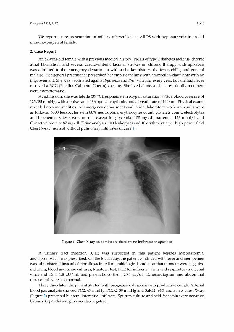
At admission, she was febrile (39 °C), eupneic with oxygen saturation 99%, a blood pressure of 125/85 mmHg, with a pulse rate of 86 bpm, arrhythmic, and a breath rate of 14 bpm. Physical exams revealed no abnormalities. At emergency department evaluation, laboratory work-up results were as follows: 6300 leukocytes with 80% neutrophils, erythrocytes count, platelets count, electrolytes and biochemistry tests were normal except for glycemia: 155 mg/dl, natremia: 123 nmol/L and C-reactive protein: 87 mg/dl. Urine analysis: 100 leukocytes and 10 erythrocytes per high-power field. Chest X-ray: normal without pulmonary infiltrates (Figure 1).
Figure 1. Chest X-ray on admission: there are no infiltrates or opacities.
A urinary tract infection (UTI) was suspected in this patient besides hyponatremia, and ciprofloxacin was prescribed. On the fourth day, the patient continued with fever and meropemen was administered instead of ciprofloxacin. All microbiological studies at that moment were negative including blood and urine cultures, Mantoux test, PCR for influenza virus and respiratory syncytial virus and TSH: 1.8 µU/mL and plasmatic cortisol: 25.5 µg/dl. Echocardiogram and abdominal ultrasound were also normal.
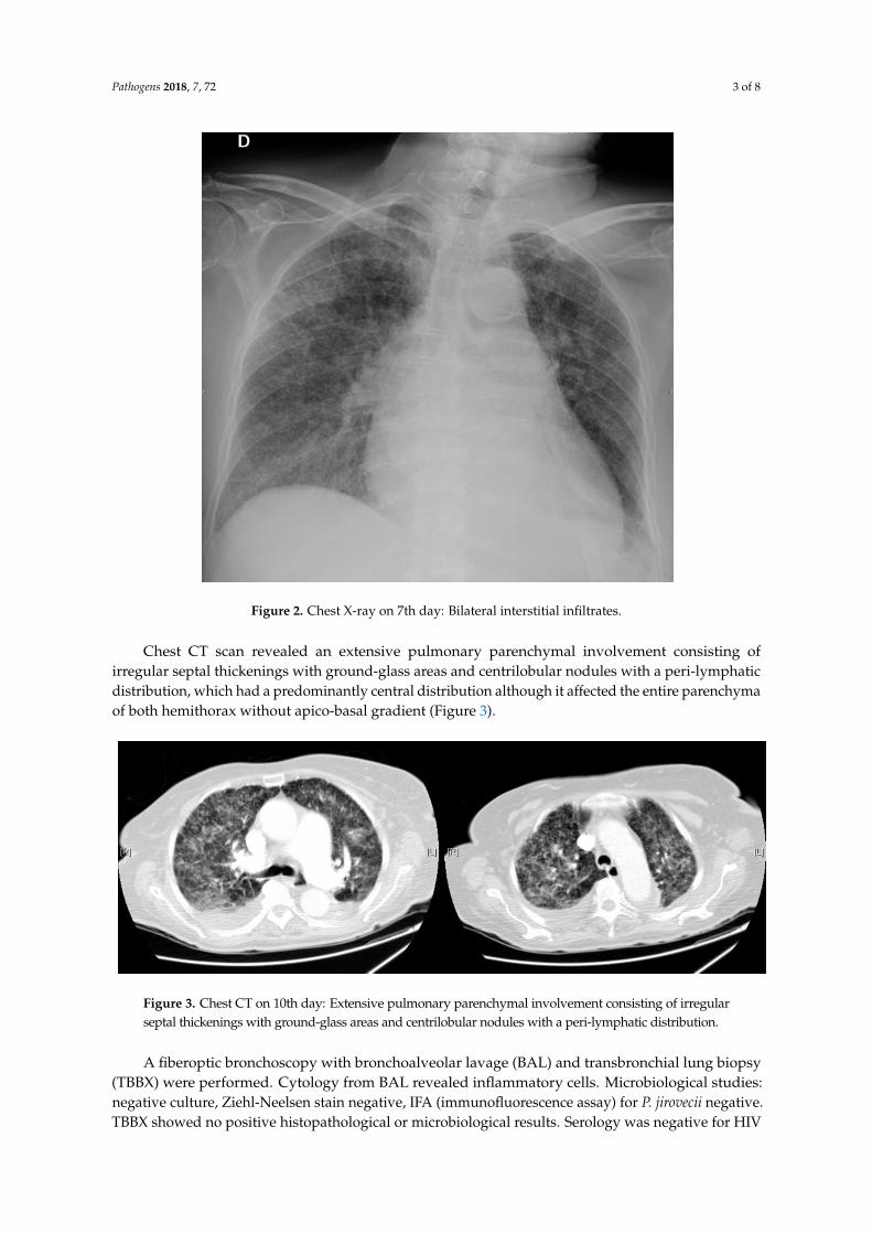
Three days later, the patient started with progressive dyspnea with productive cough. Arterial blood gas analysis showed PO2: 67 mmHg, PCO2: 39 mmHg and SatO2: 94% and a new chest X-ray (Figure 2) presented bilateral interstitial infiltrate. Sputum culture and acid-fast stain were negative. Urinary Legionella antigen was also negative.
Figure 1. Chest X-ray on admission: there are no infiltrates or opacities.
A urinary tract infection (UTI) was suspected in this patient besides hyponatremia,and ciprofloxacin was prescribed. On the fourth day, the patient continued with fever and meropemenwas administered instead of ciprofloxacin. All microbiological studies at that moment were negativeincluding blood and urine cultures, Mantoux test, PCR for influenza virus and respiratory syncytialvirus and TSH: 1.8 µU/mL and plasmatic cortisol: 25.5 µg/dl. Echocardiogram and abdominalultrasound were also normal.
Three days later, the patient started with progressive dyspnea with productive cough. Arterialblood gas analysis showed PO2: 67 mmHg, PCO2: 39 mmHg and SatO2: 94% and a new chest X-ray(Figure 2) presented bilateral interstitial infiltrate. Sputum culture and acid-fast stain were negative.Urinary Legionella antigen was also negative.
Pathogens 2018, 7, 72 3 of 8
Pathogens 2018, 7, x FOR PEER REVIEW 3 of 8
Figure 2. Chest X-ray on 7th day: Bilateral interstitial infiltrates.
Chest CT scan revealed an extensive pulmonary parenchymal involvement consisting of irregular septal thickenings with ground-glass areas and centrilobular nodules with a peri-lymphatic distribution, which had a predominantly central distribution although it affected the entire parenchyma of both hemithorax without apico-basal gradient (Figure 3).
Figure 3. Chest CT on 10th day: Extensive pulmonary parenchymal involvement consisting of irregular septal thickenings with ground-glass areas and centrilobular nodules with a peri-lymphatic distribution.
A fiberoptic bronchoscopy with bronchoalveolar lavage (BAL) and transbronchial lung biopsy (TBBX) were performed. Cytology from BAL revealed inflammatory cells. Microbiological studies: negative culture, Ziehl-Neelsen stain negative, IFA (immunofluorescence assay) for P. jirovecii negative. TBBX showed no positive histopathological or microbiological results. Serology was negative for HIV (Human Immunodeficiency virus), Cytomegalovirus (CMV), Herpes Virus 6 (HV 6), Epstein Barr virus (EBV) and Coxiella burneti.
Figure 2. Chest X-ray on 7th day: Bilateral interstitial infiltrates.
Chest CT scan revealed an extensive pulmonary parenchymal involvement consisting ofirregular septal thickenings with ground-glass areas and centrilobular nodules with a peri-lymphaticdistribution, which had a predominantly central distribution although it affected the entire parenchymaof both hemithorax without apico-basal gradient (Figure 3).
Pathogens 2018, 7, x FOR PEER REVIEW 3 of 8
Figure 2. Chest X-ray on 7th day: Bilateral interstitial infiltrates.
Chest CT scan revealed an extensive pulmonary parenchymal involvement consisting of irregular septal thickenings with ground-glass areas and centrilobular nodules with a peri-lymphatic distribution, which had a predominantly central distribution although it affected the entire parenchyma of both hemithorax without apico-basal gradient (Figure 3).
Figure 3. Chest CT on 10th day: Extensive pulmonary parenchymal involvement consisting of irregular septal thickenings with ground-glass areas and centrilobular nodules with a peri-lymphatic distribution.
A fiberoptic bronchoscopy with bronchoalveolar lavage (BAL) and transbronchial lung biopsy (TBBX) were performed. Cytology from BAL revealed inflammatory cells. Microbiological studies: negative culture, Ziehl-Neelsen stain negative, IFA (immunofluorescence assay) for P. jirovecii negative. TBBX showed no positive histopathological or microbiological results. Serology was negative for HIV (Human Immunodeficiency virus), Cytomegalovirus (CMV), Herpes Virus 6 (HV 6), Epstein Barr virus (EBV) and Coxiella burneti.
Figure 3. Chest CT on 10th day: Extensive pulmonary parenchymal involvement consisting of irregularseptal thickenings with ground-glass areas and centrilobular nodules with a peri-lymphatic distribution.
A fiberoptic bronchoscopy with bronchoalveolar lavage (BAL) and transbronchial lung biopsy(TBBX) were performed. Cytology from BAL revealed inflammatory cells. Microbiological studies:negative culture, Ziehl-Neelsen stain negative, IFA (immunofluorescence assay) for P. jirovecii negative.TBBX showed no positive histopathological or microbiological results. Serology was negative for HIV
Pathogens 2018, 7, 72 4 of 8
(Human Immunodeficiency virus), Cytomegalovirus (CMV), Herpes Virus 6 (HV 6), Epstein Barr virus(EBV) and Coxiella burneti.
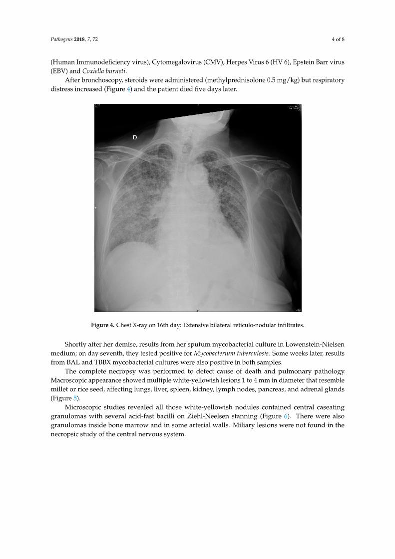
After bronchoscopy, steroids were administered (methylprednisolone 0.5 mg/kg) but respiratorydistress increased (Figure 4) and the patient died five days later.
Pathogens 2018, 7, x FOR PEER REVIEW 4 of 8
After bronchoscopy, steroids were administered (methylprednisolone 0.5 mg/kg) but respiratory distress increased (Figure 4) and the patient died five days later.
Figure 4. Chest X-ray on 16th day: Extensive bilateral reticulo-nodular infiltrates.
Shortly after her demise, results from her sputum mycobacterial culture in Lowenstein-Nielsen medium; on day seventh, they tested positive for Mycobacterium tuberculosis. Some weeks later, results from BAL and TBBX mycobacterial cultures were also positive in both samples.
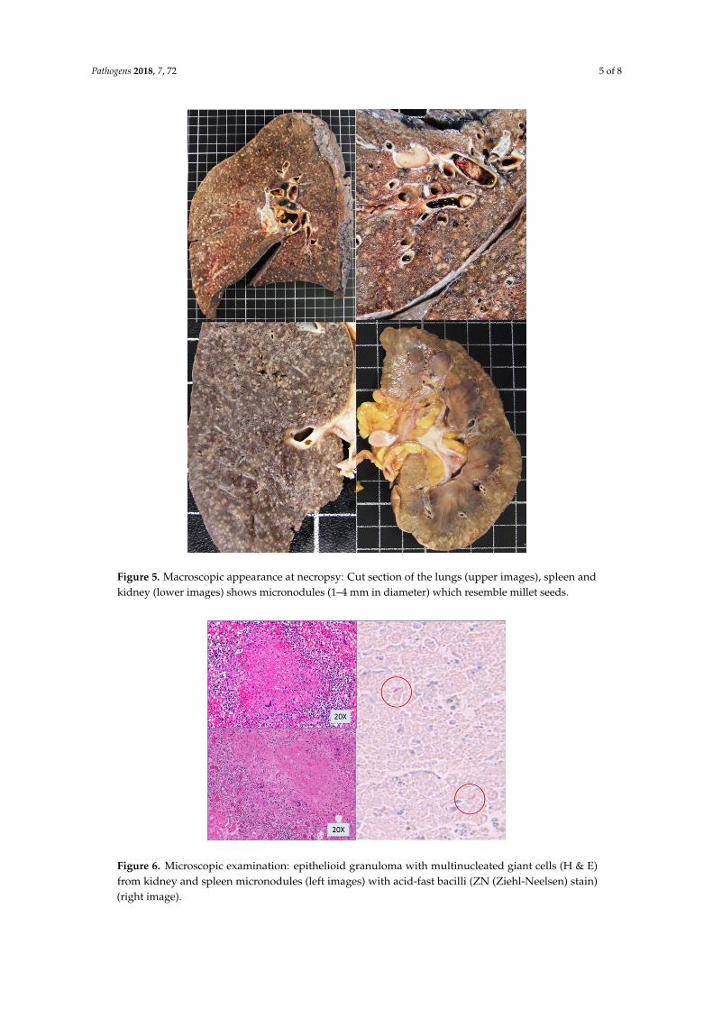
The complete necropsy was performed to detect cause of death and pulmonary pathology. Macroscopic appearance showed multiple white-yellowish lesions 1 to 4 mm in diameter that resemble millet or rice seed, affecting lungs, liver, spleen, kidney, lymph nodes, pancreas, and adrenal glands (Figure 5).
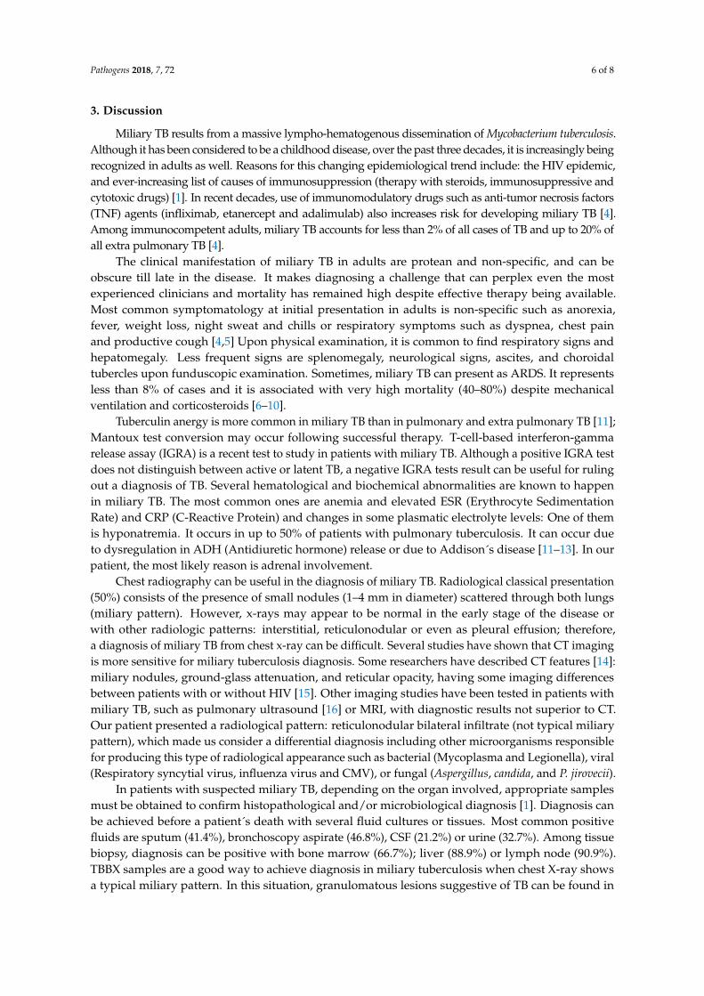
Microscopic studies revealed all those white-yellowish nodules contained central caseating granulomas with several acid-fast bacilli on Ziehl-Neelsen stanning (Figure 6). There were also granulomas inside bone marrow and in some arterial walls. Miliary lesions were not found in the necropsic study of the central nervous system.
Figure 4. Chest X-ray on 16th day: Extensive bilateral reticulo-nodular infiltrates.
Shortly after her demise, results from her sputum mycobacterial culture in Lowenstein-Nielsenmedium; on day seventh, they tested positive for Mycobacterium tuberculosis. Some weeks later, resultsfrom BAL and TBBX mycobacterial cultures were also positive in both samples.
The complete necropsy was performed to detect cause of death and pulmonary pathology.Macroscopic appearance showed multiple white-yellowish lesions 1 to 4 mm in diameter that resemblemillet or rice seed, affecting lungs, liver, spleen, kidney, lymph nodes, pancreas, and adrenal glands(Figure 5).
Microscopic studies revealed all those white-yellowish nodules contained central caseatinggranulomas with several acid-fast bacilli on Ziehl-Neelsen stanning (Figure 6). There were alsogranulomas inside bone marrow and in some arterial walls. Miliary lesions were not found in thenecropsic study of the central nervous system.
Pathogens 2018, 7, 72 5 of 8
Pathogens 2018, 7, x FOR PEER REVIEW 5 of 8
Figure 5. Macroscopic appearance at necropsy: Cut section of the lungs (upper images), spleen and kidney (lower images) shows micronodules (1–4 mm in diameter) which resemble millet seeds.
Figure 6. Microscopic examination: epithelioid granuloma with multinucleated giant cells (H & E) from kidney and spleen micronodules (left images) with acid-fast bacilli (ZN (Ziehl-Neelsen) stain) (right image).
Figure 5. Macroscopic appearance at necropsy: Cut section of the lungs (upper images), spleen andkidney (lower images) shows micronodules (1–4 mm in diameter) which resemble millet seeds.
Pathogens 2018, 7, x FOR PEER REVIEW 5 of 8
Figure 5. Macroscopic appearance at necropsy: Cut section of the lungs (upper images), spleen and kidney (lower images) shows micronodules (1–4 mm in diameter) which resemble millet seeds.
Figure 6. Microscopic examination: epithelioid granuloma with multinucleated giant cells (H & E) from kidney and spleen micronodules (left images) with acid-fast bacilli (ZN (Ziehl-Neelsen) stain) (right image).
Figure 6. Microscopic examination: epithelioid granuloma with multinucleated giant cells (H & E)from kidney and spleen micronodules (left images) with acid-fast bacilli (ZN (Ziehl-Neelsen) stain)(right image).
Pathogens 2018, 7, 72 6 of 8
3. Discussion
Miliary TB results from a massive lympho-hematogenous dissemination of Mycobacterium tuberculosis.Although it has been considered to be a childhood disease, over the past three decades, it is increasingly beingrecognized in adults as well. Reasons for this changing epidemiological trend include: the HIV epidemic,and ever-increasing list of causes of immunosuppression (therapy with steroids, immunosuppressive andcytotoxic drugs) [1]. In recent decades, use of immunomodulatory drugs such as anti-tumor necrosis factors(TNF) agents (infliximab, etanercept and adalimulab) also increases risk for developing miliary TB [4].Among immunocompetent adults, miliary TB accounts for less than 2% of all cases of TB and up to 20% ofall extra pulmonary TB [4].
The clinical manifestation of miliary TB in adults are protean and non-specific, and can beobscure till late in the disease. It makes diagnosing a challenge that can perplex even the mostexperienced clinicians and mortality has remained high despite effective therapy being available.Most common symptomatology at initial presentation in adults is non-specific such as anorexia,fever, weight loss, night sweat and chills or respiratory symptoms such as dyspnea, chest painand productive cough [4,5] Upon physical examination, it is common to find respiratory signs andhepatomegaly. Less frequent signs are splenomegaly, neurological signs, ascites, and choroidaltubercles upon funduscopic examination. Sometimes, miliary TB can present as ARDS. It representsless than 8% of cases and it is associated with very high mortality (40–80%) despite mechanicalventilation and corticosteroids [6–10].
Tuberculin anergy is more common in miliary TB than in pulmonary and extra pulmonary TB [11];Mantoux test conversion may occur following successful therapy. T-cell-based interferon-gammarelease assay (IGRA) is a recent test to study in patients with miliary TB. Although a positive IGRA testdoes not distinguish between active or latent TB, a negative IGRA tests result can be useful for rulingout a diagnosis of TB. Several hematological and biochemical abnormalities are known to happenin miliary TB. The most common ones are anemia and elevated ESR (Erythrocyte SedimentationRate) and CRP (C-Reactive Protein) and changes in some plasmatic electrolyte levels: One of themis hyponatremia. It occurs in up to 50% of patients with pulmonary tuberculosis. It can occur dueto dysregulation in ADH (Antidiuretic hormone) release or due to Addison´s disease [11–13]. In ourpatient, the most likely reason is adrenal involvement.
Chest radiography can be useful in the diagnosis of miliary TB. Radiological classical presentation(50%) consists of the presence of small nodules (1–4 mm in diameter) scattered through both lungs(miliary pattern). However, x-rays may appear to be normal in the early stage of the disease orwith other radiologic patterns: interstitial, reticulonodular or even as pleural effusion; therefore,a diagnosis of miliary TB from chest x-ray can be difficult. Several studies have shown that CT imagingis more sensitive for miliary tuberculosis diagnosis. Some researchers have described CT features [14]:miliary nodules, ground-glass attenuation, and reticular opacity, having some imaging differencesbetween patients with or without HIV [15]. Other imaging studies have been tested in patients withmiliary TB, such as pulmonary ultrasound [16] or MRI, with diagnostic results not superior to CT.Our patient presented a radiological pattern: reticulonodular bilateral infiltrate (not typical miliarypattern), which made us consider a differential diagnosis including other microorganisms responsiblefor producing this type of radiological appearance such as bacterial (Mycoplasma and Legionella), viral(Respiratory syncytial virus, influenza virus and CMV), or fungal (Aspergillus, candida, and P. jirovecii).
In patients with suspected miliary TB, depending on the organ involved, appropriate samplesmust be obtained to confirm histopathological and/or microbiological diagnosis [1]. Diagnosis canbe achieved before a patient´s death with several fluid cultures or tissues. Most common positivefluids are sputum (41.4%), bronchoscopy aspirate (46.8%), CSF (21.2%) or urine (32.7%). Among tissuebiopsy, diagnosis can be positive with bone marrow (66.7%); liver (88.9%) or lymph node (90.9%).TBBX samples are a good way to achieve diagnosis in miliary tuberculosis when chest X-ray showsa typical miliary pattern. In this situation, granulomatous lesions suggestive of TB can be found in

Pathogens 2018, 7, 72 7 of 8
up to 60% of cases [17]. Unfortunately, an important number of patients with miliary TB will not bediagnosed before a patient’s death, and it will be confirmed during autopsy [18–21].
Regarding treatment, patients with miliary TB must be promptly treated with standardanti-tuberculosis therapy, as this disease will be lethal if not treated [22]. However, there is noconsensus about optimum duration of treatment. Adjunctive corticosteroid treatment in patients withmiliary TB is considered to be beneficial in some circumstances such as TB meningitis, ARDS [8], largepericardial effusion or Addison’s disease [23].
Mortality related to miliary TB is high, reaching 25–30% of all adult cases. Delay in diagnosis,or no diagnosis, and consequently, delayed starting in specific anti-TB therapy appear to be the mostimportant factor responsible for this high mortality rate.
Although severe complications of miliary tuberculosis are frequent, mortality can be lower whereaccess to critical care intervention, anti-tuberculous therapy and corticosteroid use are possible [8–10].
This case highlights an atypical presentation of miliary tuberculosis in an old and immunocompetentfemale in a western and developed country with hyponatremia, a bilateral pulmonary interstitial infiltratethat produced her ARDS and death.
Author Contributions: Writing-Original Draft Preparation, I.P., R.G., M.C., C.G.; Writing-Review & Editing, B.H,E.P.; Visualization, B.H, E.P.; Supervision, B.H., E.P.
Funding: This research received no external funding.
Conflicts of Interest: The authors declare no conflict of interest.
References
1. Sharma, S.; Mohan, A.; Sharma, A. Challenges in the diagnosis & treatment of miliary tuberculosis. Indian J.Med. Res. 2012, 135, 703–730. [PubMed]
2. Lee, K.; Kim, J.H.; Lee, J.H.; Lee, W.-Y.; Park, M.S.; Kim, J.Y.; Kim, K.C.; Lee, M.-G.; Jung, K.-S.; Kim, Y.S.;et al. Acute respiratory distress syndrome caused by miliary tuberculosis: A multicentre survey in SouthKorea. Int. J. Tuberc. Lung Dis. 2011, 15, 1099–1103. [CrossRef] [PubMed]
3. Mohan, A.; Sharma, S.K.; Pande, J.N. Acute respiratory distress syndrome (ARDS) in miliary tuberculosis:A twelve year experience. Indian J. Chest Dis. Allied Sci. 1996, 38, 157–162. [PubMed]
4. Reichmann, M.T.; Marshall, B.G.; Cummings, F.; Elkington, P.T. Tuberculosis and TNF-inhibitors: History ofexposure should outweigh investigations. BMJ Case Rep. 2014. [CrossRef] [PubMed]
5. Sharma, S.K.; Mohan, A.; Sharma, A. Miliary tuberculosis: A new look at an old foe. J. Clin. Tuberc. OtherMycobact. Dis. 2016, 3, 13–27. [CrossRef]
6. Mert, A.; Arslan, F.; Kuyucu, T.; Koç, E.N.; Yilmaz, M.; Turan, D.; Altin, S.; Pehlivanoglu, F.; Sengoz, G.;Yildiz, D.; et al. Miliary tuberculosis. Medicine (Baltimore) 2017, 96, e5875. [CrossRef] [PubMed]
7. Singh, K.; Hyatali, S.; Giddings, S.; Singh, K.; Bhagwandass, N. Case Report Miliary Tuberculosis Presentingwith ARDS and Shock: A Case Report and Challenges in Current Management and Diagnosis. Case Rep.Crit. Care 2017. [CrossRef] [PubMed]
8. Sharma, S.K.; Mohan, A.; Banga, A.; Saha, P.K.; Guntupalli, K.K. Predictors of development and outcomein patients with acute respiratory distress syndrome due to tuberculosis. Int. J. Tuberc. Lung Dis. 2006,10, 429–435. [PubMed]
9. Boronat, M.; Arzuaga, J.A.; Diez Balda, V.; Chamorro, C.; Salas, C.; López de Letona, J.M. The adultrespiratory stress syndrome and septic shock associated with tuberculosis. Rev. Clin. Esp. 1995, 195, 160–163.[PubMed]
10. Abi-Fadel, F.; Gupta, K. Acute respiratory distress syndrome with miliary tuberculosis: A fatal combination.J. Thorac. Dis. 2013, 5, E1–E4. [PubMed]
11. Jafari, N.J.; Izadi, M.; Sarrafzadeh, F.; Heidari, A.; Ranjbar, R.; Saburi, A. Hyponatremia Due to PulmonaryTuberculosis: Review of 200 Cases. Nephrourol. Mon. 2012, 5, 687–691. [CrossRef] [PubMed]
12. Underwood, J.; Cresswell, F.; Salam, A.P.; Keeley, A.J.; Cleland, C.; John, L.; Davidson, R.N. Complications ofmiliary tuberculosis: Low mortality and predictive biomarkers from a UK cohort. BMC Infect. Dis. 2017,17, 295. [CrossRef] [PubMed]

Pathogens 2018, 7, 72 8 of 8
13. Hussain, S.F.; Irfan, M.; Abbasi, M.; Anwer, S.S.; Davidson, S.; Haqqee, R.; Khan, J.A.; Islam, M. Clinicalcharacteristics of 110 miliary tuberculosis patients from a low HIV prevalence country. Int. J. Tuberc. Lung. Dis.2004, 8, 493–499. [PubMed]
14. Giacomelli, I.L.; Neto, R.S.; Marchiori, E.; Marchiori, E.; Pereira, M.; Hochhegger, B. Chest X-ray andchest ct findings in patients diagnosed with pulmonary tuberculosis following solid organ transplantation:A systematic review. J. Bras. Pneumol. 2018, 44, 161–166. [CrossRef] [PubMed]
15. Kim, J.Y.; Jeong, Y.J.; Kim, K.I.; Lee, I.S.; Park, H.K.; Kim, Y.D.; Seok, H.I. Miliary tuberculosis: A comparisonof CT findings in HIV-seropositive and HIV-seronegative patients. Br. J. Radiol. 2010, 83, 206–211. [CrossRef][PubMed]
16. Hunter, L.; Bélard, S.; Janssen, S.; Heller, T. Miliary tuberculosis: Sonographic pattern in chest ultrasound.Infection 2016, 44, 243–246. [CrossRef] [PubMed]
17. Ibrahim, A.S.; Allangawi, M.H.; Sattar, H.A.; Mobyed, H.S.; Almohammed, A.A. Indications, diagnosticyields and complications of transbronchial biopsy over 5 years in the State of Qatar. Saudi Med. J. 2005,26, 641–645. [PubMed]
18. Gupta, M.; Lobo, F.D.; Adiga, D.S.A.; Gupta, A. A histomorphological pattern analysis of pulmonarytuberculosis in lung autopsy and surgically resected specimens. Pathol. Res. Int. 2016. [CrossRef] [PubMed]
19. Savic, I.; Trifunovic-Skodric, V.; Mitrovic, D. Clinically unrecognized miliary tuberculosis: An autopsy study.Ann. Saudi Med. 2016, 36, 42–50. [CrossRef] [PubMed]
20. Dos Santos, V.M.; Dos Santos, L.A.M. Miliary tuberculosis: The role of necropsy studies. Infez. Med. 2017,25, 162–165. [PubMed]
21. Tajiri, T.; Tate, G.; Makino, M.; Akita, H.; Omatsu, M.; Enosawa, T.; Hamatani, S.; Masunaga, A.; Kunimura, T.;Mitsuya, T.; et al. Autopsy cases of miliary tuberculosis: Clinicopathologic features including backgroundfactors. J. Nippon Med. Sch. 2011, 78, 305–311. [CrossRef] [PubMed]
22. Zahar, J.R.; Azoulay, E.; Klement, E.; De Lassence, A.; Lucet, J.C.; Regnier, B.; Schlemmer, B.; Bedos, J.P.Delayed treatment contributes to mortality in ICU patients with severe active pulmonary tuberculosis andacute respiratory failure. Intensive Care Med. 2001, 27, 513–523. [CrossRef] [PubMed]
23. Nam, S.J.; Cho, Y.J. The successful treatment of refractory respiratory failure due to miliary tuberculosis:Survival after prolonged extracorporeal membrane oxygenation support. Clin. Respir. J. 2016, 10, 393–399.[CrossRef] [PubMed]
© 2018 by the authors. Licensee MDPI, Basel, Switzerland. This article is an open accessarticle distributed under the terms and conditions of the Creative Commons Attribution(CC BY) license (http://creativecommons.org/licenses/by/4.0/).
Related Documents