Mild Traumatic Brain Injury: Mild Traumatic Brain Injury: Diagnosis and Management Diagnosis and Management Andy Jagoda, MD Andy Jagoda, MD Department of Emergency Medicine Department of Emergency Medicine Mount Sinai School of Medicine Mount Sinai School of Medicine New York, New York New York, New York

Mild Traumatic Brain Injury: Diagnosis and Management Andy Jagoda, MD Department of Emergency Medicine Mount Sinai School of Medicine New York, New York.
Dec 22, 2015
Welcome message from author
This document is posted to help you gain knowledge. Please leave a comment to let me know what you think about it! Share it to your friends and learn new things together.
Transcript

Mild Traumatic Brain Injury:Mild Traumatic Brain Injury:Diagnosis and ManagementDiagnosis and Management
Andy Jagoda, MDAndy Jagoda, MD
Department of Emergency MedicineDepartment of Emergency MedicineMount Sinai School of MedicineMount Sinai School of Medicine
New York, New YorkNew York, New York

Andy Jagoda, MD
ObjectivesObjectives• Definitions
– Glasgow coma scale score– Amnesia / loss of consciousness
• Neuroimaging– Skull radiographs– CT – MRI
• Postconcussive syndromes– Neuropsychiatric testing– Prognosis
• Management

Andy Jagoda, MD
A 50-year-old driver is in a head-on collision at approximately 15 mph. She is not wearing a seat belt and strikes her head on the windshield. The windshield does not break, and though dazed for “several seconds” she does not lose consciousness and has no amnesia. She is taken to the ED where she is alert, oriented times three, and has no complaints. She has no past medical history and is on no medications.
The CaseThe Case

Andy Jagoda, MD
Which of the Following is Not Which of the Following is Not Used to Define Mild TBI?Used to Define Mild TBI?
a. GCS >12
b. Loss of consciousness <1 hr
c. Post-traumatic amnesia <24 hrs
d. Non-focal neurologic exam
e. CT scan

What would the GCS be in the presented What would the GCS be in the presented case If on exam the patient kept her eyes case If on exam the patient kept her eyes closed but opened them to questions; closed but opened them to questions; answered questions with difficulty and was answered questions with difficulty and was confused; moved her extremities confused; moved her extremities appropriately on command?appropriately on command?
a. 15
b. 13
c. 11
d. 09
e. 07

If the presented patient had an occipital If the presented patient had an occipital laceration with no hematoma, which of the laceration with no hematoma, which of the following would be the best initial test?following would be the best initial test?
a. Skull radiographs
b. Non-contrast CT
c. Contrast CT
d. MRI
E. PET

Andy Jagoda, MD
Postconcussive Syndrome (PCS) in Postconcussive Syndrome (PCS) in Mild TBI, which of the following is true?Mild TBI, which of the following is true?
a. Early PCS occurs primarily in patients with psychiatric problems.
b. Early PCS occurs more frequently in patients involved in litigation.
c. Early PCS occurs in up to 20% of patients.
d. Late PCS occurs primarily in men.
e. Late PCS has been linked to anxiety, stress, and depression.

Andy Jagoda, MD
EpidemiologyEpidemiology
• 6 million head injury cases in the USA each year (1 in 45)– Young male predominance
• 2 million ED evaluations
• 250,000 hospitalizations (1 in 1000)
• 60,000 deaths (1 in 5000)
• Most cases are classified as mild

Andy Jagoda, MD
PathophysiologyPathophysiology
• Deceleration / rotation injury• Blood vessel disruption
– Petechial hemorrhage– Focal edema– Disruption of bridging veins
• Intra-axonal neurofilament organization / axonal swelling
• Secondary injury– Excitatory amino acids– Oxygen free radicals

Andy Jagoda, MD
Diagnosing Mild TBIDiagnosing Mild TBIMild TBI Committee of the American Congress of Mild TBI Committee of the American Congress of
Rehabilitation MedicineRehabilitation Medicine
• Alteration in mental state at the time of the accident
• LOC <30 min
• After 30 min, GCS 13-15
• Amnesia <24 hours

Andy Jagoda, MD
Classification - GCSClassification - GCS
• Eyes– 4 opens spontaneously– 3 opens to verbal– 2 opens to pain– 1 do not open
• Verbal– 5 oriented– 4 confused– 3 inappropriate– 2 incomprehensible– 1 none
• Motor– 6 obeys– 5 localizes– 4 withdraws– 3 abnormal flex– 2 extensor response– 1 none
• Scoring– Mild >12– Moderate 9-12– Severe <9

Andy Jagoda, MD
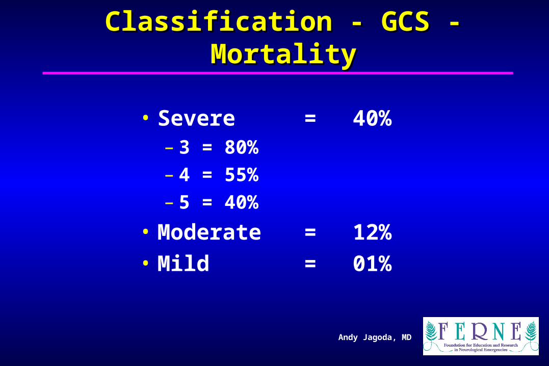
Classification - GCS - MortalityClassification - GCS - Mortality
• Developed for prognosis in severe TBI• Timing of score is not standardized• One score not sufficient - perform serial exams
– Prognosis worse if score does not improve or if it worsens
• Does not account for drugs, seizures, or metabolic problems
Andy Jagoda, MD
Classification - GCS - MortalityClassification - GCS - Mortality
• Severe = 40%– 3 = 80%– 4 = 55%– 5 = 40%
• Moderate = 12%
• Mild = 01%

What would the GCS be in the presented What would the GCS be in the presented case If on exam the patient kept her eyes case If on exam the patient kept her eyes
closed but opened them to questions; closed but opened them to questions; answered questions with difficulty and was answered questions with difficulty and was
confused; moved her extremities confused; moved her extremities appropriately on command?appropriately on command?
a. 15
b. 13
c. 11
d. 09
e. 07

Andy Jagoda, MD
Which Of The Following Is Not Which Of The Following Is Not Used To Define Mild TBI?Used To Define Mild TBI?
• a. GCS > 12
• b. Loss of consciousness < 1 hr
• c. Post-traumatic amnesia < 24 hrs
• d. Non-focal neurologic exam
• e. Normal CT scan

Andy Jagoda, MD
Use of CT in Diagnosing MTBIUse of CT in Diagnosing MTBI
• Retrospective study, 215 hospitalized patients– Mild TBI without complications– Mild TBI with complications (positive CT)– Moderate TBI
• Mild TBI patients with positive CT performed on neuropsychiatric testing like moderate TBI
Williams et al. Williams et al. Neurosurgery Neurosurgery 1990;27:422.1990;27:422.

Andy Jagoda, MD
Use of CT in Diagnosing MTBIUse of CT in Diagnosing MTBI
• Moderate group had worse function at 6 months
• Length of LOC or amnesia did not differentiate mild from moderate groups
• Depressed skull fractures without parenchymal lesions did performed as mild TABI
Williams et al. Williams et al. Neurosurgery Neurosurgery 1990;27:422.1990;27:422.

Andy Jagoda, MD
Skull Radiographs and Skull Radiographs and Intracranial LesionsIntracranial Lesions
• Retrospective review
• 207 hospitalized patients with intracranial lesions
• 63% had no skull fracture
• Skull films do not predict intracranial lesion
Cooper P, Ho V. Cooper P, Ho V. Neurosurgery Neurosurgery 1983;13:136.1983;13:136.

Andy Jagoda, MD
• Retrospective review 22,058 cases
• Patients with skull fractures, 91% did not have intracranial injury
• 51% of patients with intracranial injury did not have a skull fracture
Masters et al. NEJM 1987;316:84-91.
Skull Radiographs and Skull Radiographs and Intracranial LesionsIntracranial Lesions

Andy Jagoda, MD
Skull Radiographs and Skull Radiographs and Intracranial LesionsIntracranial Lesions
• Prospective study: 7035 patients – Not all patients received same tests– 48% lost to follow-up
• Skull fracture was associated with an intracranial injury
Masters et al. NEJM 1987;316:84-91.

Andy Jagoda, MD
Skull Radiographs and Intracranial Skull Radiographs and Intracranial LesionsLesions
• Skull fracture did not predict an intracranial injury
• Absence of a skull fracture did not rule out an intracranial injury
• Plain films are neither sensitive nor specific for intracranial injury
Masters et al. NEJM 1987;316:84-91.

Andy Jagoda, MD
Low Risk Group For Low Risk Group For Intracranial InjuryIntracranial Injury
• Asymptomatic• Headache• Dizziness• Scalp hematoma, laceration, contusion• Absence of moderate or high risk criteria,
ie, LOC or amnesia• No patients with neurologic deterioration
identified• No imaging study indicated
Masters et al. NEJM 1987;316:84-91.

Andy Jagoda, MD
Moderate Risk Group For Moderate Risk Group For Intracranial InjuryIntracranial Injury
• Loss of consciousness• Unreliable history• Progressive headache• Alcohol or drug intoxication• Age less than 2 years• Post traumatic seizure• BSF / multiple trauma / possible penetrating
trauma• CT scan recommendedMasters et al. NEJM 1987;316:84-91.

Andy Jagoda, MD
Head CT In Mild TBI Head CT In Mild TBI
• Retrospective review 1538 trauma admissions• GCS > 12; all with history of LOC or amnesia• 265 (17.2%) had intracranial lesion:
– GCS 13: 37.5%– GCS 14: 24.2%– GCS 15: 13.2%
• 58 (3.8% of total 22% of patients with positive CT) required neurosurgery
• No patient with a normal CT deterioratedStein S, Ross S. Stein S, Ross S. Ann Emerg MedAnn Emerg Med 1993;22:1193. 1993;22:1193.

Andy Jagoda, MD
Head CT In Mild TBI Head CT In Mild TBI
• Prospective study: 712 consecutive ED patients• GCS 15; history of LOC or amnesia• Nonfocal neurologic exam
– 4 object recall and digit span testing
• 67 (9.4%) had a positive head CT• 2 (.28%) required emergent neurosurgery• No statistical model could be created to classify
95% of patients into CT normal vs abnormal
Jeret et al. Jeret et al. Neurosurgery Neurosurgery 1993;32:9.1993;32:9.

Andy Jagoda, MD
Head CT In Mild TBI Head CT In Mild TBI
• 10% to 20% have a positive CT
• .2 to 4% have a neurosurgical lesion
• Patients without LOC or amnesia, normal exam, and GCS 15 do not need imaging– Direct trauma to the temporal area– Children <3 years

Andy Jagoda, MD
Head CT in Mild TBIHead CT in Mild TBI
• In patients with LOC or amnesia, there are no combination of findings that identify all patients who have a positive CT
• Patients with a normal CT can be safely discharged home

Andy Jagoda, MD
Magnetic Resonance ImagingMagnetic Resonance Imaging
• Prospective study
• 50 TBI patients; CT, MRI, neuropsych
• 72% had lesions on CT
• 80% had lesions seen on MRI– Scattered punctate lesions; Frontal temporal
regionsLevin et al. J Neurol Neurosurg Psych 1992;55:255.

Andy Jagoda, MD
Magnetic Resonance ImagingMagnetic Resonance Imaging
• MRI identified additional lesions in 52% of patients with lesions on CT
• No correlation with size of lesions and length of LOC: inconsistent relationship between lesions and neuropsych findings
Levin et al. J Neurol Neurosurg Psych 1992;55:255.

Andy Jagoda, MD
Other Diagnostic ModalitiesOther Diagnostic Modalities
• Brainstem auditory evoked potentials (BAEP)
• EEG power spectra analysis
• Singular photon emission computed tomography (SPECT)
• Positron emission tomography (PET)

If the presented patient had an occipital If the presented patient had an occipital laceration with no hematoma. Which of the laceration with no hematoma. Which of the following is the best initial test?following is the best initial test?
a. Skull radiographs
b. Non-contrast CT
c. Contrast CT
d. MRI
e. PET

Andy Jagoda, MD
PCS: Reading the LiteraturePCS: Reading the Literature• Symptom complex related to TBI
– Somatic • Headache, sleep disturbance, dizziness, nausea, fatigue,
sensitivity to light / sound
– Cognitive• Attention / concentration problems, memory problems
– Affective• Irritability, anxiety, depression, emotional lability
• Incidence in MTBI patients:– 80% at 1 month– 30% at 3 months– 15% at 12 months

Andy Jagoda, MD
PCS: Reading the LiteraturePCS: Reading the Literature
• Lack of uniformity in definitions
• Selection bias
• No controls
• No pre-injury baseline
• Lack of standardization of testing
• Attrition in follow-up

Andy Jagoda, MD
Postconcussive SyndromePostconcussive Syndrome
• Prospective study, 538 patients• MTBI, hospitalized• 3 month follow-up• 79% headaches• 59% memory dysfunction• 33% had not returned to work• Ongoing litigation did not correlate with
complaintsRimel et al. Rimel et al. NeurosurgeryNeurosurgery 1981;9:221. 1981;9:221.

Andy Jagoda, MD
Postconcussive SyndromePostconcussive Syndrome
• 587 hospitalized, uncomplicated MTBI patients• Prospective design over 1 year
– 68% lost to follow-up
• At discharge, 67% had at least one symptom• 38% had symptoms at 3 months• 23% had symptoms at 6 months• 13% had symptoms at 12 months• Presence of symptoms at hospital discharge
were not predictive of symptoms at 3 monthsAlves et al. J Head Trauma Rehab 1993;8:48.
Andy Jagoda, MD
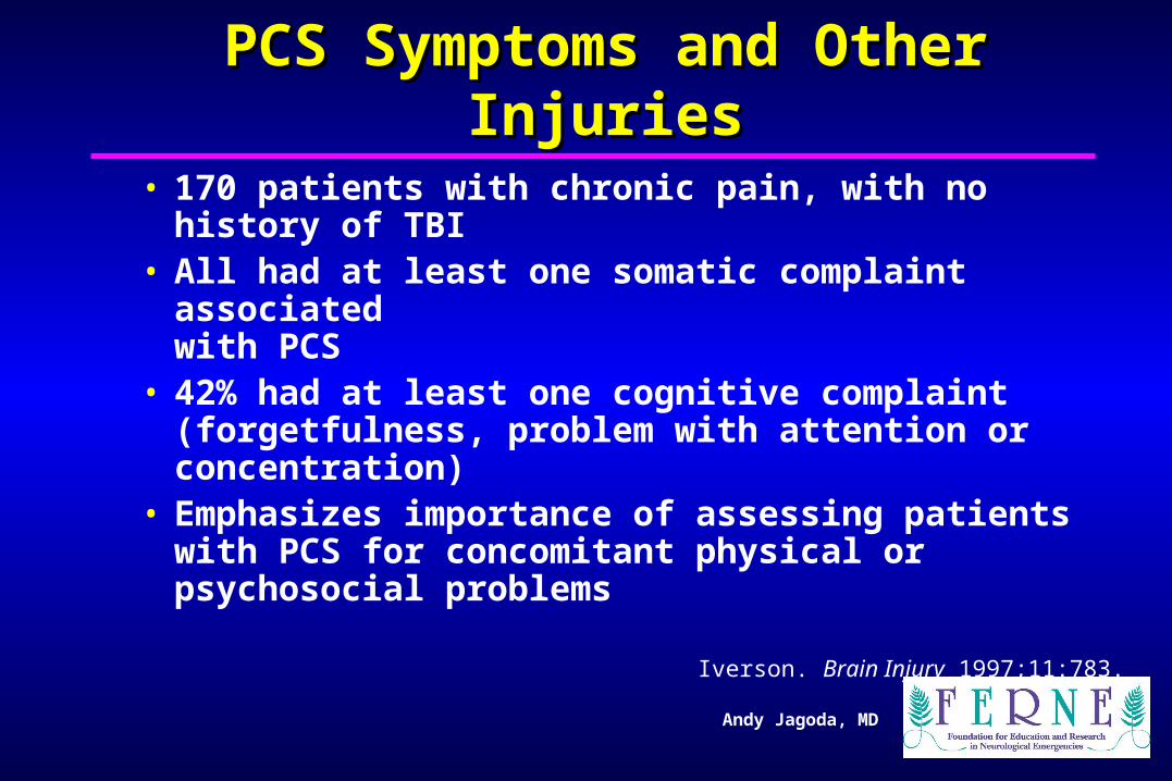
PCS Symptoms and Other InjuriesPCS Symptoms and Other Injuries• 170 patients with chronic pain, with no history of
TBI• All had at least one somatic complaint associated
with PCS• 42% had at least one cognitive complaint
(forgetfulness, problem with attention or concentration)
• Emphasizes importance of assessing patients with PCS for concomitant physical or psychosocial problems
Iverson. Brain Injury 1997;11:783.

Andy Jagoda, MD
PCS: Neuropsychiatric TestingPCS: Neuropsychiatric Testing
• No consensus on tests• Testing focuses on cognitive function:
– Attention– Information processing– Choice reaction time
• Testing demonstrates clear deficits in the first 3 months that appear independent of psychosocial factors
• Persistence of symptoms after 3 months appears to be complicated by psychosocial factors

Andy Jagoda, MD
LOC and AmnesiaLOC and Amnesia
• No correlation found between LOC and development of PCS symptoms
• No difference in neuropsych testing in patients with and without LOC
• No correlation found between length of postconcussive amnesia and PCS
Yarnell. Brain Injury 1988;2:255.
Lenninger. J Neurol Neurosurg Psych 1990;53:293.
Williams. Neurosurgery 1990;27:422.

Andy Jagoda, MD
Prognostic Predictors Of PCSPrognostic Predictors Of PCS
• Best prognosis– Young– Male– Educated– Social support
• Worse prognosis– Elderly– Female– Social / physical stressors– Substance abuse

Andy Jagoda, MD
Postconcussive Syndrome (PCS) in Mild Postconcussive Syndrome (PCS) in Mild TBI, which of the following is true?TBI, which of the following is true?
a. Early PCS occurs primarily in patients with psychiatric problems.
b. Early PCS occurs more frequently in patients involved in litigation.
c. Early PCS occurs in up to 20% of patients.
d. Late PCS occurs primarily in men.
e. Late PCS has been linked to anxiety, stress, and depression.

Andy Jagoda, MD
DispositionDisposition
• Saunders et al. Ann Emerg Med 1986;15:160.– 47 consecutive MTBI discharged from the ED– No patient could remember more than 2 of the 8
items on the home care discharge instructions
– 20% denied ever having received instructions– Third party involvement improved compliance with
instructions to 67%

Andy Jagoda, MD
DispositionDisposition
• Levitt et al. Amer J Emerg Med 1994;12:172.– 23% of MTBI patients discharged from the
ED could not remember any of their discharge instructions
• Studies emphasize importance of involving third parties in discharge process

Andy Jagoda, MD
ConclusionsConclusions• Mild TBI patients are at risk for intracranial injury
and developing the PCS• Plain film radiographs have low sensitivity and
specificity for intracranial injury and are not indicated
• 10% - 20% of mild TBI patients have abnormalities on non-contrast head CT
• .2% - 4% of mild TBI pts have a neurosurgical lesion• 80% after a mild TBI have symptoms of PCS and
should be properly counseled
Related Documents