Guide to Anticoagulant Therapy: Heparin A Statement for Healthcare Professionals From the American Heart Association Jack Hirsh, MD; Sonia S. Anand, MD; Jonathan L. Halperin, MD; Valentin Fuster, MD, PhD T hrombi are composed of fibrin and blood cells and may form in any part of the cardiovascular system, including veins, arteries, the heart, and the microcirculation. Because the relative proportion of cells and fibrin depends on hemo- dynamic factors, the proportions differ in arterial and venous thrombi. 1,2 Arterial thrombi form under conditions of high flow and are composed mainly of platelet aggregates bound together by thin fibrin strands. 3–5 In contrast, venous thrombi form in areas of stasis and are predominantly composed of red cells, with a large amount of interspersed fibrin and relatively few platelets. Thrombi that form in regions of slow to moderate flow are composed of a mixture of red cells, platelets, and fibrin and are known as mixed platelet-fibrin thrombi. 4,5 When a platelet-rich arterial thrombus becomes occlusive, stasis occurs, and the thrombus can propagate as a red stasis thrombus. As thrombi age, they undergo progres- sive structural changes. 6 Leukocytes are attracted by chemo- tactic factors released from aggregated platelets 2 or proteo- lytic fragments of plasma proteins and become incorporated into the thrombi. The aggregated platelets swell and disinte- grate and are gradually replaced by fibrin. Eventually, the fibrin clot is digested by fibrinolytic enzymes released from endothelial cells and leukocytes. The complications of throm- bosis are caused either by the effects of local obstruction of the vessel, distant embolism of thrombotic material, or, less commonly, consumption of hemostatic elements. Arterial thrombi usually form in regions of disturbed flow and at sites of rupture of an atherosclerotic plaque, which exposes the thrombogenic subendothelium to platelets and coagulation proteins; plaque rupture may also produce further narrowing due to hemorrhage into the plaque. 7–11 Nonocclu- sive thrombi may become incorporated into the vessel wall and can accelerate the growth of atherosclerotic plaques. 9,12,13 When flow is slow, the degree of stenosis is severe, or the thrombogenic stimulus is intense, the thrombi may become totally occlusive. Arterial thrombi usually occur in associa- tion with preexisting vascular disease, most commonly ath- erosclerosis; they produce clinical tissue ischemia either by obstructing flow or by embolism into the distal microcircu- lation. Activation both of blood coagulation and of platelets is important in the pathogenesis of arterial thrombosis. These 2 fundamental mechanisms of thrombogenesis are closely linked in vivo, because thrombin, a key clotting enzyme generated by blood coagulation, is a potent platelet activator, and activated platelets augment the coagulation process. Therefore, both anticoagulants and drugs that suppress plate- let function are potentially effective in the prevention and treatment of arterial thrombosis, and evidence from results of clinical trials indicates that both classes of drugs are effective. Venous thrombi usually occur in the lower limbs; although often silent, they can produce acute symptoms due to inflam- mation of the vessel wall, obstruction of flow, or embolism into the pulmonary circulation. They can produce long-term complications due to venous hypertension by damaging the venous valves. Activation of blood coagulation is the critical mechanism in pathogenesis of venous thromboembolism, whereas platelet activation is less important. Anticoagulants are therefore very effective for prevention and treatment of venous thromboembolism, and drugs that suppress platelet function are of less benefit. Intracardiac thrombi usually form on inflamed or damaged valves, on endocardium adjacent to a region of myocardial infarction (MI), in a dilated or dyskinetic cardiac chamber, or on prosthetic valves. They are usually asymptomatic when confined to the heart but may produce complications due to embolism to the cerebral or systemic circulation. Activation of blood coagulation is more important in the pathogenesis of intracardiac thrombi than platelet activation, although the latter plays a contributory role. Anticoagulants are effective for prevention and treatment of intracardiac thrombi, and in patients with prosthetic heart valves, the efficacy of antico- agulants is augmented by drugs that suppress platelet function. Widespread microvascular thrombosis is a complication of disseminated intravascular coagulation or generalized platelet This statement was approved by the American Heart Association Science Advisory and Coordinating Committee in December 2000. A single reprint is available by calling 800-242-8721 (US only) or writing the American Heart Association, Public Information, 7272 Greenville Ave, Dallas, TX 75231-4596. Ask for reprint No. 71-0203. To purchase additional reprints: up to 999 copies, call 800-611-6083 (US only) or fax 413-665-2671; 1000 or more copies, call 214-706-1466, fax 214-691-6342, or e-mail [email protected]. To make photocopies for personal or educational use, call the Copyright Clearance Center, 978-750-8400. Parts of this statement were originally published in Circulation. 1994;89:1449 –1468. This publication is an update of the 1994 statement. Parts of this statement are based on the Sixth American College of Chest Physicians Consensus Conference on Antithrombotic Therapy and have been published previously in Chest (2001;119[suppl]:64S–94S). This statement will also be published in the July 2001 issue of Arteriosclerosis, Thrombosis, and Vascular Biology. (Circulation. 2001;103:2994-3018.) © 2001 American Heart Association, Inc. Circulation is available at http://www.circulationaha.org 2994 AHA Scientific Statement by guest on September 16, 2015 http://circ.ahajournals.org/ Downloaded from

Welcome message from author
This document is posted to help you gain knowledge. Please leave a comment to let me know what you think about it! Share it to your friends and learn new things together.
Transcript

Guide to Anticoagulant Therapy: HeparinA Statement for Healthcare Professionals
From the American Heart AssociationJack Hirsh, MD; Sonia S. Anand, MD; Jonathan L. Halperin, MD; Valentin Fuster, MD, PhD
Thrombi are composed of fibrin and blood cells and mayform in any part of the cardiovascular system, including
veins, arteries, the heart, and the microcirculation. Becausethe relative proportion of cells and fibrin depends on hemo-dynamic factors, the proportions differ in arterial and venousthrombi.1,2 Arterial thrombi form under conditions of highflow and are composed mainly of platelet aggregates boundtogether by thin fibrin strands.3–5 In contrast, venous thrombiform in areas of stasis and are predominantly composed of redcells, with a large amount of interspersed fibrin and relativelyfew platelets. Thrombi that form in regions of slow tomoderate flow are composed of a mixture of red cells,platelets, and fibrin and are known as mixed platelet-fibrinthrombi.4,5 When a platelet-rich arterial thrombus becomesocclusive, stasis occurs, and the thrombus can propagate as ared stasis thrombus. As thrombi age, they undergo progres-sive structural changes.6 Leukocytes are attracted by chemo-tactic factors released from aggregated platelets2 or proteo-lytic fragments of plasma proteins and become incorporatedinto the thrombi. The aggregated platelets swell and disinte-grate and are gradually replaced by fibrin. Eventually, thefibrin clot is digested by fibrinolytic enzymes released fromendothelial cells and leukocytes. The complications of throm-bosis are caused either by the effects of local obstruction ofthe vessel, distant embolism of thrombotic material, or, lesscommonly, consumption of hemostatic elements.
Arterial thrombi usually form in regions of disturbed flowand at sites of rupture of an atherosclerotic plaque, whichexposes the thrombogenic subendothelium to platelets andcoagulation proteins; plaque rupture may also produce furthernarrowing due to hemorrhage into the plaque.7–11 Nonocclu-sive thrombi may become incorporated into the vessel walland can accelerate the growth of atherosclerotic plaques.9,12,13
When flow is slow, the degree of stenosis is severe, or thethrombogenic stimulus is intense, the thrombi may becometotally occlusive. Arterial thrombi usually occur in associa-tion with preexisting vascular disease, most commonly ath-
erosclerosis; they produce clinical tissue ischemia either byobstructing flow or by embolism into the distal microcircu-lation. Activation both of blood coagulation and of platelets isimportant in the pathogenesis of arterial thrombosis. These 2fundamental mechanisms of thrombogenesis are closelylinked in vivo, because thrombin, a key clotting enzymegenerated by blood coagulation, is a potent platelet activator,and activated platelets augment the coagulation process.Therefore, both anticoagulants and drugs that suppress plate-let function are potentially effective in the prevention andtreatment of arterial thrombosis, and evidence from results ofclinical trials indicates that both classes of drugs are effective.
Venous thrombi usually occur in the lower limbs; althoughoften silent, they can produce acute symptoms due to inflam-mation of the vessel wall, obstruction of flow, or embolisminto the pulmonary circulation. They can produce long-termcomplications due to venous hypertension by damaging thevenous valves. Activation of blood coagulation is the criticalmechanism in pathogenesis of venous thromboembolism,whereas platelet activation is less important. Anticoagulantsare therefore very effective for prevention and treatment ofvenous thromboembolism, and drugs that suppress plateletfunction are of less benefit.
Intracardiac thrombi usually form on inflamed or damagedvalves, on endocardium adjacent to a region of myocardialinfarction (MI), in a dilated or dyskinetic cardiac chamber, oron prosthetic valves. They are usually asymptomatic whenconfined to the heart but may produce complications due toembolism to the cerebral or systemic circulation. Activationof blood coagulation is more important in the pathogenesis ofintracardiac thrombi than platelet activation, although thelatter plays a contributory role. Anticoagulants are effectivefor prevention and treatment of intracardiac thrombi, and inpatients with prosthetic heart valves, the efficacy of antico-agulants is augmented by drugs that suppress plateletfunction.
Widespread microvascular thrombosis is a complication ofdisseminated intravascular coagulation or generalized platelet
This statement was approved by the American Heart Association Science Advisory and Coordinating Committee in December 2000. A single reprintis available by calling 800-242-8721 (US only) or writing the American Heart Association, Public Information, 7272 Greenville Ave, Dallas, TX75231-4596. Ask for reprint No. 71-0203. To purchase additional reprints: up to 999 copies, call 800-611-6083 (US only) or fax 413-665-2671; 1000or more copies, call 214-706-1466, fax 214-691-6342, or e-mail [email protected]. To make photocopies for personal or educational use, call theCopyright Clearance Center, 978-750-8400.
Parts of this statement were originally published inCirculation. 1994;89:1449–1468. This publication is an update of the 1994 statement.Parts of this statement are based on the Sixth American College of Chest Physicians Consensus Conference on Antithrombotic Therapy and have been
published previously inChest(2001;119[suppl]:64S–94S).This statement will also be published in the July 2001 issue ofArteriosclerosis, Thrombosis, and Vascular Biology.(Circulation. 2001;103:2994-3018.)© 2001 American Heart Association, Inc.
Circulation is available at http://www.circulationaha.org
2994
AHA Scientific Statement
by guest on September 16, 2015http://circ.ahajournals.org/Downloaded from

aggregation. Microscopic thrombi can produce tissue ische-mia, red cell fragmentation leading to a hemolytic anemia, orhemorrhage due to consumption of platelets and clottingfactors. Anticoagulants are effective in selected cases ofdisseminated intravascular coagulation.
Clinical Consequences of ThrombosisIt has been estimated that venous thromboembolism is re-sponsible for more than 300 000 hospital admissions per yearin the United States14 and that pulmonary embolism (PE)causes or contributes to death in'12% of hospitalizedpatients and is responsible for 50 000 to 250 000 deathsannually in the United States. The burden of illness producedby venous thromboembolism includes death from PE (eitheracute or, less commonly, chronic), long-term consequences ofthe postthrombotic syndrome, the need for hospitalization,complications of anticoagulant therapy, and the psychologicalimpact of a potentially chronic, recurrent illness.
Arterial thrombosis is responsible for many of the acutemanifestations of atherosclerosis and contributes to the pro-gression of atherosclerosis. The burden of illness fromatherosclerosis is enormous. As a generalized pathologicalprocess, atherosclerosis affects the arteries supplying blood tothe heart, brain, and abdomen or legs, causing acute andchronic myocardial ischemia, including sudden death, MI,unstable angina, stable angina, ischemic cardiomyopathy,chronic arrhythmia, and ischemic cerebrovascular disease(including stroke, transient ischemic attacks, and multi-infarct dementia). In addition, atherosclerosis can causerenovascular hypertension, peripheral arterial disease withresulting intermittent claudication and gangrene, and bowelischemia, and it can compound the complications of diabetesmellitus and hypertension. Thromboembolism that originatesin the heart can cause embolic stroke and peripheral embo-lism in patients with atrial fibrillation (AF), acute MI,valvular heart disease, and cardiomyopathy.
The second version of “A Guide to Anticoagulant Ther-apy” was published in 1994. Since then, the followingimportant advances have been made: (1) low-molecular-weight heparin (LMWH) preparations have become estab-lished anticoagulants for treatment of venous thrombosis andhave shown promise for the treatment of patients with acutecoronary syndromes; (2) direct thrombin inhibitors have beenevaluated in venous thrombosis and acute coronary syn-dromes; (3) important new information has been published onthe optimal dose/intensity for therapeutic anticoagulationwith coumarin anticoagulants; and (4) the dosing of heparinfor adjunctive therapy in patients with acute coronary syn-dromes has been reduced because conventional doses causeserious bleeding when combined with thrombolytic therapyor glycoprotein (GP) IIb/IIIa antagonists.
Whenever possible, the recommendations in this review ofanticoagulant therapy are based on results of well-designedclinical trials. For some indications or clinical subgroups,however, recommendations are of necessity based on lesssolid evidence and are therefore subject to revision as newinformation emerges from future studies.
Historical HighlightsHeparin was discovered by McLean in 1916.15 More than 20years later, Brinkhous and associates16 demonstrated thatheparin requires a plasma cofactor for its anticoagulantactivity; this was named antithrombin III by Abildgaard in196817 but is now referred to simply as antithrombin (AT). Inthe 1970s, Rosenberg, Lindahl, and others elucidated themechanisms responsible for the heparin/AT interaction.18–20
It is now known that the active center serine of thrombin andother coagulation enzymes is inhibited by an arginine reactivecenter on the AT molecule and that heparin binds to lysinesites on AT, producing a conformational change at thearginine reactive center that converts AT from a slow,progressive thrombin inhibitor to a very rapid inhibitor.18 ATbinds covalently to the active serine center of coagulationenzymes; heparin then dissociates from the ternary complexand can be reutilized18 (Figure 1). Subsequently, it wasdiscovered18–20 that heparin binds to and potentiates theactivity of AT through a unique glucosamine unit18–21 con-tained within a pentasaccharide sequence,22 the structure ofwhich has been confirmed. A synthetic pentasaccharide hasbeen developed and is undergoing clinical evaluation forprevention and treatment of venous thrombosis.23,24
Mechanism of Action of HeparinOnly approximately one third of an administered dose ofheparin binds to AT, and this fraction is responsible for mostof its anticoagulant effect.25,26 The remaining two thirds hasminimal anticoagulant activity at therapeutic concentrations,but at concentrations greater than those usually obtainedclinically, both high- and low-affinity heparin catalyze theAT effect of a second plasma protein, heparin cofactor II27
(Table 1).The heparin-AT complex inactivates a number of coagu-
lation enzymes, including thrombin factor (IIa) and factorsXa, IXa, XIa, and XIIa.18 Of these, thrombin and factor Xa
Figure 1. Inactivation of clotting enzymes by heparin. Top, AT-IIIis a slow inhibitor without heparin. Middle, Heparin binds toAT-III through high-affinity pentasaccharide (o) and induces aconformational change in AT-III, thereby converting AT-III from aslow to a very rapid inhibitor. Bottom, AT-III binds covalently tothe clotting enzyme, and the heparin dissociates from the com-plex and can be reused. Reprinted with permission from HirshJ, Warkentin TE, Shaughnessy SG, et al. Heparin and low-molecular-weight heparin: mechanisms of action, pharmacoki-netics, dosing, monitoring, efficacy, and safety. Chest.2001;119(1 suppl):64S–94S.
Hirsh et al Guide to Anticoagulant Therapy 2995
by guest on September 16, 2015http://circ.ahajournals.org/Downloaded from

are the most responsive to inhibition, and human thrombin is'10-fold more sensitive to inhibition by the heparin-ATcomplex than factor Xa (Figure 2). For inhibition of throm-bin, heparin must bind to both the coagulation enzyme andAT, but binding to the enzyme is less important for inhibitionof activated factor X (factor Xa; Figure 3).21 Molecules ofheparin with fewer than 18 saccharides do not bind simulta-neously to thrombin and AT and therefore are unable tocatalyze thrombin inhibition. In contrast, very small heparinfragments containing the high-affinity pentasaccharide se-quence catalyze inhibition of factor Xa by AT.28–31 Byinactivating thrombin, heparin not only prevents fibrin for-mation but also inhibits thrombin-induced activation of factorV and factor VIII.32–34
Heparin is heterogeneous with respect to molecular size,anticoagulant activity, and pharmacokinetic properties (Table2). Its molecular weight ranges from 3000 to 30 000 Da, witha mean molecular weight of 15 000 Da ('45 monosaccharidechains; Figure 4).35–37The anticoagulant activity of heparin isheterogeneous, because only one third of heparin moleculesadministered to patients have anticoagulant function, and theanticoagulant profile and clearance of heparin are influenced
by the chain length of the molecules, with the higher-molecular-weight species cleared from the circulation morerapidly than the lower-molecular-weight species. This differ-ential clearance results in accumulation of the lower-molecular-weight species, which have a lower ratio of AT toanti-factor Xa activity, in vivo. This effect is responsible fordifferences in the relationship between plasma heparin con-centration (measured in anti-factor Xa units) and the activatedpartial thromboplastin time (aPTT). The lower-molecular-weight species that are retained in vivo are measured by theanti-factor Xa heparin assay, but these have little effect on theaPTT.
In vitro, heparin binds to platelets and, depending on theexperimental conditions, can either induce or inhibit plateletaggregation.38,39 High-molecular-weight heparin fractionswith low affinity for AT have a greater effect on plateletfunction than LMWH fractions with high AT affinity40 (Table1). Heparin prolongs bleeding time in humans41 and enhancesblood loss from the microvasculature in rabbits.42–44 Theinteraction of heparin with platelets42 and endothelial cells43
may contribute to heparin-induced bleeding by a mechanismindependent of its anticoagulant effect.44
In addition to anticoagulant effects, heparin increasesvessel wall permeability,43 suppresses the proliferation ofvascular smooth muscle cells,45 and suppresses osteoblastformation and activates osteoclasts, effects that promote boneloss.46,47 Of these 3 effects, only the osteopenic effect isrelevant clinically, and all 3 are independent of the anticoag-ulant activity of heparin.48
Pharmacology of Unfractionated HeparinThe 2 preferred routes of administration of unfractionatedheparin (UFH) are continuous intravenous (IV) infusion and
Figure 2. Heparin/AT-III complex inactivates the coagulationenzymes factor XIIa, factor XIa, factor IXa, factor Xa, and throm-bin (IIa). Thrombin and factor Xa are most sensitive to theeffects of heparin/AT-III. Reprinted with permission from Hirsh J,Warkentin TE, Shaughnessy SG, et al. Heparin and low-molecular-weight heparin: mechanisms of action, pharmacoki-netics, dosing, monitoring, efficacy, and safety. Chest.2001;119(1 suppl):64S–94S.
Figure 3. Inhibition of thrombin requires simultaneous binding ofheparin to AT-III through the unique pentasaccharide sequenceand binding to thrombin through a minimum of 13 additionalsaccharide units. Inhibition of factor Xa requires binding heparinto AT-III through the unique pentasaccharide without the addi-tional requirement for binding to Xa. 5 indicates unique high-affinity pentasaccharide; 13, additional saccharide units.Reprinted with permission from Hirsh J, Warkentin TE, Shaugh-nessy SG, et al. Heparin and low-molecular-weight heparin:mechanisms of action, pharmacokinetics, dosing, monitoring,efficacy, and safety. Chest. 2001;119(1 suppl):64S–94S.
TABLE 2. Heterogeneity of Heparin
Attribute Characteristics
Molecular size Mean molecular weight515 000 Da; range, 3000to 30 000 Da
Anticoagulant activity Only one third of heparin molecules contain thehigh-affinity pentasaccharide required foranticoagulant activity
Clearance High-molecular-weight moieties are cleared morerapidly than lower-molecular-weight moieties
Reprinted with permission from Hirsh J, Warkentin TE, Shaughnessy SG, et al.Heparin and low-molecular-weight heparin: mechanisms of action, pharmacoki-netics, dosing, monitoring, efficacy, and safety. Chest. 2001;119(1 suppl):64S–94S.
TABLE 1. Antihemostatic Effects of Heparin
Effect Comment
Binds to AT-III and catalyzesinactivation of factors IIa, Xa,IXa, and XIIa
Major mechanism for anticoagulanteffect, produced by only one third ofheparin molecules (those containing theunique pentasaccharide binding AT-III)
Binds to heparin cofactor II andcatalyzes inactivation of factor IIa
Anticoagulant effect requires highconcentrations of heparin and occurs tothe same degree whether the heparinhas high or low affinity for AT-III
Binds to platelets Inhibits platelet function andcontributes to the hemorrhagic effectsof heparin. High-molecular-weightfractions have greater effect thanlow-molecular-weight fractions
Reprinted with permission from Hirsh J, Warkentin TE, Shaughnessy SG, etal. Heparin and low-molecular-weight heparin: mechanisms of action, phar-macokinetics, dosing, monitoring, efficacy, and safety. Chest. 2001;119(1suppl):64S–94S.
2996 Circulation June 19, 2001
by guest on September 16, 2015http://circ.ahajournals.org/Downloaded from
subcutaneous (SC) injection. When the SC route is selected,the initial dose must be sufficient to overcome the lowerbioavailability associated with this route of administration.49
If an immediate anticoagulant effect is required, the initialdose should be accompanied by an IV bolus injection,because the anticoagulant effect of SC heparin is delayed for1 to 2 hours.
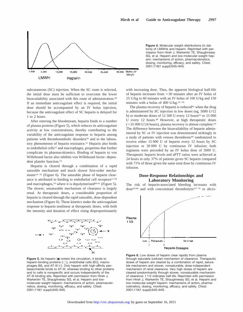
After entering the bloodstream, heparin binds to a numberof plasma proteins (Figure 5), which reduces its anticoagulantactivity at low concentrations, thereby contributing to thevariability of the anticoagulant response to heparin amongpatients with thromboembolic disorders50 and to the labora-tory phenomenon of heparin resistance.51 Heparin also bindsto endothelial cells52 and macrophages, properties that furthercomplicate its pharmacokinetics. Binding of heparin to vonWillebrand factor also inhibits von Willebrand factor–depen-dent platelet function.53
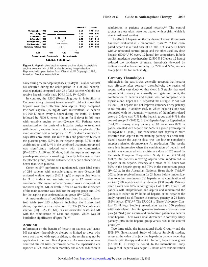
Heparin is cleared through a combination of a rapidsaturable mechanism and much slower first-order mecha-nisms54–56 (Figure 6). The saturable phase of heparin clear-ance is attributed to binding to endothelial cell receptors57,58
and macrophages,59 where it is depolymerized60,61(Figure 5).The slower, unsaturable mechanism of clearance is largelyrenal. At therapeutic doses, a considerable proportion ofheparin is cleared through the rapid saturable, dose-dependentmechanism (Figure 6). These kinetics make the anticoagulantresponse to heparin nonlinear at therapeutic doses, with boththe intensity and duration of effect rising disproportionately
with increasing dose. Thus, the apparent biological half-lifeof heparin increases from'30 minutes after an IV bolus of25 U/kg to 60 minutes with an IV bolus of 100 U/kg and 150minutes with a bolus of 400 U/kg.54–56
The plasma recovery of heparin is reduced62 when the drugis administered by SC injection in low doses (eg, 5000 U/12h) or moderate doses of 12 500 U every 12 hours63 or 15 000U every 12 hours.49 However, at high therapeutic doses(.35 000 U/24 hours), plasma recovery is almost complete.64
The difference between the bioavailability of heparin admin-istered by SC or IV injection was demonstrated strikingly ina study of patients with venous thrombosis49 randomized toreceive either 15 000 U of heparin every 12 hours by SCinjection or 30 000 U by continuous IV infusion; bothregimens were preceded by an IV bolus dose of 5000 U.Therapeutic heparin levels and aPTT ratios were achieved at24 hours in only 37% of patients given SC heparin comparedwith 71% of those given the same total dose by continuous IVinfusion.
Dose-Response Relationships andLaboratory Monitoring
The risk of heparin-associated bleeding increases withdose65,66 and with concomitant thrombolytic67–70 or abcix-
Figure 6. Low doses of heparin clear rapidly from plasmathrough saturable (cellular) mechanism of clearance. Therapeuticdoses of heparin are cleared by a combination of rapid, satura-ble mechanism and slower, nonsaturable, dose-independentmechanism of renal clearance. Very high doses of heparin arecleared predominantly through slower, nonsaturable mechanismof clearance. t 1/2 indicates half-life. Reprinted with permissionfrom Hirsh J, Warkentin TE, Shaughnessy SG, et al. Heparin andlow-molecular-weight heparin: mechanisms of action, pharma-cokinetics, dosing, monitoring, efficacy, and safety. Chest.2001;119(1 suppl):64S–94S.
Figure 4. Molecular weight distributions (in dal-tons) of LMWHs and heparin. Reprinted with per-mission from Hirsh J, Warkentin TE, ShaughnessySG, et al. Heparin and low-molecular-weight hep-arin: mechanisms of action, pharmacokinetics,dosing, monitoring, efficacy, and safety. Chest.2001;119(1 suppl):64S–94S.
Figure 5. As heparin (v) enters the circulation, it binds toheparin-binding proteins ( ), endothelial cells (EC), macro-phages (M), and AT-III (C). Only heparin with high-affinity pen-tasaccharide binds to AT-III, whereas binding to other proteinsand to cells is nonspecific and occurs independently of theAT-III binding site. Reprinted with permission from Hirsh J,Warkentin TE, Shaughnessy SG, et al. Heparin and low-molecular-weight heparin: mechanisms of action, pharmacoki-netics, dosing, monitoring, efficacy, and safety. Chest.2001;119(1 suppl):64S–94S.
Hirsh et al Guide to Anticoagulant Therapy 2997
by guest on September 16, 2015http://circ.ahajournals.org/Downloaded from

imab71,72 therapy. The risk of bleeding is also increased byrecent surgery, trauma, invasive procedures, or concomitanthemostatic defects.73 Randomized trials show a relationshipbetween the dose of heparin administered and both itsefficacy49,63,74 and its safety.71,72 Because the anticoagulantresponse to heparin varies among patients with thromboem-bolic disorders,75–78 it is standard practice to adjust the doseof heparin and monitor its effect, usually by measurement ofthe aPTT. This test is sensitive to the inhibitory effects ofheparin on thrombin, factor Xa, and factor IXa. Because thereis a relationship between heparin dose and both anticoagulanteffect and antithrombotic efficacy, it follows that there shouldbe a relationship between anticoagulant effect and antithrom-botic efficacy.
In the past, we were secure in the contention that a strongrelationship existed between the ex vivo effect of heparin onthe aPTT and its clinical effectiveness, but several lines ofevidence have challenged the strength of such a relationship.First, the initial findings supporting a tight relationshipbetween the effect of heparin on aPTT and its clinical efficacywere based on retrospective subgroup analysis of cohortstudies and are therefore subject to potential bias49,63,75–79
(Table 3). Second, the results of a randomized trial80 and 2recent meta-analyses of contemporary cohort studies81,82 callinto question the value of the aPTT as a useful predictor ofheparin efficacy in patients with venous thrombosis. Third,no direct relationship between aPTT and efficacy was ob-served in the subgroup analysis of the GUSTO-I study(Global Utilization of Streptokinase and Tissue plasminogenactivator for Occluded coronary arteries) in patients withacute MI who were treated with thrombolytic therapy fol-lowed by heparin.83 Fourth, even if the aPTT results werepredictive of clinical efficacy, the value of this test would belimited by the fact that commercial aPTT reagents varyconsiderably in responsiveness to heparin.84 Although stan-dardization can be achieved by calibration against plasmaheparin concentration (the therapeutic range is 0.2 to 0.4U/mL based on protamine titration or 0.3 to 0.7 U/mL basedon anti-factor Xa chromogenic assay), this is beyond thescope of many clinical laboratories. Heparin monitoring islikely to become less problematic in the future as LMWHreplaces UFH for most indications.85
Despite its limitations for monitoring heparin, aPTT re-mains the most convenient and most frequently used methodfor monitoring the anticoagulant response. aPTT should bemeasured'6 hours after the bolus dose of heparin, and thecontinuous IV dose should be adjusted according to the result.Various heparin dose-adjustment nomograms have been de-veloped86 (Tables 4 and 5), but none are applicable to allaPTT reagents,84 and the therapeutic range must be adapted tothe responsiveness of the reagent used. In addition, thedosage regimen should be modified when heparin is com-bined with thrombolytic therapy87 or platelet GP IIb/IIIaantagonists.72 When heparin is given by SC injection in adose of 35 000 U/24 hours in 2 divided doses,64 the antico-agulant effect is delayed'1 hour, and peak plasma levelsoccur after'3 hours.
Limitations of HeparinThe limitations of heparin are based on its pharmacokinetic,biophysical, and nonanticoagulant biological properties.88 Allare caused by the AT-independent, charge-dependent bindingproperties of heparin to proteins and surfaces. Pharmacoki-netic limitations are caused by AT-independent binding ofheparin to plasma proteins,89 proteins released from plate-lets,90 and possibly endothelial cells, resulting in the variableanticoagulant response to heparin and the phenomenon ofheparin resistance.80 AT-independent binding to macro-phages and endothelial cells also results in a dose-dependentmechanism of heparin clearance.
The biophysical limitations occur because the heparin-ATcomplex is unable to inactivate factor Xa in the prothrombi-nase complex and thrombin bound to fibrin or to subendo-thelial surfaces. The biological limitations of heparin includeosteopenia and heparin-induced thrombocytopenia (HIT).Osteopenia is caused as a result of binding of heparin toosteoblasts,46 which then release factors that activate oste-oclasts, whereas HIT results from heparin binding to platelet
TABLE 3. Relation Between Failure to Reach the TherapeuticRange for aPTT and Thromboembolic Events: SubgroupAnalyses of Prospective Studies
Study Condition Outcome Relative Risk
Hull et al49 DVT Recurrent venousthromboembolism
15.0
Basu et al79 DVT Recurrent venousthromboembolism
10.7
Turpie et al63 Acute MI Left ventricular muralthrombosis
22.2
Kaplan et al76 Acute MI Recurrent MI/angina pectoris 6.0
Camilleri et al75 Acute MI Recurrent MI/angina pectoris 13.3
Relative risk refers to the increase in event rates when patients withsubtherapeutic aPTTs are compared with those whose values were in thetherapeutic range.
TABLE 4. Protocol for Heparin Dose Adjustment
aPTT,* sRepeat Bolus
Dose, U
StopInfusion,
min
Change Infusion Rate(mL/h†) Dose(U per 24 h)
Time ofNextaPTT
,50 5000 0 13 (12880) 6 h
50–59 0 0 13 (12880) 6 h
60–85‡ 0 0 0 (0) Next morning
86–95 0 0 22 (21920) Next morning
96–120 0 30 22 (21920) 6 h
.120 0 60 24 (23840) 6 h
Starting dose of 5000 U IV bolus followed by 32 000 U per 24 hours as acontinuous infusion (40 U/mL). First aPTT performed 6 hours after bolusinjection, dosage adjustments made according to protocol, and aPTT repeatedas indicated in the far right column.
*The normal range for aPTT using the Dade Actin FS reagent is 27 to 35seconds.
†40 U/mL.‡The therapeutic range of 60 to 85 seconds corresponds to a plasma
heparin level of 0.2 to 0.4 U/mL by protamine titration or 0.35 to 0.7 U/mL interms of anti-factor Xa activity. The therapeutic range varies with responsive-ness of the aPTT reagent to heparin.
Adapted from Cruickshank et al.86
2998 Circulation June 19, 2001
by guest on September 16, 2015http://circ.ahajournals.org/Downloaded from

factor 4 (PF4), forming an epitope to which the HIT antibodybinds.91,92The pharmacokinetic and nonanticoagulant biolog-ical limitations of heparin are less evident with LMWH,93
whereas the limited ability of the heparin-AT complex toinactivate fibrin-bound thrombin and factor Xa is overcomeby several new classes of AT-independent thrombin andfactor Xa inhibitors.94
Platelets, fibrin, vascular surfaces, and plasma proteinsmodify the anticoagulant effect of heparin. Platelets limit theanticoagulant effect of heparin by protecting surface factorXa from inhibition by the heparin-AT complex95,96 and bysecreting PF4, a heparin-neutralizing protein.97 Fibrin limitsthe anticoagulant effect of heparin by protecting fibrin-boundthrombin from inhibition by heparin AT.98 Heparin binds tofibrin and bridges between fibrin and the heparin binding siteon thrombin. As a result, heparin increases the affinity ofthrombin for fibrin, and by occupying the heparin binding siteon thrombin, it protects fibrin-bound thrombin from inacti-vation by the heparin-AT complex.99,100Thrombin also bindsto subendothelial matrix proteins, where it is protected frominhibition by heparin.101 These observations explain whyheparin is less effective than the AT-independent thrombinand factor Xa inhibitors94 for preventing thrombosis at sitesof deep arterial injury in experimental animals102,103and mayexplain why hirudin is more effective than heparin in patientswith unstable angina or non–Q-wave MI.104
Clinical Use of HeparinHeparin is effective for the prevention and treatment ofvenous thrombosis and PE, for prevention of mural thrombo-sis after MI, and for treatment of patients with unstable
angina and MI. Although heparin is used to prevent acutethrombosis after coronary thrombolysis, recent reports ques-tion the benefits of heparin in this setting when patients arealso treated with aspirin (see below).
In patients with venous thromboembolism or unstableangina, the dose of heparin is usually adjusted to maintainaPTT at an intensity equivalent to a heparin level of 0.2 to 0.4U/mL as measured by protamine titration or an anti-factor Xalevel of 0.30 to 0.7 U/mL. For many aPTT reagents, this isequivalent to a ratio (patient/control aPTT) of 1.5 to 2.5. Therecommended therapeutic range49,79 is based on evidencefrom animal studies105 and supported by subgroup analysis ofprospective cohort studies involving treatment of deep veinthrombosis (DVT),49 prevention of mural thrombosis afterMI,63 and prevention of recurrent ischemia after coronarythrombolysis.75,76 Recommended heparin regimens for ve-nous and arterial thrombosis are summarized in Table 6.
Treatment of Venous ThromboembolismUse of heparin for the treatment of venous thrombosis and PEis based on results of randomized studies.106,107 The effec-tiveness and safety of heparin administered by continuous IVinfusion have been compared with intermittent IV injection in6 studies108–113 and with high-dose SC heparin in 6 stud-ies.64,114–118It is difficult to determine the optimal route ofheparin administration because different doses were used inthese studies, most of the studies were small and underpow-ered, and different criteria were used to assess efficacy andsafety. Nevertheless, the results indicate that heparin is safeand effective when appropriate doses are given. Thus, in arecent pooled analysis of 11 clinical trials in which'15 000
TABLE 5. Weight-Based Nomogram for Heparin Dosing
Initial dose 80 U/kg bolus, then 18 U z kg21 z h21
aPTT ,35 seconds (,1.23 control) 80 U/kg bolus, then 4 U z kg21 z h21
aPTT 35 to 45 seconds (1.2 to 1.53 control) 40 U/kg bolus, then 2 U z kg21 z h21
aPTT 46 to 70 seconds (1.5 to 2.33 control) No change
aPTT 71 to 90 seconds (2.3 to 33 control) Decrease infusion rate by 2 U z kg21 z h21
aPTT .90 seconds (.33 control) Interrupt infusion 1 hour, then decrease infusion rateby 3 U z kg21 z h21
Adapted from Raschke et al.74
TABLE 6. Clinical Use of Heparin
Condition Recommended Heparin Regimen
Venous thromboembolism
Prophylaxis of DVT and PE 5000 U SC every 8 or 12 hours or adjusted low-dose heparin*
Treatment of DVT 5000 U IV bolus followed by 32 000 U per 24 hours by IV infusion or 35 000 to 40 000 Uper 24 hours SC, adjusted to maintain aPTT* in the therapeutic range
Coronary heart disease
Unstable angina or acute MI without thrombolytic therapy 5000 U IV bolus followed by 32 000 U per 24 hour IV infusion adjusted to maintain aPTTin the therapeutic range
Acute MI after thrombolytic therapy† 5000 U IV bolus followed by 24 000 U per 24 hours adjusted to maintain aPTT in thetherapeutic range
*aPTT varies in responsiveness to heparin.†The role of heparin is unproven.Reprinted with permission from Hirsh J, Warkentin TE, Shaughnessy SG, et al. Heparin and low-molecular-weight heparin: mechanisms of action, pharmacokinetics,
dosing, monitoring, efficacy, and safety. Chest. 2001;119(1 suppl):64S–94S.
Hirsh et al Guide to Anticoagulant Therapy 2999
by guest on September 16, 2015http://circ.ahajournals.org/Downloaded from

patients were treated with either heparin (administered as aninitial bolus of 5000 U followed by 30 000 to 35 000 U/24hours with aPTT monitoring) or SC LMWH,119 the meanincidence of recurrent venous thromboembolism among pa-tients assigned heparin was 5.4%. The rate of major bleedingwas 1.9%, fatal recurrent venous thromboembolism occurredin 0.7%, and bleeding was fatal in 0.2% of heparin-treatedpatients. The initial dose of heparin is particularly criticalwhen heparin is administered by SC injection, because anadequate anticoagulant response is not achieved in the first 24hours unless a high starting dose is used (17 500 U SC).64
Audits of heparin monitoring practices indicate that dosageadjustments are frequently inadequate, and dosing practicescan be improved by use of a simple and effective weight-adjusted dosage regimen.74 There is evidence that a 5-daycourse of heparin is as effective as a 10-day course120,121
(Table 7). The short-course regimen has obvious appeal,reducing hospital stay and the risk of HIT. Although theshorter course of treatment can be recommended for mostpatients with venous thromboembolism, this may not beappropriate in cases of extensive iliofemoral vein thrombosisor major PE, because such patients were underrepresented inthese studies.120,121
Prophylaxis of Venous ThromboembolismHeparin in a fixed low dose of 5000 U SC every 8 or 12 hoursis an effective and safe form of prophylaxis in medical andsurgical patients at risk of venous thromboembolism. Low-dose heparin reduces the risk of venous thrombosis and fatalPE by 60% to 70%.122,123 Among general surgical patients,the incidence of fatal PE was reduced from 0.7% in controlsto 0.2% in one study (P,0.001)120 and from 0.8% to 0.3%(P,0.001) in a larger analysis that included orthopedicsurgical patients.123 There was also a small but statisticallysignificant decrease in mortality from 3.3% to 2.4% withlow-dose heparin prophylaxis (P,0.02).123 The use of low-dose heparin is associated with a small excess incidence ofwound hematoma122–124and a minimal, statistically insignif-icant increase in major bleeding but no increase in fatalbleeding. Low-dose heparin also effectively prevents venousthromboembolism in patients with MI and in those with otherserious medical disorders,125 and it reduced in-hospital mor-tality by 31% (P,0.05) in a study of 1358 general medical
patients aged.40 years.126 Although low-dose heparin isalso effective in reducing DVT after hip surgery,123 theincidence of thrombosis remains substantial (20% to 30%)and can be reduced further with either adjusted low-doseheparin127 or fixed-dose LMWH.93 Moderate-dose warfarin iseffective in patients undergoing major orthopedic surgicalprocedures,128,129but direct comparisons of low-dose heparinand warfarin have not been performed in major orthopedicsurgery.
Coronary Artery DiseaseCoronary thrombosis is important in the pathogenesis ofunstable angina, acute MI, and sudden cardiac death. It is alsoimportant in the pathogenesis of reinfarction and death inpatients with acute MI treated with thrombolytic agents orpercutaneous transluminal coronary angioplasty. In mostpatients, heparin ameliorates the thrombotic manifestations ofacute coronary syndromes, but it is no longer used as the soleantithrombotic drug in these settings. Today, heparin isalways used in combination with aspirin in potentially eligi-ble patient groups with acute myocardial ischemia,130 in thosereceiving thrombolytic therapy for evolving MI, in thosetreated with platelet GP IIb/IIIa antagonists for unstableangina,131,132 and in those undergoing high-risk coronaryangioplasty.71,72,132 When combined with aspirin,130,133
thrombolytic agents, or GP IIb/IIIa antagonists, however,heparin in full doses increases the risk of bleeding, and thedose is usually reduced in these settings.72
Unstable Angina and Non–Q-Wave MIHeparin has been evaluated in a number of randomized,double-blind, placebo-controlled clinical trials for the short-term treatment of unstable angina or non–Q-wave MI.134–137
When given alone to patients with unstable angina, heparin iseffective in preventing acute MI and recurrent angina,135–137
and when used in combination with aspirin, the results of ameta-analysis of 6 small trials suggest that the combinationalso reduces short-term rates of cardiovascular death and MIby '30% over those achieved with aspirin alone.134
Theroux et al135 compared the relative efficacy and safetyof heparin, aspirin, and their combination in 479 patients withunstable angina. Heparin was administered as an initial5000-U IV bolus, followed by IV infusion of 1000 U/h,adjusted to maintain the aPTT at 1.5 to 2.0 times the controlvalue. Treatment was initiated within 24 hours after the onsetof chest pain and continued for'6 days. The incidence of MIduring the acute period was 11.9% in the placebo group andwas reduced to 3.3% in the aspirin groups (P50.012), 0.8%in the heparin group (P,0.0001), and 1.6% in the groupgiven the combination of aspirin and heparin (P50.001). Theincidence of refractory angina (22.9% in the placebo group)was significantly reduced to 8.5% (P50.002) in the heparingroup and 10.7% in the heparin-plus-aspirin group (P50.11)but was 16.5% in the aspirin group. In a second study,138
these investigators compared the efficacy and safety ofheparin and aspirin. This was a continuation of the previousstudy in which the placebo and combination groups werediscontinued and an additional 245 patients were randomizedto receive either continuous IV heparin or oral aspirin twice
TABLE 7. Comparison of Short and Long Courses of Heparinfor Treatment of Proximal Vein Thrombosis
Gallus et al121 (n5266) Hull et al120 (n5199)
Short(4 d)
Long(9.5 d)
Short(5 d)
Long(10 d)
Recurrent VTE, %
During heparin 3.6 4.7 7.7 7.7
During warfarin 3.3 1.6
Total during treatment, % 6.9 6.3
VTE denotes venous thromboembolism.tk;2Reprinted with permission from Hirsh J, Warkentin TE, Shaughnessy SG,
et al. Heparin and low-molecular-weight heparin: mechanisms of action,pharmacokinetics, dosing, monitoring, efficacy, and safety. Chest. 2001;119(1suppl):64S–94S.
3000 Circulation June 19, 2001
by guest on September 16, 2015http://circ.ahajournals.org/Downloaded from
daily during the in-hospital phase ('6 days). Fatal or nonfatalMI occurred during the acute period in 4 of 362 heparin-treated patients compared with 23 of 362 patients who did notreceive heparin (odds ratio [OR] 0.16,P,0.005).
In contrast, the RISC (Research group in InStability inCoronary artery disease) investigators134 did not show thatheparin was more effective than aspirin. They comparedlow-dose aspirin (75 mg/d) with intermittent IV heparin(10 000 U bolus every 6 hours during the initial 24 hoursfollowed by 7500 U every 6 hours for 5 days) in 796 menwith unstable angina or non–Q-wave MI. Patients wererandomized on the basis of a factorial design to treatmentwith heparin, aspirin, heparin plus aspirin, or placebo. Themain outcome was a composite of MI or death evaluated 5days after enrollment. The rate of this end point was 6.0% inthe placebo group, 5.6% in the heparin group, 3.7% in theaspirin group, and 1.4% in the combined treatment group andwas significantly reduced only with the combination(P50.027). At 30 and 90 days, both the aspirin and aspirin-plus-heparin groups showed significantly better results thanthe placebo group, but the outcome with heparin alone was nobetter than with placebo.
Cohen et al139 performed a randomized, open-label studyof 214 patients with unstable angina or non–Q-wave MIassigned to either aspirin (162.5 mg/d) or aspirin plus heparinfor 3 to 4 days and warfarin for up to 12 weeks afterenrollment. The main outcome measure was a composite ofrecurrent angina, MI, or death. After 12 weeks, the incidenceof the main outcome was 28% for the aspirin group and 19%for the aspirin-plus-anticoagulation group (P50.09).
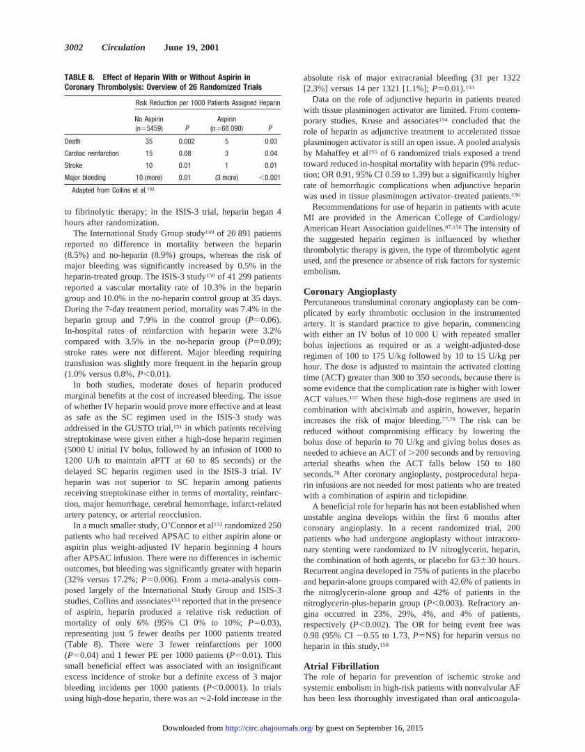
A meta-analysis of published data from 6 small random-ized trials (n51353 subjects), including the 3 describedabove, reported a risk reduction of 33% (95% confidenceinterval [CI] 22% to 56%) in cardiovascular death and MIwith the combination of UFH and aspirin, which was ofborderline significance (Figure 7).130
Acute MIInformation on the benefit of heparin in patients with acuteMI not given thrombolytic therapy is limited to those whowere not treated with aspirin either, so the results may not beapplicable to current clinical practice. An overview of ran-domized clinical trials performed before the reperfusion erareported a 17% reduction in mortality and a 22% reduction in
reinfarction in patients assigned heparin.140 The controlgroups in these trials were not treated with aspirin, which isnow considered routine.
The effect of heparin on the incidence of mural thrombosishas been evaluated in 2 randomized trials.141,142 One com-pared heparin in a fixed dose of 12 500 U SC every 12 hourswith an untreated control group, and the other used low-doseheparin (5000 U SC every 12 hours) for comparison. In bothstudies, moderate-dose heparin (12 500 U SC every 12 hours)reduced the incidence of mural thrombosis detected by2-dimensional echocardiography by 72% and 58%, respec-tively (P,0.05 for each study).
Coronary ThrombolysisAlthough in the past it was generally accepted that heparinwas effective after coronary thrombolysis, the results ofrecent studies cast doubt on this view. In 3 studies that usedangiographic patency as a usually surrogate end point, thecombination of heparin and aspirin was not compared withaspirin alone. Topol et al143 reported that a single IV bolus of10 000 U of heparin did not improve coronary artery patencyat 90 minutes. In another trial, in which heparin alone wascompared with no treatment,144 patency of the infarct-relatedartery at 2 days was 71% in the heparin group and 44% in thecontrol group (P,0.023). In the Heparin-Aspirin ReperfusionTrial,145 coronary artery patency at 18 hours was 82% inpatients treated with heparin and 52% in a group given aspirin80 mg/d (P,0.0002). The conclusion that heparin is moreeffective than aspirin in maintaining patency has been criti-cized because the aspirin dose was too low to completelysuppress platelet thromboxane A2 production. The resultswere less impressive when the combination of heparin andaspirin was compared with aspirin in a dose of 325 mg/d. Inthe sixth European Cooperative Study Group (ECSG-6)trial,77 687 patients receiving aspirin were randomized toheparin or no heparin. Patency at a mean of 81 hours was80% in the heparin group and 75% in the comparison group(P,0.01). In the Australian National Heart Study Trial,146
202 patients received heparin for 24 hours before randomiza-tion to either continuous IV heparin or a combination ofaspirin (300 mg/d) and dipyridamole (300 mg/d). Patencyafter 1 week was 80% in both groups. Col et al147 treated 128patients with streptokinase and aspirin and randomized thepatients to either an IV bolus of heparin or no heparin; thestudy reported no difference in coronary patency at 24 hours(86% versus 87%).147 The DUCCS-1 (Duke University Clin-ical Cardiology Studies) investigators treated 250 patientswith anisoylated plasminogen–streptokinase activator com-plex (APSAC) and aspirin and randomized patients to heparinor no heparin. There was a small difference in coronary arterypatency (80% in the heparin group versus 74% in the controlgroup).148
Two large trials, the International Study Group149 and theISIS-3150 (International Study of Infarct Survival) studies,assessed the value of adjunctive heparin in patients receivingthrombolytic therapy and aspirin. In both, heparin was given(12 500 U SC every 12 hours). In the International StudyGroup trial, heparin was begun 12 hours after randomization
Figure 7. Heparin plus aspirin versus aspirin alone in unstableangina: relative risk of MI or death during hospitalization.Reprinted with permission from Oler et al.130 Copyright 1996,American Medical Association.
Hirsh et al Guide to Anticoagulant Therapy 3001
by guest on September 16, 2015http://circ.ahajournals.org/Downloaded from
to fibrinolytic therapy; in the ISIS-3 trial, heparin began 4hours after randomization.
The International Study Group study149 of 20 891 patientsreported no difference in mortality between the heparin(8.5%) and no-heparin (8.9%) groups, whereas the risk ofmajor bleeding was significantly increased by 0.5% in theheparin-treated group. The ISIS-3 study150 of 41 299 patientsreported a vascular mortality rate of 10.3% in the heparingroup and 10.0% in the no-heparin control group at 35 days.During the 7-day treatment period, mortality was 7.4% in theheparin group and 7.9% in the control group (P50.06).In-hospital rates of reinfarction with heparin were 3.2%compared with 3.5% in the no-heparin group (P50.09);stroke rates were not different. Major bleeding requiringtransfusion was slightly more frequent in the heparin group(1.0% versus 0.8%,P,0.01).
In both studies, moderate doses of heparin producedmarginal benefits at the cost of increased bleeding. The issueof whether IV heparin would prove more effective and at leastas safe as the SC regimen used in the ISIS-3 study wasaddressed in the GUSTO trial,151 in which patients receivingstreptokinase were given either a high-dose heparin regimen(5000 U initial IV bolus, followed by an infusion of 1000 to1200 U/h to maintain aPTT at 60 to 85 seconds) or thedelayed SC heparin regimen used in the ISIS-3 trial. IVheparin was not superior to SC heparin among patientsreceiving streptokinase either in terms of mortality, reinfarc-tion, major hemorrhage, cerebral hemorrhage, infarct-relatedartery patency, or arterial reocclusion.
In a much smaller study, O’Connor et al152 randomized 250patients who had received APSAC to either aspirin alone oraspirin plus weight-adjusted IV heparin beginning 4 hoursafter APSAC infusion. There were no differences in ischemicoutcomes, but bleeding was significantly greater with heparin(32% versus 17.2%;P50.006). From a meta-analysis com-posed largely of the International Study Group and ISIS-3studies, Collins and associates133 reported that in the presenceof aspirin, heparin produced a relative risk reduction ofmortality of only 6% (95% CI 0% to 10%;P50.03),representing just 5 fewer deaths per 1000 patients treated(Table 8). There were 3 fewer reinfarctions per 1000(P50.04) and 1 fewer PE per 1000 patients (P50.01). Thissmall beneficial effect was associated with an insignificantexcess incidence of stroke but a definite excess of 3 majorbleeding incidents per 1000 patients (P,0.0001). In trialsusing high-dose heparin, there was an'2-fold increase in the
absolute risk of major extracranial bleeding (31 per 1322[2.3%] versus 14 per 1321 [1.1%];P50.01).153
Data on the role of adjunctive heparin in patients treatedwith tissue plasminogen activator are limited. From contem-porary studies, Kruse and associates154 concluded that therole of heparin as adjunctive treatment to accelerated tissueplasminogen activator is still an open issue. A pooled analysisby Mahaffey et al155 of 6 randomized trials exposed a trendtoward reduced in-hospital mortality with heparin (9% reduc-tion; OR 0.91, 95% CI 0.59 to 1.39) but a significantly higherrate of hemorrhagic complications when adjunctive heparinwas used in tissue plasminogen activator–treated patients.156
Recommendations for use of heparin in patients with acuteMI are provided in the American College of Cardiology/American Heart Association guidelines.87,156The intensity ofthe suggested heparin regimen is influenced by whetherthrombolytic therapy is given, the type of thrombolytic agentused, and the presence or absence of risk factors for systemicembolism.
Coronary AngioplastyPercutaneous transluminal coronary angioplasty can be com-plicated by early thrombotic occlusion in the instrumentedartery. It is standard practice to give heparin, commencingwith either an IV bolus of 10 000 U with repeated smallerbolus injections as required or as a weight-adjusted-doseregimen of 100 to 175 U/kg followed by 10 to 15 U/kg perhour. The dose is adjusted to maintain the activated clottingtime (ACT) greater than 300 to 350 seconds, because there issome evidence that the complication rate is higher with lowerACT values.157 When these high-dose regimens are used incombination with abciximab and aspirin, however, heparinincreases the risk of major bleeding.77,78 The risk can bereduced without compromising efficacy by lowering thebolus dose of heparin to 70 U/kg and giving bolus doses asneeded to achieve an ACT of.200 seconds and by removingarterial sheaths when the ACT falls below 150 to 180seconds.78 After coronary angioplasty, postprocedural hepa-rin infusions are not needed for most patients who are treatedwith a combination of aspirin and ticlopidine.
A beneficial role for heparin has not been established whenunstable angina develops within the first 6 months aftercoronary angioplasty. In a recent randomized trial, 200patients who had undergone angioplasty without intracoro-nary stenting were randomized to IV nitroglycerin, heparin,the combination of both agents, or placebo for 63630 hours.Recurrent angina developed in 75% of patients in the placeboand heparin-alone groups compared with 42.6% of patients inthe nitroglycerin-alone group and 42% of patients in thenitroglycerin-plus-heparin group (P,0.003). Refractory an-gina occurred in 23%, 29%, 4%, and 4% of patients,respectively (P,0.002). The OR for being event free was0.98 (95% CI20.55 to 1.73,P5NS) for heparin versus noheparin in this study.158
Atrial FibrillationThe role of heparin for prevention of ischemic stroke andsystemic embolism in high-risk patients with nonvalvular AFhas been less thoroughly investigated than oral anticoagula-
TABLE 8. Effect of Heparin With or Without Aspirin inCoronary Thrombolysis: Overview of 26 Randomized Trials
Risk Reduction per 1000 Patients Assigned Heparin
No Aspirin(n55459) P
Aspirin(n568 090) P
Death 35 0.002 5 0.03
Cardiac reinfarction 15 0.08 3 0.04
Stroke 10 0.01 1 0.01
Major bleeding 10 (more) 0.01 (3 more) ,0.001
Adapted from Collins et al.153
3002 Circulation June 19, 2001
by guest on September 16, 2015http://circ.ahajournals.org/Downloaded from

tion with warfarin. It is likely that heparin represents aneffective alternative to warfarin for antithrombotic prophy-laxis, because both anticoagulants decrease hemostatic acti-vation associated with atrial stasis in patients with this cardiacrhythm disturbance.159 Heparin is sometimes given as analternative to oral anticoagulation perioperatively in patientswith chronic AF who are undergoing elective surgery, but noconsensus has emerged regarding when and how to substituteheparin in this situation.160
Patients with AF who have sustained recent cerebralischemic events are among those at highest risk of thrombo-embolism ('12% per year). Oral anticoagulation reduces therisk by two thirds, similar to the benefit achieved in primaryprevention. When oral anticoagulation is contraindicated,aspirin is a much less effective alternative.161 How rapidlyand intensively to initiate anticoagulation after a cerebralischemic event is controversial, however, considering thathemorrhagic transformation might worsen the neurologicaldeficit.162,163
In a study of 231 patients with nonvalvular AF and acutestroke, heparin was administered IV or SC in doses adjustedto an aPTT 1.5 to 2.0 times control values.164Delay before theinitiation of heparin therapy was,6 hours from the onset ofsymptoms in 74 patients and 6 to 48 hours in 157 patients.In-hospital mortality was 9%, hemorrhagic worsening oc-curred in 3% of patients, and stroke recurred early in 2% ofpatients. Neurological recovery was associated with ageyounger than 70 years (OR 0.2), normal baseline computedtomography (CT)-scan findings (OR 8.9), and early heparintreatment (OR 1.7, 95% CI 1.1 to 2.5), even though targetedaPTT ratios were achieved at 24 hours in fewer than 50% ofpatients. Stroke recurrence was associated with lower meanaPTT ratios, but higher ratios were observed in patients withsymptomatic bleeding, especially on the day bleeding oc-curred. Neither age, initial stroke severity, blood pressure, orbaseline CT findings predicted hemorrhagic worsening.Functional recovery was improved sooner when heparin wasadministered early, but close monitoring of aPTT was neces-sary to lessen the risk of hemorrhagic complications.
Hemorrhagic transformation after acute ischemic stroke iscompounded by thrombolytic therapy, but the impact ofheparin can only be inferred. The Multicenter Acute StrokeTrial-Europe (MAST-E) study165 evaluated the safety andefficacy of streptokinase administered IV within 6 hours ofstroke onset. Among 310 patients, 159 (51%) had evidence ofhemorrhagic transformation on CT scan, but only 23% ofthese were symptomatic. The relative risk of hemorrhagictransformation after streptokinase in this trial was in the samerange as in other trials of thrombolytic therapy for acutestroke. Multivariate secondary analysis found that patientswith symptomatic hemorrhagic transformation were morelikely to have AF and less likely to have received heparintreatment.165
To minimize the risk of thromboembolism after electricalcardioversion of AF or flutter, therapeutic anticoagulationshould be established for at least 3 weeks before and for 4weeks after cardioversion when the dysrhythmia has persistedlonger than 2 days or when the duration is unknown. Warfarinis usually used during the outpatient phase.166,167 A more
recent approach uses transesophageal echocardiography todemonstrate the absence of thrombi in the left atrium and leftatrial appendage. If no thrombus is evident, heparin antico-agulation may be initiated before pharmacological or electri-cal cardioversion, followed by warfarin therapy for 1 monthafter cardioversion. This treatment algorithm has a safetyprofile similar to that of conventional therapy and minimizesboth the period of anticoagulation and the duration of AFbefore cardioversion, but no outcome superiority has beenestablished.168
A similar rationale underlies the use of heparin in conjunc-tion with radiofrequency catheter ablation of cardiactachyarrhythmias. A review of the literature over the last 10years found an overall incidence of reported thromboemboliccomplications of 0.6% associated with radiofrequency cath-eter ablation. The risk is increased (to 1.8% to 2%) whenablation is performed in the left heart, but this increase is lessthan when the indication is ventricular tachycardia (2.8%).169
For the ablation of AF, creation of extensive left atrial lesionshas been associated with a high rate of thromboembolicstroke, despite administration of IV heparin and modulatedelectromagnetic energy. Adjuvant platelet inhibitor therapy toreduce the risk of thromboembolism in this specializedsituation is under investigation.170
Heparin-Induced ThrombocytopeniaHIT is an antibody-mediated adverse reaction to heparin thatcan result in venous or arterial thrombosis. Diagnosis of HITis based on both clinical and serological features.171,172
Manifestations of the HIT syndrome include an otherwiseunexplained fall in platelet count$50%, even if the nadirremains above 1503109/L, or skin lesions at heparin injectionsites173,174accompanied by HIT antibody formation. The fallin platelet count almost always occurs between day 5 and day15 after introduction of heparin but can develop earlier inpatients exposed to heparin during the previous 3 months.The frequency of HIT varies in different clinical set-tings175,176such that the risk of venous thrombosis from HITis higher in high-risk surgical patients175 than in medicalpatients.176
The HIT antigen is a multimolecular complex between PF4and heparin.91,92,177–179HIT antibodies bind to regions of thePF4 molecule that have been conformationally modified byits interaction with heparin. The increased propensity tothrombosis in HIT is probably mediated by thrombin gener-ated as a result of in vivo platelet activation,180,181 as aconsequence of interaction between heparin/PF4/IgG im-mune complexes with Fc receptors on platelets.182 A mini-mum of 12 to 14 saccharides are required to form theantigenic complex with PF4,178,179so heparin molecules witha molecular weight greater than'4000 Da have the potentialto cause HIT, and HIT occurs less commonly with LMWHthan with UFH.183,184
DiagnosisTwo main classes of laboratory assays have been developedto detect HIT antibodies,185,186activation assays and antigenassays. The use of washed platelets rather than platelet-richplasma derived from normal donors increases the reliability
Hirsh et al Guide to Anticoagulant Therapy 3003
by guest on September 16, 2015http://circ.ahajournals.org/Downloaded from

of activation assays. Of the various activation assays avail-able, those that use washed platelets and platelet serotoninrelease187 or heparin-induced platelet activation188,189 aremost accurate.173 Antigen assays, now commercially avail-able, that are based on detecting antibodies against PF4 boundto heparin188 or polyvinylsulfonate190 respond to clinicallyinsignificant antibodies more often than do activationassays.175
TreatmentIf HIT is suspected on clinical grounds and the patient eitherhas thrombosis or is at risk of developing thrombosis, heparinshould be stopped and replaced with lepirudin (Refludan).Although the diagnosis should be confirmed as soon aspractical, treatment should not be delayed. Warfarin shouldnot be used alone, because a recent report suggests this canaggravate the thrombotic process. Lepirudin is a hirudinderivative that does not exhibit cross-reactivity and is man-ufactured by recombinant technology.191 Its use in HIT hasbeen approved by the Food and Drug Administration on thebasis of 2 prospective cohort studies192,193 that comparedtreatment of HIT-associated thrombosis with lepirudin versushistorical controls. An IV infusion is used for rapid therapeu-tic anticoagulation, beginning with a bolus loading dose of0.4 mg/kg IV followed by a maintenance dose of 15 mg · kg21
· h21, with adjustments to maintain aPTT at 1.5 to 2.5 timesthe median of the normal laboratory range.
In the absence of overt thrombosis, cessation of heparin haslong been the cornerstone of management of HIT, but severalstudies have shown that simply stopping heparin may beinadequate because of the high risk of overt thrombosis in theweek after interruption of heparin.192–197 Treatment withhirudin should therefore be considered in all patients withHIT who remain at risk of thrombosis, including postopera-tive patients and those with sepsis. Recombinant hirudin(lepirudin) should be used until the platelet count has recov-ered (Table 9). This should also be considered for patientswith acute HIT without thrombosis (isolated HIT), becausethere is a high risk for subsequent clinically evident throm-bosis in these patients. Warfarin should not be used alone totreat acute HIT complicated by DVT because of the risk ofvenous limb gangrene. When given to patients adequatelyanticoagulated with lepirudin, warfarin appears safe in acuteHIT, but it is prudent to delay starting warfarin until theplatelet count has risen above 1003109/L.
Low-Molecular-Weight HeparinsHistorical PerspectiveThe development of LMWHs for clinical use was stimulated by3 main observations. These are that compared with UFH,LMWH has reduced anti-factor IIa activity relative to anti-factor
Xa activity,26,198 more favorable benefit-risk ratios199–204 inexperimental animals, and superior pharmacokinetic proper-ties.205–210Of these potential advantages, only the pharmacoki-netic properties are of clear clinical importance.93,211
LMWH fractions prepared from standard commercial-grade heparin have progressively less effect on the aPTT asthey are reduced in molecular size, while still inhibitingactivated factor X (factor Xa).26,198 The aPTT activity ofheparin reflects mainly its anti-factor IIa activity. The disas-sociation of anti-factor Xa activity from AT (IIa) activity(expressed as an aPTT measurement), described in 1976,26
challenged the prevailing biophysical model for the antico-agulant effect of heparin, which predicted that any heparinmolecule, irrespective of chain length, would catalyze theinactivation of serine protease coagulation enzymes equallyprovided it contained the high-affinity binding site for AT.The explanation for the difference in anticoagulant profilebetween LMWH and heparin was elucidated in subsequentstudies (Table 10).29,30,212–216
Bleeding in Experimental AnimalsEarly evidence that LMWH produces less microvascularbleeding than heparin in experimental models199–204has notbeen borne out by recent large randomized trials in theprevention and treatment of venous thrombosis, treatment ofPE, or treatment of unstable angina. In these studies, LMWHand heparin have shown similar low rates of bleeding (seebelow).
Pharmacokinetic PropertiesIn the 1980s, a number of investigators205–210 reported thatLMWH preparations had a longer plasma half-life and betterbioavailability at low doses than heparin, as well as a morepredictable dose response.217 These findings provided therationale for comparing unmonitored weight-adjustedLMWH with aPTT-monitored heparin in patients with estab-lished DVT and in patients with unstable angina.
Structure and PharmacologyLMWHs are derived from heparin by chemical or enzymaticdepolymerization to yield fragments approximately one thirdthe size of heparin. The various LMWHs approved for use inEurope, Canada, and the United States are shown in Table 11.Because they are prepared by different methods of depoly-merization, they differ to some extent in pharmacokineticproperties and anticoagulant profile and are not clinically
TABLE 9. Treatment Protocol for Lepirudin (RecombinantHirudin) in HIT
For rapid therapeutic anticoagulation (IV infusion):
Loading dose 0.4 mg/kg bolus IV
Maintenance 0.15 mg z kg21 z h21 IV, with adjustments tomaintain aPTT 1.5–2.5 times median normal range
TABLE 10. Relationship Between Molecular Weight of HeparinFractions and Anticoagulant Activity
HeparinOligosaccharides
MolecularWeight,
Da
AnticoagulantActivity
(Anti-Xa)
AnticoagulantActivity(Anti-IIa)
8 2400 1.30 Nil
12 3600 2.58 Nil
16 4800 1.60 Nil
18 5400 0.95 0.51
24 7200 1.30 1.21
Data from Lane et al.29
3004 Circulation June 19, 2001
by guest on September 16, 2015http://circ.ahajournals.org/Downloaded from

interchangeable. LMWHs have a mean molecular weight of4500 to 5000 Da with a distribution of 1000 to 10 000 Da.
Depolymerization of heparin yields low-molecular-weightfragments with reduced binding to proteins or cells (Table12). Indeed, all of the anticoagulant, pharmacokinetic, andother biological differences between UFH and LMWH can beexplained by the relatively lower binding properties ofLMWH. Thus, compared with UFH, LMWHs have reducedability to inactivate thrombin because the smaller fragmentscannot bind simultaneously to AT and thrombin. On the otherhand, because bridging between AT and factor Xa is less criticalfor anti-factor Xa activity, the smaller fragments inactivatefactor Xa almost as well as larger molecules.35,218–220Re-duced binding to plasma proteins is responsible for the morepredictable dose-response relationship of LMWHs.89 Lessbinding to macrophages and endothelial cells increases theplasma half-life211 of LMWH compared with UFH, whereasreduced binding to platelets and PF4 may explain the lowerincidence of HIT.40,90,183Finally, reduced binding of LMWHto osteoblasts results in less activation of osteoclasts and lessbone loss.46,47 LMWHs are cleared principally by the renalroute, and their biological half-life is prolonged in patientswith renal failure.221–223
Anticoagulant EffectsLike UFH, LMWHs produce their major anticoagulant effectby activating AT. The interaction with AT is mediated by aunique pentasaccharide sequence21,224 found on fewer thanone third of LMWH molecules. Because a minimum chainlength of 18 saccharides (including the pentasaccharide se-quence) is required for the formation of ternary complexesbetween heparin chains, AT, and thrombin, only the 25% to50% of LMWH species that are above this critical chain
length are able to inactivate thrombin. In contrast, all LMWHchains containing the high-affinity pentasaccharide catalyzethe inactivation of factor Xa (Figure 3). Because virtually allheparin molecules contain at least 18 saccharide units,213,214
heparin has an anti-factor Xa to anti-factor IIa ratio of 1:1. Incontrast, commercial LMWHs have anti-factor Xa to anti-IIaratios between 2:1 and 4:1, depending on their molecular sizedistribution.
LMWHs have been evaluated in a large number of ran-domized clinical trials and have been found to be safe andeffective for prevention and treatment of venous thrombosis.More recently, LMWH preparations have also been evaluatedin patients with acute PE and those with unstable angina.
Prevention of Venous ThrombosisLMWHs were first evaluated for the prevention of venousthrombosis in high-risk surgical patients in the mid-1980s,and there is now extensive experience with their use for thisindication. In patients undergoing general surgery and inhigh-risk medical patients, low doses of LMWH administeredSC once daily are at least as effective and safe as low-doseUFH administered SC 2 or 3 times daily. LMWH has becomethe anticoagulant of choice for the prevention of venousthrombosis during major orthopedic surgery and inanticoagulant-eligible patients after major trauma. The risk ofbleeding with LMWH is small and comparable to that withlow-dose heparin.
General SurgeryLMWHs were effective and safe in 2 well-designed random-ized trials. One trial225 in 4498 patients showed a statisticallysignificant reduction in thromboembolic mortality in favor ofLMWH (0.07%) compared with a UFH control group
TABLE 11. Commercial LMWH and a Heparinoid: Methods of Preparation
Agent Manufacturer Method of Preparation
Nadroparin calcium (Fraxiparin) Sanofi Nitrous acid depolymerization
Enoxaparin sodium (Lovenox/Clexane) Rhone-Poulenc Rorer Benzylation followed by alkaline depolymerization
Dalteparin (Fragmin) Kabi Nitrous acid depolymerization
Ardeparin (Normiflo) Wyeth-Ayerst Peroxidative depolymerization
Tinzaparin (Innohep) Leo Laboratories Enzymatic depolymerization with heparinase
Reviparin (Clivarine) Knoll Nitrous acid depolymerization
Danaparoid sodium (Orgaran) NV Organon Prepared from animal gut mucosa; contains heparin sulfate (84%),dermatan sulfate (12%), and chondroitin sulfate (4%)
Reprinted with permission from Hirsh J, Warkentin TE, Shaughnessy SG, et al. Heparin and low-molecular-weight heparin:mechanisms of action, pharmacokinetics, dosing, monitoring, efficacy, and safety. Chest. 2001;119(1 suppl):64S–94S.
TABLE 12. Biological Consequences of Reduced Binding of LMWH to Proteins and Cells
Binding Target Biological Effects Clinical Consequences
Thrombin Reduced anti-IIa to anti-Xa ratio Unknown
Proteins More predictable anticoagulant response Monitoring of anticoagulant effect unnecessary
Macrophages Cleared through renal mechanism Longer plasma half-life. Once-daily SC treatment effective
Platelets Reduced incidence of heparin-dependent antibody Reduced incidence of HIT
Osteoblasts Reduced activation of osteoclasts Lower incidence of osteopenia
Reprinted with permission from Hirsh J, Warkentin TE, Shaughnessy SG, et al. Heparin and low-molecular-weight heparin:mechanisms of action, pharmacokinetics, dosing, monitoring, efficacy, and safety. Chest. 2001;119(1 suppl):64S–94S.
Hirsh et al Guide to Anticoagulant Therapy 3005
by guest on September 16, 2015http://circ.ahajournals.org/Downloaded from

(0.36%). A meta-analysis226 of randomized trials comparinglow-dose heparin with LMWH concluded that there wereminimal differences between the 2 forms of prophylaxis.
Orthopedic SurgeryCompared with placebo, LMWH produced a risk reductionfor all thrombi and for proximal vein thrombi between 70%and 79%. This reduction occurred without an increase inmajor bleeding in 2 studies227,228and with a small increase inminor bleeding in a third,229 but all were too small to excludea modest increase in major bleeding with LMWH. LMWHhas been compared with a variety of other methods ofprophylaxis, including low-dose heparin,230–232adjusted-doseheparin,233,234dextran,235,236and warfarin.237 In most studiesperformed in North America, the LMWH was started 12 to 24hours postoperatively, increasing the acceptance of prophy-laxis among orthopedic surgeons and anesthesiologists con-cerned about the risk of spinal cord hematoma with prophy-lactic LMWH in patients undergoing spinal anesthesia. Insuch cases, the first dose of LMWH should be delayed untilafter the epidural catheter has been removed; when this is notfeasible, the catheter should be removed at least 8 hours afterthe last dose of LMWH. In addition, other drugs that impairhemostasis (such as nonsteroidal anti-inflammatory agents)should be avoided.
Anderson et al238performed a meta-analysis of randomizedstudies comparing LMWH with either fixed low-dose oradjusted-dose heparin. The observed incidence of venousthrombosis was 15.9% in the LMWH group and 21.7% in theheparin group (P50.01), and there was a significant reduc-tion in the incidence of proximal venous thrombosis in theLMWH group (5.4% versus 12.5%;P,0.0001). There wasno difference in the incidence of bleeding between the 2groups (Table 13). These results are comparable to those ofan earlier meta-analysis.226
Two studies compared LMWH and low-dose heparin forprevention of venous thrombosis after elective total knee
arthroplasty. In one, LMWH was more effective than hepa-rin239; the incidence of venous thrombosis was 24.6% in theLMWH group and 34.2% in the heparin group (P50.02). Inthe other,240 the incidence of venous thrombosis was 23% inthe LMWH group and 27% in the heparin group. There wasno difference in the incidence of bleeding in either study.
LMWH preparations have been compared with warfarinand other oral anticoagulants in 6 studies involving high-riskorthopedic surgical patients.241–246The LMWH preparationstested showed efficacy equal to warfarin in patients undergo-ing elective hip replacement, but LMWHs appeared moreeffective than oral anticoagulants in patients undergoingmajor knee surgery (Table 14). In a number of these studies,LMWH was associated with a small but significant increasein major bleeding.
Hip FractureTwo randomized trials have been performed with danaparoidsodium in patients with hip fracture. In one,204 the incidenceof thrombosis was 13% in patients given danaparoid sodium(Orgaran) and 35% in patients given dextran (P,0.001).Blood transfusion requirements were significantly higher inthe dextran group. In the other,247 venous thrombosis oc-curred in 27.8% of the patients treated with danaparoidsodium and in 44.8% of patients in the aspirin group(P50.028). There was no difference in bleeding between the2 groups.
Multiple TraumaGeerts and colleagues248 compared LMWH (enoxaparin so-dium [Lovenox/Clexane]; 30 mg SC every 12 hours) withlow-dose heparin (5000 U SC every 12 hours), started within36 hours of injury in victims of multiple trauma. Theincidence of venous thrombosis was 44% in the 136 patientsallocated to the low-dose heparin group and 31% among 129patients in the LMWH group (P50.014). The incidence ofproximal venous thrombosis was 15% in the low-dose hepa-
TABLE 13. Meta-Analysis of Randomized Studies Comparing LMWH With UFH inPatients Undergoing Elective Hip Surgery
FactorProximal Venous
ThrombosisTotal VenousThrombosis
MajorBleeding
MinorBleeding PE
Common OR* 0.40 0.70 0.64 0.90 0.30
95% CI 0.28–0.59 0.53–0.92 0.34–1.23 0.61–1.33 0.09–1.02
*A common OR ,1.0 favors LMWH over heparin.Data from Anderson et al.238
TABLE 14. Controlled Trials With LMWH in Patients Undergoing Elective Total Knee Arthroplasty
Author LMWH DVTProximal
DVTMajor
Hemorrhage Control DVTProximal
DVTMajor
Hemorrhage
Hull et al244 Tinzaparin (Innohep) 116/258 (45%) 20/258 (18%) 9/317 (3%) Warfarin 152/277 (55%) 34/277 (12%) 3/324 (1%)
RD Heparin Group241 Ardeparin (Normiflo) 41/149 (28%) 7/149 (5%) N/A Warfarin 60/147 (41%) 15/147 (10%) NA
Leclerc et al245 Enoxaparin sodium (Lovenox/Clexane) 8/41 (19%) 0/41 (0%) 0/66 (0%) Placebo 35/54 (65%) 11/54 (20%) 1/65 (2%)
Spiro et al242 Enoxaparin sodium (Lovenox/Clexane) 41/108 (38%) 3/108 (3%) 9/173 (5%) Warfarin 72/122 (59%) 16/122 (13%) 4/176 (2%)
Fauno et al240 Enoxaparin sodium (Lovenox/Clexane) 29/92 (23%) 3/92 (3%) NA Heparin 25/93 (27%) 5/93 (5%) NA
Colwell et al239 Enoxaparin sodium (Lovenox/Clexane) 54/145 (37%) 4/145 (2%) 3/28 (1%) Heparin 74/143 (52%) 22/143 (15%) 3/225 (1%)
Adapted from Colwell et al.239
3006 Circulation June 19, 2001
by guest on September 16, 2015http://circ.ahajournals.org/Downloaded from

rin group and 6% in the LMWH group (P50.09).249 Majorbleeding occurred in 0.6% of patients treated with heparinand 2.9% of those treated with LMWH.
Summary of LMWH in Orthopedic Surgeryand TraumaOverall, LMWHs appear effective for prevention of venousthromboembolism, and they appear safe for use in high-riskpatients undergoing major orthopedic surgical procedures.Compared with placebo, the relative risk reduction for allthrombi and for proximal vein thrombi is'70%. LMWHs aremore effective than low-dose heparin, at least as effective aswarfarin, and more effective than dextran or aspirin inpatients undergoing total hip arthroplasty, and they are moreeffective than warfarin, aspirin, or dextran in patients under-going major knee surgery. Similarly, LMWHs are moreeffective than aspirin in patients with hip fracture. The risk ofbleeding with LMWH is comparable to that with low-doseheparin or warfarin.
NeurosurgeryIn a recent study250 comparing LMWH plus compressionstockings with compression stockings alone, those assignedto the LMWH group had a risk reduction of 48%. There wasno difference in major bleeding.
Medical PatientsLMWHs have been compared with placebo in 2 studies ofpatients with ischemic stroke251,252and have been comparedwith low-dose heparin in 2 studies.253,254 Compared withplacebo, LMWHs reduce the risk of venous thrombosisbetween 40% and 86% without an increase in clinicallyimportant bleeding. In studies comparing LMWHs withheparin, patients randomized to receive LMWH showed astatistically significant .70% relative risk reduction inthrombosis.118,259
Treatment of Venous ThromboembolismA number of LMWH preparations have been compared withheparin in patients with venous thrombosis and/or PE inwell-designed studies. The results of studies published before1995 that used 4 different preparations have been summa-rized in a meta-analysis255 in which the data for each of the 4LMWHs were pooled separately (Tables 15 and 16). All 4LMWH preparations were as effective and safe as IV heparin,and the rates of recurrent thromboembolism and majorbleeding were similar with all of the LMWHs. It is notewor-thy that the LMWHs were administered by SC injection inunmonitored, weight-adjusted doses, whereas heparin wasmonitored by the aPTT.
Since publication of the pooled analysis in 1995, 5 additionallarge randomized trials have been completed,256–2602 in patientswith venous thrombosis,257,2581 in patients with venous thrombosisor PE,259 1 in patients with PE,256 and 1 in patients with proximalvein thrombosis who were also randomized to inferior vena cavafilter insertion.260The design of 2 of the studies257,258capitalized onthe more predictable anticoagulant response to LMWH by encour-aging patients assigned to LMWH treatment at home, whereasthose assigned to heparin were treated conventionally in the hospitalwith a continuous IV infusion. The results, shown in Table 17,indicate that out-of-hospital administration of LMWH to eligiblepatients with DVT is as effective and safe as IV heparin adminis-tered in the hospital. Both studies excluded patients with symptom-atic PE or a history of recent previous venous thrombosis. Toaddress this study deficiency, the investigators collaborated in theCOLUMBUS study,259 in which 1021 patients with venous throm-bosis or PE were randomly assigned to treatment with either SCLMWH (riviparin sodium) or adjusted-dose IV UFH for 8 days.Warfarin was started concomitantly and continued for 3 months.The mean hospital stay was 3 days shorter in patients assigned toLMWH, whereas the incidence of recurrent thromboembolism,bleeding, and mortality was similar in both groups.
TABLE 16. Incidence of Major Bleeding Complications During Initial Treatmentand for 48 Hours After Heparin Cessation: UFH vs LMWH
Agent
Patients, n (%)Relative Risk Reduction
(95% CI) PLMWH UFH
Nadroparin (Fraxiparin) 4/446 (0.9) 10/436 (2.3) 59 (216–86) 0.09
Tinzaparin (Logiparin) 1/213 (0.5) 11/219 (5.0) 91 ,0.01
Enoxaparin (Clexane) 5/314 (1.6) 3/320 (0.9) 270 (2580–58) .0.2
Dalteparin (Fragmin) 2/433 (0.5) 5/464 (1.0) 55 (299–90) .0.2
Data from Kuijer et al.255
TABLE 15. LMWH vs Heparin for Treatment of DVT: Symptomatic RecurrentVenous Thromboembolism During Initial Treatment and After 3 to 6 Months
Agent
Patients, n (%)Relative Risk Reduction
(95% CI) PLMWH UFH
Nadroparin (Fraxiparin) 20/361 (5.5) 32/355 (9.0) 40 (25–66) 0.07
Tinzaparin (Logiparin) 6/213 (2.8) 15/219 (6.9) 59 (21–83) 0.07
Enoxaparin (Clexane) 13/314 (4.1) 20/320 (6.3) 35 (232–68) 0.23
Dalteparin (Fragmin) 16/322 (5.0) 8/339 (2.4) 2110 (2374–7) 0.07
Data from Kuijer et al.255
Hirsh et al Guide to Anticoagulant Therapy 3007
by guest on September 16, 2015http://circ.ahajournals.org/Downloaded from
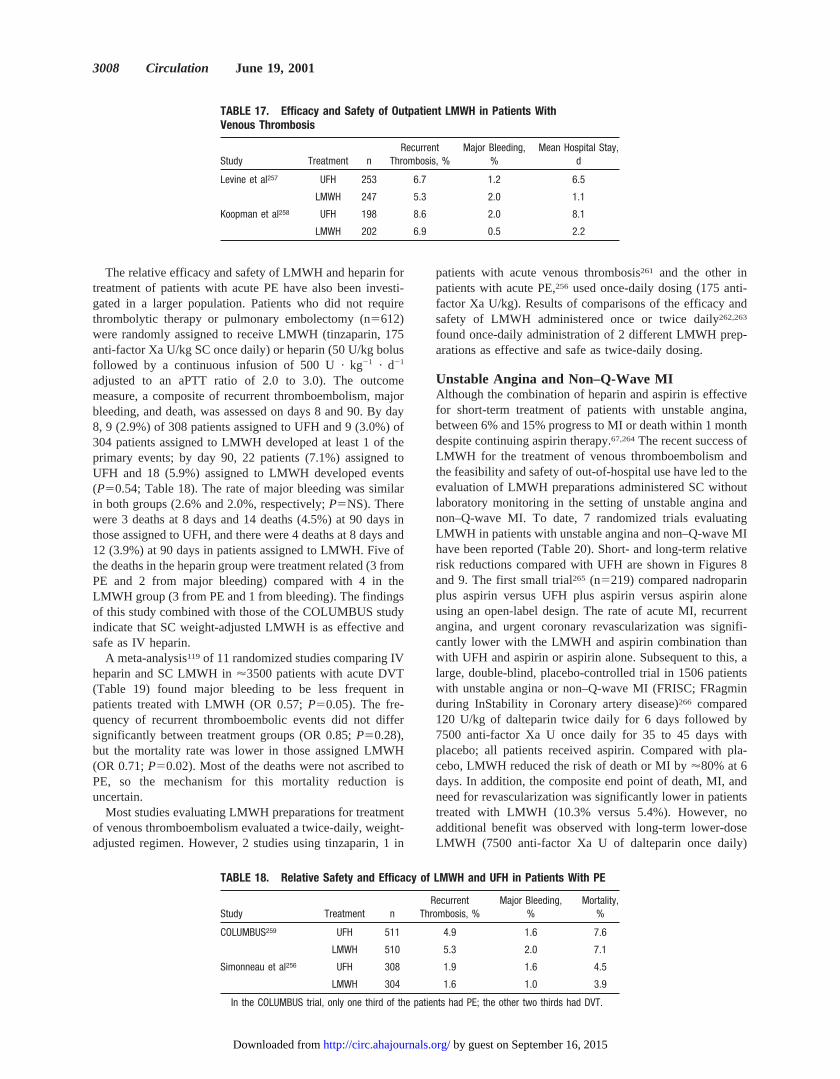
The relative efficacy and safety of LMWH and heparin fortreatment of patients with acute PE have also been investi-gated in a larger population. Patients who did not requirethrombolytic therapy or pulmonary embolectomy (n5612)were randomly assigned to receive LMWH (tinzaparin, 175anti-factor Xa U/kg SC once daily) or heparin (50 U/kg bolusfollowed by a continuous infusion of 500 U · kg21 · d21
adjusted to an aPTT ratio of 2.0 to 3.0). The outcomemeasure, a composite of recurrent thromboembolism, majorbleeding, and death, was assessed on days 8 and 90. By day8, 9 (2.9%) of 308 patients assigned to UFH and 9 (3.0%) of304 patients assigned to LMWH developed at least 1 of theprimary events; by day 90, 22 patients (7.1%) assigned toUFH and 18 (5.9%) assigned to LMWH developed events(P50.54; Table 18). The rate of major bleeding was similarin both groups (2.6% and 2.0%, respectively;P5NS). Therewere 3 deaths at 8 days and 14 deaths (4.5%) at 90 days inthose assigned to UFH, and there were 4 deaths at 8 days and12 (3.9%) at 90 days in patients assigned to LMWH. Five ofthe deaths in the heparin group were treatment related (3 fromPE and 2 from major bleeding) compared with 4 in theLMWH group (3 from PE and 1 from bleeding). The findingsof this study combined with those of the COLUMBUS studyindicate that SC weight-adjusted LMWH is as effective andsafe as IV heparin.
A meta-analysis119 of 11 randomized studies comparing IVheparin and SC LMWH in'3500 patients with acute DVT(Table 19) found major bleeding to be less frequent inpatients treated with LMWH (OR 0.57;P50.05). The fre-quency of recurrent thromboembolic events did not differsignificantly between treatment groups (OR 0.85;P50.28),but the mortality rate was lower in those assigned LMWH(OR 0.71;P50.02). Most of the deaths were not ascribed toPE, so the mechanism for this mortality reduction isuncertain.
Most studies evaluating LMWH preparations for treatmentof venous thromboembolism evaluated a twice-daily, weight-adjusted regimen. However, 2 studies using tinzaparin, 1 in
patients with acute venous thrombosis261 and the other inpatients with acute PE,256 used once-daily dosing (175 anti-factor Xa U/kg). Results of comparisons of the efficacy andsafety of LMWH administered once or twice daily262,263
found once-daily administration of 2 different LMWH prep-arations as effective and safe as twice-daily dosing.
Unstable Angina and Non–Q-Wave MIAlthough the combination of heparin and aspirin is effectivefor short-term treatment of patients with unstable angina,between 6% and 15% progress to MI or death within 1 monthdespite continuing aspirin therapy.67,264The recent success ofLMWH for the treatment of venous thromboembolism andthe feasibility and safety of out-of-hospital use have led to theevaluation of LMWH preparations administered SC withoutlaboratory monitoring in the setting of unstable angina andnon–Q-wave MI. To date, 7 randomized trials evaluatingLMWH in patients with unstable angina and non–Q-wave MIhave been reported (Table 20). Short- and long-term relativerisk reductions compared with UFH are shown in Figures 8and 9. The first small trial265 (n5219) compared nadroparinplus aspirin versus UFH plus aspirin versus aspirin aloneusing an open-label design. The rate of acute MI, recurrentangina, and urgent coronary revascularization was signifi-cantly lower with the LMWH and aspirin combination thanwith UFH and aspirin or aspirin alone. Subsequent to this, alarge, double-blind, placebo-controlled trial in 1506 patientswith unstable angina or non–Q-wave MI (FRISC; FRagminduring InStability in Coronary artery disease)266 compared120 U/kg of dalteparin twice daily for 6 days followed by7500 anti-factor Xa U once daily for 35 to 45 days withplacebo; all patients received aspirin. Compared with pla-cebo, LMWH reduced the risk of death or MI by'80% at 6days. In addition, the composite end point of death, MI, andneed for revascularization was significantly lower in patientstreated with LMWH (10.3% versus 5.4%). However, noadditional benefit was observed with long-term lower-doseLMWH (7500 anti-factor Xa U of dalteparin once daily)
TABLE 17. Efficacy and Safety of Outpatient LMWH in Patients WithVenous Thrombosis
Study Treatment nRecurrent
Thrombosis, %Major Bleeding,
%Mean Hospital Stay,
d
Levine et al257 UFH 253 6.7 1.2 6.5
LMWH 247 5.3 2.0 1.1
Koopman et al258 UFH 198 8.6 2.0 8.1
LMWH 202 6.9 0.5 2.2
TABLE 18. Relative Safety and Efficacy of LMWH and UFH in Patients With PE
Study Treatment nRecurrent
Thrombosis, %Major Bleeding,
%Mortality,
%
COLUMBUS259 UFH 511 4.9 1.6 7.6
LMWH 510 5.3 2.0 7.1
Simonneau et al256 UFH 308 1.9 1.6 4.5
LMWH 304 1.6 1.0 3.9
In the COLUMBUS trial, only one third of the patients had PE; the other two thirds had DVT.
3008 Circulation June 19, 2001
by guest on September 16, 2015http://circ.ahajournals.org/Downloaded from

compared with placebo. After 4 to 5 months of follow-up, therates of death and MI in the placebo and dalteparin groupswas 15.3% and 14.0%, respectively (P50.41), and the ratesof death, MI, or revascularization were 43.6% and 42.7%,respectively (P50.18). This study established the short-termvalue of LMWH (dalteparin) for treatment of unstable anginaand non–Q-wave MI and added to the data in support of abeneficial effect of heparin and aspirin over aspirin alone inthis patient population. However, no effect of moderate-doseLMWH was observed over the long term.
In a third study (FRIC; FRagmin In unstable Coronaryartery disease),267 which used an open, randomized design,dalteparin (120 anti-factor Xa U/kg twice daily) was com-pared with heparin (5000-U bolus followed by 1000-U/hcontinuous infusion for 6 days) in 1492 patients with unstableangina or non–Q-wave infarction. This was followed in asecond phase by a double-blind study comparing LMWH at adose of 7500 U/d with placebo. All patients received aspirin.Both treatment regimens were equivalent in terms of efficacyand safety. At 6 days, the composite outcome of death, MI, orrecurrent angina occurred in 7.6% of the heparin group and9.3% of the LMWH group, whereas the corresponding ratesof the composite of death or MI were 3.6% and 3.9%,respectively. Between days 6 and 45, the rate of death, MI, or
recurrent angina was 12.3% in both groups. There was nodifference in major bleeding, which was infrequent.
The ESSENCE trial (Efficacy and Safety of SubcutaneousEnoxaparin in Non-Q-wave Coronary Events)268 is 1 of 2studies that compared enoxaparin with heparin. Patients(n53171) with unstable angina or non–Q-wave MI wererandomized in a double-blind fashion to 1 mg/kg SC (100anti-factor Xa U) of enoxaparin every 12 hours or UFH,administered as an IV bolus followed by a continuousinfusion, for 2 to 8 days; the median duration of treatment inboth groups was 2.6 days (Table 20). There was a significant17% risk reduction in the primary end point of death, MI, orrecurrent angina at 14 days with LMWH (P50.019) and a15% risk reduction at 30 days (P50.016). This differencewas accounted for mainly by a lower incidence of recurrentangina in patients assigned to LMWH. There was no differ-ence in the incidence of major bleeding at 30 days (6.5% withLMWH versus 7.0% with heparin), but total bleeding wasmore frequent with the LMWH group (18.4% versus 14.2%),primarily because of bruising at injection sites. At 1 year, thedifference in the composite end point remained significant(P50.022).269
TIMI-11B (Thrombolysis In Myocardial Infarction 11B)270
was a double-blind study comparing enoxaparin (bolus fol-lowed by SC injections every 12 hours for 3 to 8 days) and IVheparin (administered for at least 3 days) in 3910 patientswith unstable angina or non–Q-wave MI. The primary out-come was death, MI, or urgent revascularization at 43 days.Patients assigned to LMWH continued SC treatment at alower dose until day 43, whereas patients assigned initially toheparin received placebo. There was an 18% relative riskreduction in events at 14 days (P50.029) and a 12% riskreduction at 43 days (P50.048). The absolute risk reductionswere 2.4% and 2.3% at 14 and 43 days, respectively. Thedifference in treatment duration between UFH (3 days) andenoxaparin (43 days) and the validity of comparing eventrates at 14 days render these data questionable. In addition,the lack of effectiveness of enoxaparin beyond 14 days issurprising and fails to establish a role for long-term use ofLMWH in this patient population.271
TABLE 19. LMWH vs Heparin for Treatment of DVT:Meta-Analysis
TotalSubjects,
n
SummaryOdds
Reduction*NNT,
n
FrequencyWith
UFH, %
Major bleeding 3674 0.57† 164 1.9
RTE 3566 0.85 114 5.4
Mortality (overall) 3566 0.7† 61 6.8
NNT indicates number of patients needed to treat to prevent 1 event thatwould occur during alternate treatment; RTE, recurrent thromboembolism.
*A summary odds reduction ,1.0 favors LMWH; odds reduction .1.0 favorsUFH.
†P,0.05.Adapted from Gould et al.119
TABLE 20. Trials of LMWH in Patients With Acute Coronary Syndromes Without ST-Segment Elevation on the ECG
Trial n Short-Term Comparison* Long-Term Comparison†
Gurfinkel et al265 219 Nadroparin 214 U SC BID vs placebo N/A
FRISC266 1506 Dalteparin 120 U/kg SC BID vs placebo Dalteparin 7500 U SC QD vs placebo
Klein et al (FRIC)267 1482 Dalteparin 120 U/kg SC BID vs UFH 5000 U B, 1000 U/hIV then 12 500 U SC BID
Dalteparin 7500 U SC QD vs placebo
Cohen et al (ESSENCE)268 3171 Enoxaparin 1 mg/kg SC BID 2–8 d vs UFH 5000 U aPTT55–85 seconds
N/A
Antman et al (TIMI-11B)270 3912 Enoxaparin 3000 U B, 100 U/kg BID vs UFH 70 U B, 15U/kg aPTT 1.5–2.03
Enoxaparin 4500–6000 U SC BID vs placebo
Leizorivicz (FRAXIS)272 3468 Nadroparin 86 U SC BID vs UFH 5000 B 1250 U/h targetlocal aPTT
Nadroparin 86 U SC vs placebo
FRISC-II273 2457 Dalteparin 120 U/kg BID vs placebo Dalteparin 5000 or 7500 U SC BID vsplacebo
B indicates bolus.*In-hospital phase.†One to 3 months.
Hirsh et al Guide to Anticoagulant Therapy 3009
by guest on September 16, 2015http://circ.ahajournals.org/Downloaded from

Nadroparin was also evaluated in the FRAXIS (FRAXi-parine in Ischaemic Syndrome) trial.272 Patients with unstableangina or non–Q-wave infarction were randomly assigned toreceive full-dose SC nadroparin (every 12 hours on days 1through 6, then placebo from day 7 to day 14), sustained SCnadroparin (every 12 hours for 14 days), or initial IV heparin(on days 1 through 6, followed by placebo until day 14). Theincidence of the primary outcome (cardiovascular death, MI,and refractory or recurrent angina) was no different amongthe 3 groups at 6 days, and there was no difference betweenshort- and long-term treatment with LMWH by 14 days.
Another recent trial evaluated long-term administration ofLMWH compared with placebo. FRISC-II (Fragmin and FastRevascularisation during InStability in Coronary artery dis-ease) was a randomized, placebo-controlled trial of dalteparinin 2267 patients with unstable angina and non–Q-wave-MI.273 All patients received dalteparin 120 IU/kg every 12hours for the acute phase (5 to 7 days) and then wererandomized to dalteparin 5000 to 7500 IU every 12 hours orplacebo for 90 days. The primary outcome was the compositeof death and MI at 3 months. The primary outcome rates didnot differ significantly between the dalteparin and placebogroups (6.7% versus 8.0%,P50.17), but the rates of majorand minor bleeding were significantly higher in patients whoreceived dalteparin (3.3% versus 1.5%,P,0.01 and 23.0%versus 8.4%,P,0.001), respectively. A meta-analysis of the2 trials of enoxaparin (ESSENCE268 and TIMI-11B270)showed that compared with UFH, enoxaparin produced a20% relative reduction in rates of death and MI during thefirst 7 to 14 days of treatment.
The reason for the observed differences across trials thatevaluated short-term LMWH compared with UFH in thepresence of aspirin (the FRIC and FRAXIS versus ESSENCEand TIMI-11B studies) is not clear. Potential explanationsinclude true therapeutic differences between the LMWHagents and differences in trial design, administration of UFH,and patient population, or the play of chance. To determinedefinitively whether enoxaparin is superior to other LMWHpreparations would require head-to-head comparisons within1 or more trials.
When all trials that compared short-term LMWH withUFH were pooled (n512 171), an OR of 0.85 (95% CI 0.70to 1.04) was derived, which suggests a modest 15% reductionwith LMWH over UFH (Figure 8). Data from'10 000patients in long-term trials do not indicate a benefit ofLMWH over placebo in reducing MI or death (OR 1.04; 95%CI 0.79 to 1.37; Figure 9). It is of interest to consider theseresults with LMWH in unstable angina and non–Q-wave MIin light of experience with platelet GP IIb/IIIa antagonists anddirect thrombin inhibitors. Because LMWH has not beencompared directly with either of these classes of antithrom-botic agents, however, only indirect inferences are possible,and these may be misleading.
Heparin has been compared with the synthetic GP IIb/IIIablocker, iamifiban, in the PARAGON trial (Platelet IIb/IIIaAntagonism for the Reduction of Acute coronary syndromeevents in a Global Organization Network)274 and withtirofiban in the PRISM trial (Platelet Receptor Inhibition inIschemic Syndrome Management).275 When used alone, nei-ther GP IIb/IIIa antagonist was more effective than heparin.The PURSUIT trial (Platelet Glycoprotein IIb/IIIa in Unsta-ble Angina Receptor Suppression Using Integrilin Thera-py)276 evaluated 9450 patients and showed that a 72-hourinfusion of Integrilin was associated with a 15% to 20%relative reduction in death and MI. Both the CAPTURE trial(C7E3 fab AntiPlatelet Therapy in Unstable REfractoryangina),132 which evaluated abciximab in 1265 patients, andthe PRISM-PLUS trial,131 which evaluated tirofiban in 1915patients, focused on high-risk patients with unstable coronaryartery disease. In both studies, GP IIb/IIIa inhibitors produceda 30% to 50% relative reduction in death or MI with treatmentbefore and during revascularization.
Hirudin, a bivalent direct thrombin inhibitor, was evaluatedin the OASIS-2 (Organization to Assess Strategies for Ische-mic Syndromes) trial,104 a randomized study of 10 141patients with unstable coronary artery disease assigned toeither 72 hours of IV hirudin or standard heparin. There wasa 10% to 20% relative reduction in the incidence of death orMI with hirudin in the first 3 to 7 days, but this was associatedwith an increase in bleeding (major bleeding in 1.2% versus0.7% of patients and minor bleeding in 7.6% versus 4.5% ofpatients with hirudin and heparin, respectively).
Thus, 3 new classes of antithrombotic agents—LMWH(such as enoxaparin), platelet GP IIb/IIIa antagonists (such asabciximab), and thrombin inhibitors (such as hirudin)—areavailable for treatment of patients with unstable angina andnon–Q-wave infarction. It appears that it is necessary tocombine GP IIb/IIIa antagonists with heparin to achieveoptimal efficacy. Because the efficacy of these new anti-
Figure 8. Unstable angina in LMWH vs UFH meta-analysis ofshort-term trials for death/recurrent MI.
Figure 9. Long-term trials comparing LMWH vs UFH in patientswith acute coronary syndromes and outcome of death/MI.
3010 Circulation June 19, 2001
by guest on September 16, 2015http://circ.ahajournals.org/Downloaded from

thrombotic agents is limited to the initial period of acutetreatment, the challenge is to develop safe and effectiveregimens that require simple or no monitoring and that areconvenient for outpatient use to reduce the 15% risk of newMI over 3 months.
Q-Wave MIExperience with LMWH in patients with acute Q-wave MI islimited to 2 small studies in which the majority of patientsreceived thrombolytic therapy. The Fragmin in Acute Myo-cardial Infarction (FRAMI) study277 enrolled 776 patientswith acute anterior MI in a randomized, double-blind com-parison of LMWH (dalteparin at 150 U/kg SC twice dailyduring hospitalization) with placebo. Thrombolytic therapy(streptokinase) and aspirin were administered to 91.5% and97.6% of patients, respectively. The mean time to the start oftreatment was'12 hours in both the dalteparin and placebogroups. The primary end point was the composite of leftventricular mural thrombus formation diagnosed by echocar-diography and systemic arterial embolism by day 92. Of the517 patients with echocardiograms available for analysis,thrombus formation, embolism, or both developed in 59(21.9%) of 270 patients in the placebo group and 35 (14.2%)of 247 patients receiving dalteparin (P50.03). Benefit waspredominantly a consequence of decreased left ventricularthrombus formation. The relative risk of thrombus formationwith LMWH treatment was 0.63 (95% CI 0.43 to 0.92,P50.02). Analyses of all randomized patients revealed nosignificant difference between treatments with respect toarterial embolism (6 versus 5 patients, respectively), reinfarc-tion (8 versus 6 patients), or death (23 patients in each group).LMWH therapy was associated with an increased risk of bothmajor (2.9% versus 0.3%,P50.006) and minor (14.8%versus 1.8%,P,0.001) hemorrhage. One nonfatal and 2 fatalcerebral hemorrhages (verified by CT scan) occurred in theLMWH group. Thus, although LMWH reduced left ventric-ular thrombus formation in patients with acute anterior MI, itsuse was associated with a significantly increased risk ofmajor hemorrhage, possibly a consequence of concomitantthrombolytic therapy and a higher dose of dalteparin thanused in either the FRISC or FRIC studies.
In a small study278 of 103 streptokinase-treated patientsrandomly assigned to enoxaparin (40 mg/d for 25 days) orplacebo within 5 days of acute MI, 2 (4.3%) of 43 patients inthe enoxaparin group developed recurrent MI within 30 dayscompared with 12 (20%) of 60 patients receiving placebo(P50.02). The BIOMACS II study279 (Biochemical Markersin Acute Coronary Syndromes), a phase III clinical trial, iscurrently in progress in Scandinavia to address this issue.
Coronary AngioplastyStudies in laboratory animals indicating that LMWH sup-presses neointimal proliferation after arterial balloon inju-ry280,281 prompted clinical trials to evaluate the effect ofLMWH on the rate of restenosis after angioplasty. In theEnoxaparin Restenosis after Angioplasty (ERA) trial,282 pa-tients were randomly assigned to receive either 40 mg ofenoxaparin or placebo SC once daily for 1 month aftersuccessful coronary angioplasty. Angiographic or clinical
restenosis occurred in 51% of the 231 patients receivingplacebo and 52% of the 227 patients receiving enoxaparin(P50.625). Although major bleeding was more common inthe enoxaparin group, the rate of major bleeding did not differsignificantly. The Enoxaparin and MaxEPA for the Preven-tion of Angioplasty Restenosis (EMPAR) study283 randomlyallocated 653 patients to either enoxaparin (30 mg SC twicedaily) or placebo for 6 weeks after successful angioplasty,with randomization to either fish oil or control a median of 6days earlier. Quantitative coronary angiography revealed nosignificant difference in the rate of restenosis either perpatient or per lesion. The results of these 2 negative studiesleave little doubt as to the lack of efficacy of enoxaparin inpreventing restenosis when used in doses of up to 60 mg/d(6000 anti-factor Xa U/d) for 6 weeks.
Atrial FibrillationThe multicenter, randomized, double-blind Heparin in AcuteEmbolic Stroke Trial (HAEST)284 found no evidence thatLMWH is superior to aspirin for treatment of acute ischemicstroke in patients with AF. In that study, either the LMWH,dalteparin (100 U/kg SC twice daily), or aspirin (160 mg/d)was started within 30 hours of stroke onset in 449 patientswith AF and acute ischemic stroke. The frequency of recur-rent ischemic stroke during the first 14 days was 8.5% indalteparin-allocated patients versus 7.5% in aspirin-allocatedpatients (OR 1.13, 95% CI 0.57 to 2.24). There was no benefitof dalteparin compared with aspirin in reducing cerebralhemorrhage (12% versus 14%), progression of symptomswithin the first 48 hours (11% versus 8%), or death (9%versus 7%, allP5NS) or functional outcome at 14 days or 3months.
LMWH has also been used in patients with AF as anadjunct to the strategy of transesophageal echocardiography–guided cardioversion but has not been specifically evaluatedin a controlled trial. In one observational series,285 242patients referred for cardioversion of AF or flutter withoutprior anticoagulation were examined by transesophagealechocardiography. Those subjected to prompt cardioversion(n5162; mean age 62 years) were younger than others treatedconventionally with warfarin before cardioversion (n580;mean age 67 years;P,0.05) and more often had “lone” AFor flutter without associated heart disease (53% versus 34%,P,0.05). Dalteparin was administered together with warfarinbefore early cardioversion of these low-risk patients andcontinued until the international normalized ratio reached thetherapeutic range. Although no ischemic events were ob-served, more systematic experience must be gained in AFpatients across a broader range of intrinsic thromboembolicrisk before LMWH can be routinely advocated beforecardioversion.
ConclusionsLMWH preparations are at least as effective and safe as UFHand more convenient, although they have the disadvantage ofexpense. The higher cost of the drug itself cannot be consid-ered in isolation, however, because savings from SC admin-istration and reduced hospital stay offset this. One appealingfeature of LMWH is a more predictable dose response
Hirsh et al Guide to Anticoagulant Therapy 3011
by guest on September 16, 2015http://circ.ahajournals.org/Downloaded from

relative to UFH, which translates clinically into weight-adjusted dosing without laboratory monitoring. The onlystudy that compared the predictability of the dose response ofLMWH with that of UFH173demonstrated less variability, butthis was not abolished entirely. The efficacy and safety ofLMWH might be improved by monitoring anti-factor Xalevels, but the anticipated improvement in clinical outcomewould likely be marginal and balanced by inconvenience andadded expense. Weight-adjusted dosing could be misleadingin patients with renal insufficiency and in obese patients.Further studies are required to determine whether monitoringis necessary in such patients. Based on current information,however, LMWH preparations should be administered withweight-adjusted dosing in the majority of patients.
References1. Badimon L, Badimon JJ, Fuster V. Pathogenesis of thrombosis. In:
Fuster V, Verstraete M, eds.Thrombosis in Cardiovascular Disorders.Philadelphia, Pa: WB Saunders Co; 1992:17–39.
2. Harker LA, Mann KG. Thrombosis and fibrinolysis. In: Fuster V,Verstraete M, eds.Thrombosis in Cardiovascular Disorders. Phila-delphia, Pa: WB Saunders Co; 1992:1–16.
3. Baumgartner HR. The role of blood flow in platelet adhesion, fibrindeposition and formation of mural thrombi.Microvasc Res. 1973;5:167–179.
4. Badimon L, Badimon JJ. Mechanism of arterial thrombosis in non-parallel streamlines: platelet thrombi grow on the apex of stenoticseverely injured vessel wall: experimental study in the pig model.J ClinInvest. 1989;84:1134–1144.
5. Lassila R, Badimon JJ, Vallabhajosula S, et al. Dynamic monitoring ofplatelet deposition on severely damaged vessel wall in flowing blood:effects of different stenoses on thrombus growth.Arteriosclerosis. 1990;10:306–315.
6. Jorgensen L, Rowsell HC, Hovig T, et al. Resolution and organization ofplatelet-rich mural thrombi in carotid arteries of swine.Am J Pathol.1967;51:681–719.
7. Constantinides P. Plaque fissures in human coronary thrombosis.JAtheroscler Res. 1966;6:1–17.
8. Fuster V, Badimon L, Badimon JJ, et al. The pathogenesis of coronaryartery disease and the acute coronary syndromes, part I.N Engl J Med.1992;326:242–250.
9. Richardson PD, Davies MJ, Born GVR. Influence of plaque configu-ration and stress distribution on fissuring of coronary atheroscleroticplaques.Lancet. 1989;2:941–944.
10. Falk E. Morphologic features of unstable atherothrombotic plaquesunderlying acute coronary syndromes.Am J Cardiol. 1989;663:114E–120E.
11. Falk E. Unstable angina with fatal outcome: dynamic coronarythrombosis leading to infarction and/or sudden death: autopsy evidenceof recurrent mural thrombosis with peripheral embolization culminatingin total vascular occlusion.Circulation. 1985;71:699–708.
12. Davies MJ, Thomas AC, Knapman PA, et al. Intramyocardial plateletaggregation in patients with unstable angina suffering sudden ischemiccardiac death.Circulation. 1986;73:418–427.
13. Roberts WC, Buja LM. The frequency and significance of coronaryarterial thrombi and other observations in fatal acute myocardialinfarction: a study of 107 necropsy patients.Am J Med. 1972;52:425–443.
14. Anderson FA, Wheeler HB, Goldberg RJ, et al. A population-basedperspective of the incidence and case-fatality rates of deep veinthrombosis and pulmonary embolism: the Worcester DVT Study.ArchIntern Med. 1991;151:933–938.
15. McLean J. The thromboplastic action of cephalin.Am J Physiol. 1916;41:250–257.
16. Brinkhous KM, Smith HP, Warner ED, et al. The inhibition of bloodclotting: an unidentified substance which acts in conjunction withheparin to prevent the conversion of prothrombin into thrombin.Am JPhysiol. 1939;125:683–687.
17. Abildgaard U. Highly purified antithrombin 3 with heparin cofactoractivity prepared by disc electrophoresis.Scand J Clin Lab Invest.1968;21:89–91.
18. Rosenberg RD, Bauer KA. The heparin-antithrombin system: a naturalanticoagulant mechanism. In: Colman RW, Hirsh J, Marder VJ, SalzmanEW, eds.Hemostasis and Thrombosis: Basic Principles and ClinicalPractice.3rd ed. Philadelphia, Pa: JB Lippincott Co; 1994:837–860.
19. Rosenberg RD, Lam L. Correlation between structure and function ofheparin.Proc Natl Acad Sci U S A. 1979;76:1218–1222.
20. Lindahl U, Backstrom G, Hook M, et al. Structure of the antithrombin-binding site of heparin.Proc Natl Acad Sci U S A. 1979;76:3198–3202.
21. Casu B, Oreste P, Torri G, et al. The structure of heparin oligosaccharidefragments with high anti-(factor Xa) activity containing the minimalantithrombin III-binding sequence: chemical and13C nuclear-magnetic-resonance studies.Biochem J. 1981;97:599–609.
22. Choay J, Lormeau JC, Petitou M, et al. Structural studies on a biolog-ically active hexasaccharide obtained from heparin.Ann N Y Acad Sci.1981;370:644–649.
23. Vuillemenot A, Schiele F, Meneveau N, et al. Efficacy of a syntheticpentasaccharide, a pure factor Xa inhibitor, as an antithrombotic agent:a pilot study in the setting of coronary angioplasty.Thromb Haemost.1999;81:214–220.
24. Herbert JM, Herault JP, Bernat A, et al. Biochemical and pharmaco-logical properties of SANORG 34006, a potent and long-acting syntheticpentasaccharide.Blood. 1998;91:4197–4205.
25. Lam LH, Silbert JE, Rosenberg RD. The separation of active andinactive forms of heparin.Biochem Biophys Res Commun. 1976;69:570–577.
26. Andersson LO, Barrowcliffe TW, Holmer E, et al. Anticoagulant prop-erties of heparin fractionated by affinity chromatography onmatrix-bound antithrombin III and by gel filtration.Thromb Res. 1976;9:575–583.
27. Tollefsen DM, Majerus DW, Blank MK. Heparin cofactor II: purifi-cation and properties of a heparin-dependent inhibitor of thrombin inhuman plasma.J Biol Chem. 1982;257:2162–2169.
28. Lindahl U, Thunberg L, Backstrom G, et al. Extension and structuralvariability of the antithrombin-binding sequence in heparin.J BiolChem. 1984;259:12368–12376.
29. Lane DA, Denton J, Flynn AM, et al. Anticoagulant activities of heparinoligosaccharides and their neutralization by platelet factor 4.Biochem J.1984;218:725–732.
30. Oosta GM, Gardner WT, Beeler DL, et al. Multiple functional domainsof the heparin molecule.Proc Natl Acad Sci U S A. 1981;78:829–833.
31. Nesheim ME. A simple rate law that describes the kinetics of theheparin-catalyzed reaction between antithrombin III and thrombin.J Biol Chem. 1983;258:14708–14717.
32. Ofosu FA, Sie P, Modi GJ, et al. The inhibition of thrombin-dependentpositive feedback reactions is critical to the expression of anticoagulanteffects of heparin.Biochem J. 1987;243:579–588.
33. Ofosu FA, Hirsh J, Esmon CT, et al. Unfractionated heparin inhibitsthrombin-catalysed amplification reactions of coagulation more effi-ciently than those catalysed by factor Xa.Biochem J. 1989;257:143–150.
34. Beguin S, Lindhout T, Hemker HC. The mode of action of heparin inplasma.Thromb Haemost. 1988;60:457–462.
35. Andersson LO, Barrowcliffe TW, Holmer E, et al. Molecular weightdependency of the heparin potentiated inhibition of thrombin and acti-vated factor X: effect of heparin neutralization in plasma.Thromb Res.1979;5:531–541.
36. Harenberg J. Pharmacology of low molecular weight heparins.SeminThromb Hemost. 1990;16:12–18.
37. Johnson EA, Mulloy B. The molecular weight range of mucosal heparinpreparations.Carbohydr Res. 1976;51:119–127.
38. Eika C. Inhibition of thrombin-induced aggregation of human plateletsby heparin.Scand J Haematol. 1971;8:216–222.
39. Kelton JG, Hirsh J. Bleeding associated with antithrombotic therapy.Semin Hematol. 1980;17:259–291.
40. Salzman EW, Rosenberg RD, Smith MH, et al. Effect of heparin andheparin fractions on platelet aggregation.J Clin Invest. 1980;65:64–73.
41. Heiden D, Mielke CH, Rodvien R. Impairment by heparin of primaryhaemostasis and platelet (14C)5-hydroxytryptamine release.Br JHaematol. 1977;36:427–436.
42. Fernandez F, Nguyen P, Van Ryn J, et al. Hemorrhagic doses of heparinand other glycosaminoglycans induce a platelet defect.Thromb Res.1986;43:491–495.
43. Blajchman MA, Young E, Ofosu FA. Effects of unfractionated heparin,dermatan sulfate and low molecular weight heparin on vessel wallpermeability in rabbits.Ann N Y Acad Sci. 1989;556:245–254.
3012 Circulation June 19, 2001
by guest on September 16, 2015http://circ.ahajournals.org/Downloaded from

44. Ockelford PA, Carter CJ, Cerskus A, et al. Comparison of the in vivohemorrhagic and antithrombotic effects of a low antithrombin III affinityheparin fraction.Thromb Res. 1982;27:679–690.
45. Clowes AW, Karnovsky MJ. Suppression by heparin of smooth musclecell proliferation in injured arteries.Nature. 1977;265:625–626.
46. Shaughnessy SG, Young E, Deschamps P, et al. The effects of lowmolecular weight and standard heparin on calcium loss from the fetal ratcalvaria.Blood. 1995;86:1368–1373.
47. Bhandari M, Hirsh J, Weitz JI, et al. The effects of standard and lowmolecular weight heparin on bone nodule formation in vitro.ThrombHaemost. 1998;80:413–417.
48. Castellot JJ, Favreau LV, Karnovsky MJ, et al. Inhibition of vascularsmooth muscle cell growth by endothelial cell-derived heparin: possible roleof a platelet endoglycosidase.J Biol Chem. 1982;257:11256–11260.
49. Hull RD, Raskob GE, Hirsh J, et al. Continuous intravenous heparincompared with intermittent subcutaneous heparin in the initial treatmentof proximal-vein thrombosis.N Engl J Med. 1986;315:1109–1114.
50. Hirsh J, van Aken WG, Gallus AS, et al. Heparin kinetics in venousthrombosis and pulmonary embolism.Circulation. 1976;53:691–695.
51. Young E, Prins M, Levine MN, et al. Heparin binding to plasmaproteins, an important mechanism for heparin resistance.ThrombHaemost. 1992;67:639–643.
52. Barzu T, Molho P, Tobelem G, et al. Binding and endocytosis of heparinby human endothelial cells in culture.Biochem Biophys Acta. 1985;845:196–203.
53. Sobel M, McNeill PM, Carlson PL, et al. Heparin inhibition of vonWillebrand factor-dependent platelet function in vitro and in vivo.J ClinInvest. 1991;87:1787–1793.
54. de Swart CA, Nijmeyer B, Roelofs JM, et al. Kinetics of intravenouslyadministered heparin in normal humans.Blood. 1982;60:1251–1258.
55. Olsson P, Lagergren H, Ek S. The elimination from plasma of intrave-nous heparin: an experimental study on dogs and humans.Acta MedScand. 1963;173:619–630.
56. Bjornsson TD, Wolfram KM, Kitchell BB. Heparin kinetics determinedby three assay methods.Clin Pharmacol Ther. 1982;31:104–113.
57. Glimelius B, Busch C, Hook M. Binding of heparin on the surface ofcultured human endothelial cells.Thromb Res. 1978;12:773–782.
58. Mahadoo J, Heibert L, Jaques LB. Vascular sequestration of heparin.Thromb Res. 1978;12:79–90.
59. Friedman Y, Arsenis C. Studies on the heparin sulphamidase activityfrom rat spleen: intracellular distribution and characterization of theenzyme.Biochem J. 1974;139:699–708.
60. Dawes J, Popper DS. Catabolism of low-dose heparin in man.ThrombRes. 1979;14:845–860.
61. McAllister BM, Demis DJ. Heparin metabolism: isolation and charac-terization of uroheparin.Nature. 1966;212:293–294.
62. Bara L, Billaud E, Gramond G, et al. Comparative pharmacokinetics oflow molecular weight heparin (PK 10169) and unfractionated heparinafter intravenous and subcutaneous administration.Thromb Res. 1985;39:631–636.
63. Turpie AG, Robinson JG, Doyle DJ, et al. Comparison of high-dose withlow-dose subcutaneous heparin to prevent left ventricular muralthrombosis in patients with acute transmural anterior myocardialinfarction.N Engl J Med. 1989;320:352–357.
64. Pini M, Pattachini C, Quintavalla R, et al. Subcutaneous vs intravenousheparin in the treatment of deep venous thrombosis: a randomizedclinical trial. Thromb Haemost. 1990;64:222–226.
65. Levine MN, Hirsh J, Kelton JG. Heparin-induced bleeding. In: LaneDA, Lindahl U, eds.Heparin: Chemical and Biological Properties,Clinical Applications. London, UK: Edward Arnold; 1989:517–532.
66. Morabia A. Heparin doses and major bleedings.Lancet. 1986;1:1278–1279.
67. GUSTO IIA Investigators. Randomized trial of intravenous heparinversus recombinant hirudin for acute coronary syndromes.Circulation.1994;90:1631–1637.
68. Antman EM, and the TIMI 9A Investigators. Hirudin in acute myo-cardial infarction: safety report from the Thrombolysis and ThrombinInhibition in Myocardial Infarction (TIMI) 9A trial.Circulation. 1994;90:1624–1630.
69. GUSTO IIB Investigators, Topol E, Califf R, et al. A comparison ofrecombinant hirudin with heparin for the treatment of acute coronarysyndromes.N Engl J Med. 1996;335:775–782.
70. Antman EM, and the TIMI 9B Investigators. Hirudin in acute myo-cardial infarction: Thrombolysis and Thrombin Inhibition in MyocardialInfarction (TIMI) 9B trial. Circulation. 1996;94:911–921.
71. EPIC Investigators. Use of a monoclonal antibody directed against theplatelet glycoprotein IIb/IIIa receptor in high-risk coronary angioplasty.N Engl J Med. 1994;330:956–961.
72. EPILOG Investigators. Platelet glycoprotein IIb/IIIa receptor blockadeand low-dose heparin during percutaneous coronary revascularization.N Engl J Med. 1997;336:1689–1696.
73. Landefeld CS, Cook EF, Flatley M, et al. Identification and preliminaryvalidation of predictors of major bleeding in hospitalized patientsstarting anticoagulant therapy.Am J Med. 1987;82:703–723.
74. Raschke RA, Gollihare B, Peirce JC. The effectiveness of implementingthe weight-based heparin nomogram as a practice guideline.Arch InternMed. 1996;156:1645–1649.
75. Camilleri JF, Bonnet JL, Bouvier JL, et al. Thrombolyse intraveineuse dansl’infarctus du myocarde: influence de la qualite de l’antiocoagulation sur letaux de recidives precoces d’angor ou d’infarctus.Arch Mal Coeur. 1988;81:1037–1041.
76. Kaplan K, Davison R, Parker M, et al. Role of heparin after intravenousthrombolytic therapy for acute myocardial infarction.Am J Cardiol. 1987;59:241–244.
77. de Bono DP, Simoons ML, Tijssen J, et al, and the European Coop-erative Study Group. Effect of early intravenous heparin on coronarypatency, infarct size, and bleeding complications after alteplasethrombolysis: results of a randomised double blind European Coop-erative Study Group trial.Br Heart J. 1992;67:122–128.
78. Arnout J, Simoons M, de Bono D, et al. Correlation between level ofheparinization and patency of the infarct-related coronary artery aftertreatment of acute myocardial infarction with alteplase (rt-PA).J AmColl Cardiol. 1992;20:513–519.
79. Basu D, Gallus A, Hirsh J, et al. A prospective study of the value ofmonitoring heparin treatment with the activated partial thromboplastintime. N Engl J Med. 1972;287:324–327.
80. Levine MN, Hirsh J, Gent M, et al. A randomized trial comparingactivated thromboplastin time with heparin assay in patients with acutevenous thromboembolism requiring large daily doses of heparin.ArchIntern Med. 1994;154:49–56.
81. Anand S, Ginsberg JS, Kearon C, et al. The relation between theactivated partial thromboplastin time response and recurrence in patientswith venous thrombosis treated with continuous intravenous heparin.Arch Intern Med. 1996;156:1677–1681.
82. Anand SS, Bates S, Ginsberg JS, et al. Recurrent venous thrombosis andheparin therapy: an evaluation of the importance of early activatedpartial thromboplastin times.Arch Intern Med. 1999;159:2029–2032.
83. GUSTO Investigators. An international randomized trial comparing fourthrombolytic strategies for acute myocardial infarction.N Engl J Med.1993;329:673–682.
84. Brill-Edwards P, Ginsberg JS, Johnston M, et al. Establishing a thera-peutic range for heparin therapy.Ann Intern Med. 1993;119:104–109.
85. Hirsh J. Low-molecular-weight heparin: a review of the results of recentstudies of the treatment of venous thromboembolism and unstableangina.Circulation. 1998;98:1575–1582.
86. Cruickshank MK, Levine MN, Hirsh J, et al. A standard heparinnomogram for the management of heparin therapy.Arch Intern Med.1991;151:333–337.
87. Ryan TJ, Antman EM, Brooks NH, et al. 1999 update: ACC/AHAguidelines for the management of patients with acute myocardialinfarction: a report of the American College of Cardiology/AmericanHeart Association Task Force on Practice Guidelines (Committee onManagement of Acute Myocardial Infarction).J Am Coll Cardiol. 1999;34:890–911.
88. Hirsh J, Warkentin TE, Raschke R, et al. Heparin and low-molecular-weight heparin: mechanisms of action, pharmacokinetics, dosing con-siderations, monitoring, efficacy, and safety.Chest. 1998;114:489S–510S.
89. Young E, Wells P, Holloway S, et al. Ex-vivo and in-vitro evidence thatlow molecular weight heparins exhibit less binding to plasma proteinsthan unfractionated heparin.Thromb Haemost. 1994;71:300–304.
90. Lane DA. Heparin binding and neutralizing protein. In: Lane DA,Lindahl U, eds.Heparin: Chemical and Biological Properties, ClinicalApplications.London, UK: Edward Arnold; 1989:363–374.
91. Visentin GP, Ford SE, Scott JP, et al. Antibodies from patients withheparin-induced thrombocytopenia/thrombosis are specific for plateletfactor 4 complexed with heparin or bound to endothelial cells.J ClinInvest. 1994;93:81–88.
92. Greinacher A, Potzsch B, Amiral J, et al. Heparin-associated thrombo-cytopenia: isolation of the antibody and characterization of a multimo-
Hirsh et al Guide to Anticoagulant Therapy 3013
by guest on September 16, 2015http://circ.ahajournals.org/Downloaded from

lecular PF4-heparin complex as the major antigen.Thromb Haemost.1994;71:247–251.
93. Hirsh J, Levine MN. Low molecular weight heparin.Blood. 1992;79:1–17.
94. Hirsh J, Weitz JI. New antithrombotic agents.Lancet. 1999;353:1431–1436.
95. Marciniak E. Factor Xa inactivation by antithrombin 3: evidence forbiological stabilization of factor Xa by factor V-phospholipid complex.Br J Haematol. 1973;24:391–400.
96. Walker FJ, Esmon CT. The effects of phospholipid and factor Va on theinhibition of factor Xa by antithrombin III.Biochem Biophys ResCommun. 1979;90:641–647.
97. Lane DA, Pejler G, Flynn AM, et al. Neutralization of heparin-relatedsaccharides by histidine-rich glycoprotein and platelet factor 4.J BiolChem. 1986;261:3980–3986.
98. Weitz JI, Hudoba M, Massel D, et al. Clot-bound thrombin is protectedfrom inhibition by heparin-antithrombin III but is susceptible to inacti-vation by antithrombin III-independent inhibitors.J Clin Invest. 1990;86:385–391.
99. Hogg PJ, Jackson CM. Fibrin monomer protects thrombin from inacti-vation by heparin-antithrombin III: implications for heparin efficacy.Proc Natl Acad Sci U S A. 1989;86:3619–3623.
100. Becker DL, Fredenburgh JC, Stafford AR, et al. Exosites 1 and 2 areessential for protection of fibrin-bound thrombin from heparin-catalyzedinhibition by antithrombin and heparin cofactor II.J Biol Chem. 1999;274:6226–6233.
101. Bar-Shavit R, Eldor A, Vlodavsky I. Binding of thrombin to subendo-thelial extracellular matrix: protection and expression of functionalproperties.J Clin Invest. 1989;84:1096–1104.
102. Heras M, Chesebro JH, Penny WJ, et al. Effects of thrombin inhibitionon the development of acute platelet-thrombus deposition during angio-plasty in pigs: heparin versus recombinant hirudin, a specific thrombininhibitor. Circulation. 1989;79:657–665.
103. Agnelli G, Pascucci C, Cosmi B, et al. The comparative effects ofrecombinant hirudin (CGP 39393) and standard heparin on thrombusgrowth in rabbits.Thromb Haemost. 1990;63:204–207.
104. OASIS-2 Investigators. Effect of recombinant hirudin (lepirudin)compared with heparin on death, myocardial infarction, refractoryangina, and revascularisation procedures in patients with acute myo-cardial ischaemia without ST elevation: a randomised trial.Lancet.1999;353:429–438.
105. Chiu HM, Hirsh J, Yung WL, et al. Relationship between the antico-agulant and antithrombotic effects of heparin in experimental venousthrombosis.Blood. 1977;49:171–184.
106. Barritt DW, Jordon SC. Anticoagulant drugs in the treatment of pulmo-nary embolism: a controlled trial.Lancet. 1960;1:1309–1312.
107. Brandjes DP, Heijboer H, Buller HR, et al. Acenocoumarol and heparincompared with acenocoumarol alone in the initial treatment of proximalvein thrombosis.N Engl J Med. 1992;327:1485–1489.
108. Salzman EW, Deykin D, Shapiro RM, et al. Management of heparintherapy: controlled prospective trial.N Engl J Med. 1975;292:1046–1050.
109. Glazier RL, Corwell EB. Randomized prospective trial of continuous vsintermittent heparin therapy.JAMA. 1976;236:1365–1367.
110. Mant MJ, O’Brien BD, Thong KL, et al. Haemorrhagic complications ofheparin therapy.Lancet. 1977;1:1133–1135.
111. Wilson JR, Lampman J. Heparin therapy: a randomized prospectivestudy.Am Heart J. 1979;97:155–158.
112. Fagher B, Lundh B. Heparin treatment of deep vein thrombosis: effectsand complications after continuous or intermittent heparin adminis-tration.Acta Med Scand. 1981;210:357–361.
113. Wilson JE, Bynum LJ, Parkey RW. Heparin therapy in venous throm-boembolism.Am J Med. 1981;70:808–816.
114. Krahenbuhl B, Simon CA, Bouvier CA, et al. Traitement heparinique:comparaison entre les voies d’admistration intraveineuse et souscutanee.Schweiz Med Wochenschr. 1979;109:1322–1325.
115. Doyle DJ, Turpie AG, Hirsh J, et al. Adjusted subcutaneous heparin orcontinuous intravenous heparin in patients with acute deep vein throm-bosis: a randomized trial. Ann Intern Med. 1987;107:441–445.
116. Bentley PG, Kakkar VV, Scully MF, et al. An objective study ofalternative methods of heparin administration.Thromb Res. 1980;18:177–187.
117. Andersson G, Fagrell B, Holmgren K, et al. Subcutaneous adminis-tration of heparin: a randomised comparison with intravenous adminis-
tration of heparin to patients with deep vein thrombosis.Thromb Res.1982;27:631–639.
118. Walker MG, Shaw JW, Thomson GJ, et al. Subcutaneous calciumheparin versus intravenous sodium heparin in treatment of establishedacute deep vein thrombosis of the legs: a multicentre prospective ran-domized trial.Br Med J. 1987;294:1189–1192.
119. Gould MK, Dembitzer AD, Doyle RL, et al. Low molecular weightheparins compared with unfractionated heparin for the treatment ofacute deep venous thrombosis: a meta-analysis of randomized controlledtrials. Ann Intern Med. 1999;130:800–809.
120. Hull RD, Raskob GE, Rosenbloom D, et al. Heparin for 5 days ascompared with 10 days in the initial treatment of proximal venousthrombosis.N Engl J Med. 1990;322:1260–1264.
121. Gallus A, Jackaman J, Tillett J, et al. Safety and efficacy of warfarinstarted early after submassive venous thrombosis or pulmonaryembolism.Lancet. 1986;2:1293–1296.
122. Clagett GP, Reisch JS. Prevention of venous thromboembolism ingeneral surgical patients: results of meta-analysis.Ann Surg. 1988;208:227–240.
123. Collins R, Scrimgeour A, Yusuf S, et al. Reduction in fatal pulmonaryembolism and venous thrombosis by perioperative administration ofsubcutaneous heparin: overview of results of randomized trials ingeneral, orthopedic, and urologic surgery.N Engl J Med. 1988;318:1162–1173.
124. Kakkar VV, Corrigan TP, Fossard DP. Prevention of fatal postoperativepulmonary embolism by low doses of heparin: an international multi-centre trial.Lancet. 1975;2:45–51.
125. Gallus AS. Overview of the management of thrombotic disorders.SeminThromb Hemost. 1989;15:99–110.
126. Halkin H, Goldberg J, Modan M, et al. Reduction of mortality in generalmedical inpatients by low-dose heparin prophylaxis.Ann Intern Med.1982;96:561–565.
127. Leyvraz PF, Richard J, Bachmann F. Adjusted versus fixed-dose sub-cutaneous heparin in the prevention of deep vein thrombosis after totalhip replacement.N Engl J Med. 1983;309:954–958.
128. Powers PJ, Gent M, Jay RM, et al. A randomized trial of less intensepostoperative warfarin or aspirin therapy in the prevention of venousthromboembolism after surgery for fractured hip.Arch Intern Med.1989;149:771–774.
129. Francis CW, Marder VJ, Evarts CM, et al. Two-step warfarin therapy:prevention of postoperative venous thrombosis without excessivebleeding.JAMA. 1983;249:374–378.
130. Oler A, Whooley MA, Oler J, et al. Adding heparin to aspirin reducesthe incidence of myocardial infarction and death in patients withunstable angina: a meta-analysis.JAMA. 1996;276:811–815.
131. PRISM PLUS Study Investigators. Inhibition of the platelet glycoproteinIIb/IIIa receptor with tirofiban in unstable angina and non–Q-wavemyocardial infarction.N Engl J Med. 1998;338:1488–1497.
132. CAPTURE Investigators. Randomised placebo-controlled trial ofabciximab before and during coronary intervention in refractoryunstable angina: the CAPTURE study.Lancet. 1997;349:1429–1435.
133. Collins R, Peto R, Baigent C, et al. Aspirin, heparin, and fibrinolytictherapy in suspected acute myocardial infarction.N Engl J Med. 1997;336:847–860.
134. The RISC Group. Risk of myocardial infarction and death duringtreatment with low dose aspirin and intravenous heparin in men withunstable coronary artery disease.Lancet. 1990;336:827–830.
135. Theroux P, Ouimet H, McCans J, et al. Aspirin, heparin, or both to treatacute unstable angina.N Engl J Med. 1988;319:1105–1111.
136. Theroux P, Waters D, Lam J, et al. Reactivation of unstable angina afterthe discontinuation of heparin.N Engl J Med. 1992;327:141–145.
137. Neri Serneri GG, Gensini GF, Poggesi L, et al. Effect of heparin, aspirin,or alteplase in reduction of myocardial ischaemia in refractory unstableangina.Lancet. 1990;335:615–618.
138. Theroux P, Waters D, Qiu S, et al. Aspirin versus heparin to preventmyocardial infarction during the acute phase of unstable angina.Circu-lation. 1993;99:2045–2048.
139. Cohen M, Adams PC, Parry G, et al. Combination antithrombotictherapy in unstable rest angina and non–Q-wave infarction in nonprioraspirin users: primary end points analysis from the ATACS trial: Anti-thrombotic Therapy in Acute Coronary Syndromes Research Group.Circulation. 1994;89:81–88.
140. MacMahon S, Collins R, Knight C, et al. Reduction in major morbidityand mortality by heparin in acute myocardial infarction.Circulation.1988;78(suppl II):II-98. Abstract.
3014 Circulation June 19, 2001
by guest on September 16, 2015http://circ.ahajournals.org/Downloaded from

141. Neri Serneri GG, Roveli F, Gensini GF, et al. Effectiveness of low-doseheparin in prevention of myocardial reinfarction.Lancet. 1987;1:937–942.
142. The SCATI (Studio sulla Calciparina nell’Angina e nella ThrombosiVentricolare nell’Infarto) Group. Randomised controlled trial of subcu-taneous calcium-heparin in acute myocardial infarction.Lancet. 1989;2:182–186.
143. Topol EJ, George BS, Kereiakes DJ, et al, and the TAMI Study Group.A randomized controlled trial of intravenous tissue plasminogen activa-tor and early intravenous heparin in acute myocardial infarction.Circu-lation. 1989;79:281–286.
144. Bleich SD, Nichols TC, Schumacher RR, et al. Effect of heparin oncoronary arterial patency after thrombolysis with tissue plasminogenactivator in acute myocardial infarction.Am J Cardiol. 1990;66:1412–1417.
145. Hsia J, Hamilton WP, Kleiman N, et al, for the Heparin-Aspirin Reper-fusion Trial (HART) Investigators. A comparison between heparin andlow-dose aspirin as adjunctive therapy with tissue plasminogen activatorfor acute myocardial infarction.N Engl J Med. 1990;323:1433–1437.
146. The Australian National Heart Study Trial. A randomized comparison oforal aspirin/dipyridamole versus intravenous heparin after rtPA for acutemyocardial infarction.Circulation. 1989;80(suppl II):II-114. Abstract.
147. Col J, Decoster O, Hanique G, et al. Infusion of heparin conjunct tostreptokinase accelerates reperfusion of acute myocardial infarction:results of a double blind randomized study (OSIRIS).Circulation. 1992;(suppl):I-259. Abstract.
148. O’Connor CM, Meese R, Carney R, et al. A randomized trial of intra-venous heparin in conjunction with anistreplase (anisolyated plasmino-gen streptokinase activator complex) in acute myocardial infarction: theDuke University Clinical Cardiology Study (DUCCS).J Am CollCardiol. 1994;29:11–18.
149. The International Study Group. In-hospital mortality and clinical courseof 20,891 patients with suspected acute myocardial infarction ran-domized between alteplase and streptokinase with or without heparin.Lancet. 1990;336:71–75.
150. ISIS-3 Collaborative Group. ISIS-3: randomised comparison of strep-tokinase vs tissue plasminogen activator vs anistreplase and of aspirinplus heparin vs aspirin alone among 41,299 cases of suspected acutemyocardial infarction.Lancet. 1992;339:753–770.
151. The GUSTO Investigators. An international randomized trial comparingfour thrombolytic strategies for acute myocardial infarction.N EnglJ Med. 1993;329:673–682.
152. O’Connor C, for the DUCCS Study Group. Duke University ClinicalCardiology Studies (DUCCS-1). Presented at: American Heart Asso-ciation; 1992; Anaheim, Calif.
153. Collins R, MacMahon S, Flather M, et al. Clinical effects of antico-agulant therapy in suspected acute myocardial infarction: systematicoverview of randomised trials.BMJ. 1996;313:652–659.
154. Kruse KR, Califf RM, Ohman EM. Adjunctive therapies. In:Thrombo-lytic Therapy: A Critical Review of Clinical Trials. 19XX:459–471.
155. Mahaffey KW, Granger CB, Collins R, et al. Overview of randomizedtrials of intravenous heparin in patients with acute myocardial infarctiontreated with thrombolytic therapy.Am J Cardiol. 1996;77:551–556.
156. Ryan TJ, Anderson JL, Antman EM, et al. ACC/AHA guidelines for themanagement of patients with acute myocardial infarction: a report of theAmerican College of Cardiology/American Heart Association TaskForce on Practice Guidelines (Committee on Management of AcuteMyocardial Infarction).J Am Coll Cardiol. 1996;28:1328–1428.
157. Nairns CR, Hillegass WBJ, Nelson CL, et al. Relation between activatedclotting time during angioplasty and abrupt closure.Circulation. 1996;93:667–671.
158. Doucet S, Malekianpour M, Theroux P, et al. Randomized trial com-paring intravenous nitroglycerin and heparin for treatment of unstableangina secondary to restenosis after coronary artery angioplasty.Circu-lation. 2000;101:955–961.
159. Mitusch R, Siemens HJ, Garbe M, et al. Detection of a hypercoagulablestate in nonvalvular atrial fibrillation and the effect of anticoagulanttherapy.Thromb Haemost. 1996;75:219–223.
160. Douketis JD, Crowther MA, Cherian SS. Perioperative anticoagulationin patients with chronic atrial fibrillation who are undergoing electivesurgery: results of a physician survey.Can J Cardiol. 2000;16:326–330.
161. Koudstaal PJ, Koudstaal A. Secondary stroke prevention in atrial fibril-lation: indications, risks, and benefits.J Thromb Thrombolysis. 1999;7:61–65.
162. Babikian VL, Kase CS, Pessin MS, et al. Intracerebral hemorrhage in strokepatients anticoagulated with heparin.Stroke. 1989;20:1500–1503.
163. Albers GW, Bittar N, Young L, et al. Clinical characteristics andmanagement of acute stroke in patients with atrial fibrillation admittedto US university hospitals.Neurology. 1997;48:1598–1604.
164. Chamorro A, Vila N, Ascaso C, et al. Heparin in acute stroke with atrialfibrillation: clinical relevance of very early treatment.Arch Neurol.1999;56:1098–1102.
165. Jaillard A, Cornu C, Durieux A, et al. Hemorrhagic transformation inacute ischemic stroke: the MAST-E study.Stroke. 1999;30:1326–1332.
166. Kerber RE. Transthoracic cardioversion of atrial fibrillation and flutter:standard techniques and new advances.Am J Cardiol. 1996;78:22–26.
167. Mayet J, More RS, Sutton GC. Anticoagulation for cardioversion ofatrial arrhythmias.Eur Heart J. 1998;19:548–552.
168. Manning WJ, Silverman DI, Keighley CS, et al. Transesophageal echo-cardiographically facilitated early cardioversion from atrial fibrillationusing short-term anticoagulation: final results of a prospective 4.5-yearstudy.J Am Coll Cardiol. 1995;25:1354–1361.
169. Zhou L, Keane D, Reed G, et al. Thromboembolic complications ofcardiac radiofrequency catheter ablation: a review of the reportedincidence, pathogenesis and current research directions.J CardiovascElectrophysiol. 1999;10:611–620.
170. Keane D, Zhou L, Ruskin J. Catheter ablation of atrial fibrillation.SeminInterv Cardiol. 1997;2:251–265.
171. Warkentin TE, Chong BH, Greinacher A. Heparin-induced thrombocy-topenia: towards consensus.Thromb Haemost. 1998;79:1–7.
172. Warkentin TE. Heparin-induced thrombocytopenia: a clinicopathologicsyndrome.Thromb Haemost. 1999;82(suppl):439–447.
173. Warkentin TE, Levine MN, Hirsh J, et al. Formation of heparin-inducedthrombocytopenia IgG without thrombocytopenia: analysis of a clinicaltrial. Blood. 1995;86(suppl 1):537a. Abstract.
174. Warkentin TE. Heparin-induced skin lesions.Br J Haematol. 1996;92:494–497.
175. Warkentin TE, Sheppard JA, Horsewood P, et al. Impact of the patientpopulation on the risk of heparin-induced thrombocytopenia.Blood.2000;96:1703–1708.
176. Lee DP, Warkentin TE. Frequency of heparin-induced thrombocy-topenia. In: Warkentin TE, Greinacher A, eds.Heparin-Induced Throm-bocytopenia.New York, NY: Marcel Dekker; 2000:81–112.
177. Amiral J, Bridey F, Dreyfus M, et al. Platelet factor 4 complexed toheparin is the target for antibodies generated in heparin-induced throm-bocytopenia.Thromb Haemost. 1992;68:95–96.
178. Amiral J, Meyer D. Heparin-dependent antigens in heparin-inducedthrombocytopenia. In: Warkentin TE, Greinacher A, eds.Heparin-Induced Thrombocytopenia. New York, NY: Marcel Dekker; 2000:127–138.
179. Visentin GP, Bacsi S, Aster RH. Molecular immunopathogenesis ofheparin-induced thrombocytopenia. In: Warkentin TE, Greinacher A,eds. Heparin-Induced Thrombocytopenia. New York, NY: MarcelDekker; 2000:139–154.
180. Chong BH, Murray B, Berndt MC, et al. Plasma P-selectin is increased inthrombotic consumptive platelet disorders.Blood. 1994;83:1535–1541.
181. Warkentin TE, Hayward CP, Boshkov LK, et al. Sera from patients withheparin-induced thrombocytopenia generate platelet-derived micropar-ticles with procoagulant activity: an explanation for the thromboticcomplications of heparin-induced thrombocytopenia.Blood. 1994;84:3691–3699.
182. Kelton JG, Sheridan D, Santos A, et al. Heparin-induced thrombocy-topenia: laboratory studies.Blood. 1988;72:925–930.
183. Warkentin TE, Levine MN, Hirsh J, et al. Heparin-induced thrombocy-topenia in patients treated with low-molecular-weight heparin or unfrac-tionated heparin.N Engl J Med. 1995;332:1330–1335.
184. Funk S, Eichler P, Albrecht D, et al. Heparin-induced thrombocytopenia(HIT) in orthopedic patients: a prospective cohort trial comparing UFHand LMWH. Ann Hematol. 2000;79(suppl 1):A92. Abstract.
185. Warkentin TE, Greinacher A. Laboratory testing for heparin-inducedthrombocytopenia. In: Warkentin TE, Greinacher A, eds.Heparin-Induced Thrombocytopenia. New York, NY: Marcel Dekker; 2000:211–244.
186. Griffiths E, Dzik WH. Assays for heparin-induced thrombocytopenia.Transfus Med. 1997;7:1–11.
187. Warkentin TE, Hayward CP, Smith CA, et al. Determinants of donorplatelet variability when testing for heparin-induced thrombocytopenia.J Lab Clin Med. 1992;120:371–379.
Hirsh et al Guide to Anticoagulant Therapy 3015
by guest on September 16, 2015http://circ.ahajournals.org/Downloaded from
188. Greinacher A, Amiral J, Dummel V, et al. Laboratory diagnosis ofheparin-associated thrombocytopenia and comparison of platelet aggre-gation test, heparin-induced platelet activation test, and platelet factor4/heparin enzyme-linked immunosorbent assay.Transfusion. 1994;34:381–385.
189. Eichler P, Budde U, Haas S, et al. First workshop for detection ofheparin-induced antibodies: validation of the heparin-induced platelet-activation test (HIPA) in comparison with a PF4/heparin ELISA.Thromb Haemost. 1999;81:625–629.
190. Collins JL, Aster RH, Moghaddam M, et al. Diagnostic testing forheparin-induced thrombocytopenia (HIT): an enhanced platelet factor 4complex enzyme linked immunosorbent assay (PF ELISA).Blood.1997;90(suppl 1):461a. Abstract.
191. Greinacher A. Recombinant hirudin for the treatment of heparin-inducedthrombocytopenia. In: Warkentin TE, Greinacher A, eds.Heparin-Induced Thrombocytopenia. New York, NY: Marcel Dekker; 2000:313–338.
192. Greinacher A, Völpel H, Janssens U, et al, for the HIT InvestigatorsGroup. Recombinant hirudin (lepirudin) provides safe and effectiveanticoagulation in patients with heparin-induced thrombocytopenia: aprospective study.Circulation. 1999;99:73–80.
193. Greinacher A, Janssens U, Berg G, et al, for the Heparin-AssociatedThrombocytopenia Study (HAT) Investigators. Lepirudin (recombinanthirudin) for parenteral anticoagulation in patients with heparin-inducedthrombocytopenia.Circulation. 1999;100:587–593.
194. Greinacher A, Eichler P, Lubenow N, et al. Heparin-induced thrombo-cytopenia with thromboembolic complications: meta-analysis of 2 pros-pective trials to assess the value of parenteral treatment with lepirudinand its therapeutic aPTT range.Blood. 2000;96:846–851.
195. Warkentin TE, Kelton JG. A 14-year study of heparin-induced throm-bocytopenia.Am J Med. 1996;101:502–507.
196. Boon DM, Michiels JJ, Stibbe J, et al. Heparin-induced thrombocy-topenia and antithrombotic therapy.Lancet. 1994;344:1296. Letter.
197. Wallis DE, Workman DL, Lewis BE, et al. Failure of early heparincessation as treatment for heparin-induced thrombocytopenia.Am JMed. 1999;106:629–635.
198. Johnson EA, Kirkwood TB, Stirling Y, et al. Four heparin preparations:anti-Xa potentiating effect of heparin after subcutaneous injection.Thromb Haemost. 1976;35:586–591.
199. Carter CJ, Kelton JG, Hirsh J, et al. The relationship between thehemorrhagic and antithrombotic properties of low molecular weightheparins in rabbits.Blood. 1982;59:1239–1245.
200. Esquivel CO, Bergqvist D, Bjorck CG, et al. Comparison betweencommercial heparin, low-molecular weight heparin and pentosan poly-sulfate on hemostasis and platelets in vivo.Thromb Res. 1982;28:389–399.
201. Cade JF, Buchanan MR, Boneu B, et al. A comparison of the anti-thrombotic and haemorrhagic effects of low molecular weight heparinfractions: the influence of the method of preparation.Thromb Res.1984;35:613–625.
202. Holmer E, Mattsson C, Nilsson S. Anticoagulant and antithromboticeffects of heparin and low molecular weight heparin fragments inrabbits.Thromb Res. 1982;25:475–485.
203. Andriuoli G, Mastacchi R, Barbanti M, et al. Comparison of the anti-thrombotic and hemorrhagic effects of heparin and a new low molecularweight heparin in the rat.Haemostasis. 1985;15:324–330.
204. Bergqvist D, Nilsson B, Hedner U, et al. The effects of heparinfragments of different molecular weights on experimental thrombosisand haemostasis.Thromb Res. 1985;38:589–601.
205. Frydman AM, Bara L, LeRoux Y, et al. The antithrombotic activity andpharmacokinetics of enoxaparin, a low molecular weight heparin inhumans given single subcutaneous doses of 20 up to 80 mg.J ClinPharmacol. 1988;28:609–618.
206. Briant L, Caranobe C, Saivin S, et al. Unfractionated heparin andCY216: pharmacokinetics and bioavailabilities of the anti-factor Xa andIIa: effects after intravenous and subcutaneous injection in rabbits.Thromb Haemost. 1989;61:348–353.
207. Bratt G, Tornebohm E, Widlund L, et al. Low molecular weight heparin(KABI 2165, FRAGMIN): pharmacokinetics after intravenous and sub-cutaneous administration in human volunteers.Thromb Res. 1986;42:613–620.
208. Matzsch T, Bergqvist D, Hedner U, et al. Effects of an enzymaticallydepolymerized heparin as compared with conventional heparin inhealthy volunteers.Thromb Haemost. 1987;57:97–101.
209. Bara L, Samama M. Pharmacokinetics of low molecular weightheparins.Acta Chir Scand Suppl. 1988;543:65–72.
210. Bradbrook ID, Magnani HN, Moelker HC, et al. ORG 10172: a lowmolecular weight heparinoid anticoagulant with a long half life in man.Br J Clin Pharmacol. 1987;23:667–675.
211. Weitz JI. Low-molecular-weight heparins.N Engl J Med. 1997;337:688–698.
212. Jordan RE, Oosta GM, Gardner WT, et al. The kinetics of hemostaticenzyme-antithrombin interactions in the presence of low molecularweight heparin.J Biol Chem. 1980;255:10081–10090.
213. Holmer E, Kurachi K, Soderstrom G. The molecular-weight dependenceof the rate-enhancing effect of heparin on the inhibition of thrombin,factor Xa, factor IXa, factor XIa, factor XIIa and kallikrein by anti-thrombin.Biochem J. 1981;193:395–400.
214. Holmer E, Soderberg K, Bergqvist D, et al. Heparin and its lowmolecular weight derivatives: anticoagulant and antithrombotic prop-erties.Haemostasis. 1986;16(suppl 2):1–7.
215. Griffith MJ. Heparin-catalyzed inhibitor/protease reactions: kineticevidence for a common mechanism of action of heparin.Proc Natl AcadSci U S A. 1983;80:5460–5464.
216. Pletcher CH, Nelsestuen GL. Two-substrate reaction model for theheparin-catalyzed bovine antithrombin/protease reaction.J Biol Chem.1983;258:1086–1091.
217. Handeland GF, Abildgaard GF, Holm HA, et al. Dose adjusted heparintreatment of deep venous thrombosis: a comparison of unfractionatedand low molecular weight heparin.Eur J Clin Pharmacol. 1990;39:107–112.
218. Rosenberg RD, Jordon RE, Favreau LV, et al. Highly active heparinspecies with multiple binding sites for antithrombin.Biochem BiophysRes Commun. 1979;86:1319–1324.
219. Danielsson A, Raub E, Lindahl U, et al. Role of ternary complexes inwhich heparin binds both antithrombin and proteinase in the accelerationof the reactions between antithrombin and thrombin or factor Xa.J BiolChem. 1986;261:15467–15473.
220. Jordan RE, Favreau LV, Braswell EH, et al. Heparin with two bindingsites for antithrombin or platelet factor 4.J Biol Chem. 1982;257:400–406.
221. Boneu B, Caranobe C, Cadroy Y, et al. Pharmacokinetic studies ofstandard unfractionated heparin and low molecular weight heparins inthe rabbit.Semin Thromb Hemost. 1988;14:18–27.
222. Caranobe C, Barret A, Gabaig AM, et al. Disappearance of circulatinganti-Xa activity after intravenous injection of standard heparin and oflow molecular weight heparin (CY216) in normal and nephrectomizedrabbits.Thromb Res. 1985;40:129–133.
223. Palm M, Mattsson C. Pharmacokinetics of heparin and low molecularweight heparin fragment (Fragmin) in rabbits with impaired renal ormetabolic clearance.Thromb Haemost. 1987;58:932–935.
224. Choay J, Petitou M, Lormeau JC, et al. Structure-activity relationship inheparin: a synthetic pentasaccharide with high affinity for antithrombinIII and eliciting high anti-factor Xa activity.Biochem Biophys ResCommun. 1983;116:492–499.
225. Pezzuoli G, Neri Serneri GG, Settembrini P, et al, and the STEP-StudyGroup. Prophylaxis of fatal pulmonary embolism in general surgeryusing low molecular weight heparin CY216: a multicenter, double-blind,randomized, controlled clinical trial versus placebo.Int Surg. 1989;74:205–210.
226. Nurmohamed MT, Rosendaal FR, Buller HR, et al. Low-molecular-weight heparin versus standard heparin in general and orthopaedicsurgery: a meta-analysis.Lancet. 1992;340:152–156.
227. Leclerc JR, Geerts WH, Desjardins L, et al. Prevention of deep veinthrombosis after major knee surgery: a randomized, double-blind trialcomparing a low molecular weight heparin fragment (enoxaparin) toplacebo.Thromb Haemost. 1992;67:417–423.
228. Turpie AGG, Levine MN, Hirsh J, et al. A randomized controlled trialof a low molecular weight heparin (enoxaparin) to prevent deep veinthrombosis in patients undergoing elective hip surgery.N Engl J Med.1986;315:925–929.
229. Hoek JA, Nurmohamed MT, Hamelynck KJ, et al. Prevention of deepvein thrombosis following total hip replacement by low molecularweight heparinoid.Thromb Haemost. 1992;67:28–32.
230. Levine MN, Hirsh J, Gent M, et al. Prevention of deep vein thrombosisafter elective hip surgery: a randomized trial comparing low molecularweight heparin with standard unfractionated heparin.Ann Intern Med.1991;114:545–551.
3016 Circulation June 19, 2001
by guest on September 16, 2015http://circ.ahajournals.org/Downloaded from

231. Planes A, Vochelle N, Mazas F, et al. Prevention of postoperativevenous thrombosis: a randomized trial comparing unfractionated heparinwith low molecular weight heparin in patients undergoing total hipreplacement.Thromb Haemost. 1988;60:407–410.
232. Eriksson BI, Kalebo P, Anthymyr BA, et al. Prevention of deep-veinthrombosis and pulmonary embolism after total hip replacement: com-parison of low-molecular-weight heparin and unfractionated heparin.J Bone Joint Surg. 1991;73A:484–493.
233. Leyvraz PF, Bachmann F, Hoek J, et al. Prevention of deep veinthrombosis after hip replacement: randomized comparison betweenunfractionated heparin and low molecular weight heparin.BMJ. 1991;303:543–548.
234. Dechavanne M, Ville D, Berruyer M, et al. Randomized trial of lowmolecular weight heparin (Kabi 2165) versus adjusted dose subcu-taneous standard heparin in the prophylaxis of deep vein thrombosisafter elective hip surgery.Haemostasis. 1989;1:5–12.
235. Bergqvist D, Kettunen K, Fredin H, et al. Thromboprophylaxis inpatients with hip fracture: a prospective, randomized, comparative studybetween Org 10172 and dextran 70.Surgery. 1991;109:617–622.
236. Borris LC, Hauch O, Jorgensen LN, et al, for the Danish EnoxaparinStudy Group. Low-molecular-weight heparin (enoxaparin) vs dextran70: the prevention of postoperative deep vein thrombosis after total hipreplacement.Arch Intern Med. 1991;151:1621–1624.
237. Heit J, Kessler C, Mammen E, et al, for the RD Heparin Study Group.Efficacy of RD heparin (a LMWH) and warfarin for prevention ofdeep-vein thrombosis after hip or knee replacement.Blood. 1991;778:187A. Abstract.
238. Anderson DR, O’Brien BJ, Levine MN, et al. Efficacy and cost oflow-molecular-weight heparin compared with standard heparin for theprevention of deep vein thrombosis after total hip arthroplasty.AnnIntern Med. 1993;119:1105–1112.
239. Colwell CW, Spiro TE, Trowbridge AA, et al. Efficacy and safety ofenoxaparin versus unfractionated heparin for prevention of deep venousthrombosis after elective knee arthroplasty: Enoxaparin Clinical TrialGroup.Clin Orthop. 1995;321:19–27.
240. Fauno P, Suomalainen O, Rehnberg V, et al. Prophylaxis for the pre-vention of venous thromboembolism after total knee arthroplasty: acomparison between unfractionated and low-molecular-weight heparin.J Bone Joint Surg Am. 1994;76A:1814–1818.
241. RD Heparin Arthroplasty Group. RD heparin compared with warfarinfor prevention of venous thromboembolic disease following total hip orknee arthroplasty.J Bone Joint Surg. 1994;76A:1174–1185.
242. Spiro TE, Johnson GJ, Christie MJ, and the Enoxaparin Clinical TrialGroup. Efficacy and safety of enoxaparin to prevent deep venousthrombosis after hip replacement surgery.Ann Intern Med. 1994;121:81–89.
243. Heit JA, Berkowitz SD, Bona R, for the Ardeparin Athroplasty StudyGroup. Efficacy and safety of low molecular weight heparin (Ardeparinsodium) compared to warfarin for the prevention of venous thrombo-embolism after total knee replacement surgery: a double-blind dose-ranging study.Thromb Haemost. 1997;77:32–38.
244. Hull R, Raskob G, Pineo G, et al. A comparison of subcutaneouslow-molecular-weight heparin with warfarin sodium for prophylaxisagainst deep-vein thrombosis after hip or knee implantation.N EnglJ Med. 1993;19:1370–1376.
245. Leclerc JR, Geerts WH, Desjardins L, et al. Prevention of venousthromboembolism (VTE) after knee arthroplasty: a randomizeddouble-blind trial comparing enoxaparin with warfarin sodium.AnnIntern Med. 1996;124:619–626.
246. Hamulyak K, Lensing AW, van der Meer J, et al, for the FraxiparineOral Anticoagulant Study Group. Subcutaneous low-molecular weightheparin or oral anticoagulants for the prevention of deep-veinthrombosis in elective hip and knee replacement?Thromb Haemost.1995;74:1428–1431.
247. Gent M, Hirsh J, Ginsberg JS, et al. Low molecular weight heparinoidOrgaran is more effective than aspirin in the prevention of venousthromboembolism after surgery for hip fracture.Circulation. 1996;93:80–84.
248. Geerts WH, Code KI, Jay RM, et al. A prospective study of venousthromboembolism after major trauma.N Engl J Med. 1994;331:1601–1606.
249. Geerts WH, Jay RM, Code KI, et al. A comparison of low-dose heparinwith low-molecular-weight heparin as prophylaxis against venousthromboembolism after major trauma.N Engl J Med. 1996;335:701–707.
250. Agnelli G, Piovella F, Buoncristiani P, et al. Enoxaparin plus com-pression stockings compared with compression stockings alone in theprevention of venous thromboembolism after elective neurosurgery.N Engl J Med. 1998;339:80–85.
251. Prins MH, Den Ottolander GJH, Gelsema R, et al. Deep vein thrombosisprophylaxis with a low molecular weight heparin (Kabi 2165) in strokepatients.Thromb Haemost. 1987;58(suppl):117. Abstract.
252. Turpie AG, Levine MN, Hirsh J, et al. Double-blind randomized trial ofORG 10172 low-molecular-weight heparinoid in prevention ofdeep-vein thrombosis in thrombotic stroke.Lancet. 1987;1:523–526.
253. Green D, Lee MY, Lim AC, et al. Prevention of thromboembolism afterspinal cord injury using low-molecular-weight heparin.Ann Intern Med.1990;113:571–574.
254. Turpie AG, Gent M, Cote R, et al. A low-molecular-weight heparinoidcompared with unfractionated heparin in the prevention of deep veinthrombosis in patients with acute ischemic stroke: a randomized,double-blind study.Ann Intern Med. 1992;117:353–357.
255. Kuijer PMM, Prins MH, Buller HR. Low-molecular-weight heparins:treatment of venous thromboembolism. In: Sasahara AA, Loscalzo J,eds.Advances in Therapeutic Agents in Thrombosis and Thrombolysis.New York, NY: Marcel Dekker; 1997:129–147.
256. Simonneau G, Sors H, Charbonnier B, et al, for the THESEE StudyGroup. A comparison of low-molecular-weight heparin with unfrac-tionated heparin for acute pulmonary embolism: the THESEE StudyGroup: Tinzaparine ou Heparine Standard: Evaulations dans l’EmbolicPulmonaire.N Engl J Med. 1997;337:663–669.
257. Levine M, Gent M, Hirsh J, et al. A comparison of low molecular weightheparin administered primarily at home with unfractionated heparinadministered in the hospital for proximal deep-vein thrombosis.N EnglJ Med. 1996;334:677–681.
258. Koopman MM, Prandoni P, Piovella F, et al, and the TASMAN StudyGroup. Treatment of venous thrombosis with intravenous unfractionatedheparin administered in the hospital as compared with subcutaneouslow-molecular-weight heparin administered at home.N Engl J Med.1996;334:682–687.
259. The COLUMBUS Investigators. Low-molecular-weight heparin in thetreatment of patients with venous thromboembolism.N Engl J Med.1997;337:657–662.
260. Decousus H, Leizorovicz A, Parent F, et al. A clinical trial of vena cavalfilters in the prevention of pulmonary embolism in patients withproximal deep-vein thrombosis: Prevention du Risque d’Embolie Pul-monaire par Interruption Cave Study Group.N Engl J Med. 1998;338:409–415.
261. Hull RD, Raskob GE, Pineo GF, et al. Subcutaneous low-molecular-weight heparin compared with continuous intravenous heparin in thetreatment of proximal-vein thrombosis.N Engl J Med. 1992;326:975–982.
262. Charbonnier BA, Fiessinger JN, Banga JD, et al, for the FRAXODIGroup. Comparison of a once daily with a twice daily subcutaneous lowmolecular weight heparin regimen in the treatment of deep veinthrombosis.Thromb Haemost. 1998;79:897–901.
263. Merli G, Spiro TE, Olsson CG, et al. Subcutaneous enoxaparin once ortwice daily compared with intravenous unfractionated heparin fortreatment of venous thromboembolic disease.Ann Intern Med. 2001;134:191–202.
264. Patrono C, Coller B, Dalen JE, et al. Platelet-active drugs: the rela-tionships among dose, effectiveness, and side effects.Chest. 1998;114:470S–488S.
265. Gurfinkel EP, Manos EJ, Mejail RI, et al. Low molecular weight heparinversus regular heparin or aspirin in the treatment of unstable angina andsilent ischemia.J Am Coll Cardiol. 1995;26:313–318.
266. FRISC Study Group. Low-molecular-weight heparin during instabilityin coronary artery disease: Fragmin during instability in CoronaryArtery Disease (FRISC) Study Group.Lancet. 1996;347:561–568.
267. Klein W, Buchwald A, Hillis S, et al. Comparison of low molecularweight heparin with unfractionated heparin acutely and with placebo for6 weeks in the management of unstable coronary artery disease: Fragminin Unstable Coronary Artery Disease Study (FRIC).Circulation. 1997;96:61–68.
268. Cohen M, Demers C, Gurfinkel EP, for the ESSENCE Trial. A com-parison of low-molecular-weight heparin with unfractionated heparin forunstable coronary artery disease: Efficacy and Safety of SubcutaneousEnoxaparin in Non-Q-wave Coronary Events Study Group.N EnglJ Med. 1997;337:447–452.
Hirsh et al Guide to Anticoagulant Therapy 3017
by guest on September 16, 2015http://circ.ahajournals.org/Downloaded from
269. Cohen M, Demers C, Lelouer F, et al. One year follow up of theESSENCE trial (enoxaparin versus heparin in unstable coronary arterydisease).J Am Coll Cardiol. 1998;31:79A. Abstract.
270. Antman EM, McCabe CH, Gurfinkel EP, et al, for the TIMI-11BInvestigators. Enoxaparin prevents death and cardiac ischemic events inunstable angina/non–Q-wave myocardial infarction: results of theThrombolysis In Myocardial Infarction (TIMI) 11B Trial.Circulation.1999;100:1593–1601.
271. Antman EM, Cohen M, Radley R, et al. Assessment of the treatmenteffect of enoxaparin for unstable angina/non-Q myocardial infarction:the TIMI 11B-ESSENCE meta-analysis.Circulation. 1999;100:1602–1608.
272. The FRAXIS Study Group. Comparison of two treatment durations (6days and 14 days) of a low molecular weight heparin with a 6-daytreatment of unfractionated heparin in the initial management ofunstable angina or non-Q-wave myocardial infarction: FRAXIS(FRAXiparine in Ischaemic Syndrome).Eur Heart J. 1999;20:1553–1562.
273. FRISC II Investigators. Long-term low-molecular-mass heparin inunstable coronary-artery disease: FRISC II prospective randomised mul-ticentre study: Fragmin and Fast Revascularisation during InStability inCoronary artery disease.Lancet. 1999;354:701–707.
274. International, randomized, controlled trial of lamifiban (a platelet gly-coprotein IIb/IIIa inhibitor), heparin, or both in unstable angina: thePARAGON Investigators: Platelet IIb/IIIa Antagonism for theReduction of Acute coronary syndrome events in a Global OrganizationNetwork.Circulation. 1998;97:2386–2395.
275. A comparison of aspirin plus tirofiban with aspirin plus heparin forunstable angina: Platelet Receptor Inhibition in Ischemic SyndromeManagement (PRISM) Study Investigators.N Engl J Med. 1998;228:1498–1505.
276. PURSUIT Trial Investigators. Inhibition of platelet glycoprotein IIb/IIIawith eptifibatide in patients with acute coronary syndromes: thePURSUIT Trial Investigators: Platelet Glycoprotein IIb/IIIa in UnstableAngina Receptor Suppression Using Integrilin Therapy.N Engl J Med.1998;339:436–443.
277. Kontny F, Dale J, Abildgaard U, et al. Randomized trial of lowmolecular weight heparin (dalteparin) in prevention of left ventricularthrombus formation and arterial embolism after acute anterior myo-cardial infarction: the Fragmin in Acute Myocardial Infarction (FRAMI)Study.J Am Coll Cardiol. 1997;39:962–969.
278. Glick A, Kornowski R, Michowich Y, et al. Reduction of reinfarctionand angina with use of low-molecular-weight heparin therapy afterstreptokinase (and heparin) in acute myocardial infarction.Am JCardiol. 1996;77:1145–1148.
279. Frostfeldt G. Low molecular weight heparin (dalteparin) as adjuvanttreatment of thrombolysis in acute myocardial infarction: a pilot study:Biochemical Markers in Acute Coronary Syndromes (BIOMACS II).J Am Coll Cardiol. 1999;33:627–633.
280. Hanke H, Oberhoff M, Hanke S, et al. Inhibition of cellular proliferationafter experimental balloon angioplasty by low-molecular-weightheparin.Circulation. 1992;85:1548–1556.
281. Buchwald AB, Unterberg C, Nebendahl K, et al. Low-molecular-weightheparin reduces neointimal proliferation after coronary stent implan-tation in hypercholesterolemic minipigs.Circulation. 1992;86:531–537.
282. Faxon DP, Spiro TE, Minor S, et al. Low molecular weight heparin inprevention of restenosis after angioplasty: results of the Enoxaparin inRestenosis (ERA) Trial.Circulation. 1994;90:908–914.
283. Cairns JA, Gill J, Morton B, et al, and the EMPAR Collaborators. Fishoils and low-molecular-weight heparin for the reduction of restenosisafter percutaneous transluminal coronary angioplasty.Circulation.1996;94:1553–1560.
284. Berge E, Abdelnoor M, Nakstad PH, et al, and the HAEST Study Group.Heparin in Acute Embolic Stroke Trial: low molecular-weight heparinversus aspirin in patients with acute ischaemic stroke and atrial fibril-lation: a double-blind randomised study.Lancet. 2000;355:1205–1210.
285. Roijer A, Eskilsson J, Olsson B. Transoesophageal echocardiography-guided cardioversion of atrial fibrillation or flutter: selection of alow-risk group for immediate cardioversion.Eur Heart J. 2000;21:837–847.
KEY WORDS: AHA Scientific Statementn anticoagulantsn heparin
3018 Circulation June 19, 2001
by guest on September 16, 2015http://circ.ahajournals.org/Downloaded from
Jack Hirsh, Sonia S. Anand, Jonathan L. Halperin and Valentin FusterAmerican Heart Association
Guide to Anticoagulant Therapy: Heparin: A Statement for Healthcare Professionals From the
Print ISSN: 0009-7322. Online ISSN: 1524-4539 Copyright © 2001 American Heart Association, Inc. All rights reserved.
is published by the American Heart Association, 7272 Greenville Avenue, Dallas, TX 75231Circulation doi: 10.1161/01.CIR.103.24.2994
2001;103:2994-3018Circulation.
http://circ.ahajournals.org/content/103/24/2994Wide Web at:
The online version of this article, along with updated information and services, is located on the World
http://circ.ahajournals.org//subscriptions/
is online at: Circulation Information about subscribing to Subscriptions:
http://www.lww.com/reprints Information about reprints can be found online at: Reprints:
document. and Rights Question and Answer
Permissionsthe middle column of the Web page under Services. Further information about this process is available in thethe online version of the published article for which permission is being requested is located, click Request Permissions in
can be obtained via RightsLink, a service of the Copyright Clearance Center, not the Editorial Office. OnceCirculation Requests for permissions to reproduce figures, tables, or portions of articles originally published inPermissions:
by guest on September 16, 2015http://circ.ahajournals.org/Downloaded from
Related Documents