Miglustat therapy in the French cohort of paediatric patients with Niemann-Pick disease type C. B´ en´ edicte H´ eron, Vassili Valayannopoulos, Julien Baruteau, Brigitte Chabrol, H´ el` ene Ogier, Philippe Latour, Dries Dobbelaere, Didier Eyer, Fran¸ cois Labarthe, H´ el` ene Maurey, et al. To cite this version: B´ en´ edicte H´ eron, Vassili Valayannopoulos, Julien Baruteau, Brigitte Chabrol, H´ el` ene Ogier, et al.. Miglustat therapy in the French cohort of paediatric patients with Niemann-Pick disease type C.. Orphanet Journal of Rare Diseases, BioMed Central, 2012, 7 (1), pp.36. <10.1186/1750-1172-7-36>. <inserm-00723766> HAL Id: inserm-00723766 http://www.hal.inserm.fr/inserm-00723766 Submitted on 13 Aug 2012 HAL is a multi-disciplinary open access archive for the deposit and dissemination of sci- entific research documents, whether they are pub- lished or not. The documents may come from teaching and research institutions in France or abroad, or from public or private research centers. L’archive ouverte pluridisciplinaire HAL, est destin´ ee au d´ epˆ ot et ` a la diffusion de documents scientifiques de niveau recherche, publi´ es ou non, ´ emanant des ´ etablissements d’enseignement et de recherche fran¸cais ou ´ etrangers, des laboratoires publics ou priv´ es.

Welcome message from author
This document is posted to help you gain knowledge. Please leave a comment to let me know what you think about it! Share it to your friends and learn new things together.
Transcript

Miglustat therapy in the French cohort of paediatric
patients with Niemann-Pick disease type C.
Benedicte Heron, Vassili Valayannopoulos, Julien Baruteau, Brigitte Chabrol,
Helene Ogier, Philippe Latour, Dries Dobbelaere, Didier Eyer, Francois
Labarthe, Helene Maurey, et al.
To cite this version:
Benedicte Heron, Vassili Valayannopoulos, Julien Baruteau, Brigitte Chabrol, Helene Ogier,et al.. Miglustat therapy in the French cohort of paediatric patients with Niemann-Pickdisease type C.. Orphanet Journal of Rare Diseases, BioMed Central, 2012, 7 (1), pp.36.<10.1186/1750-1172-7-36>. <inserm-00723766>
HAL Id: inserm-00723766
http://www.hal.inserm.fr/inserm-00723766
Submitted on 13 Aug 2012
HAL is a multi-disciplinary open accessarchive for the deposit and dissemination of sci-entific research documents, whether they are pub-lished or not. The documents may come fromteaching and research institutions in France orabroad, or from public or private research centers.
L’archive ouverte pluridisciplinaire HAL, estdestinee au depot et a la diffusion de documentsscientifiques de niveau recherche, publies ou non,emanant des etablissements d’enseignement et derecherche francais ou etrangers, des laboratoirespublics ou prives.

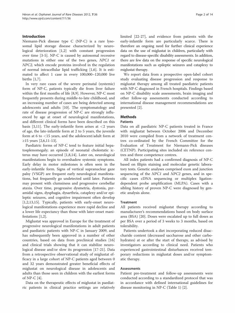
RESEARCH Open Access
Miglustat therapy in the French cohort ofpaediatric patients with Niemann-Pickdisease type CBénédicte Héron1,2*, Vassili Valayannopoulos2,3, Julien Baruteau2,4, Brigitte Chabrol2,5, Hélène Ogier2,6,
Philippe Latour2,7, Dries Dobbelaere2,8, Didier Eyer9, François Labarthe10, Hélène Maurey11, Jean-Marie Cuisset12,
Thierry Billette de Villemeur1,2,13, Frédéric Sedel2,14 and Marie T Vanier2,7,15
Abstract
Background: Niemann-Pick disease type C (NP-C) is a rare neurovisceral lysosomal lipid storage disease
characterized by progressive neurological deterioration. Published data on the use of miglustat in paediatric
patients in clinical practice settings are limited. We report findings from a prospective open-label study in the
French paediatric NP-C cohort.
Methods: Data on all paediatric NP-C patients treated with miglustat in France between October 2006 and
December 2010 were compiled. All patients had a confirmed diagnosis of NP-C, and received miglustat therapy
according to manufacturer’s recommendations. Pre-treatment and follow-up assessments were conducted
according to a standardized protocol.
Results: Twenty children were enrolled; 19 had NPC1 gene mutations and 1 had NPC2 gene mutations. The median
age at diagnosis was 1.5 years, and the median age at miglustat initiation was 6.0 years. Eight NPC1 patients had
the early-infantile, eight had the late-infantile, and three had the juvenile-onset forms of NP-C. A history of
hepatosplenomegaly and/or other cholestatic symptoms was recorded in all 8 early-infantile onset patients, 3/8
late-infantile patients, and 1/3 juvenile onset patients. Brain imaging indicated white matter abnormalities in most
patients. The median (range) duration of miglustat therapy was 1.3 (0.6–2.3) years in early-infantile, 1.0 (0.8–5.0) year
in late-infantile, and 1.0 (0.6–2.5) year in juvenile onset patients. NP-C disability scale scores indicated either
stabilization or improvement of neurological manifestations in 1/8, 6/8, and 1/3 NPC1 patients in these subgroups,
respectively. There were no correlations between brain imaging findings and disease course. Mild-to-moderate
gastrointestinal disturbances were frequent during the first 3 months of miglustat therapy, but were easily
managed with dietary modifications and/or anti-propulsive medication.
Conclusions: Miglustat can improve or stabilize neurological manifestations in paediatric patients with the late-
infantile and juvenile-onset forms of NP-C. Among early-infantile onset patients, a shorter delay between
neurological disease onset and miglustat initiation was associated with an initial better therapeutic outcome in one
patient, but miglustat did not seem to modify overall disease course in this subgroup. More experience is required
with long-term miglustat therapy in early-infantile onset patients treated from the very beginning of neurological
manifestations.
Keywords: Niemann-Pick disease type C, Paediatric, Miglustat
* Correspondence: [email protected] Référence des Maladies Lysosomales, Neuropédiatrie, CHU Trousseau,
APHP, 75 571, Paris Cedex 12, France2Committee for the Evaluation of Treatment for Niemann-Pick diseases
(CETNP), Paris, France
Full list of author information is available at the end of the article
© 2012 Héron et al.; licensee BioMed Central Ltd. This is an Open Access article distributed under the terms of the CreativeCommons Attribution License (http://creativecommons.org/licenses/by/2.0), which permits unrestricted use, distribution, andreproduction in any medium, provided the original work is properly cited.
Héron et al. Orphanet Journal of Rare Diseases 2012, 7:36
http://www.ojrd.com/content/7/1/36
IntroductionNiemann-Pick disease type C (NP-C) is a rare lyso-
somal lipid storage disease characterized by neuro-
logical deterioration [1,2] with constant progression
over time [3-5]. NP-C is caused by autosomal recessive
mutations in either one of the two genes, NPC1 or
NPC2, which encode proteins involved in the regulation
of normal intracellular lipid trafficking [1,6]. It is esti-
mated to affect 1 case in every 100,000–120,000 live
births [1,7].
In very rare cases of the severe perinatal (systemic)
form of NP-C, patients typically die from liver failure
within the first months of life [8,9]. However, NP-C most
frequently presents during middle-to-late childhood, and
an increasing number of cases are being detected among
adolescents and adults [10]. The symptomatology and
rate of disease progression of NP-C are strongly influ-
enced by age at onset of neurological manifestations,
and different clinical forms have been described on this
basis [3,11]. The early-infantile form arises at <2 years
of age, the late-infantile form at 2 to 5 years, the juvenile
form at 6 to <15 years, and the adolescent/adult form at
≥15 years [2,6,11-13].
Paediatric forms of NP-C tend to feature initial hepa-
tosplenomegaly; an episode of neonatal cholestatic ic-
terus may have occurred [1,8,14]. Later on, neurological
manifestations begin to overshadow systemic symptoms.
Early delay in motor milestones is often seen in the
early-infantile form. Signs of vertical supranuclear gaze
palsy (VSGP) are frequent early neurological manifesta-
tions, but frequently go undetected until later. Patients
may present with clumsiness and progressive cerebellar
ataxia. Over time, progressive dysmetria, dystonia, pyr-
amidal signs, dysphagia, dysarthria, cataplexy and/or epi-
leptic seizures, and cognitive impairment often develop
[1,2,13,15]. Typically, patients with early-onset neuro-
logical manifestations experience more rapid decline and
a lower life expectancy than those with later-onset mani-
festations [1,2].
Miglustat was approved in Europe for the treatment of
progressive neurological manifestations in adult patients
and paediatric patients with NP-C in January 2009, and
has subsequently been approved in a number of other
countries, based on data from preclinical studies [16]
and clinical trials showing that it can stabilize neuro-
logical disease and/or slow its progression [17-21]. Data
from a retrospective observational study of miglustat ef-
ficacy in a large cohort of NP-C patients aged between 0
and 32 years demonstrated greater beneficial effects of
miglustat on neurological disease in adolescents and
adults than those seen in children with the earliest forms
of NP-C [4].
Data on the therapeutic effects of miglustat in paediat-
ric patients in clinical practice settings are relatively
limited [22-27], and evidence from patients with the
early-infantile form are particularly scarce. There is
therefore an ongoing need for further clinical experience
data on the use of miglustat in children, particularly with
regard to disease-specific disability assessments. In addition,
there are few data on the response of specific neurological
manifestations such as epileptic seizures and cataplexy to
miglustat therapy.
We report data from a prospective open-label cohort
study evaluating disease progression and response to
miglustat therapy among all treated paediatric patients
with NP-C diagnosed in French hospitals. Findings based
on NP-C disability scale assessments, brain imaging and
other follow-up assessments conducted according to
international disease management recommendations are
presented [2].
MethodsPatients
Data on all paediatric NP-C patients treated in France
with miglustat between October 2006 and December
2010 were compiled from a network of treatment cen-
tres co-ordinated by the French Committee for the
Evaluation of Treatment for Niemann-Pick diseases
(CETNP). Participating sites included six reference cen-
tres and three competence centres.
All index patients had a confirmed diagnosis of NP-C
based on filipin staining and molecular genetic labora-
tory tests. Genetic analyses comprised exon and junction
sequencing of the NPC1 and NPC2 genes, and in spe-
cific cases cDNA sequencing or multiplex ligation-
dependent probe amplification (MLPA). Cases with a
sibling history of proven NP-C were diagnosed by gen-
etic analysis alone.
Treatment
All patients received miglustat therapy according to
manufacturer’s recommendations based on body surface
area (BSA) [20]. Doses were escalated up to full doses as
per BSA over a period of 3 weeks to 3 months, based on
tolerability.
Patients undertook a diet incorporating reduced disac-
charide content (decreased saccharose and other carbo-
hydrates) at or after the start of therapy, as advised by
investigators according to clinical need. Patients who
experienced gastrointestinal disturbances received tem-
porary reductions in miglustat doses and/or symptom-
atic therapy.
Assessments
Patient pre-treatment and follow-up assessments were
conducted according to a standardized protocol that was
in accordance with defined international guidelines for
disease monitoring in NP-C (Table 1) [2].
Héron et al. Orphanet Journal of Rare Diseases 2012, 7:36 Page 2 of 14
http://www.ojrd.com/content/7/1/36

Pre-treatment assessments included medical histories of
systemic and neurological manifestations, clinical exami-
nations, abdominal ultrasound, chest X-ray, electromyog-
raphy, and laboratory tests (including haematology, liver
markers and optional plasma chitotriosidase activities).
Specific examinations were video-recorded.
Regular assessments of seizures (with electro-encephalog-
raphy [EEG]) and cataplexy were conducted, when required.
Other evaluations for characteristic NP-C manifestations
included psychometric testing of neuropsychological im-
pairment, ophthalmological examination including saccadic
eye movement (SEM) abnormalities, and changes in hearing
based on brainstem auditory evoked potentials (BAEP).
Cerebral imaging was conducted based on magnetic
resonance imaging (MRI) [28], which included T1,
FLAIR and T2-weighted sequences. When possible,
magnetic resonance spectroscopy (MRS) was also
conducted (often under general anaesthesia) with a
single voxel at long (TR1500 ms/TE 135 or 144 ms)
and short (TE 28 ms) echo times in the centrum
semi-ovale. The surfaces of metabolite peaks (N-
acetyl-aspartate [NAA], creatine [Cr] and choline
[Cho]) were integrated, and the NAA/Cr, Cho/Cr and
Cho/NAA ratios were calculated and compared with
normal values in age-matched children.
Patient scores on a published NP-C specific disability
scale [11], which assesses four key parameters of
neurological disease progression (ambulation, manipula-
tion, language, swallowing) were measured before treat-
ment and at multiple time points during follow up. A
modified version of the original scale was used, which
assigns scores from 0 (best) to 1 unit (worst), with equal
weighting for each parameter [4].
No statistical analyses were performed as this was an
open-label study with no pre-defined hypotheses. De-
scriptive statistics were used to describe observed clin-
ical changes. All results are stratified according to
established forms of neurological disease in NP-C, based
on age at neurological disease onset [2].
ResultsPatients
A total of 20 children born between 1994 and 2010 were
included in the study (11 females and 9 males), among
whom 19 had mutations in the NPC1 gene and 1 had
mutations in the NPC2 gene. Cases 12 and 13 are
siblings.
All patients were fully genotyped (Table 2); in 17/19
families the genotypes of the patients’ parents were also
characterised to establish segregation of alleles.
Among the NPC1 patients, eight were classified as
having the early-infantile form, eight as having the late-
infantile form, and three as the juvenile form, as defined
by the age of neurological onset [12]. Overall, the
Table 1 Standard assessments at treatment start and during follow up
Assessment Treatment start Frequency
General
• Complete physical examination ✔ Every 3–6 months
Clinical parameters of neurological disease
• NP-C functional disability scale ✔ Every 6–12 months
• Video recording ✔ Every 6–12 months
• Epileptic seizures ✔ Every 6–12 months
• Narcolepsy/cataplexy ✔ Every 6–12 months
Other measures
• Psychometric evaluations ✔ Every 6–12 months
• Hearing ✔ Every 6–12 months
• Abdominal ultrasound ✔ Depending on initialfindings
• Chest X-ray ✔ Initial assessment anddepending on clinicalevolution
Laboratory parameters
• Liver function ✔ Every 6–12 months
• Haematology (blood counts) ✔ Every 6–12 months
• Plasma chitotriosidase (optional) ✔ Initial assessment
Cerebral imaging
• MRI or MRS (magnetic resonance spectroscopy)* ✔ Every 12 months
*Determination of Cho/NAA ratio optional.
Héron et al. Orphanet Journal of Rare Diseases 2012, 7:36 Page 3 of 14
http://www.ojrd.com/content/7/1/36
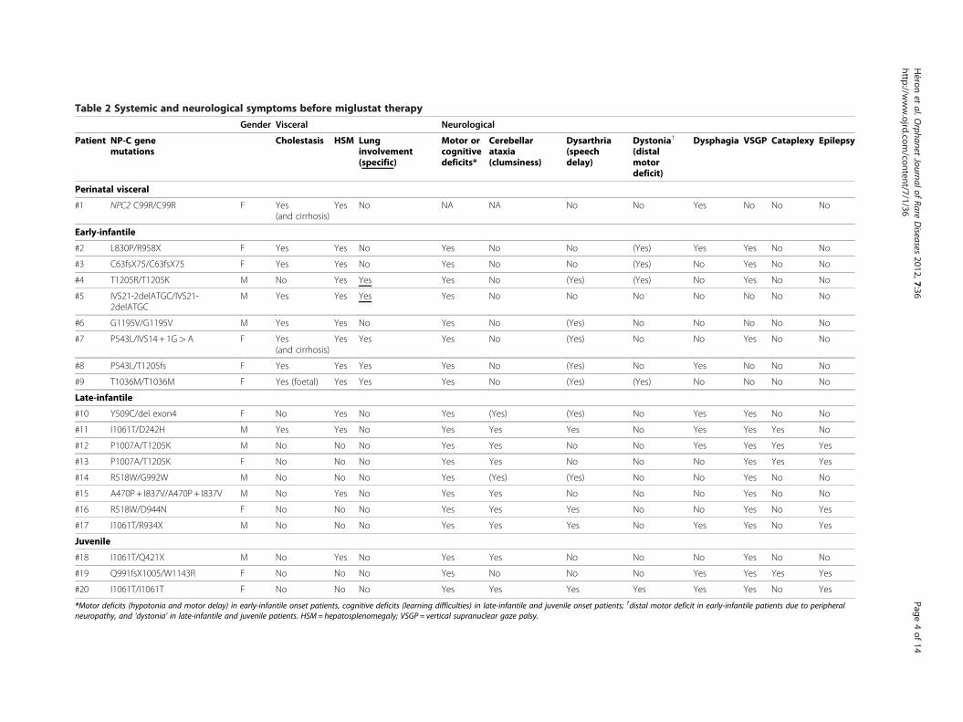
Table 2 Systemic and neurological symptoms before miglustat therapy
Gender Visceral Neurological
Patient NP-C genemutations
Cholestasis HSM Lunginvolvement(specific)
Motor orcognitivedeficits*
Cerebellarataxia(clumsiness)
Dysarthria(speechdelay)
Dystonia†
(distalmotordeficit)
Dysphagia VSGP Cataplexy Epilepsy
Perinatal visceral
#1 NPC2 C99R/C99R F Yes(and cirrhosis)
Yes No NA NA No No Yes No No No
Early-infantile
#2 L830P/R958X F Yes Yes No Yes No No (Yes) Yes Yes No No
#3 C63fsX75/C63fsX75 F Yes Yes No Yes No No (Yes) No Yes No No
#4 T1205R/T1205K M No Yes Yes Yes No (Yes) (Yes) No Yes No No
#5 IVS21-2delATGC/IVS21-2delATGC
M Yes Yes Yes Yes No No No No No No No
#6 G1195V/G1195V M Yes Yes No Yes No (Yes) No No No No No
#7 P543L/IVS14+ 1G>A F Yes(and cirrhosis)
Yes Yes Yes No (Yes) No No Yes No No
#8 P543L/T1205fs F Yes Yes Yes Yes No (Yes) No Yes No No No
#9 T1036M/T1036M F Yes (foetal) Yes Yes Yes No (Yes) (Yes) No No No No
Late-infantile
#10 Y509C/del exon4 F No Yes No Yes (Yes) (Yes) No Yes Yes No No
#11 I1061T/D242H M Yes Yes No Yes Yes Yes No Yes Yes Yes No
#12 P1007A/T1205K M No No No Yes Yes No No Yes Yes Yes Yes
#13 P1007A/T1205K F No No No Yes Yes No No No Yes Yes Yes
#14 R518W/G992W M No No No Yes (Yes) (Yes) No No Yes No No
#15 A470P+ I837V/A470P+ I837V M No Yes No Yes Yes No No No Yes No No
#16 R518W/D944N F No No No Yes Yes Yes No No Yes No Yes
#17 I1061T/R934X M No No No Yes Yes Yes No Yes Yes No Yes
Juvenile
#18 I1061T/Q421X M No Yes No Yes Yes No No No Yes No No
#19 Q991fsX1005/W1143R F No No No Yes No No No Yes Yes Yes Yes
#20 I1061T/I1061T F No No No Yes Yes Yes Yes Yes Yes No Yes
*Motor deficits (hypotonia and motor delay) in early-infantile onset patients, cognitive deficits (learning difficulties) in late-infantile and juvenile onset patients; †distal motor deficit in early-infantile patients due to peripheral
neuropathy, and ‘dystonia’ in late-infantile and juvenile patients. HSM=hepatosplenomegaly; VSGP = vertical supranuclear gaze palsy.
Héronet
al.Orphanet
JournalofRare
Disea
ses2012,7:36
Page4of14
http
://www.ojrd
.com/co
ntent/7
/1/36
median (range) age at neurological disease onset was 3.0
years (5 months to 7 years). The median (range) age at
diagnosis was 1.5 years (prenatal to 14 years), and the
median (range) age at start of miglustat therapy was 6.0
years (2 months to 14.8 years).
Pre-treatment disease history
Patient with perinatal visceral disease
Patient 1 developed cholestasis and hepatosplenomegaly
at 1 month of age. This patient was homozygous for the
well described NPC2 mutation, p.C99R, which has previ-
ously been associated with either a perinatal lethal dis-
ease or an early-infantile neurological form in the same
sibship [29,30]. No disability scale or brain imaging
assessments were performed in this child, but clinical
examination revealed hypotonia, due either to the sever-
ity of his liver disease or to early cerebral disease. Haem-
atopoietic stem cell transplantation was declined due to
his poor general condition.
Patients with early-infantile neurological onset
All eight patients with early-infantile neurological dis-
ease onset had a history of hepatosplenomegaly, and all
but one had prolonged neonatal cholestasis; liver biopsy
revealed evidence of cirrhosis in two patients (Table 2).
One patient (#7) had severe portal hypertension with
oesophageal varices (but no digestive bleeding) at 5
months of age. Five patients had a history of pulmonary
involvement, but only two (#4 and #5) had specific al-
veolar or interstitial pulmonary disease detected by chest
X-ray at 6 months of age and confirmed by bronchoal-
veolar lavage revealing accumulation of foamy macro-
phages: patient 4 needed oxygen therapy up to 15
months of age, and currently has frequent pulmonary
infections; patient 5 had frequent bronchitis with inter-
stitial signs on chest X-ray around 6 months of age and
then developed alveolar proteinosis with progressive re-
spiratory failure. Patient 6 had repeated pulmonary infec-
tions from birth to 3 months of age. Patient 8 had sub-
acute aspiration pneumonia at 2 years of age without spe-
cific biological signs on bronchopulmonary lavage. Patient
9 had bronchopulmonary dysplasia presumably due to pre-
maturity, and which required non-invasive ventilation up to
6 months of age.
Neurological manifestations among early-infantile
patients appeared between 5 and 12 months of age, and
included initial hypotonia, delayed motor development
and swallowing disorders. VSGP was observed at 9, 18,
and 24 months of age in five patients, but no patients
had cataplexy. One patient had pronounced dysphagia
and subsequent feeding difficulties at 5 months of age,
requiring enteral feeding with nasogastric tube followed
by gastrostomy aged 9 months. Four patients exhibited
clinical signs of peripheral neuropathy, which included
distal motor deficit, dysesthesia and diminished osteo-
tendinous reflexes. In each case a myelinic neuropathy
was confirmed by electrodiagnostic testing.
Filipin staining, performed in fibroblasts from each pa-
tient, invariably showed a massive accumulation of unester-
ified cholesterol in perinuclear vesicles (classic phenotype).
The NPC1 mutant allele p.I1061T was not observed in this
age subgroup, in good accordance with our previous obser-
vations [6,30,31]. The p.P543L mutation (present in
patients 7 and 8) has previously been reported to lead to an
early-infantile form of NP-C [29]. Mutations p.G1195V and
p.L830P were detected in one patient each; to our know-
ledge these mutations have not previously been reported.
Patients with late-infantile neurological onset
Three of the eight late-infantile onset patients had
splenomegaly, among whom only one also had a his-
tory of neonatal cholestasis (#11 – his elder brother
died from foetal hydrops due to NP-C). No patients
in this subgroup exhibited pulmonary disease. Neuro-
logical manifestations appeared between 2 and 5 years
of age, and included initial slow motor function and
clumsiness or ataxia, delayed language development,
and behavioural disturbances with relational troubles.
All patients exhibited VSGP, and 5/8 patients had
cataplexy and/or epilepsy. Because of pronounced
dysphagia and related feeding difficulties, gastrostomy
tube and discontinuous enteral feeding became
mandatory before initiation of miglustat in patient 12
(at 12 years of age) and patient 17 (at 10 years of
age). Another patient (#14) underwent gastrostomy a
few weeks after miglustat initiation because he
refused oral treatment due to its bitter taste.
A ‘classic’ filipin staining result was observed in fibro-
blasts from all cases except patient 16, including patient
12 who had one p.P1007A allele, which is usually asso-
ciated with a ‘variant’ filipin staining pattern [31]. The
p.1061 T mutation constituted 2/14 of the mutant alleles
among 7 unrelated patients in this age subgroup. Muta-
tions detected in patients 10 and 15 have not previously
been described. Two patients (#14 and #16) were hetero-
zygous for the p.R518W mutation, which in the homozy-
gous state has previously been reported in adult-onset
patients [10].
Patients with juvenile neurological onset
Only one juvenile-onset patient had visceral symptoms:
enlarged spleen without a history of neonatal cholestasis.
No patients in this subgroup exhibited pulmonary dis-
ease. Neurological manifestations started between 5 and
7 years of age in all three juvenile-onset patients, and
included praxis disorders, a cerebello-dystonic syn-
drome, cognitive decline and swallowing disorders (but
no psychiatric signs). All patients had VSGP. Two
Héron et al. Orphanet Journal of Rare Diseases 2012, 7:36 Page 5 of 14
http://www.ojrd.com/content/7/1/36
patients (#’s 19 and 20) experienced epileptic seizures
and cataplectic episodes before miglustat therapy, at the
ages of 9 years and 12 years, respectively. At the age of 5
years, one patient (#18) diagnosed earlier based on vis-
ceral symptoms was found to have deafness, a probable
early sign of the disease.
As regards biochemical phenotype and genotype, a
‘classic’ filipin pattern was found in all juvenile-onset
patients. The p.1061 T mutation constituted 3/6 of the
mutant alleles detected in this age subgroup.
Miglustat therapy and effects on neurological disease
Key age milestones (periods before and with neuro-
logical manifestations, age at diagnosis and duration of
miglustat therapy) are summarized in Figure 1. Details
of miglustat therapy and changes in neurological disease
status are summarized in Table 3.
Patients with early-infantile neurological onset
The median (range) interval between the onset of neuro-
logical manifestations and initiation of miglustat therapy
was 14 (4–34) months, and the median (range) age at
miglustat start was 25 (9–43) months. Early-infantile
patients received miglustat for a median (range) of 16
(8–27) months.
Based on disability scale assessments, key neurological
parameters were stabilized in 1/8 (13%) patients (Figure 2a).
Patient 6, who started miglustat aged 3.6 years, was essen-
tially stabilized throughout 18 months on miglustat. In
spite of showing initial neurological signs at 9 months of
age, the evolution of disease before miglustat therapy in
this patient was similar to that seen in the late-infantile
onset form (i.e. a slower disease progression). Patient 2,
who had the earliest neurologic onset in this group (at 5
months), started miglustat at the age of 9 months and
showed consistent improvement throughout 15 months
of therapy, but began to deteriorate after Month 18. By
Month 22 of therapy her disability score was similar to
that at initiation of miglustat.
During a mean treatment period of approximately 18
months, disability scale scores increased (indicating dis-
ease progression) in five patients in spite of slight
improvements in interactions, tonus and/or salivation.
All but one exhibited pyramidal tract involvement.
Three patients required discontinuous enteral feeding by
nasogastric tube (#7, persistence of severe portal hyper-
tension with oesophageal varices contra-indicated the
gastrostomy) or by gastrostomy tube (#’s 3 and 8) after
10 to 12 months of miglustat therapy, and one patient
(#4) developed epilepsy at the age of 32 months. NP-C
disability scale data were not obtained for patient 5,
whose steady respiratory worsening was associated with
neurological disease progression during a 13-month
period of therapy; the severity of his respiratory condi-
tion precluded disability scale assessments.
Patients with late-infantile neurological onset
The median (range) interval between the onset of neuro-
logical manifestations and initiation of miglustat therapy
was 4.2 (1.0–9.6) years, and the median (range) age at
miglustat start was 8.0 (4.0–12.6) years. The median
(range) treatment duration at last evaluation was 1.0
(0.8–5.0) years.
Disability scale scores indicated improvement/stabilization
of neurological parameters in 6/8 (75%) patients in this
subgroup (Figure 2b). Patients 11 and 17 both improved
during 12 months of miglustat therapy. Patient 17,
whose epilepsy was very active before miglustat, showed
a global improvement and became free of seizures
after months 5 on miglustat. Patient 16 showed initial
Figure 1 Key age milestones for NPC1 patients. Each horizontal bar represents age milestones for an individual patient. Data included only for
NPC1 patient; *Patient 5 died aged 2 years and 9 months, and patient 12 died aged 13 years 11 months.
Héron et al. Orphanet Journal of Rare Diseases 2012, 7:36 Page 6 of 14
http://www.ojrd.com/content/7/1/36

deterioration during the first month of treatment, and
improved later on. Patient 15 showed stabilization
during 12 months on miglustat before treatment was
stopped because of adverse events. Two patients (#’s
10 and 14) showed overall improvement, but epileptic
and cataplectic episodes started 44 months after and
just after miglustat initiation, respectively. In patient
14, seizures and cataplectic episodes are controlled
using symptomatic medications. In patient 10, neuro-
logical deterioration began when epilepsy became
medication-resistant (persistence of one to three short
tonic seizures per day despite antiepileptic polyther-
apy). Patient 12 exhibited pronounced worsening dur-
ing the initial 6 months of treatment, but appeared
stabilized (albeit at a high disability score) after 12
months. Patient 13 experienced difficulties to ingest
miglustat powder due to its bitter taste and worsened
during therapy, showing more active epilepsy: miglustat
was discontinued after 9 months.
Patients with juvenile neurological onset
The median (range) interval between the onset of neuro-
logical manifestations and initiation of miglustat therapy
was 4.3 (1.5–7.75) years, and the median (range) age at
miglustat start was 9.8 (7.0–14.8) years. The median
(range) treatment duration at last evaluation was 1.0
(0.6–2.5) years.
Disability scale scores indicated improvement of
neurological manifestations in one of the three patients
(Figure 2c). Patient 20 showed initial improvement then
stabilized. Cataplectic episodes began in this patient at
Month 22; her cataplectic episodes and epilepsy are cur-
rently controlled using symptomatic therapy. Patient 19
worsened during the first 3 months of miglustat therapy
Table 3 Miglustat therapy and neurological evolution during follow up
Patient Age at onset ofneurologicalmanifestations
Age at start ofmiglustattherapy
Miglustatdose(mg/day)
Duration ofmiglustattherapy
Disease evolution*
Perinatal visceral
#1 2 months 2 months 50 2 months NA†
Early infantile
#2 5 months 9 months 100–150-100 22 months Initially improvedthen worsened
#3 6 months 2 years 1 month 200 18 months Worsened
#4 7 months 2 years 3 months 150–300–150 27 months Worsened
#5 9 months 20 months 200 13 months Worsened†
#6 9 months 3 years 7 months 200–300–150 18 months Stabilized
#7 10 months 2 years 2 months 200–150–200 12 months Worsened
#8 12 months 2 years 50–100 12 months Worsened
#9 12 months 2 years 100–200 8 months Worsened
Late infantile
#10 2 years 4 years 250 60 months Stabilized
#11 3 years 8 years 100–300 12 months Improved
#12 3 years 12 years 7 months 600–400 12 months Worsened†
#13 4 years 6 months 8 years 400 9 months WorsenedŦ
#14 5 years 6 years 150–250 36 months Worsened transientlythen stabilized
#15 5 years 7 years 9 months 200–300 12 months StabilizedŦ
#16 5 years 9 years 10 months 400–200–400 9 months Worsened transientlythen improved
#17 5 years 10 years 3 months 300 12 months Improved
Juvenile
#18 5–6 years 7 years 300 12 months WorsenedŦ
#19 5–6 years 9 years 9 months 400 7 months Worsened then stable
#20 6–8 years 14 years 9 months 600 30 months Improved then stable
†Patient 1 died aged 4 months, patient 5 died aged 2 years and 9 months, and patient 12 died aged 13 years 11 months; *disease evolution based on NP-C disability
scale [11] and global clinical judgment; NA: not applicable due to young age Ŧmiglustat treatment was stopped in patient #’s 13, 15 and 18 after 9, 12 and 12 months,
respectively.
Héron et al. Orphanet Journal of Rare Diseases 2012, 7:36 Page 7 of 14
http://www.ojrd.com/content/7/1/36

and then appeared stable between 4 and 7 months of
treatment, showing less dystonia but more swallowing
difficulties. This patient’s epilepsy became more active
during miglustat treatment but was stabilized following
alteration of antiepileptic therapy. Patient 18 displayed
worsening of neurological manifestations during 12
months of miglustat therapy before treatment was
stopped.
Electrophysiological findings
Patients with early-infantile neurological onset
Slight non-specific abnormalities such as bioccipital slow
waves or slow background activity were identified in
EEG analyses before and during miglustat therapy. Rare
posterior spikes were observed in patient 4 when he
developed epilepsy aged 32 months, after 5 months on
miglustat therapy.
Hearing was normal in all patients in this subgroup,
although BAEP showed prolonged latencies in two
patients aged 2 and 3 years.
Pre-existing electromyographic signs of myelinic neur-
opathy in four patients did not worsen after 12–24
months in four patients. Clinical signs of myelinic
neuropathy worsened in patient 2 after 22 months on
miglustat, which correlated with viral infection and sim-
ultaneous losses in the ability to stand and language, as
well as complete dysphagia. This worsening neuropathy
is considered as being related to disease progression.
Patients with late-infantile neurological onset
Several non-specific signs were observed in EEG assess-
ments. EEG findings were normal or showed slow waves
or a slow background activity in two patients without
epilepsy (#’s 11 and 15), and in two patients (#’s 10 and
14) before epilepsy. Various types of electro-clinic sei-
zures were observed before miglustat start (patients 12,
13, 16 and 17), or during miglustat treatment (patients
10 and 14), including atypical or myoclonic absence,
generalized tonic-clonic, and focal seizures. After the
start of epilepsy, EEG abnormalities were more active,
showing focal, multifocal or generalized interictal spikes
or spike-waves.
Late-infantile onset patients did not develop hearing im-
pairment, although BAEP showed prolonged latencies in
four patients aged between 8 and 9.5 years (#’s 11, 13, 14
and 16). Findings were normal in the three other patients.
Figure 2 Changes over time in individual patient composite scores on the NP-C disability scale during miglustat treatment. Patients
with a) early-infantile, b) late-infantile and c) juvenile-onset disease; data included for NPC1 patients only; no disability scale data available from
early-infantile patient 5, who died aged 2 years and 9 months; *patient 12 died at 13 years 11 months of age.
Héron et al. Orphanet Journal of Rare Diseases 2012, 7:36 Page 8 of 14
http://www.ojrd.com/content/7/1/36
Patients with juvenile neurological onset
Juvenile-onset patients exhibited a similar profile of EEG
abnormalities as that seen in patients with late-infantile
onset. For one patient (#18), early-onset deafness at the
age of 5 years was considered a sign of neurological dis-
ease onset, and required a hearing prosthesis. No hear-
ing loss was observed in the other two juvenile patients.
Imaging findings
Findings from MRI and MRS assessments before therapy
and at follow up are summarized in Table 4.
Patients with early-infantile neurological onset
The seven living early-infantile patients showed white-
matter abnormalities indicative of delayed myelination
or demyelination before miglustat therapy, with three
showing atrophy in the periventricular or subcortical
regions or the corpus callosum, and two also showing
slight atrophy of the cerebellar vermis or peduncles
(Table 4). Patient 9 had a normal MRI at 5 months of
age before developmental delay was noted at 12 months.
These white matter abnormalities remained stable in
patients 8 and 9, but worsened slightly during miglustat
treatment in the three other patients (#’s 3, 4 and 7),
who experienced clinical worsening. The two patients
who showed initial improvement (#2) or clinical stabilization
(#6) had stable MRI abnormalities and normal MRS at pre-
treatment assessment and at 12-month follow up. Patient 3,
who showed clinical worsening, also had normal MRS find-
ings at pre-treatment.
Pre-treatment MRS showed low NAA and high Cho
peaks in 2/5 patients (#’s 4 and 9). Low NAA peaks were
noted at Months 8–24 of miglustat therapy in four
patients (#’s 4, 7, 8 and 9) who showed clinical worsen-
ing, with normal Cho peaks in patients 4 and 8.
Patients with late-infantile neurological onset
Before starting miglustat, three late-infantile onset
patients (#’s 11, 14 and 17) had normal brain MRI find-
ings at 6, 8 and 10 years of age, respectively. Five
patients had slight periventricular or more generalized
white matter abnormalities on MRI (#’s 10, 12, 13, 15,
and 16), with cortical or slight cerebellar atrophy also
seen in two patients (#’s 15 and 16).
Four patients developed cortical, subcortical or cere-
bellar signs of atrophy after the commencement of
miglustat therapy, two of whom (#’s 10 and 14) experi-
enced stabilization of neurological symptoms, while
patients 12 and 13 worsened. Follow-up MRI findings
are not yet available for four patients (#’s 11 and 17 who
had normal findings before therapy, and #’s 15 and 16).
MRS findings were normal before treatment in
patients 10, 12, 13, 14 and 16, and showed a low NAA
peak in patient 15. At follow up, patient 10 showed a
persistent low NAA peak. Patient 14 had a transient low
NAA peak at Month 18 on miglustat, and MRS findings
became normal at Month 24. A high Cho peak was also
noted at Month 12 in patient 10 and at Month 18 in pa-
tient 14, which contrasted with a slight improvement of
symptoms.
Patients with juvenile neurological onset
Magnetic resonance imaging analysis revealed slight
periventricular white matter abnormalities in patient 18
before therapy and at Month 12 of follow up during
miglustat therapy, but MRS findings were normal. Peri-
ventricular white matter abnormalities were associated
with cortical atrophy in patient 19 before therapy, but
no MRI follow-up is yet available for this patient, who
has so far received 7 months of miglustat treatment. In
patient 20, imaging findings were normal before miglu-
stat at the age of 14 years and 9 months, but follow-up
analysis showed slight cortical atrophy after 12 months
of miglustat therapy and a high Cho/NAA ratio after 30
months, associated with clinical stabilization.
Safety and tolerability
Fifteen out of 20 patients (75%) in this paediatric cohort
experienced adverse effects that were considered related
to miglustat therapy, including diarrhoea, abdominal
pain, anorexia and weight loss. Gastrointestinal adverse
events occurred mostly during the first 3 months of
miglustat therapy, and usually resolved during continued
therapy at lower doses, following institution of a
disaccharide-free diet and/or administration of symp-
tomatic treatment (e.g. loperamide).
Most adverse events were mild or moderate in sever-
ity, except in three cases where adverse events led to dis-
continuation of miglustat therapy. Asthenia and/or
persistent diarrhoea motivated a decision to stop miglu-
stat therapy after 1 year in one late-infantile onset pa-
tient (#15) and one juvenile-onset patient (#18). These
adverse events resolved without clinical sequelae after
withdrawal of miglustat.
Patient 13 experienced persistent diarrhoea, but refused
the recommended disaccharide-free diet and became
anorexic: miglustat was subsequently discontinued after 9
months of therapy. One late-infantile onset patient (#14)
refused to ingest oral miglustat powder due to its bitter
taste, and a gastrostomy was conducted to enable drug ad-
ministration. This patient subsequently received a normal
oral diet and miglustat was well tolerated. Miglustat com-
mercial 100-mg capsules were repackaged in smaller cap-
sules of 50 mg to allow easier swallowing for patient 7.
Patient 9 remains strongly constipated on miglustat treat-
ment in spite of laxative medications. Patient 20 presented
at the age of 16 years (1 year after starting miglustat ther-
apy) with rectal fistula that persisted despite many
Héron et al. Orphanet Journal of Rare Diseases 2012, 7:36 Page 9 of 14
http://www.ojrd.com/content/7/1/36
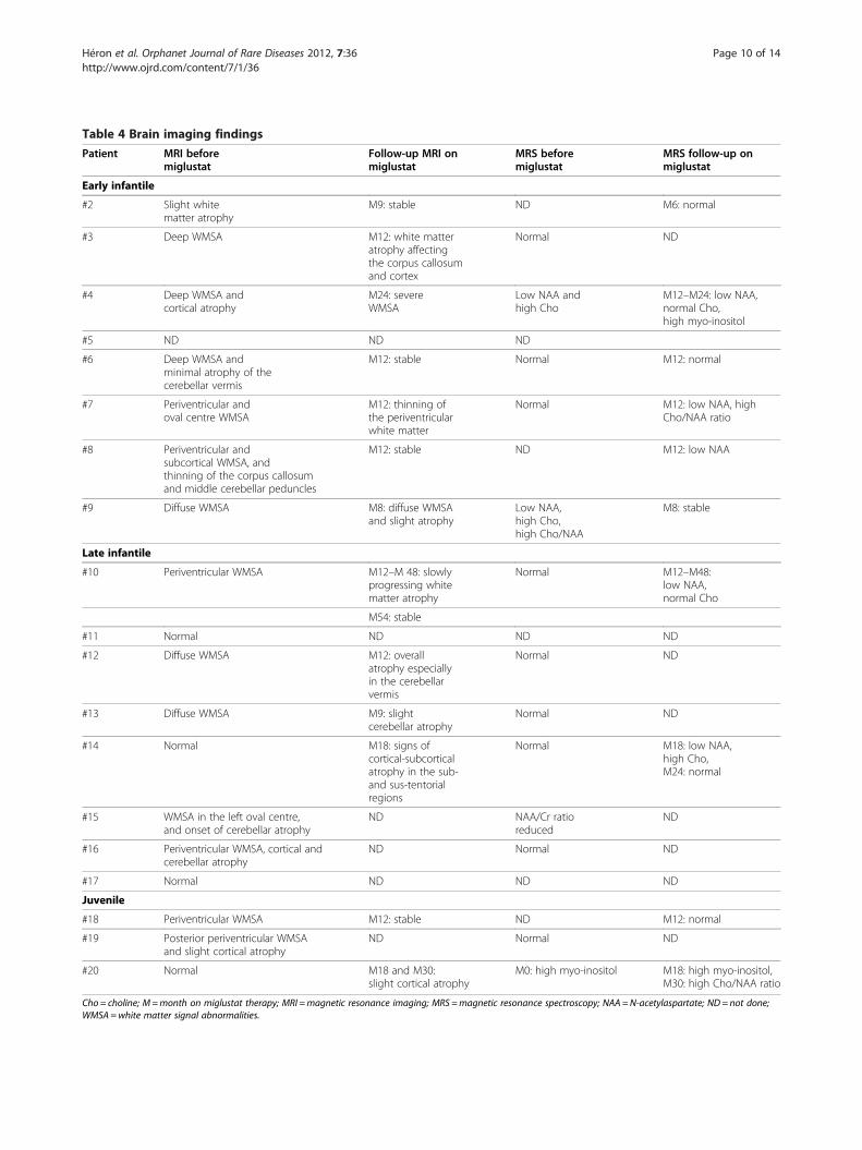
Table 4 Brain imaging findings
Patient MRI beforemiglustat
Follow-up MRI onmiglustat
MRS beforemiglustat
MRS follow-up onmiglustat
Early infantile
#2 Slight whitematter atrophy
M9: stable ND M6: normal
#3 Deep WMSA M12: white matteratrophy affectingthe corpus callosumand cortex
Normal ND
#4 Deep WMSA andcortical atrophy
M24: severeWMSA
Low NAA andhigh Cho
M12–M24: low NAA,normal Cho,high myo-inositol
#5 ND ND ND
#6 Deep WMSA andminimal atrophy of thecerebellar vermis
M12: stable Normal M12: normal
#7 Periventricular andoval centre WMSA
M12: thinning ofthe periventricularwhite matter
Normal M12: low NAA, highCho/NAA ratio
#8 Periventricular andsubcortical WMSA, andthinning of the corpus callosumand middle cerebellar peduncles
M12: stable ND M12: low NAA
#9 Diffuse WMSA M8: diffuse WMSAand slight atrophy
Low NAA,high Cho,high Cho/NAA
M8: stable
Late infantile
#10 Periventricular WMSA M12–M 48: slowlyprogressing whitematter atrophy
Normal M12–M48:low NAA,normal Cho
M54: stable
#11 Normal ND ND ND
#12 Diffuse WMSA M12: overallatrophy especiallyin the cerebellarvermis
Normal ND
#13 Diffuse WMSA M9: slightcerebellar atrophy
Normal ND
#14 Normal M18: signs ofcortical-subcorticalatrophy in the sub-and sus-tentorialregions
Normal M18: low NAA,high Cho,M24: normal
#15 WMSA in the left oval centre,and onset of cerebellar atrophy
ND NAA/Cr ratioreduced
ND
#16 Periventricular WMSA, cortical andcerebellar atrophy
ND Normal ND
#17 Normal ND ND ND
Juvenile
#18 Periventricular WMSA M12: stable ND M12: normal
#19 Posterior periventricular WMSAand slight cortical atrophy
ND Normal ND
#20 Normal M18 and M30:slight cortical atrophy
M0: high myo-inositol M18: high myo-inositol,M30: high Cho/NAA ratio
Cho = choline; M=month on miglustat therapy; MRI =magnetic resonance imaging; MRS =magnetic resonance spectroscopy; NAA =N-acetylaspartate; ND= not done;
WMSA =white matter signal abnormalities.
Héron et al. Orphanet Journal of Rare Diseases 2012, 7:36 Page 10 of 14
http://www.ojrd.com/content/7/1/36

treatments. She was later discovered to have Crohn’s dis-
ease, which was subsequently controlled with symptom-
atic therapy (mesalamine) to allow continued miglustat
treatment.
Three patients died during follow up. The patient with
perinatal visceral disease (i.e. NPC2; #1) developed cho-
lestasis and hepatosplenomegaly at 1 month of age, and
died aged 4 months (after 2 months of miglustat ther-
apy) due to liver failure. One early-infantile onset patient
(#5) died aged 2 years and 9 months due to respiratory
failure with alveolar proteinosis, which was considered
to be associated with neurological disease progression
during the patient’s 13-month period of miglustat ther-
apy. One late-infantile onset patient (#12) died aged 13
years and 11 months due to aspiration pneumonia.
DiscussionAssessments of key parameters of neurological disease
progression based on the published NP-C disability scale
indicated either stabilization or improvement of neuro-
logical manifestations in 1/8 early-infantile, 6/8 late-
infantile, and 1/3 juvenile-onset NP-C patients who
received miglustat in this multicentre, open-label cohort
study. Beneficial therapeutic effects were seen more fre-
quently in patients with late-infantile/juvenile neuro-
logical disease onset than in those with early-infantile
onset.
In agreement with previous reports, visceral disease
(prolonged neonatal cholestasis and/or hepatosplenome-
galy) was more prevalent among early-infantile onset
patients than in later-onset patients in this paediatric co-
hort [1,26]. Neonatal cholestasis healed in all NPC1
patients before miglustat therapy, but portal hyperten-
sion persisted in one patient during miglustat treatment.
While visceral disease was not a focus of disease moni-
toring in this study, miglustat did not appear to have any
effect on hepatosplenomegaly (data not shown) or pul-
monary disease (patient 5 died from alveolo-interstitial
complications after 13 months of miglustat treatment).
While the NPC2 patient had cholestasis and hepatos-
plenomegaly from 1 month of age (before initiation of
miglustat), she developed hepatic failure during miglu-
stat therapy. It is not possible to ascertain whether
miglustat might have worsened the liver disease in this
patient as a similar course of disease is quite common in
NPC2 [29].
In general, our findings appear in line with those from
previous clinical trial data [4,18,19] and case reports
[23,25,27] on the effects of miglustat on neurological
disease manifestations in paediatric NP-C patients.
Based on clinical assessments in a 24-month study of
miglustat in children aged 4–12 years, Patterson et al.
reported stabilization of SEM, an accepted marker of
early neurological deterioration in NP-C, in 67% of
patients throughout therapy [18]. Ambulation (measured
using the standard ambulation index) was stabilized in
80% of patients, and swallowing (patients’ ability to swal-
low various substances) remained stable in 90% [18].
Based on the same NP-C disability scale as that
employed in the current study, a retrospective analysis
of miglustat efficacy in 66 NP-C patients aged between 0
and 32 years (mean ± SD, 9.7 ± 7.6 years) showed that
ambulation, manipulation, language and swallowing
were stabilized or improved in 75% of patients during an
average of 18 months of therapy [4]. Stratification of
patients according to age indicated that beneficial effects
were greater in juvenile, adolescent and adult patients
than in those with disease onset before 6 years of age
[4].
Pineda et al. reported clinical experience with the use
of miglustat in a paediatric cohort of 16 Spanish NP-C
patients, comprising five with the early-infantile form,
four with the late-infantile form, and seven with the ju-
venile form [26]. As in the current study, efficacy assess-
ments were based on an NP-C specific disability scale,
albeit a modified version that included scores for the
presence of epilepsy and ocular movements. Similar to
our findings, the Spanish cohort study indicated that
patients with the late-infantile and juvenile-onset forms
were more likely to show improvements or stabilization
of neurological disease during miglustat therapy com-
pared with patients with severe, early-infantile onset
[26]. However, Spanish early-infantile onset patients who
showed deterioration during miglustat therapy were at
an advanced stage of the disease before starting therapy,
while those with a better evolution had started therapy
at the youngest ages. Overall, the Spanish data suggested
that better treatment effects might be expected when
treatment was initiated early, before “irreversible neuro-
logical damage” [2,26].
In our cohort, there did not appear to be as strong a
correlation between patients’ disability scale scores be-
fore treatment and subsequent changes during therapy.
However, a short interval between neurological disease
onset and the start of miglustat therapy and/or young
age at treatment start was associated with a better initial
therapeutic outcome in one early-infantile onset patient:
in patient 2, who initially improved, the delay between
neurological disease onset and initiation of miglustat
was only 4 months, and miglustat was commenced at 9
months of age. Other early-infantile patients, four of
whom worsened, had a mean (range) delay between
neurological disease onset and start of treatment of 1.8
(0.9–2.8) years, and a mean (range) age at treatment
start of 2.3 (range 1.7–3.6) years. In the late-infantile
and juvenile-onset patients the mean (range) delay to
therapy was 3.8 (1.0–7.8) years in patients who were
stable or improved after treatment, and 4.8 (1.5–9.6)
Héron et al. Orphanet Journal of Rare Diseases 2012, 7:36 Page 11 of 14
http://www.ojrd.com/content/7/1/36

years for those who worsened. These observations ap-
pear to support the argument for starting miglustat
treatment earlier, as soon as possible after the onset of
neurological symptoms and especially in the early-
infantile onset patients.
Data from the current cohort are in line with previous
data on the high prevalence of epilepsy and cataplexy in
late-infantile and juvenile (but not early-infantile) onset
patients [1,2]. Published data on the possible therapeutic
effect of miglustat on cataplexy and epilepsy are very
scarce. Zarowski et al. have previously reported a
complete cessation of cataplectic activity in a young
male patient with juvenile-onset NP-C [32]. In our study,
miglustat did not appear to prevent the occurrence of,
or to systematically improve, cataplexy or epilepsy
among the small number of patients in the late-infantile
and juvenile-onset subgroups. Limited data from the
Spanish paediatric cohort study indicated that the onset
of epilepsy and its resistance to symptomatic pharmaco-
therapy may result in worsening of patients’ scores on
the NP-C disability scale [26]. In our series, the presence
of pre-existing epilepsy, or its onset during miglustat
therapy, did not appear to affect neurological outcome
when seizures were stabilized using anti-epileptic therap-
ies. Anti-epileptic drugs employed included sodium val-
proate, lamotrigin and levetiracetam. Carbamazepine,
oxcarbazepine and vigabatrin were avoided as they could
promote myoclonias. Phenytoin was also not used in
order to avoid possible cerebellar adverse effects.
Auditory acuity remained stable in this series, and no
patient experienced worsening of electrical peripheral
neuropathy during miglustat therapy.
Magnetic resonance imaging showed white matter abnor-
malities in NPC1 patients with each of the age-at-onset
forms. In general, discrete posterior periventricular white
matter abnormalities were followed by more diffuse
changes resembling delayed myelination or demyelination
among these patients. Cortical or subcortical atrophy
tended to appear first in the infantile-onset forms (although
it was also present in later-onset forms). Cerebellar atrophy
was present in relatively few cases (two early-infantile and
three late-infantile patients).
Magnetic resonance spectroscopy identified some ab-
normalities, including low NAA and/or high Cho with
high Cho/NAA ratio. At short echo time, a high myo-
inositol peak was observed before therapy and at Month
18 of follow-up in patient 4 (worsened) and patient 20
(stabilized). It is known that progressive neurodegenera-
tive diseases are associated with a decrease of the NAA
peak, which is considered to be a marker of neuronal
viability, and by an increase of the Cho peak, which is
considered to be a marker of membrane destruction or
gliosis. Nevertheless there was no consistent pattern of
change over time or in response to miglustat in our
series. A low NAA peak was associated with cerebral at-
rophy in 8/18 cases but not with clinical worsening in all
of these. This contrasts with a previously published case
series based on three adult NP-C patients treated with
miglustat for 24 months, where mild clinical improve-
ment or stabilization concurrent with sustained
decreases in cerebral Cho/NAA ratio were observed
[24].
While our imaging findings are of value in that they
add to the relatively limited amount of published data
from longitudinal imaging studies in paediatric NP-C
patients, it is notable that there were no apparent cor-
relations between MRI or MRS findings and clinical
disease course during miglustat therapy. It is possible
that methodological and data limitations in our cohort
preclude a definitive conclusion on the utility of this
imaging technique. MRS analyses for French paediatric
patients were conducted at several different sites by
several technicians, and according to varied analysis
protocols. MRS data follow up beyond 24 months were
only available for 4/20 patients, which makes it difficult
to assess long-term changes. However our findings do
not favour the use of high Cho peak or Cho/NAA
ratios as objective markers of therapeutic effect in
paediatric patients, as has been proposed for adult
patients [24].
A possible correlation between the evolution of neuro-
logical manifestations (based on changes in NP-C dis-
ability scores) and cerebral hypometabolism (measured
using positron emission tomography [PET]) was previ-
ously reported based on data from Spanish juvenile- and
infantile-onset patients treated with miglustat [26]. Cere-
bral hypometabolism was stabilized when miglustat
appeared to slow the progression of neurological symp-
toms, and progressive hypometabolism correlated with
increasing disability scores [26]. Nevertheless, PET is un-
likely to be of practical use for routine clinical monitor-
ing due to the limited availability of equipment.
Mild or moderate gastrointestinal disturbances were
frequent during miglustat therapy, but usually resolved
within the first 3 months of treatment. In addition,
gastrointestinal adverse events were easily managed in
most cases by the adoption of dietary alterations, by pro-
gressive initiation of miglustat treatment, or by the use
of symptomatic therapy (e.g. loperamide). In particular,
dietary modifications such as reduced consumption of
dietary sucrose, maltose and lactose have been shown to
improve the gastrointestinal tolerability of miglustat, and
to reduce the magnitude of any changes in body weight,
particularly if initiated at or before the start of therapy
[33,34]. Finally, observed factors that appear to contrib-
ute to reduced treatment compliance among the young-
est patients include the bitter taste of oral miglustat
therapy, and the lack of a paediatric galenic form.
Héron et al. Orphanet Journal of Rare Diseases 2012, 7:36 Page 12 of 14
http://www.ojrd.com/content/7/1/36

A decision to stop miglustat was taken for two late-
infantile patients and one juvenile patient because of
persistent adverse events (e.g. asthenia or anorexia) and
clinical judgment of insufficient beneficial effects on dis-
ease progression. Such choices are made on a case-by-
case basis with collaborative discussions between med-
ical staff and parents, as well as detailed consideration of
patients’ clinical evolution and quality of life, well in line
with the updated recommendations from an expert
panel [35].
In spite of the small number of patients and the rela-
tively short period of follow up in the French paediatric
NP-C cohort, and in recognition of the invariably pro-
gressive course of neurological deterioration in untreated
patients [3,5], we conclude that miglustat can improve
or stabilize neurological disease progression in paediatric
patients with NP-C, particularly those with the late-
infantile and juvenile-onset forms. Our data from early-
infantile onset patients, who generally exhibit greater
symptom severity and more rapid progression of neuro-
logical manifestations, indicate that commencement of
miglustat at approximately 2 years of age has no sus-
tained global effect on the natural course of the disease.
A shorter delay between the onset of neurological mani-
festations and the start of miglustat therapy was asso-
ciated with a better initial therapeutic outcome in one
early infantile-onset patient in this cohort. However, this
patient later exhibited worsening of neurological disease
after 2 years of age. More clinical experience in early-
infantile onset patients treated at the very beginning of
their neurological disease over a longer period is
required to more fully assess the therapeutic effects of
miglustat in this group.
Current guidelines for the clinical management of NP-C
propose that miglustat treatment should be initiated at
the onset of neurological signs [2,35]. However, among
patients with early-infantile NP-C, the invariable occur-
rence and high frequency of systemic symptoms particu-
larly neonatal cholestasis and hepatosplenomegaly, often
leads to a diagnosis before the appearance of neurologic
signs or developmental delay. Regular and thorough clin-
ical examination of these patients, if possible combined
with cerebral MRI, could detect neurological problems at
their very beginning, leading to earlier initiation of treat-
ment with miglustat.
Competing interests
The authors declare that they have no competing interests.
Acknowledgements
The authors are grateful to the following clinicians who referred patients to
relevant reference centres: Dr Alice Kuster and Dr Marie Bru, Nantes; Dr Maria
Peralta, Mulhouse; Dr Patrick Boutard, Caen; Dr Stéphanie Torre, Rouen. The
authors also thank Pr Nathalie Boddaert and Dr Marie Thérèse Abi Warde,
who respectively performed MRI analyses and assisted with collection of
data from patients of Necker Hospital, Paris. This study was enabled through
the collaboration of treating physicians forming the CETNP. Matthew Reilly
PhD, associated with InTouch Medical Ltd, provided medical writing
assistance in the preparation of this manuscript, funded by Actelion
Pharmaceuticals Ltd.
Author details1Centre Référence des Maladies Lysosomales, Neuropédiatrie, CHU Trousseau,
APHP, 75 571, Paris Cedex 12, France. 2Committee for the Evaluation of
Treatment for Niemann-Pick diseases (CETNP), Paris, France. 3Centre
Référence des Maladies Héréditaires du Métabolisme de l’Enfant et de
l’Adulte (MaMEA), Necker-Enfants Malades, APHP, Paris, France. 4Maladies
Métaboliques, Hôpital des Enfants, CHU, Toulouse, France. 5Centre Référence
des Maladies Héréditaires du Métabolisme, CHU La Timone, APHM, Marseille,
France. 6Centre Référence des Maladies Héréditaires du Métabolisme, CHU
Robert Debré, APHP, Paris, France. 7Laboratoire Gillet-Mérieux, CBPE, Hospices
Civils de Lyon, Lyon, France. 8Centre Référence des Maladies Héréditaires du
Métabolisme CHRU, Lille, France. 9Pédiatrie, CHU, Hautepierre, Strasbourg,
France. 10Médecine Pédiatrique & INSERM U921, CHRU de Tours, Université
François Rabelais, Tours, France. 11Neuropédiatrie, CHU, Kremlin
BicêtreAPHPFrance. 12Neurologie Pédiatrique, CHRU, Lille, France. 13Université
Pierre et Marie Curie, Paris VI, France. 14Fédération des Maladies du Système
Nerveux, Salpêtrière APHP, Paris, France. 15Inserm Unité 820, Université
Lyon-1, Lyon, France.
Authors’ contributions
MTV, FS, TBV, BH, BC and HO conceived the study and participated in its
design. BH coordinated the study. BH, MTV and TBV drafted the manuscript.
MTV and PL carried out the filipin and molecular genetic studies. All authors
participated in collection of data, and have read and approved the final
manuscript.
Disclosures
BH has received travel expenses, and been invited to meetings funded and
organized by Actelion Pharmaceuticals Ltd., Biomarin, Genzyme Corporation
and Shire HGT, and has received presentation honoraria from Actelion
Pharmaceuticals Ltd. MTV has received travel expenses, carried out paid and
unpaid consultancy work, and presentation honoraria from Actelion
Pharmaceuticals Ltd., and has received travel expenses, presentation
honoraria, and been invited to meetings funded and organized by Genzyme
Corporation and Shire HGT. VV has received travel expenses, research grants
and presentation honoraria from Actelion Pharmaceuticals Ltd. FS has
received travel expenses, carried out paid consultancy work, and received
presentation honoraria from Actelion Pharmaceuticals Ltd. BC has received
travel expenses and presentation honoraria from Actelion Pharmaceuticals
Ltd. DE, JB and JMC have received travel expenses and presentation
honoraria from Actelion Pharmaceuticals Ltd. PL has received presentation
honoraria from Actelion Pharmaceuticals France. FL has received travel
expenses from Merck Serono and Genzyme Corporation. TBV has received
funds for an association (ASEP: for health and progress in paediatrics) from
Shire HGT and Genzyme Corporation. DD, HO and HM declare that they
have no competing interests.
Received: 19 February 2012 Accepted: 7 June 2012
Published: 7 June 2012
References
1. Vanier MT: Niemann-Pick disease type C. Orphanet J Rare Dis 2010, 5:16.
2. Wraith JE, Baumgartner MR, Bembi B, Covanis A, Levade T, Mengel E, Pineda
M, Sedel F, Topcu M, Vanier MT, et al: Recommendations on the diagnosis
and management of Niemann-Pick disease type C. Mol Genet Metab 2009,
98:152–165.
3. Wraith JE, Guffon N, Rohrbach M, Hwu WL, Korenke GC, Bembi B, Luzy C,
Giorgino R, Sedel F: Natural history of Niemann-Pick disease type C in a
multicentre observational retrospective cohort study. Mol Genet Metab
2009, 98:250–254.
4. Pineda M, Wraith JE, Mengel E, Sedel F, Hwu WL, Rohrbach M, Bembi B,
Walterfang M, Korenke GC, Marquardt T, et al: Miglustat in patients with
Niemann-Pick disease Type C (NP-C): a multicenter observational
retrospective cohort study. Mol Genet Metab 2009, 98:243–249.
5. Yanjanin NM, Velez JI, Gropman A, King K, Bianconi SE, Conley SK, Brewer
CC, Solomon B, Pavan WJ, Arcos-Burgos M, et al: Linear clinical
Héron et al. Orphanet Journal of Rare Diseases 2012, 7:36 Page 13 of 14
http://www.ojrd.com/content/7/1/36
progression, independent of age of onset, in Niemann-Pick disease, type
C. Am J Med Genet B Neuropsychiatr Genet 2010, 153B:132–140.
6. Vanier MT, Millat G: Niemann-Pick disease type C. Clin Genet 2003,
64:269–281.
7. Poupetova H, Ledvinova J, Berna L, Dvorakova L, Kozich V, Elleder M: The
birth prevalence of lysosomal storage disorders in the Czech Republic:
comparison with data in different populations. J Inherit Metab Dis 2010,
33:387–396.
8. Vanier MT, Wenger DA, Comly ME, Rousson R, Brady RO, Pentchev PG:
Niemann-Pick disease group C: clinical variability and diagnosis based
on defective cholesterol esterification. A collaborative study on 70
patients. Clin Genet 1988, 33:331–348.
9. Spiegel R, Raas-Rothschild A, Reish O, Regev M, Meiner V, Bargal R, Sury V,
Meir K, Nadjari M, Hermann G, et al: The clinical spectrum of fetal
Niemann-Pick type C. Am J Med Genet A 2009, 149A:446–450.
10. Imrie J, Dasgupta S, Besley GT, Harris C, Heptinstall L, Knight S, Vanier MT,
Fensom AH, Ward C, Jacklin E, et al: The natural history of Niemann-Pick
disease type C in the UK. J Inherit Metab Dis 2007, 30:51–59.
11. Iturriaga C, Pineda M, Fernandez-Valero EM, Vanier MT, Coll MJ: Niemann-
Pick C disease in Spain: clinical spectrum and development of a
disability scale. J Neurol Sci 2006, 249:1–6.
12. Vanier MT, Pentchev P, Rodriguez-Lafrasse C, Rousson R: Niemann-Pick
disease type C: an update. J Inherit Metab Dis 1991, 14:580–595.
13. Heron B, Ogier H: Niemann-Pick type C disease: clinical presentations in
pediatric patients. Arch Pediatr 2010, 17(Suppl 2):S45–S49.
14. Kelly DA, Portmann B, Mowat AP, Sherlock S, Lake BD: Niemann-Pick
disease type C: diagnosis and outcome in children, with particular
reference to liver disease. J Pediatr 1993, 123:242–247.
15. Sevin M, Lesca G, Baumann N, Millat G, Lyon-Caen O, Vanier MT, Sedel F:
The adult form of Niemann-Pick disease type C. Brain 2007, 130:120–133.
16. Zervas M, Somers KL, Thrall MA, Walkley SU: Critical role for
glycosphingolipids in Niemann-Pick disease type C. Curr Biol 2001,
11:1283–1287.
17. Lachmann RH, te Vruchte D, Lloyd-Evans E, Reinkensmeier G, Sillence DJ,
Fernandez-Guillen L, Dwek RA, Butters TD, Cox TM, Platt FM: Treatment
with miglustat reverses the lipid-trafficking defect in Niemann-Pick
disease type C. Neurobiol Dis 2004, 16:654–658.
18. Patterson MC, Vecchio D, Jacklin E, Abel L, Chadha-Boreham H, Luzy C,
Giorgino R, Wraith JE: Long-term miglustat therapy in children with
Niemann-Pick disease type C. J Child Neurol 2010, 25:300–305.
19. Patterson MC, Vecchio D, Prady H, Abel L, Wraith JE: Miglustat for
treatment of Niemann-Pick C disease: a randomised controlled study.
Lancet Neurol 2007, 6:765–772.
20. Miglustat (Zavesca) Summary of Product Characteristics., . http://
eudrapharm.eu/eudrapharm/showDocument?
documentId = 299255761182430949.
21. Wraith JE, Vecchio D, Jacklin E, Abel L, Chadha-Boreham H, Luzy C, Giorgino
R, Patterson MC: Miglustat in adult and juvenile patients with Niemann-
Pick disease type C: Long-term data from a clinical trial. Mol Genet Metab
2010, 99:351–357.
22. Paciorkowski AR, Westwell M, Ounpuu S, Bell K, Kagan J, Mazzarella C,
Greenstein RM: Motion analysis of a child with Niemann-Pick disease
type C treated with miglustat. Mov Disord 2008, 23:124–128.
23. Chien YH, Lee NC, Tsai LK, Huang AC, Peng SF, Chen SJ, Hwu WL:
Treatment of Niemann-Pick disease type C in two children with
miglustat: initial responses and maintenance of effects over 1 year. J
Inherit Metab Dis 2007, 30:826.
24. Galanaud D, Tourbah A, Lehericy S, Leveque N, Heron B, Billette De
Villemeur T, Guffon N, Feillet F, Baumann N, Vanier MT, Sedel F: 24
month-treatment with miglustat of three patients with Niemann-Pick
disease type C: follow up using brain spectroscopy. Mol Genet Metab
2009, 96:55–58.
25. Santos ML, Raskin S, Telles DS, Lohr A Jr, Liberalesso PB, Vieira SC,
Cordeiro ML: Treatment of a child diagnosed with Niemann-Pick
disease type C with miglustat: a case report in Brazil. J Inherit Metab
Dis 2008, 31(Suppl 2):S357–S361.
26. Pineda M, Perez-Poyato MS, O’Callaghan M, Vilaseca MA, Pocovi M,
Domingo R, Portal LR, Perez AV, Temudo T, Gaspar A, et al: Clinical
experience with miglustat therapy in pediatric patients with Niemann-
Pick disease type C: a case series. Mol Genet Metab 2010, 99:358–366.
27. Fecarotta S, Amitrano M, Romano A, Della Casa R, Bruschini D, Astarita L,
Parenti G, Andria G: The videofluoroscopic swallowing study shows a
sustained improvement of dysphagia in children with Niemann-Pick
disease type C after therapy with miglustat. Am J Med Genet A 2011,
155A:540–547.
28. Tedeschi G, Bonavita S, Barton NW, Betolino A, Frank JA, Patronas NJ, Alger
JR, Schiffmann R: Proton magnetic resonance spectroscopic imaging in
the clinical evaluation of patients with Niemann-Pick type C disease. J
Neurol Neurosurg Psychiatry 1998, 65:72–79.
29. Verot L, Chikh K, Freydiere E, Honore R, Vanier MT, Millat G: Niemann-
Pick C disease: functional characterization of three NPC2 mutations
and clinical and molecular update on patients with NPC2. Clin Genet
2007, 71:320–330.
30. Millat G, Bailo N, Molinero S, Rodriguez C, Chikh K, Vanier MT: Niemann-Pick
C disease: use of denaturing high performance liquid chromatography
for the detection of NPC1 and NPC2 genetic variations and impact on
management of patients and families. Mol Genet Metab 2005, 86:220–232.
31. Millat G, Marcais C, Tomasetto C, Chikh K, Fensom AH, Harzer K, Wenger DA,
Ohno K, Vanier MT: Niemann-Pick C1 disease: correlations between NPC1
mutations, levels of NPC1 protein, and phenotypes emphasize the
functional significance of the putative sterol-sensing domain and of the
cysteine-rich luminal loop. Am J Hum Genet 2001, 68:1373–1385.
32. Zarowski M, Steinborn B, Gurda B, Dvorakova L, Vlaskova H, Kothare SV:
Treatment of cataplexy in Niemann-Pick disease type C with the use of
miglustat. Eur J Paediatr Neurol 2011, 15:84–87.
33. Champion H, Ramaswami U, Imrie J, Lachmann RH, Gallagher J, Cox TM,
Wraith JE: Dietary modifications in patients receiving miglustat. J Inherit
Metab Dis 2010. doi:10.1007/s10545-008-0923-9. Epub ahead of print.
34. Belmatoug N, Burlina A, Giraldo P, Hendriksz CJ, Kuter DJ, Mengel E,
Pastores GM: Gastrointestinal disturbances and their management in
miglustat-treated patients. J Inherit Metab Dis 2011, 34:991–1001.
35. Patterson MC, Hendriksz CJ, Walterfang M, Sedel F, Vanier MT, Wijburg F,
NP-C Guidelines Working Group: Recommendations for the diagnosis and
management of Niemann-Pick disease type C: an update. Mol Genet
Metab 2012, 106:330–344.
doi:10.1186/1750-1172-7-36Cite this article as: Héron et al.: Miglustat therapy in the French cohortof paediatric patients with Niemann-Pickdisease type C. Orphanet Journal of Rare Diseases 2012 7:36.
Submit your next manuscript to BioMed Centraland take full advantage of:
• Convenient online submission
• Thorough peer review
• No space constraints or color figure charges
• Immediate publication on acceptance
• Inclusion in PubMed, CAS, Scopus and Google Scholar
• Research which is freely available for redistribution
Submit your manuscript at www.biomedcentral.com/submit
Héron et al. Orphanet Journal of Rare Diseases 2012, 7:36 Page 14 of 14
http://www.ojrd.com/content/7/1/36
Related Documents