Middle East respiratory syndrome coronavirus: a comprehensive review Mahmoud M. Shehata 1 , Mokhtar R. Gomaa 1 , Mohamed A. Ali 1 , Ghazi Kayali (✉) 2 1 Center of Scientific Excellence for Influenza Viruses, National Research Centre, Giza, Egypt; 2 Department of Infectious Diseases, St. Jude Children’ s Research Hospital, Memphis, TN 38105, USA © Higher Education Press and Springer-Verlag Berlin Heidelberg 2016 Abstract The Middle East respiratory syndrome coronavirus was first identified in 2012 and has since then remained uncontrolled. Cases have been mostly reported in the Middle East, however travel-associated cases and outbreaks have also occurred. Nosocomial and zoonotic transmission of the virus appear to be the most important routes. The infection is severe and highly fatal thus necessitating rapid and efficacious interventions. Here, we performed a comprehensive review of published literature and summarized the epidemiology of the virus. In addition, we summarized the virological aspects of the infection and reviewed the animal models used as well as vaccination and antiviral tested against it. Keywords MERS; coronavirus; review Introduction Coronaviruses (CoV) became known to cause human disease in the twentieth century. HCoV-229E and HCoV- OC43 were discovered in the 1960s and shown to cause respiratory infections in humans [1,2]. With the emergence of SARS-CoV in 2003 [3], two other human coronaviruses were discovered, HCoV NL63, HCoV HKU1 [4]. In 2012, a new type of coronavirus was detected as the cause of severe respiratory illness in humans. The first case was a 60-year-old male from Saudi Arabia admitted to hospital with acute respiratory illness leading to pneumonia and acute renal failure. The virus initially named as human corona virus-EMC [5], is currently known as the Middle East respiratory syndrome coronavirus (MERS-CoV) [6]. Virology of MERS-CoV Classification and nomenclature of MERS-CoV Phylogenetically, MERS-CoV is a lineage C β coronavirus (β-CoV) and is closely related to bat coronaviruses HKU4 and HKU5. The rooted phylogenetic analysis showed that MERS-CoV had an amino acid sequence identity less than 90% to all other known CoVs [7]. The virus initially named by many different working groups as novel coronavirus, human coronavirus EMC, human b corona- virus 2c EMC, human b coronavirus 2c England-Qatar, human b coronavirus 2C Jordan-N3, and b coronavirus England 1, which represented the places where the first complete viral genome was sequenced (Erasmus Medical Center, Rotterdam, The Netherlands) or where the first laboratory-confirmed cases were identified or managed (Jordan, Qatar, and England) was later named as MERS- CoV by the coronaviruses study groups of ICTV [5,6,8]. General virology of MERS-CoV MERS-CoV is an enveloped virus with a positive sense RNA genome. Coronavirus genomes range between 25 to 32 kb in size. The complete sequence of HCoV-EMC-2012 resulted in 30 119 nucleotides sequence [7]. Coronavirus genomes are polycistronic with large replicase open reading frames ORF1a and ORF1b which are subsequently cleaved into 15 or 16 nonstructural proteins (NSPs). The region downstream of ORF1b encode smaller genes including the spike (S), envelope (E), membrane (M), and nucleocapsid (N) structural protein [9–11]. The functional receptor for MERS-CoV is the Dipeptidyl peptidase 4 (DPP4) which is present on human non- ciliated bronchial epithelial cells surfaces [12]. The DPP4 protein displays high amino acid sequence conservation REVIEW Received October 1, 2015; accepted November 18, 2015 Correspondence: [email protected] Front. Med. 2016, 10(2): 120–136 DOI 10.1007/s11684-016-0430-6

Welcome message from author
This document is posted to help you gain knowledge. Please leave a comment to let me know what you think about it! Share it to your friends and learn new things together.
Transcript

Middle East respiratory syndrome coronavirus: a comprehensivereview
Mahmoud M. Shehata1, Mokhtar R. Gomaa1, Mohamed A. Ali1, Ghazi Kayali (✉)2
1Center of Scientific Excellence for Influenza Viruses, National Research Centre, Giza, Egypt; 2Department of Infectious Diseases, St. JudeChildren’s Research Hospital, Memphis, TN 38105, USA
© Higher Education Press and Springer-Verlag Berlin Heidelberg 2016
Abstract The Middle East respiratory syndrome coronavirus was first identified in 2012 and has since thenremained uncontrolled. Cases have been mostly reported in the Middle East, however travel-associated cases andoutbreaks have also occurred. Nosocomial and zoonotic transmission of the virus appear to be the most importantroutes. The infection is severe and highly fatal thus necessitating rapid and efficacious interventions. Here, weperformed a comprehensive review of published literature and summarized the epidemiology of the virus. Inaddition, we summarized the virological aspects of the infection and reviewed the animal models used as well asvaccination and antiviral tested against it.
Keywords MERS; coronavirus; review
Introduction
Coronaviruses (CoV) became known to cause humandisease in the twentieth century. HCoV-229E and HCoV-OC43 were discovered in the 1960s and shown to causerespiratory infections in humans [1,2]. With the emergenceof SARS-CoV in 2003 [3], two other human coronaviruseswere discovered, HCoV NL63, HCoV HKU1 [4]. In 2012,a new type of coronavirus was detected as the cause ofsevere respiratory illness in humans. The first case was a60-year-old male from Saudi Arabia admitted to hospitalwith acute respiratory illness leading to pneumonia andacute renal failure. The virus initially named as humancorona virus-EMC [5], is currently known as the MiddleEast respiratory syndrome coronavirus (MERS-CoV) [6].
Virology of MERS-CoV
Classification and nomenclature of MERS-CoV
Phylogenetically, MERS-CoV is a lineage C β coronavirus(β-CoV) and is closely related to bat coronaviruses HKU4and HKU5. The rooted phylogenetic analysis showed thatMERS-CoV had an amino acid sequence identity less than
90% to all other known CoVs [7]. The virus initiallynamed by many different working groups as novelcoronavirus, human coronavirus EMC, human b corona-virus 2c EMC, human b coronavirus 2c England-Qatar,human b coronavirus 2C Jordan-N3, and b coronavirusEngland 1, which represented the places where the firstcomplete viral genome was sequenced (Erasmus MedicalCenter, Rotterdam, The Netherlands) or where the firstlaboratory-confirmed cases were identified or managed(Jordan, Qatar, and England) was later named as MERS-CoV by the coronaviruses study groups of ICTV [5,6,8].
General virology of MERS-CoV
MERS-CoV is an enveloped virus with a positive senseRNA genome. Coronavirus genomes range between 25 to32 kb in size. The complete sequence of HCoV-EMC-2012resulted in 30 119 nucleotides sequence [7]. Coronavirusgenomes are polycistronic with large replicase openreading frames ORF1a and ORF1b which are subsequentlycleaved into 15 or 16 nonstructural proteins (NSPs). Theregion downstream of ORF1b encode smaller genesincluding the spike (S), envelope (E), membrane (M),and nucleocapsid (N) structural protein [9–11]. Thefunctional receptor for MERS-CoV is the Dipeptidylpeptidase 4 (DPP4) which is present on human non-ciliated bronchial epithelial cells surfaces [12]. The DPP4protein displays high amino acid sequence conservation
REVIEW
Received October 1, 2015; accepted November 18, 2015
Correspondence: [email protected]
Front. Med. 2016, 10(2): 120–136DOI 10.1007/s11684-016-0430-6

across different species, including the sequence that wasobtained from bat cells. Cell lines susceptibility studiesshowed that MERS-CoV infected several human cell lines,including histiocytes as well as respiratory, kidney,intestinal, and liver cells [13]. The range of tissue tropismin vitro was broader than that for any other known humancoronavirus [14]. MERS-CoV can also infect nonhumanprimate, porcine, bat, civet, rabbit, and horse cell lines allpossessing the DPP4 receptor [15].
MERS-CoV replication cycle
The replication cycle of MERS-CoV consists of numeroussteps as illustrated by Lu et al. [30].
Viral receptor attachment
The MERS-CoV S protein is a class I fusion proteincomposed of two subunits: the amino N-terminal receptorbinding S1 and carboxyl C-terminal membrane fusion S2subunits. The S1/S2 junction is a protease cleavage sitewhich is responsible for membrane fusion activation, virusentry, and syncytium formation. The S1 C domain containsthe receptor binding domain (RBD), and an N domain [13].Neutralizing monoclonal antibodies against the RBD mayinhibit virus entry into cells and receptor-dependentsyncytium formation in cell culture, hence vaccinescontaining the RBD induced high levels of neutralizingantibodies in mice and rabbits [16–18].DPP4 is the cell key functional receptor for the MERS-
CoV S protein [19]. MERS-CoV is the first CoV that hasbeen identified to use DPP4 as a receptor [19,20]. DPP4has important roles in glucose metabolism, T cellactivation, chemotaxis modulation, cell adhesion, andapoptosis [19,21].
Membrane fusion
The S2 subunit contains five domains: a fusion peptide, theheptad repeat 1 (HR1) and HR2 domains, a trans-membrane domain, and a cytoplasmic domain, whichform the stalk region of S protein that facilitates fusion ofthe viral and cell membranes [22,23]. The binding of theS1 subunit to the cellular receptor triggers conformationalchanges in the S2 subunit, which inserts its fusion peptideinto the target cell membrane to form a six-helix bundlefusion core between the HR1 and HR2 domains thatapproximates the viral and cell membranes for fusion.MERS-CoV utilizes many pathways for membrane fusiondepending on available host proteases, such as transmem-brane protease serine protease 2 (TMPRSS2), trypsin,chymotrypsin, elastase, thermolysin, endoproteinase Lys-C, and human airway trypsin-like protease. Proteasescleave the S protein into the S1 and S2 subunits to activatethe MERS-CoV S protein for endosome-independent host
cell entry at the plasma membrane [24–26]. In addition tothe pervious fusion proteases furin has been identifiedrecently to play an essential role in the MERS-CoV Sprotein cleavage activation into their biologically activeforms [27,28].
Disassembly, genome replication and expression
After cell entry, the virion particle disassembles to releasethe nucleocapsid and viral RNA into the cytoplasm forexpression of viral polyproteins pp1a and pp1ab. Double-membrane vesicles and convoluted membranes are formedby the attachment of the hydrophobic domains of theMERS-CoV replication machinery to the limiting mem-brane of auto-phagosomes [29]. The viral polyproteinspp1a and pp1ab are cleaved by papain-like protease and3C-like protease into nsp1 to nsp16 [7,30,31]. These non-structural proteins form the replication-transcription com-plex, which regulates transcription and viral proteinexpression [29].
Assembly and release of the new viruses
After the production of abundant viral RNA and structuraland accessory proteins, the N protein binds to the genomicRNA in the cytoplasm to form the helical nucleocapsid(viral core). The viral core is enveloped by buddingthrough intracellular membranes between the endoplasmicreticulum and Golgi apparatus [32]. The S, E, and Mproteins are transported to the budding virion, where thenucleocapsid probably interacts with M protein to generatethe basic structure and complexes with the S and E proteinsto induce viral budding and release from the Golgiapparatus [33]. MERS-CoV replication cycle is completedby releasing the progeny virions through the cellmembrane via exocytosis pathway.
Animal models for MERS-CoV infection
Mice
MERS-CoV strain HCoV-EMC/2012 was inoculated tothree different mouse strains (immunocompetent BALB/cmice, 129S6/SvEv and innate immune-deficient 129/STAT1–/– mice) intranasally. No significant weight losswas observed and infectious virus could not be detected inthe lungs. Only moderate pathological lesions wereobserved in the lungs. Hence no viral replication wasobserved in these strains of mice [34].Zhao et al. developed a mouse model transduced with a
recombinant adenovirus vector expressing hDPP4 (Ad5-hDPP4) in lung tissue. Inoculation of MERS-CoV in thesemice resulted in MERS-CoV replication but withoutmortality. Young mice cleared from MERS-CoV in 6‒8days and old mice in 10‒14 days. Perivascular and
Mahmoud M. Shehata et al. 121

peribronchial lymphoid infiltration was observed, withprogression to an interstitial pneumonia postinfection [35].In another study, transgenic mice expressing hDPP4
were susceptible to MERS-CoV infection. Infectious viruswas isolated from lung and brain tissue and weight losswas observed [36]. Pascal et al. developed humanizedtransgenic mouse. No mortality or clinical signs wasobserved but interstitial pneumonia and significant lungdisease were observed histopathologically, suggesting thathumanized DPP4 mouse is a model for MERS-CoVinfection in which pathological changes resembles MERS-CoV infection in humans [37,38].
Non-human primate models
The rhesus macaque was the first animal model used forMERS-CoV infection as it possessed DPP4 receptor[38,39]. In infected animals, an increase in respiratoryrates, body temperature, cough and reduced appetite wasobserved with mild to moderate severity. Infectious virusisolated only from the lower respiratory tract. Viral RNAwas detected in the conjunctiva, nasal mucosa, tonsils,pharynx, trachea, bronchus and lungs. Mild to markedinterstitial pneumonia with dark red lesions appeared inlungs. Seroconversion of neutralizing antibodies began at 7dpi and increased in titer with time. The development of atransient pneumonia, rapid replication, and tropism ofMERS-CoV for the lower respiratory tract resembled theseverity of the disease observed in humans [38,40,41].Similarly, the common marmoset was shown to possess
the DPP4 receptor [42]. Radiographic imaging showedmild to severe bilateral interstitial infiltration and extensivebronchointertitial pneumonia in infected animals. Infec-tious virus was detected in lower and upper respiratorytract tissue and viral RNA was detected in nasal mucosa,oropharyngeal swabs, blood, conjunctiva, lymph nodes,gastrointestinal tract, kidney, heart, adrenal gland, liver,spleen, brain and lungs [42].
Other models
Inoculation of Syrian hamsters and ferrets with MERS-CoV did not result in infection [12,43].Rabbits may be used as a model to study pathogenesis,
transmission, and disease control strategies of MERS-CoVin vivo as they seroconvert and shed virus after inoculation[44].
Epidemiology of MERS-CoV
In September 2012, a novel coronavirus infection wasnoted in ProMed Mail [45]. The virus was isolated fromthe sputum of a 60-year-old Saudi male, who was admittedto a hospital with pneumonia and acute kidney injury in
June 2012. A few days later, another report appeareddescribing an almost identical virus detected in a patient inQatar with acute respiratory syndrome and acute kidneyinjury. The patient had a recent travel history to SaudiArabia and then traveled to UK for further medical care[5,46,47]. Two cases from Jordan (April 2012) wereretrospectively diagnosed as MERS patients. Since thattime, more than 1542 cases of MERS-CoV infection havebeen reported including 544 deaths [48]. The actualnumber of cases could be higher than those reported[49]. An outbreak of more than 180 confirmed casesincluding 36 deaths occurred in South Korea in May andJune 2015. The median age of Korean cases were 55 years(range: 16 to 87 years), 60% were men, and 14% werehealth care professionals. The index case was a 68-year-oldmale who had recently traveled to several countries in theArabian Peninsula [50].
Case definitions, clinical manifestation, and diagnosis
Case definitions
MERS-CoV infection cases were classified by the WorldHealth Organization (WHO) [51], the US Centers forDisease Control and Prevention (CDC), and the Ministryof Health of Saudi Arabia (MOHSA) as asymptomatic,mild, severely symptomatic, or mortal. Cases may beclassified into suspected, probable, and confirmed [52,53].
Confirmed case
Any person with laboratory confirmation of infection withMERS-CoV irrespective of clinical signs and symptoms isconsidered as a confirmed case. WHO criteria forlaboratory confirmation require detection of viral RNAor acute and convalescent serology. The presence ofnucleic acid can be confirmed by positive results from atleast two sequence-specific rRT-PCRs or a singlesequence-specific rRT-PCR test and direct sequencingfrom a separate genomic target [54]. A case confirmationby serological methods requires demonstration of sero-conversion in two samples collected at least 14 days apartusing at least one screening assay (enzyme-linkedimmunoassay, immunofluorescence assay) and a neutrali-zation assay.
Probable case
A probable case is defined by the following criteria, afebrile acute respiratory illness as pneumonia or acuterespiratory distress syndrome, direct contact with aconfirmed MERS-CoV case and unavailability of MERS-CoV testing or results being inconclusive for a singleinadequate specimen.
122 MERS-coronavirus review

Suspected case
Any person who developed a fever and pneumonia or acuterespiratory distress syndrome with a history of travel tocountries in or near the Arabian Peninsula within 14 daysbefore symptom onset or was in contact with a travelerfrom this region who developed a febrile respiratory illnessis considered as a MERS-CoV suspected case.
Diagnosis
The WHO, CDC, and MOHSA recommended laboratorydiagnostics for MERS-CoV infection [6,9,51,55,56].MERS-CoV cases must be confirmed by at least twopositive qRT-PCR tests on two different specific genomicregions or single positive qRT-PCR with a sequence ofanother positive genome fragment [57]. The WHOalgorithm for testing MERS-CoV relies on qRT-PCR andsequencing [58]. Available real-time tests include an assaytargeting the RNA upstream of the E gene (upE) as ahighly sensitive screening assay and three confirmatoryassays targeting open reading frames (ORF 1a and 1b) and/or N gene. The ORF 1a assay is of equal sensitivity to theupE assay. The ORF 1b assay is less sensitive but is usefulfor confirmation. These assays are specific for MERS-CoVand have not shown cross-reactivity with other respiratoryhuman coronaviruses. For sequencing, two target genes,the RNA-dependent RNA polymerase (RdRp, present inORF 1b) and N genes are enough to confirm the existenceof MERS-CoV RNA in the samples of a patient [57].Several serologic assays including immunofluorescence
assays, protein microarray assay, enzyme-linked immuno-sorbent assay (ELISA) have been developed for thedetection of MERS-CoV antibodies [57,59–61]. Anypositive test by one of these assays should be confirmedwith a neutralization assay. Single serological result maybe valuable for definition of probable case and should befollowed by further testing for confirmation of MERS-CoVinfection [62–64].
Clinical manifestation of MERS-CoV infection
Incubation period of MERS-CoV infections was studied byAssiri et al. in 2013. The median incubation period was 5.2days (95% CI 1.9‒14.7 days) [65]. In another report fromFrance of a secondary case, a patient who shared a roomwith an infected patient, the incubation period wasestimated at 9 to 12 days [66]. In the recent outbreak inSouth Korea during May/June 2015, the median incuba-tion period was 6.3 days [67]. WHO and CDC recom-mended that individuals that returned from the ArabianPeninsula and other affected countries must be evaluatedfor MERS-CoV infections up to at least 14 days [68].Clinical features of MERS-CoV infections range from
asymptomatic cases to mildly ill, severe pneumonia, acute
respiratory distress syndrome, septic shock and mortal withmulti-organ failure (Table 1) [64,65]. Many other clinicalfeatures such as gastrointestinal symptoms (anorexia,nausea, vomiting, abdominal pain, diarrhea), pericarditis,and disseminated intravascular coagulation were reported[65,69,70].Specific clinical conditions (comorbidities) were appar-
ently proportionate with high severity of MERS-CoVinfections. A study by Assiri et al. in Saudi Arabia showedthat of a total of 47 patients with MERS-CoV infection in2013, 45 (96%) had underlying clinical conditions,including diabetes mellitus (68%), hypertension (34%),chronic cardiac disease (28%), and chronic kidney disease(49%) [65]. This high rate of comorbidities must beinterpreted with some caution, since diabetes mellitus iscommon in Saudi Arabia, and because approximately halfof those 47 were part of an outbreak in a hemodialysis unit,where rates of comorbidities might be high due to chronickidney disease [65,71]. In another study, being on dialysis,diabetes mellitus, and age > 50 years was associated withmortality [72]. In this study, testing positive for MERS-CoV in a plasma sample was a predictor of severe outcome[72].Younger adults and children appeared to be less
susceptible to MERS-CoV infection. Only one studydescribed MERS-CoV infection in children [73]. All ofthose children were discovered during contact investiga-tions of older patients. Only 2 of 11 children developedsymptoms of MERS-CoV infection. These two childrenhad underlying conditions (cystic fibrosis and Downsyndrome). The other 9 children were asymptomatic.There are few reports of MERS-CoV infections in pregnantwomen. A five-month pregnant female developed vaginalbleeding and abdominal pain after one week, thendelivered a stillborn infant [74]. Another case in theUnited Arab Emirates was near term phase, she gave birthto an apparently healthy baby, and died after delivery [52].Mild and asymptomatic MERS-CoV infections have
been reported, a majority of whom were identified amongthe contacts of patients [62,75,76]. In a report fromMOHSA, more than 3000 contacts of patients werescreened using qRT-PCR and seven healthcare workerswith MERS-CoV infection were identified, two of whomwere asymptomatic and five of whom had mild upperrespiratory tract symptoms [75].
Human-to-human transmission
Epidemiological and virological studies were conducted inattempts to determine person to person transmission ofMERS-CoV. They studied case clustering in householdand hospital outbreaks in the UK, Tunisia, Italy, and inhealthcare facilities in Saudi Arabia, France, Iran, andlately in South Korea. Those studies provided strongevidence that human-to-human transmission occurs
Mahmoud M. Shehata et al. 123

[70,77–80]. The number of contacts infected by indivi-duals with confirmed infections, however, appears to belimited [62], except the outbreak of South Korea in May/June 2015, where most cases were secondary and some
cases were tertiary infections [67,81]. Secondary casesoften were milder or symptomless [62]. Possible modes oftransmission include droplet and close contact transmis-sion, air borne transmission, and fomite transmission [82].
Table 1 Clinical features for MERS-CoV patientsFirst cases reported April, 2012 (Zarqa, Jordan)
June, 2012 (Jeddah, Saudi Arabia)
Incubation period
Mean (d) 5.2 (1.9–14.7)
Range (d) 2–13
Patient characteristics
Adults 98%
Children 2%
Age range (year) 1–94
Average age (year) Median 50
Sex ratio (male/female) 64.5%/35.5%
Mortality
Overall CFR 35.35%
CFR in patients with comorbidities 60%
Disease progression
Time from onset to ventilatory support Median 7 days
Time from onset to death Median 11.5 days
Presenting symptoms
Fever (>38°C) 98%
Chills or rigors 87%
Cough 83%
Dry 56%
Productive 44%
Hemoptysis 17%
Headache 11%
Myalgia 32%
Malaise 38%
Shortness of breath 72%
Nausea 21%
Vomiting 21%
Diarrhea 26%
Sore throat 14%
Rhinorrhea 6%
Comorbidities 76%
Dead subjects with underlying conditions 86%
Asymptomatic or recovered subjects with underlying conditions 42%
Laboratory results
Chest radiography abnormalities 90%–100%
Leucopenia (<4.0�109 cells per L) 14%
Lymphopenia (<1.5�109 cells per L) 32%
Thrombocytopenia (<140�109 platelets per L) 36%
High lactate dehydrogenase 48%
High alanine aminotransferase 11%
High aspartate aminotransferase 14%
CFR, case-fatality rate; MERS,Middle East respiratory syndrome.
124 MERS-coronavirus review
The majority of all laboratory-confirmed secondarycases have been associated with healthcare settings [82].The majority of cases of Jeddah, Saudi Arabia hospitaloutbreak during the spring of 2014 were acquired throughhuman-to-human transmission due to systematic weak-nesses in infection control [76]. Secondary transmissionrates were assessed within households and the transmissionrate was around 4%, suggesting that the actual number ofinfection is greater than reported [62].During the outbreak in South Korea during May/June
2015, 25 secondary infections were associated with theindex case, who was hospitalized from May 15 to May 17and 11 were tertiary [83]. The median incubation periodwas six days for secondary cases and six days for tertiarycases. This outbreak also clearly demonstrated roles of“superspreaders,” who may be responsible for a highproportion of cases [83]. For instance, a single patientinfected more than 70 other people while being treated inthe emergency room of a hospital in South Korea for threedays, 27‒29 May 2015.Transmissibility and epidemic potential studies of
MERS-CoV revealed that the reproduction number (R0)of patients infected with MERS-CoV ranged between 0.6to 0.69 [84,85]. The finding of an R0 < 1 suggests thatMERS-CoV does not yet have pandemic potential. Otherstudy suggested that R0 values might reach to 0.8 to 1.3 inthe absence of infection control [49]. Shedding periods ofMERS-CoV in humans was reported to be long as viruseswere detected in lower respiratory samples of symptomaticpatients for more than two weeks [86]. At instances,prolonged shedding for 6 weeks was detected in anasymptomatic healthcare worker. These findings raiseconcerns that asymptomatic persons could transmit infec-tion to others in a silent manner [87].
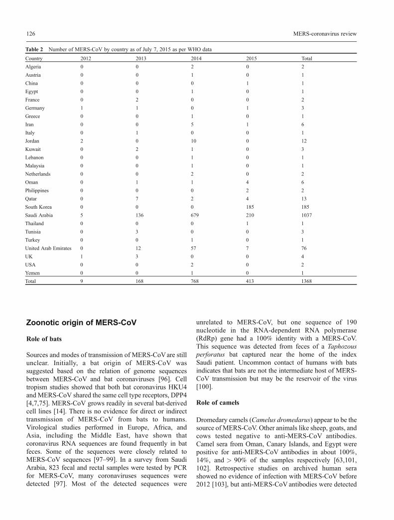
Geographic distribution of MERS-CoV cases
The majority of cases have occurred in Saudi Arabia andUnited Arab Emirates [88–90]. Many cases have also beenreported outside the Arabian Peninsula in North Africa,Europe, Asia, and North America as shown in Table 2.Almost all cases reported outside the Arabian Peninsulahad a travel history to it.
Notable outbreaks and clusters
The first cluster was in October/November 2012 in fourmen of the same family in Riyadh, Saudi Arabia, two ofwhom died [75]. The second cluster was reported in Jordanin April 2012 involving 10 healthcare workers exposed tofatal patients. In addition, seven surviving hospital contactsseroconverted suggesting that they had MERS-CoVinfection [91]. The third cluster was reported in UK duringJanuary/ February 2013. An English resident had a travel
history to Saudi Arabia and Pakistan in January, developeda severe respiratory illness, and tested positive for bothMERS-CoV and H1N1 influenza A, and died in March2013 after infecting several contacts [92].A cluster of 43 cases of MERS-CoV was reported in Al-
Hasa in Saudi Arabia during April 2013. All those caseswere directly linked to human to human contact in thesame hospital. There were only two confirmed cases ofhealthcare workers, and three family members weredetected by a survey of over 200 household contacts thatvisited this hospital [77]. In France, May 2013, an infectionof MERS-CoV was reported in a patient who recentlytraveled to the United Arab Emirates. A second case whoshared the hospital room with the first case tested positive.The first patient died and the second patient was criticallyill. A survey of 100 healthcare workers found no otherinfections with MERS-CoV, despite the lack of use ofpersonal protective equipment [70].A surge in MERS-CoV cases was reported in Saudi
Arabia and the United Arab Emirates during March andApril 2014 [55,76]. The majority of cases were associatedwith hospital-based outbreaks Jeddah, Riyadh, Tabuk, andMadinah in Saudi Arabia as well as in Al Ain, and AbuDhabi in United Arab Emirates. Cases included severalhealthcare workers, visitors, patients, and ambulance staff.Person to person transmission was confirmed in > 75% ofcases. The majority of infected health care workersdeveloped mild symptomatic or asymptomatic infection,but about 15% had severe illness or died [93].The recent outbreak of South Korea occurred in May
2015. The index case was a man who had recently traveledto Bahrain, the United Arab Emirates, Saudi Arabia, andQatar [55]. As of late July 2015, > 180 secondary caseswere reported including 36 death and many cases had beenreported among household and hospital contacts [55,67].In China, one case occurred in a man who traveled toChina from Korea following exposure to two relatives withMERS-CoV infection [55].
Disease seasonality
In spite of reporting of MERS-CoV infections throughoutthe year, some evidence on disease seasonality occurred.The first identified cases of MERS-CoV infection werereported in April and June 2012 [5,46,47]. A high increasein cases was reported in April and May 2013 followed by asurge in case reporting in April and May 2014. Increase incase reporting in March to May 2013 were attributed toinfection from infected young camels [94,95], but theincrease in 2014 in Saudi Arabia and in South Korea in2015 were due to gaps in infection control in hospitals.Small peaks in case reporting occurred in September andNovember of 2013 and 2014. The epicurve of infection isshown in Fig. 1.
Mahmoud M. Shehata et al. 125
Zoonotic origin of MERS-CoV
Role of bats
Sources and modes of transmission of MERS-CoVare stillunclear. Initially, a bat origin of MERS-CoV wassuggested based on the relation of genome sequencesbetween MERS-CoV and bat coronaviruses [96]. Celltropism studies showed that both bat coronavirus HKU4and MERS-CoV shared the same cell type receptors, DPP4[4,7,75]. MERS-CoV grows readily in several bat-derivedcell lines [14]. There is no evidence for direct or indirecttransmission of MERS-CoV from bats to humans.Virological studies performed in Europe, Africa, andAsia, including the Middle East, have shown thatcoronavirus RNA sequences are found frequently in batfeces. Some of the sequences were closely related toMERS-CoV sequences [97–99]. In a survey from SaudiArabia, 823 fecal and rectal samples were tested by PCRfor MERS-CoV, many coronaviruses sequences weredetected [97]. Most of the detected sequences were
unrelated to MERS-CoV, but one sequence of 190nucleotide in the RNA-dependent RNA polymerase(RdRp) gene had a 100% identity with a MERS-CoV.This sequence was detected from feces of a Taphozousperforatus bat captured near the home of the indexSaudi patient. Uncommon contact of humans with batsindicates that bats are not the intermediate host of MERS-CoV transmission but may be the reservoir of the virus[100].
Role of camels
Dromedary camels (Camelus dromedarus) appear to be thesource of MERS-CoV. Other animals like sheep, goats, andcows tested negative to anti-MERS-CoV antibodies.Camel sera from Oman, Canary Islands, and Egypt werepositive for anti-MERS-CoV antibodies in about 100%,14%, and > 90% of the samples respectively [63,101,102]. Retrospective studies on archived human serashowed no evidence of infection with MERS-CoV before2012 [103], but anti-MERS-CoV antibodies were detected
Table 2 Number of MERS-CoV by country as of July 7, 2015 as per WHO data
Country 2012 2013 2014 2015 Total
Algeria 0 0 2 0 2
Austria 0 0 1 0 1
China 0 0 0 1 1
Egypt 0 0 1 0 1
France 0 2 0 0 2
Germany 1 1 0 1 3
Greece 0 0 1 0 1
Iran 0 0 5 1 6
Italy 0 1 0 0 1
Jordan 2 0 10 0 12
Kuwait 0 2 1 0 3
Lebanon 0 0 1 0 1
Malaysia 0 0 1 0 1
Netherlands 0 0 2 0 2
Oman 0 1 1 4 6
Philippines 0 0 0 2 2
Qatar 0 7 2 4 13
South Korea 0 0 0 185 185
Saudi Arabia 5 136 679 210 1037
Thailand 0 0 0 1 1
Tunisia 0 3 0 0 3
Turkey 0 0 1 0 1
United Arab Emirates 0 12 57 7 76
UK 1 3 0 0 4
USA 0 0 2 0 2
Yemen 0 0 1 0 1
Total 9 168 768 413 1368
126 MERS-coronavirus review

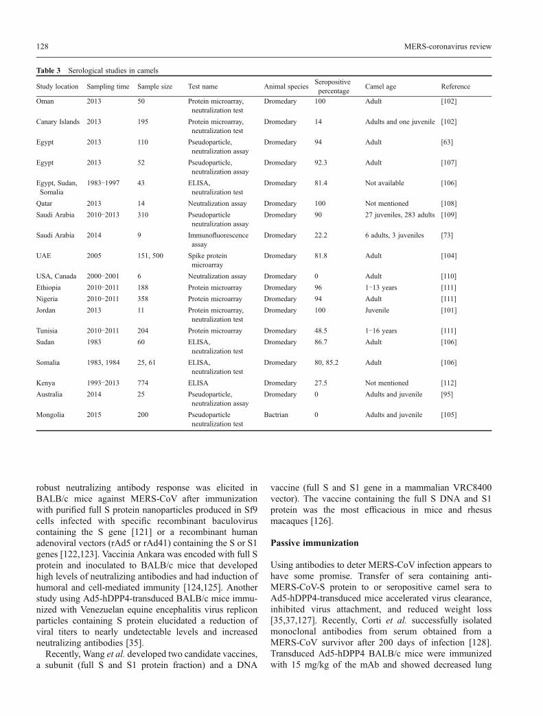
in archived camel sera in Saudi Arabia in 1993 [95], andUnited Arab Emirates in 2003 [104], indicating circulationof MERS-CoV in camels for many years. Bactrian camelsin Mongolia tested negative for MERS-CoV antibodies[105].Serologic studies from around the Middle East
suggested that camels are one of the sources of MERS-CoV as > 90% of adult camels tested positive and hadhigh titers of antibodies. Seropositivity was different injuvenile camels and was usually lower than in adults.These results suggested that MERS-CoV infections incamels occurred in young ages followed by frequentboosting [94,95,102,104]. Camels in other parts of theworld, far from the Middle East like in Europe, Australia,and the Americas do not have MERS-CoV antibodies andhave no evidence of infection [106]. Table 3 summarizescamel serologic studies.In a study aimed to evaluate virus infectivity and
shedding in camels, three adult dromedary camels wereinoculated with MERS-CoV intratracheally, intranasally,and conjunctivally. Those camels shed large quantities ofvirus from the upper respiratory tract and infectious viruswas detected in nasal secretions for 7 days post-inoculationand viral RNA for up to 35 days post-inoculation [113].Human infections with MERS-CoV were linked to
camels. The first evidence was a study in Saudi Arabia inwhich the MERS-CoV full genome sequences of isolatesfrom a man with fatal infection and from one of his camelswere identical. This patient had a direct contact with hisdeceased camels some days before the onset of symptoms.
These results suggested that MERS-CoV can infectdromedary camels and can be transmitted from them tohumans by direct close contact [86]. In other studies,phylogenetic analyses of camel and human isolates of theMERS-CoV genome demonstrated that the viruses werehighly identical or in some cases were similar to each other[107,108,114].Seroepidemiological studies shown low prevalence of
MERS-CoV antibodies in humans in Saudi Arabia[103,115]. A survey of 10 009 individuals representativeof the general population of Saudi Arabia resulted in 15seropositive subjects (0.15%), however, seropositivityincreased 15‒23 folds in camel-exposed individuals [78].In a separate report, 7 of 87 camel shepherds and 140slaughterhouse workers (3.1%) tested positive for MERS-CoV antibodies [103]. An overview of MERS-CoVtransmission routes is illustrated in Fig.2.
Vaccines and antivirals
Vaccines
The development of an effective vaccine is critical forprevention of a MERS-CoV pandemic. Some investigatorshave indicated that the RBD protein of MERS-CoV Sprotein is a good candidate antigen as a subunit vaccine.Various RBD fragments showed the highest DPP4 bindingaffinity and induced the highest-titer of IgG Ab andneutralizing Ab in mice and rabbits [17,18,116–120]. A
Fig. 1 Epidemiological curve of MERS-CoV up to week 23, 2015 as per WHO data.
Mahmoud M. Shehata et al. 127
robust neutralizing antibody response was elicited inBALB/c mice against MERS-CoV after immunizationwith purified full S protein nanoparticles produced in Sf9cells infected with specific recombinant baculoviruscontaining the S gene [121] or a recombinant humanadenoviral vectors (rAd5 or rAd41) containing the S or S1genes [122,123]. Vaccinia Ankara was encoded with full Sprotein and inoculated to BALB/c mice that developedhigh levels of neutralizing antibodies and had induction ofhumoral and cell-mediated immunity [124,125]. Anotherstudy using Ad5-hDPP4-transduced BALB/c mice immu-nized with Venezuelan equine encephalitis virus repliconparticles containing S protein elucidated a reduction ofviral titers to nearly undetectable levels and increasedneutralizing antibodies [35].Recently, Wang et al. developed two candidate vaccines,
a subunit (full S and S1 protein fraction) and a DNA
vaccine (full S and S1 gene in a mammalian VRC8400vector). The vaccine containing the full S DNA and S1protein was the most efficacious in mice and rhesusmacaques [126].
Passive immunization
Using antibodies to deter MERS-CoV infection appears tohave some promise. Transfer of sera containing anti-MERS-CoV-S protein to or seropositive camel sera toAd5-hDPP4-transduced mice accelerated virus clearance,inhibited virus attachment, and reduced weight loss[35,37,127]. Recently, Corti et al. successfully isolatedmonoclonal antibodies from serum obtained from aMERS-CoV survivor after 200 days of infection [128].Transduced Ad5-hDPP4 BALB/c mice were immunizedwith 15 mg/kg of the mAb and showed decreased lung
Table 3 Serological studies in camels
Study location Sampling time Sample size Test name Animal speciesSeropositivepercentage
Camel age Reference
Oman 2013 50 Protein microarray,neutralization test
Dromedary 100 Adult [102]
Canary Islands 2013 195 Protein microarray,neutralization test
Dromedary 14 Adults and one juvenile [102]
Egypt 2013 110 Pseudoparticle,neutralization assay
Dromedary 94 Adult [63]
Egypt 2013 52 Pseudoparticle,neutralization assay
Dromedary 92.3 Adult [107]
Egypt, Sudan,Somalia
1983‒1997 43 ELISA,neutralization test
Dromedary 81.4 Not available [106]
Qatar 2013 14 Neutralization assay Dromedary 100 Not mentioned [108]
Saudi Arabia 2010‒2013 310 Pseudoparticleneutralization assay
Dromedary 90 27 juveniles, 283 adults [109]
Saudi Arabia 2014 9 Immunofluorescenceassay
Dromedary 22.2 6 adults, 3 juveniles [73]
UAE 2005 151, 500 Spike proteinmicroarray
Dromedary 81.8 Adult [104]
USA, Canada 2000‒2001 6 Neutralization assay Dromedary 0 Adult [110]
Ethiopia 2010‒2011 188 Protein microarray Dromedary 96 1‒13 years [111]
Nigeria 2010‒2011 358 Protein microarray Dromedary 94 Adult [111]
Jordan 2013 11 Protein microarray,neutralization test
Dromedary 100 Juvenile [101]
Tunisia 2010‒2011 204 Protein microarray Dromedary 48.5 1‒16 years [111]
Sudan 1983 60 ELISA,neutralization test
Dromedary 86.7 Adult [106]
Somalia 1983, 1984 25, 61 ELISA,neutralization test
Dromedary 80, 85.2 Adult [106]
Kenya 1993‒2013 774 ELISA Dromedary 27.5 Not mentioned [112]
Australia 2014 25 Pseudoparticle,neutralization assay
Dromedary 0 Adults and juvenile [95]
Mongolia 2015 200 Pseudoparticleneutralization test
Bactrian 0 Adults and juvenile [105]
128 MERS-coronavirus review

viral titers, no weight loss, and decreased peribronchiallymphoid infiltration [128].
Antivirals
No approved antivirals for use against MERS-CoVinfection are yet available. The first approach performedwhen a new unknown virus like MERS-CoV emerges istesting drugs used as antiviral for similar viruses[29,129,130]. Type I interferons and ribavirine combina-tion exhibited acceptable results in cell culture and rhesusmacaques by decreasing the host inflammatory response,replication of virus, and improved clinical outcome[129,131,132]. A human cohort study in Saudi Arabiashowed that treatment with combination of ribavirin andinterferon-α2b to 5 did not improve clinical outcomes butthis may have been due to late treatment or due to theimmunocompromised state of the patients [133]. In aretrospective study of 20 MERS-CoV infected patientstreated with ribavirin and interferon α-2a, results showed14-day and 28-day survival was improved by 70% and28% in the treated group as compared to an untreatedgroup [134].The second approach is screening of approved drugs
with known safety profiles and transcriptional signatures indifferent cell lines. Several drugs, including antiparasitics,neurotransmitters, antibacterials, inhibitors of clathrin-mediated endocytosis estrogen receptor, lipid or sterolmetabolism, protein processing, and DNA synthesis or
repair were tested on culture cells [119,135–139].Lopinavir-ritonavir combined with pegylated interferonand ribavirin therapy showed improved outcomes ininfected marmosets [140].The third approach involves in vitro inhibition of S
protein to block virus entry into host cells using designedantiviral peptides targeting the HR2 domain of the S2subunit of the MERS-CoV and preventing the interactionbetween the HR1 and HR2 domains required for theformation of the heterologous six-helix bundle in viralfusion core formation [22,23]. Other drugs that act asinhibitors for viral proteases and helicase to suppressMERS-CoV infection were tested [141–145]. Otherinvestigators studied inhibition of MERS-CoV infectionby competitive inhibition of DPP4 cell receptor usingcompounds such as sitagliptin, vildagliptin, and saxaglip-tin [19,146].
Conclusions
More than three years have passed since the first detectionof MERS-CoV human infection and the virus, uncon-trolled, continues to cause major outbreaks in the MiddleEast. The recent outbreak in Korea demonstrated that asingle index case can lead to 185 more infections in a shortperiod of time, hence raising questions about the accuracyof the number of cases being reported in the Middle East.Furthermore, the Korean outbreak confirmed the highfatality rate of MERS-CoV infection as being true rather
Fig. 2 Zoonotic transmission of newly emerged MERS-CoV.
Mahmoud M. Shehata et al. 129

than overestimated in case only the more severe cases aredetected. In all, public health, veterinary health, andresearch efforts need to be consolidated in order to answerthe following high priority questions:- What is the true extent of human infection with MERS-
CoV?- What antivirals and vaccines are to be used in humans?- What infection control measures are needed in
healthcare settings to prevent nosocomial outbreaks?- What measures need to be in place in order to prevent
zoonotic infections from camels?- Is it possible to control the virus in the camel
population and if so, how?- Are there other animal species involved in the MERS-
CoV transmission cycle?
Acknowledgements
This work was funded by the National Institute of Allergy
and Infectious Diseases, National Institutes of Health, Departmentof Health and Human Services, under contract number
HHSN272201400006C, the Egyptian Science and TechnologyDevelopment Fund, under contract number 5175, and supportedby the American Lebanese Syrian Associated Charities (ALSAC).
Compliance with ethics guidelines
Mahmoud M. Shehata, Mokhtar R. Gomaa, Mohamed A. Ali, andGhazi Kayali declare that they have no conflict of interest. This
manuscript is a review article and does not involve a researchprotocol requiring approval by the relevant institutional review
board or ethics committee.
References
1. Hamre D, Procknow JJ. A new virus isolated from the humanrespiratory tract. Proc Soc Exp Biol Med 1966; 121(1): 190–193
2. McIntosh K, Dees JH, Becker WB, Kapikian AZ, Chanock RM.Recovery in tracheal organ cultures of novel viruses from patientswith respiratory disease. Proc Natl Acad Sci USA 1967; 57(4):933–940
3. Peiris JS, Lai ST, Poon LL, Guan Y, Yam LY, Lim W, Nicholls J,Yee WK, YanWW, Cheung MT, Cheng VC, Chan KH, Tsang DN,
Yung RW, Ng TK, Yuen KY; SARS study group. Coronavirus as apossible cause of severe acute respiratory syndrome. Lancet 2003;361(9366): 1319–1325
4. Woo PC, Lau SK, Lam CS, Lau CC, Tsang AK, Lau JH, Bai R,Teng JL, Tsang CC, Wang M, Zheng BJ, Chan KH, Yuen KY.Discovery of seven novel Mammalian and avian coronaviruses inthe genus deltacoronavirus supports bat coronaviruses as the genesource of alphacoronavirus and betacoronavirus and aviancoronaviruses as the gene source of gammacoronavirus and
deltacoronavirus. J Virol 2012; 86(7): 3995–4008
5. Zaki AM, van Boheemen S, Bestebroer TM, Osterhaus AD,Fouchier RA. Isolation of a novel coronavirus from a man with
pneumonia in Saudi Arabia. N Engl J Med 2012; 367(19): 1814–1820
6. de Groot RJ, Baker SC, Baric RS, Brown CS, Drosten C, EnjuanesL, Fouchier RA, Galiano M, Gorbalenya AE, Memish ZA,Perlman S, Poon LL, Snijder EJ, Stephens GM,Woo PC, Zaki AM,Zambon M, Ziebuhr J. Middle East respiratory syndromecoronavirus (MERS-CoV): announcement of the CoronavirusStudy Group. J Virol 2013; 87(14): 7790–7792
7. van Boheemen S, de Graaf M, Lauber C, Bestebroer TM, Raj VS,Zaki AM, Osterhaus AD, Haagmans BL, Gorbalenya AE, SnijderEJ, Fouchier RA. Genomic characterization of a newly discoveredcoronavirus associated with acute respiratory distress syndrome inhumans. MBio 2012; 3(6): e00473-12
8. Cotten M, Watson SJ, Kellam P, Al-Rabeeah AA, Makhdoom HQ,Assiri A, Al-Tawfiq JA, Alhakeem RF, Madani H, AlRabiah FA,
Al Hajjar S, Al-nassir WN, Albarrak A, Flemban H, Balkhy HH,Alsubaie S, Palser AL, Gall A, Bashford-Rogers R, Rambaut A,Zumla AI, Memish ZA. Transmission and evolution of the MiddleEast respiratory syndrome coronavirus in Saudi Arabia: adescriptive genomic study. Lancet 2013; 382(9909): 1993–2002
9. Bialek SR, Allen D, Alvarado-Ramy F, Arthur R, Balajee A, BellD, Best S, Blackmore C, Breakwell L, Cannons A, Brown C,Cetron M, Chea N, Chommanard C, Cohen N, Conover C, CrespoA, Creviston J, Curns AT, Dahl R, Dearth S, DeMaria A, Echols F,
Erdman DD, Feikin D, Frias M, Gerber SI, Gulati R, Hale C,Haynes LM, Heberlein-Larson L, Holton K, Ijaz K, Kapoor M,Kohl K, Kuhar DT, Kumar AM, Kundich M, Lippold S, Liu L,Lovchik JC, Madoff L, Martell S, Matthews S, Moore J, MurrayLR, Onofrey S, Pallansch MA, Pesik N, Pham H, Pillai S, PontonesP, Pringle K, Pritchard S, Rasmussen S, Richards S, Sandoval M,Schneider E, Schuchat A, Sheedy K, Sherin K, Swerdlow DL,Tappero JW, Vernon MO, Watkins S, Watson J; Centers forDisease Control and Prevention (CDC). First confirmed cases ofMiddle East respiratory syndrome coronavirus (MERS-CoV)infection in the United States, updated information on the
epidemiology of MERS-CoV infection, and guidance for thepublic, clinicians, and public health authorities—May 2014.MMWR Morb Mortal Wkly Rep 2014; 63(19): 431–436
10. Gorbalenya AE, Enjuanes L, Ziebuhr J, Snijder EJ. Nidovirales:evolving the largest RNA virus genome. Virus Res 2006; 117(1):17–37
11. Ziebuhr J, Snijder EJ, Gorbalenya AE. Virus-encoded proteinasesand proteolytic processing in the Nidovirales. J Gen Virol 2000; 81(Pt 4): 853–879
12. Raj VS, Smits SL, Provacia LB, van den Brand JM, Wiersma L,Ouwendijk WJ, Bestebroer TM, Spronken MI, van Amerongen G,Rottier PJ, Fouchier RA, Bosch BJ, Osterhaus AD, Haagmans BL.Adenosine deaminase acts as a natural antagonist for dipeptidylpeptidase 4-mediated entry of the Middle East respiratorysyndrome coronavirus. J Virol 2014; 88(3): 1834–1838
13. Chan JF, Chan KH, Choi GK, To KK, Tse H, Cai JP, Yeung ML,Cheng VC, Chen H, Che XY, Lau SK, Woo PC, Yuen KY.Differential cell line susceptibility to the emerging novel humanbetacoronavirus 2c EMC/2012: implications for disease pathogen-esis and clinical manifestation. J Infect Dis 2013; 207(11): 1743–1752
14. Müller MA, Raj VS, Muth D, Meyer B, Kallies S, Smits SL,Wollny R, Bestebroer TM, Specht S, Suliman T, Zimmermann K,
130 MERS-coronavirus review
Binger T, Eckerle I, Tschapka M, Zaki AM, Osterhaus AD,Fouchier RA, Haagmans BL, Drosten C. Human coronavirus EMCdoes not require the SARS-coronavirus receptor and maintainsbroad replicative capability in mammalian cell lines. MBio 2012; 3(6): e00515-12
15. Meyer B, García-Bocanegra I, Wernery U, Wernery R, Sieberg A,Müller MA, Drexler JF, Drosten C, Eckerle I. Serologic assessmentof possibility for MERS-CoV infection in equids. Emerg Infect Dis2015; 21(1): 181–182
16. Du L, Zhao G, Kou Z, Ma C, Sun S, Poon VK, Lu L, Wang L,Debnath AK, Zheng BJ, Zhou Y, Jiang S. Identification of areceptor-binding domain in the S protein of the novel humancoronavirus Middle East respiratory syndrome coronavirus as anessential target for vaccine development. J Virol 2013; 87(17):9939–9942
17. Mou H, Raj VS, van Kuppeveld FJ, Rottier PJ, Haagmans BL,Bosch BJ. The receptor binding domain of the new Middle Eastrespiratory syndrome coronavirus maps to a 231-residue region inthe spike protein that efficiently elicits neutralizing antibodies. JVirol 2013; 87(16): 9379–9383
18. Ma C, Wang L, Tao X, Zhang N, Yang Y, Tseng CT, Li F, Zhou Y,Jiang S, Du L. Searching for an ideal vaccine candidate amongdifferent MERS coronavirus receptor-binding fragments—theimportance of immunofocusing in subunit vaccine design. Vaccine
2014; 32(46): 6170–6176
19. Raj VS, Mou H, Smits SL, Dekkers DH, Müller MA, Dijkman R,Muth D, Demmers JA, Zaki A, Fouchier RA, Thiel V, Drosten C,Rottier PJ, Osterhaus AD, Bosch BJ, Haagmans BL. Dipeptidylpeptidase 4 is a functional receptor for the emerging humancoronavirus-EMC. Nature 2013; 495(7440): 251–254
20. Chan JF, To KK, Tse H, Jin DY, Yuen KY. Interspeciestransmission and emergence of novel viruses: lessons from batsand birds. Trends Microbiol 2013; 21(10): 544–555
21. Lambeir AM, Durinx C, Scharpé S, De Meester I. Dipeptidyl-peptidase IV from bench to bedside: an update on structuralproperties, functions, and clinical aspects of the enzyme DPP IV.Crit Rev Clin Lab Sci 2003; 40(3): 209–294
22. Lu L, Liu Q, Zhu Y, Chan KH, Qin L, Li Y, Wang Q, Chan JF, DuL, Yu F, Ma C, Ye S, Yuen KY, Zhang R, Jiang S. Structure-baseddiscovery of Middle East respiratory syndrome coronavirus fusioninhibitor. Nat Commun 2014; 5: 3067
23. Gao J, Lu G, Qi J, Li Y, Wu Y, Deng Y, Geng H, Li H, Wang Q,Xiao H, Tan W, Yan J, Gao GF. Structure of the fusion core andinhibition of fusion by a heptad repeat peptide derived from the Sprotein of Middle East respiratory syndrome coronavirus. J Virol
2013; 87(24): 13134–13140
24. Gierer S, Bertram S, Kaup F, Wrensch F, Heurich A, Krämer-KühlA, Welsch K, Winkler M, Meyer B, Drosten C, Dittmer U, vonHahn T, Simmons G, Hofmann H, Pöhlmann S. The spike proteinof the emerging betacoronavirus EMC uses a novel coronavirusreceptor for entry, can be activated by TMPRSS2, and is targetedby neutralizing antibodies. J Virol 2013; 87(10): 5502–5511
25. Gierer S, Müller MA, Heurich A, Ritz D, Springstein BL, KarstenCB, Schendzielorz A, Gnirß K, Drosten C, Pöhlmann S. Inhibitionof proprotein convertases abrogates processing of the middleeastern respiratory syndrome coronavirus spike protein in infectedcells but does not reduce viral infectivity. J Infect Dis 2015; 211(6):
889–897
26. Qian Z, Dominguez SR, Holmes KV. Role of the spikeglycoprotein of human Middle East respiratory syndromecoronavirus (MERS-CoV) in virus entry and syncytia formation.PLoS ONE 2013; 8(10): e76469
27. Millet JK, Whittaker GR. Host cell entry of Middle East respiratorysyndrome coronavirus after two-step, furin-mediated activation of
the spike protein. Proc Natl Acad Sci USA 2014; 111(42): 15214–15219
28. Thomas G. Furin at the cutting edge: from protein traffic toembryogenesis and disease. Nat Rev Mol Cell Biol 2002; 3(10):753–766
29. de Wilde AH, Raj VS, Oudshoorn D, Bestebroer TM, van
Nieuwkoop S, Limpens RW, Posthuma CC, van der Meer Y,Bárcena M, Haagmans BL, Snijder EJ, van den Hoogen BG.MERS-coronavirus replication induces severe in vitro cytopathol-ogy and is strongly inhibited by cyclosporin A or interferon-αtreatment. J Gen Virol 2013; 94(Pt 8): 1749–1760
30. Lu L, Liu Q, Du L, Jiang S. Middle East respiratory syndromecoronavirus (MERS-CoV): challenges in identifying its source andcontrolling its spread. Microbes Infect 2013; 15(8-9): 625–629
31. Lei J, Mesters JR, Drosten C, Anemüller S, Ma Q, Hilgenfeld R.Crystal structure of the papain-like protease of MERS coronavirusreveals unusual, potentially druggable active-site features. Anti-viral Res 2014; 109: 72–82
32. Narayanan K, Maeda A, Maeda J, Makino S. Characterization ofthe coronavirus M protein and nucleocapsid interaction in infected
cells. J Virol 2000; 74(17): 8127–8134
33. Stadler K, Masignani V, Eickmann M, Becker S, Abrignani S,Klenk HD, Rappuoli R. SARS—beginning to understand a newvirus. Nat Rev Microbiol 2003; 1(3): 209–218
34. Coleman CM, Matthews KL, Goicochea L, Frieman MB. Wild-type and innate immune-deficient mice are not susceptible to the
Middle East respiratory syndrome coronavirus. J Gen Virol 2014;95(Pt 2): 408–412
35. Zhao J, Li K, Wohlford-Lenane C, Agnihothram SS, Fett C, ZhaoJ, Gale MJ Jr, Baric RS, Enjuanes L, Gallagher T, McCray PB Jr,Perlman S. Rapid generation of a mouse model for Middle Eastrespiratory syndrome. Proc Natl Acad Sci USA 2014; 111(13):4970–4975
36. Agrawal AS, Garron T, Tao X, Peng BH, Wakamiya M, Chan TS,Couch RB, Tseng CT. Generation of a transgenic mouse model ofMiddle East respiratory syndrome coronavirus infection anddisease. J Virol 2015; 89(7): 3659–3670
37. Pascal KE, Coleman CM, Mujica AO, Kamat V, Badithe A,Fairhurst J, Hunt C, Strein J, Berrebi A, Sisk JM, Matthews KL,Babb R, Chen G, Lai KM, Huang TT, Olson W, Yancopoulos GD,Stahl N, Frieman MB, Kyratsous CA. Pre- and postexposure
efficacy of fully human antibodies against Spike protein in a novelhumanized mouse model of MERS-CoV infection. Proc Natl AcadSci USA 2015; 112(28): 8738–8743
38. Munster VJ, de Wit E, Feldmann H. Pneumonia from humancoronavirus in a macaque model. N Engl J Med 2013; 368(16):1560–1562
39. van Doremalen N, Miazgowicz KL, Milne-Price S, Bushmaker T,Robertson S, Scott D, Kinne J, McLellan JS, Zhu J, Munster VJ.Host species restriction of Middle East respiratory syndrome
Mahmoud M. Shehata et al. 131

coronavirus through its receptor, dipeptidyl peptidase 4. J Virol2014; 88(16): 9220–9232
40. de Wit E, Rasmussen AL, Falzarano D, Bushmaker T, Feldmann F,Brining DL, Fischer ER, Martellaro C, Okumura A, Chang J, ScottD, Benecke AG, Katze MG, Feldmann H, Munster VJ. MiddleEast respiratory syndrome coronavirus (MERS-CoV) causestransient lower respiratory tract infection in rhesus macaques.Proc Natl Acad Sci USA 2013; 110(41): 16598–16603
41. Yao Y, Bao L, Deng W, Xu L, Li F, Lv Q, Yu P, Chen T, Xu Y,Zhu H, Yuan J, Gu S, Wei Q, Chen H, Yuen KY, Qin C. An animalmodel of MERS produced by infection of rhesus macaques withMERS coronavirus. J Infect Dis 2014; 209(2): 236–242
42. Falzarano D, de Wit E, Feldmann F, Rasmussen AL, Okumura A,Peng X, Thomas MJ, van Doremalen N, Haddock E, Nagy L,LaCasse R, Liu T, Zhu J, McLellan JS, Scott DP, Katze MG,
Feldmann H, Munster VJ. Infection with MERS-CoV causes lethalpneumonia in the common marmoset. PLoS Pathog 2014; 10(8):e1004250
43. de Wit E, Prescott J, Baseler L, Bushmaker T, Thomas T,Lackemeyer MG, Martellaro C, Milne-Price S, Haddock E,Haagmans BL, Feldmann H, Munster VJ. The Middle Eastrespiratory syndrome coronavirus (MERS-CoV) does not replicatein Syrian hamsters. PLoS ONE 2013; 8(7): e69127
44. Haagmans BL, van den Brand JM, Provacia LB, Raj VS, StittelaarKJ, Getu S, de Waal L, Bestebroer TM, van Amerongen G, VerjansGM, Fouchier RA, Smits SL, Kuiken T, Osterhaus AD.Asymptomatic Middle East respiratory syndrome coronavirusinfection in rabbits. J Virol 2015; 89(11): 6131–6135
45. ProMed-Mail: Novel coronavirus—Saudi Arabia: human isolate.Archive Number: 20120920.1302733. Published Date: 2012-09-
20
46. Wise J. Patient with new strain of coronavirus is treated in intensivecare at London hospital. BMJ 2012; 345: e6455
47. WHO. Novel coronavirus infection in the United Kingdom. 2012
48. WHO. Middle East respiratory syndrome coronavirus (MERS-CoV)—Saudi Arabia. August 21, 2015. Available from: www.who.int/csr/don/21-august-2015-mers-saudi-arabia/en/. AccessedAugust 25, 2015
49. Cauchemez S, Fraser C, Van Kerkhove MD, Donnelly CA, RileyS, Rambaut A, Enouf V, van der Werf S, Ferguson NM. MiddleEast respiratory syndrome coronavirus: quantification of the extentof the epidemic, surveillance biases, and transmissibility. LancetInfect Dis 2014; 14(1): 50–56
50. WHO. MERS-CoV in the Republic of Korea at a glance. 2015
51. WHO. Laboratory testing for Middle East respiratory syndromecoronavirus — Interim recommendations (revised). 2014
52. WHO. Revised case definition for reporting to WHO—MiddleEast respiratory syndrome coronavirus—Interim case definition asof 14 July 2014. 2014
53. CDC. Middle east respiratory syndrome (MERS), case definition.2015
54. WHO. Middle East respiratory syndrome coronavirus, casedefinition for reporting to WHO, interim case definition as of 14July 2015. 2015
55. WHO. Middle East respiratory syndrome coronavirus (MERS-CoV): summary of current situation, literature update and riskassessment. 2015
56. CDC. Interim guidelines for collecting, handling, and testing
clinical specimens from patients under investigation (PUIs) forMiddle East respiratory syndrome coronavirus (MERS-CoV).2014; Version 2.1
57. Corman VM,Müller MA, Costabel U, Timm J, Binger T, Meyer B,Kreher P, Lattwein E, Eschbach-Bludau M, Nitsche A, Bleicker T,Landt O, Schweiger B, Drexler JF, Osterhaus AD, Haagmans BL,Dittmer U, Bonin F, Wolff T, Drosten C. Assays for laboratoryconfirmation of novel human coronavirus (hCoV-EMC) infections.Euro Surveill 2012; 17(49): 20334
58. WHO. Laboratory testing for Middle East respiratory syndromecoronavirus. 2014
59. Chan KH, Chan JF, Tse H, Chen H, Lau CC, Cai JP, Tsang AK,Xiao X, To KK, Lau SK, Woo PC, Zheng BJ, Wang M, Yuen KY.Cross-reactive antibodies in convalescent SARS patients’ seraagainst the emerging novel human coronavirus EMC (2012) by
both immunofluorescent and neutralizing antibody tests. J Infect2013; 67(2): 130–140
60. Buchholz U, Müller MA, Nitsche A, Sanewski A, Wevering N,Bauer-Balci T, Bonin F, Drosten C, Schweiger B, Wolff T, MuthD, Meyer B, Buda S, Krause G, Schaade L, Haas W. Contactinvestigation of a case of human novel coronavirus infectiontreated in a German hospital, October-November 2012. EuroSurveill 2013; 18(8): 20406
61. Reusken C, Mou H, Godeke GJ, van der Hoek L, Meyer B, MüllerMA, Haagmans B, de Sousa R, Schuurman N, Dittmer U, RottierP, Osterhaus A, Drosten C, Bosch BJ, Koopmans M. Specificserology for emerging human coronaviruses by protein microarray.Euro Surveill 2013; 18(14): 20441
62. Drosten C, Meyer B, Müller MA, Corman VM, Al-Masri M,
Hossain R, Madani H, Sieberg A, Bosch BJ, Lattwein E, Alhakeem
RF, Assiri AM, Hajomar W, Albarrak AM, Al-Tawfiq JA, ZumlaAI, Memish ZA. Transmission of MERS-coronavirus in householdcontacts. N Engl J Med 2014; 371(9): 828–835
63. Perera RA, Wang P, Gomaa MR, El-Shesheny R, Kandeil A,Bagato O, Siu LY, Shehata MM, Kayed AS, Moatasim Y, Li M,Poon LL, Guan Y, Webby RJ, Ali MA, Peiris JS, Kayali G.Seroepidemiology for MERS coronavirus using microneutralisa-tion and pseudoparticle virus neutralisation assays reveal a high
prevalence of antibody in dromedary camels in Egypt, June 2013.Euro Surveill 2013; 18(36): 20574
64. WHO. Laboratory testing for Middle East respiratory syndromecoronavirus. 2013
65. Assiri A, Al-Tawfiq JA, Al-Rabeeah AA, Al-Rabiah FA, Al-HajjarS, Al-Barrak A, Flemban H, Al-Nassir WN, Balkhy HH, Al-
Hakeem RF, Makhdoom HQ, Zumla AI, Memish ZA. Epidemio-logical, demographic, and clinical characteristics of 47 cases ofMiddle East respiratory syndrome coronavirus disease from SaudiArabia: a descriptive study. Lancet Infect Dis 2013; 13(9): 752–761
66. Mailles A, Blanckaert K, Chaud P, van der Werf S, Lina B, Caro V,Campese C, Guéry B, Prouvost H, Lemaire X, Paty MC,Haeghebaert S, Antoine D, Ettahar N, Noel H, Behillil S, HendricxS, Manuguerra JC, Enouf V, La Ruche G, Semaille C, Coignard B,
Lévy-Bruhl D, Weber F, Saura C, Che D; Investigation Team. Firstcases of Middle East respiratory syndrome coronavirus (MERS-CoV) infections in France, investigations and implications for the
132 MERS-coronavirus review

prevention of human-to-human transmission, France, May 2013.Euro Surveill 2013; 18(24): 20502
67. Cowling BJ, Park M, Fang VJ, Wu P, Leung GM, Wu JT.Preliminary epidemiological assessment of MERS-CoV outbreakin South Korea, May to June 2015. Euro Surveill 2015; 20(25):21163
68. CDC. Update: severe respiratory illness associated with Middle
East respiratory syndrome coronavirus (MERS-CoV)—world-wide, 2012–2013. 2013
69. Memish ZA, Zumla AI, Al-Hakeem RF, Al-Rabeeah AA,Stephens GM. Family cluster of Middle East respiratory syndromecoronavirus infections. N Engl J Med 2013; 368(26): 2487–2494
70. Guery B, Poissy J, el Mansouf L, Séjourné C, Ettahar N, Lemaire
X, Vuotto F, Goffard A, Behillil S, Enouf V, Caro V, Mailles A,Che D, Manuguerra JC, Mathieu D, Fontanet A, van der Werf S;MERS-CoV Study Group. Clinical features and viral diagnosis oftwo cases of infection with Middle East respiratory syndromecoronavirus: a report of nosocomial transmission. Lancet 2013;381(9885): 2265–2272
71. Drosten C. Is MERS another SARS? Lancet Infect Dis 2013; 13(9): 727–728
72. Shalhoub S, Farahat F, Al-Jiffri A, Simhairi R, Shamma O, SiddiqiN, Mushtaq A. IFN-α2a or IFN-β1a in combination with ribavirinto treat Middle East respiratory syndrome coronavirus pneumonia:a retrospective study. J Antimicrob Chemother 2015; 70(7): 2129–2132
73. Memish ZA, Al-Tawfiq JA, Assiri A, AlRabiah FA, Al Hajjar S,
Albarrak A, Flemban H, Alhakeem RF, Makhdoom HQ, AlsubaieS, Al-Rabeeah AA. Middle East respiratory syndrome coronavirusdisease in children. Pediatr Infect Dis J 2014; 33(9): 904–906
74. Payne DC, Iblan I, Alqasrawi S, Al Nsour M, Rha B, Tohme RA,Abedi GR, Farag NH, Haddadin A, Al Sanhouri T, Jarour N,Swerdlow DL, Jamieson DJ, Pallansch MA, Haynes LM, GerberSI, Al Abdallat MM; Jordan MERS-CoV Investigation Team.Sabri N, Al Azhari M, Khazali H, Al Maayah M, Bilbeisi A,Dawood N, Al Zubi B, Meflih J, Mounts T, Fitzner J, Eltom A,
Mafi A, Miao C, Caidi H, Trivedi S, Kamili S, Hall AJ, Curns A,Moore J, Pham H, Zimmerman C, Farnon E, Giorgi G, Gerber R,Kuhar D. Stillbirth during infection with Middle East respiratorysyndrome coronavirus. J Infect Dis 2014; 209(12): 1870–1872
75. Memish ZA, Zumla AI, Assiri A. Middle East respiratorysyndrome coronavirus infections in health care workers. N EnglJ Med 2013; 369(9): 884–886
76. Oboho IK, Tomczyk SM, Al-Asmari AM, Banjar AA, Al-Mugti H,Aloraini MS, Alkhaldi KZ, Almohammadi EL, Alraddadi BM,Gerber SI, Swerdlow DL, Watson JT, Madani TA. 2014 MERS-CoV outbreak in Jeddah—a link to health care facilities. N Engl JMed 2015; 372(9): 846–854
77. Assiri A, McGeer A, Perl TM, Price CS, Al Rabeeah AA,Cummings DA, Alabdullatif ZN, Assad M, Almulhim A,Makhdoom H, Madani H, Alhakeem R, Al-Tawfiq JA, Cotten
M, Watson SJ, Kellam P, Zumla AI, Memish ZA; KSA MERS-CoV Investigation Team. Hospital outbreak of Middle Eastrespiratory syndrome coronavirus. N Engl J Med 2013; 369(5):407–416
78. Müller MA, Meyer B, Corman VM, Al-Masri M, Turkestani A,Ritz D, Sieberg A, Aldabbagh S, Bosch BJ, Lattwein E, Alhakeem
RF, Assiri AM, Albarrak AM, Al-Shangiti AM, Al-Tawfiq JA,Wikramaratna P, Alrabeeah AA, Drosten C, Memish ZA. Presenceof Middle East respiratory syndrome coronavirus antibodies inSaudi Arabia: a nationwide, cross-sectional, serological study.Lancet Infect Dis 2015; 15(6): 629
79. Abroug F, Slim A, Ouanes-Besbes L, Hadj Kacem MA, DachraouiF, Ouanes I, Lu X, Tao Y, Paden C, Caidi H, Miao C, Al-HajriMM, Zorraga M, Ghaouar W, BenSalah A, Gerber SI; WorldHealth Organization Global Outbreak Alert and Response Network
Middle East Respiratory Syndrome Coronavirus InternationalInvestigation Team. Family cluster of Middle East respiratorysyndrome coronavirus infections, Tunisia, 2013. Emerg Infect Dis2014; 20(9): 1527–1530
80. Hui DS, Perlman S, Zumla A. Spread of MERS to South Korea andChina. Lancet Respir Med 2015; 3(7): 509–510
81. WHO. Summary and risk assessment of current situation inRepublic of Korea and China. 2015
82. CDC. Health Alert Network. Notice to health care providers:updated Guidelines for Evaluation of Severe Respiratory IllnessAssociated with Middle East respiratory syndrome coronavirus(MERS-CoV). 2013
83. Park HY, Lee EJ, Ryu YW, Kim Y, Kim H, Lee H, Yi SJ.Epidemiological investigation of MERS-CoV spread in a singlehospital in South Korea, May to June 2015. Euro Surveill 2015; 20(25): 21169
84. Breban R, Riou J, Fontanet A. Interhuman transmissibility ofMiddle East respiratory syndrome coronavirus: estimation ofpandemic risk. Lancet 2013; 382(9893): 694–699
85. Bauch CT, Oraby T. Assessing the pandemic potential of MERS-CoV. Lancet 2013; 382(9893): 662–664
86. Azhar EI, El-Kafrawy SA, Farraj SA, Hassan AM, Al-Saeed MS,Hashem AM, Madani TA. Evidence for camel-to-human transmis-
sion of MERS coronavirus. N Engl J Med 2014; 370(26): 2499–2505
87. Al-Gethamy M, Corman VM, Hussain R, Al-Tawfiq JA, DrostenC, Memish ZA. A case of long-term excretion and subclinicalinfection with Middle East respiratory syndrome coronavirus in ahealthcare worker. Clin Infect Dis 2015; 60(6): 973–974
88. Rha B, Rudd J, Feikin D, Watson J, Curns AT, Swerdlow DL,Pallansch MA, Gerber SI; Centers for Disease Control andPrevention (CDC). Update on the epidemiology of Middle Eastrespiratory syndrome coronavirus (MERS-CoV) infection, and
guidance for the public, clinicians, and public health author-ities—January 2015. MMWRMorbMortal Wkly Rep 2015; 64(3):61–62
89. Zumla A, Hui DS, Perlman S. Middle East respiratory syndrome.Lancet 2015; 386(9997): 995–1007
90. WHO. Middle East respiratory syndrome coronavirus (MERS-
CoV): summary of current situation, literature update and riskassessment—as of 5 February 2015. 2015
91. Al-Abdallat MM, Payne DC, Alqasrawi S, Rha B, Tohme RA,Abedi GR, Al Nsour M, Iblan I, Jarour N, Farag NH, Haddadin A,Al-Sanouri T, Tamin A, Harcourt JL, Kuhar DT, Swerdlow DL,Erdman DD, Pallansch MA, Haynes LM, Gerber SI; JordanMERS-CoV Investigation Team.Sabri N, Al Azhari M, Khazali H,Al Maayah M, Bilbeisi A, Dawood N, Al Zubi B, Meflih J,Mounds T, Fitzner J, Eltom A, MafiA, Miao C, Caidi H, Trivedi S,
Mahmoud M. Shehata et al. 133
Kamili S, Hall AJ, Curns A, Moore J, Pham H, Zimmerman C,Farnon E, Giorgi G, Gerber R. Hospital-associated outbreak ofMiddle East respiratory syndrome coronavirus: a serologic,epidemiologic, and clinical description. Clin Infect Dis 2014; 59(9): 1225–1233
92. Bermingham A, Chand MA, Brown CS, Aarons E, Tong C,Langrish C, Hoschler K, Brown K, Galiano M, Myers R, PebodyRG, Green HK, Boddington NL, Gopal R, Price N, Newsholme W,Drosten C, Fouchier RA, Zambon M. Severe respiratory illness
caused by a novel coronavirus, in a patient transferred to the UnitedKingdom from the Middle East, September 2012. Euro Surveill2012; 17(40): 20290
93. WHO. WHO risk assessment. Middle East respiratory syndromecoronavirus (MERS-CoV). 2014
94. Hemida MG, Chu DK, Poon LL, Perera RA, Alhammadi MA, Ng
HY, Siu LY, Guan Y, Alnaeem A, Peiris M. MERS coronavirus indromedary camel herd, Saudi Arabia. Emerg Infect Dis 2014; 20(7): 1231–1234
95. Hemida MG, Perera RA, Al Jassim RA, Kayali G, Siu LY, Wang P,Chu KW, Perlman S, Ali MA, Alnaeem A, Guan Y, Poon LL, SaifL, Peiris M. Seroepidemiology of Middle East respiratorysyndrome (MERS) coronavirus in Saudi Arabia (1993) andAustralia (2014) and characterisation of assay specificity. EuroSurveill 2014; 19(23): 20828
96. Corman VM, Ithete NL, Richards LR, Schoeman MC, Preiser W,Drosten C, Drexler JF. Rooting the phylogenetic tree of middleEast respiratory syndrome coronavirus by characterization of aconspecific virus from an African bat. J Virol 2014; 88(19): 11297–11303
97. Memish ZA, Mishra N, Olival KJ, Fagbo SF, Kapoor V, Epstein
JH, Alhakeem R, Durosinloun A, Al Asmari M, Islam A, KapoorA, Briese T, Daszak P, Al Rabeeah AA, Lipkin WI. Middle Eastrespiratory syndrome coronavirus in bats, Saudi Arabia. EmergInfect Dis 2013; 19(11): 1819–1823
98. Annan A, Baldwin HJ, Corman VM, Klose SM, Owusu M,Nkrumah EE, Badu EK, Anti P, Agbenyega O, Meyer B, OppongS, Sarkodie YA, Kalko EK, Lina PH, Godlevska EV, Reusken C,Seebens A, Gloza-Rausch F, Vallo P, Tschapka M, Drosten C,
Drexler JF. Human betacoronavirus 2c EMC/2012-related virusesin bats, Ghana and Europe. Emerg Infect Dis 2013; 19(3): 456–459
99. Ithete NL, Stoffberg S, Corman VM, Cottontail VM, Richards LR,Schoeman MC, Drosten C, Drexler JF, Preiser W. Close relative ofhuman Middle East respiratory syndrome coronavirus in bat, SouthAfrica. Emerg Infect Dis 2013; 19(10): 1697–1699
100. The Lancet Infectious Diseases. Need for global cooperation in
control of MERS-CoV. Lancet Infect Dis 2013; 13(8): 639
101. Reusken CB, Ababneh M, Raj VS, Meyer B, Eljarah A,Abutarbush S, Godeke GJ, Bestebroer TM, Zutt I, Muller MA,Bosch BJ, Rottier PJ, Osterhaus AD, Drosten C, Haagmans BL,Koopmans MP. Middle East respiratory syndrome coronavirus(MERS-CoV) serology in major livestock species in an affectedregion in Jordan, June to September 2013. Euro Surveill 2013; 18(50): 20662
102. Reusken CB, Haagmans BL, Müller MA, Gutierrez C, Godeke GJ,Meyer B, Muth D, Raj VS, Smits-De Vries L, Corman VM,Drexler JF, Smits SL, El Tahir YE, De Sousa R, van Beek J,Nowotny N, van Maanen K, Hidalgo-Hermoso E, Bosch BJ,Rottier P, Osterhaus A, Gortázar-Schmidt C, Drosten C, Koop-
mans MP. Middle East respiratory syndrome coronavirus neu-tralising serum antibodies in dromedary camels: a comparativeserological study. Lancet Infect Dis 2013; 13(10): 859–866
103. Aburizaiza AS, Mattes FM, Azhar EI, Hassan AM, Memish ZA,Muth D, Meyer B, Lattwein E, Müller MA, Drosten C.Investigation of anti-middle East respiratory syndrome antibodiesin blood donors and slaughterhouse workers in Jeddah andMakkah, Saudi Arabia, fall 2012. J Infect Dis 2014; 209(2):243–246
104. Meyer B, Müller MA, Corman VM, Reusken CB, Ritz D, GodekeGJ, Lattwein E, Kallies S, Siemens A, van Beek J, Drexler JF,Muth D, Bosch BJ, Wernery U, Koopmans MP, Wernery R,Drosten C. Antibodies against MERS coronavirus in dromedarycamels, United Arab Emirates, 2003 and 2013. Emerg Infect Dis2014; 20(4): 552–559
105. Chan SM, Damdinjav B, Perera RA, Chu DK, Khishgee B,Enkhbold B, Poon LL, Peiris M. Absence of MERS-Coronavirusin Bactrian Camels, Southern Mongolia, November 2014. EmergInfect Dis 2015; 21(7): 1269–1271
106. Mller MA, Corman VM, Jores J, Meyer B, Younan M, Liljander A,Bosch BJ, Lattwein E, Hilali M, Musa BE, Bornstein S, Drosten C.MERS coronavirus neutralizing antibodies in camels, EasternAfrica, 1983–1997. Emerg Infect Dis 2014; 20(12): 2093–2095
107. Chu DK, Poon LL, Gomaa MM, Shehata MM, Perera RA, AbuZeid D, El Rifay AS, Siu LY, Guan Y, Webby RJ, Ali MA, PeirisM, Kayali G. MERS coronaviruses in dromedary camels, Egypt.Emerg Infect Dis 2014; 20(6): 1049–1053
108. Haagmans BL, Al Dhahiry SH, Reusken CB, Raj VS, Galiano M,Myers R, Godeke GJ, Jonges M, Farag E, Diab A, Ghobashy H,Alhajri F, Al-Thani M, Al-Marri SA, Al Romaihi HE, Al Khal A,
Bermingham A, Osterhaus AD, AlHajri MM, Koopmans MP.Middle East respiratory syndrome coronavirus in dromedarycamels: an outbreak investigation. Lancet Infect Dis 2014; 14(2):140–145
109. Hemida MG, Perera RA, Wang P, Alhammadi MA, Siu LY, Li M,Poon LL, Saif L, Alnaeem A, Peiris M. Middle East RespiratorySyndrome (MERS) coronavirus seroprevalence in domestic live-stock in Saudi Arabia, 2010 to 2013. Euro Surveill 2013; 18(50):
20659
110. Alexandersen S, Kobinger GP, Soule G, Wernery U. Middle Eastrespiratory syndrome coronavirus antibody reactors among camelsin Dubai, United Arab Emirates, in 2005. Transbound Emerg Dis2014; 61(2): 105–108
111. Reusken CB, Messadi L, Feyisa A, Ularamu H, Godeke GJ,Danmarwa A, Dawo F, Jemli M, Melaku S, Shamaki D, Woma Y,
Wungak Y, Gebremedhin EZ, Zutt I, Bosch BJ, Haagmans BL,Koopmans MP. Geographic distribution of MERS coronavirusamong dromedary camels, Africa. Emerg Infect Dis 2014; 20(8):1370–1374
112. Corman VM, Jores J, Meyer B, Younan M, Liljander A, Said MY,Gluecks I, Lattwein E, Bosch BJ, Drexler JF, Bornstein S, DrostenC, Müller MA. Antibodies against MERS coronavirus indromedary camels, Kenya, 1992–2013. Emerg Infect Dis 2014;
20(8): 1319–1322
113. Adney DR, van Doremalen N, Brown VR, Bushmaker T, Scott D,de Wit E, Bowen RA, Munster VJ. Replication and shedding of
MERS-CoV in upper respiratory tract of inoculated dromedarycamels. Emerg Infect Dis 2014; 20(12): 1999–2005
134 MERS-coronavirus review
114. Alagaili AN, Briese T, Mishra N, Kapoor V, Sameroff SC, Burbelo
PD, de Wit E, Munster VJ, Hensley LE, Zalmout IS, Kapoor A,Epstein JH, Karesh WB, Daszak P, Mohammed OB, Lipkin WI.Middle East respiratory syndrome coronavirus infection indromedary camels in Saudi Arabia. MBio 2014; 5(2):e00884–e14
115. Gierer S, Hofmann-Winkler H, Albuali WH, Bertram S, Al-Rubaish AM, Yousef AA, Al-Nafaie AN, Al-Ali AK, Obeid OE,Alkharsah KR, Pöhlmann S. Lack of MERS coronavirusneutralizing antibodies in humans, eastern province, Saudi Arabia.
Emerg Infect Dis 2013; 19(12): 2034–2036
116. Ma C, Li Y, Wang L, Zhao G, Tao X, Tseng CT, Zhou Y, Du L,Jiang S. Intranasal vaccination with recombinant receptor-bindingdomain of MERS-CoV spike protein induces much stronger localmucosal immune responses than subcutaneous immunization:Implication for designing novel mucosal MERS vaccines. Vaccine2014; 32(18): 2100–2108
117. Jiang L, Wang N, Zuo T, Shi X, Poon KM, Wu Y, Gao F, Li D,Wang R, Guo J, Fu L, Yuen KY, Zheng BJ, Wang X, Zhang L.Potent neutralization of MERS-CoV by human neutralizingmonoclonal antibodies to the viral spike glycoprotein. Sci TranslMed 2014; 6(234): 234ra59
118. Du L, Kou Z, Ma C, Tao X, Wang L, Zhao G, Chen Y, Yu F, TsengCT, Zhou Y, Jiang S. A truncated receptor-binding domain of
MERS-CoV spike protein potently inhibits MERS-CoV infectionand induces strong neutralizing antibody responses: implication fordeveloping therapeutics and vaccines. PLoS ONE 2013; 8(12):e81587
119. Zhao G, Du L, Ma C, Li Y, Li L, Poon VK, Wang L, Yu F, ZhengBJ, Jiang S, Zhou Y. A safe and convenient pseudovirus-basedinhibition assay to detect neutralizing antibodies and screen forviral entry inhibitors against the novel human coronavirus MERS-CoV. Virol J 2013; 10(1): 266
120. Lan J, Deng Y, Chen H, Lu G, Wang W, Guo X, Lu Z, Gao GF,Tan W. Tailoring subunit vaccine immunity with adjuvantcombinations and delivery routes using the Middle East respiratorycoronavirus (MERS-CoV) receptor-binding domain as an antigen.PLoS ONE 2014; 9(11): e112602
121. Coleman CM, Liu YV, Mu H, Taylor JK, Massare M, Flyer DC,
Glenn GM, Smith GE, Frieman MB. Purified coronavirus spikeprotein nanoparticles induce coronavirus neutralizing antibodies inmice. Vaccine 2014; 32(26): 3169–3174
122. Kim E, Okada K, Kenniston T, Raj VS, AlHajri MM, Farag EA,AlHajri F, Osterhaus AD, Haagmans BL, Gambotto A. Immuno-genicity of an adenoviral-based Middle East Respiratory Syn-drome coronavirus vaccine in BALB/c mice. Vaccine 2014; 32(45): 5975–5982
123. Guo X, Deng Y, Chen H, Lan J, Wang W, Zou X, Hung T, Lu Z,Tan W. Systemic and mucosal immunity in mice elicited by asingle immunization with human adenovirus type 5 or 41 vector-based vaccines carrying the spike protein of Middle Eastrespiratory syndrome coronavirus. Immunology 2015; 145(4):476–484
124. Song F, Fux R, Provacia LB, Volz A, Eickmann M, Becker S,
Osterhaus AD, Haagmans BL, Sutter G. Middle East respiratorysyndrome coronavirus spike protein delivered by modifiedvaccinia virus Ankara efficiently induces virus-neutralizing anti-
bodies. J Virol 2013; 87(21): 11950–11954
125. Volz A, Kupke A, Song F, Jany S, Fux R, Shams-Eldin H, SchmidtJ, Becker C, Eickmann M, Becker S, Sutter G. Protective efficacyof recombinant Modified Vaccinia virus Ankara (MVA) deliveringMiddle East Respiratory Syndrome coronavirus spike glycopro-tein. J Virol 2015; 89(16): 8651–8656
126. Wang L, Shi W, Joyce MG, Modjarrad K, Zhang Y, Leung K, Lees
CR, Zhou T, Yassine HM, Kanekiyo M, Yang ZY, Chen X, BeckerMM, Freeman M, Vogel L, Johnson JC, Olinger G, Todd JP, BagciU, Solomon J, Mollura DJ, Hensley L, Jahrling P, Denison MR,Rao SS, Subbarao K, Kwong PD, Mascola JR, Kong WP, GrahamBS. Evaluation of candidate vaccine approaches for MERS-CoV.Nat Commun 2015; 6: 7712
127. Zhao J, Perera RA, Kayali G, Meyerholz D, Perlman S, Peiris M.Passive immunotherapy with dromedary immune serum in an
experimental animal model for Middle East respiratory syndromecoronavirus infection. J Virol 2015; 89(11): 6117–6120
128. Corti D, Zhao J, Pedotti M, Simonelli L, Agnihothram S, Fett C,Fernandez-Rodriguez B, Foglierini M, Agatic G, Vanzetta F,Gopal R, Langrish CJ, Barrett NA, Sallusto F, Baric RS, Varani L,Zambon M, Perlman S, Lanzavecchia A. Prophylactic andpostexposure efficacy of a potent human monoclonal antibodyagainst MERS coronavirus. Proc Natl Acad Sci USA 2015; 112(33): 10473–10478
129. Falzarano D, de Wit E, Martellaro C, Callison J, Munster VJ,Feldmann H. Inhibition of novel β coronavirus replication by acombination of interferon-α2b and ribavirin. Sci Rep 2013; 3:1686
130. Frausto SD, Lee E, Tang H. Cyclophilins as modulators of viralreplication. Viruses 2013; 5(7): 1684–1701
131. Chan JF, Chan KH, Kao RY, To KK, Zheng BJ, Li CP, Li PT, DaiJ, Mok FK, Chen H, Hayden FG, Yuen KY. Broad-spectrumantivirals for the emerging Middle East respiratory syndromecoronavirus. J Infect 2013; 67(6): 606–616
132. Falzarano D, de Wit E, Rasmussen AL, Feldmann F, Okumura A,Scott DP, Brining D, Bushmaker T, Martellaro C, Baseler L,
Benecke AG, Katze MG, Munster VJ, Feldmann H. Treatmentwith interferon-α2b and ribavirin improves outcome in MERS-CoV-infected rhesus macaques. Nat Med 2013; 19(10): 1313–1317
133. Al-Tawfiq JA, Momattin H, Dib J, Memish ZA. Ribavirin andinterferon therapy in patients infected with the Middle Eastrespiratory syndrome coronavirus: an observational study. Int JInfect Dis 2014; 20: 42–46
134. Omrani AS, Saad MM, Baig K, Bahloul A, Abdul-Matin M,Alaidaroos AY, Almakhlafi GA, Albarrak MM, Memish ZA,Albarrak AM. Ribavirin and interferon α-2a for severe Middle Eastrespiratory syndrome coronavirus infection: a retrospective cohortstudy. Lancet Infect Dis 2014; 14(11): 1090–1095
135. Josset L, Menachery VD, Gralinski LE, Agnihothram S, Sova P,Carter VS, Yount BL, Graham RL, Baric RS, Katze MG. Cell host
response to infection with novel human coronavirus EMC predictspotential antivirals and important differences with SARS corona-virus. MBio 2013; 4(3): e00165–e13
136. Kindrachuk J, Ork B, Hart BJ, Mazur S, Holbrook MR, FriemanMB, Traynor D, Johnson RF, Dyall J, Kuhn JH, Olinger GG,Hensley LE, Jahrling PB. Antiviral potential of ERK/MAPK and
Mahmoud M. Shehata et al. 135

PI3K/AKT/mTOR signaling modulation for Middle East respira-tory syndrome coronavirus infection as identified by temporalkinome analysis. Antimicrob Agents Chemother 2015; 59(2):1088–1099
137. Liu Q, Xia S, Sun Z, Wang Q, Du L, Lu L, Jiang S. Testing ofMiddle East respiratory syndrome coronavirus replication inhibi-tors for the ability to block viral entry. Antimicrob AgentsChemother 2015; 59(1): 742–744
138. de Wilde AH, Jochmans D, Posthuma CC, Zevenhoven-Dobbe JC,van Nieuwkoop S, Bestebroer TM, van den Hoogen BG, Neyts J,Snijder EJ. Screening of an FDA-approved compound libraryidentifies four small-molecule inhibitors of Middle East respiratorysyndrome coronavirus replication in cell culture. AntimicrobAgents Chemother 2014; 58(8): 4875–4884
139. Hart BJ, Dyall J, Postnikova E, Zhou H, Kindrachuk J, Johnson
RF, Olinger GG Jr, Frieman MB, Holbrook MR, Jahrling PB,Hensley L. Interferon-β and mycophenolic acid are potentinhibitors of Middle East respiratory syndrome coronavirus incell-based assays. J Gen Virol 2014; 95(Pt 3): 571–577
140. Chan JF, Yao Y, Yeung ML, Deng W, Bao L, Jia L, Li F, Xiao C,Gao H, Yu P, Cai JP, Chu H, Zhou J, Chen H, Qin C, Yuen KY.Treatment with lopinavir/ritonavir or interferon-β1b improvesoutcome of MERS-CoV infection in a nonhuman primate model ofcommon marmoset. J Infect Dis 2015; 212(12): 1904–1913
141. Wu A, Wang Y, Zeng C, Huang X, Xu S, Su C, Wang M, Chen Y,
Guo D. Prediction and biochemical analysis of putative cleavagesites of the 3C-like protease of Middle East respiratory syndromecoronavirus. Virus Res 2015; 208: 56–65
142. Kilianski A, Mielech AM, Deng X, Baker SC. Assessing activityand inhibition of Middle East respiratory syndrome coronaviruspapain-like and 3C-like proteases using luciferase-based biosen-sors. J Virol 2013; 87(21): 11955–11962
143. Adedeji AO, Singh K, Kassim A, Coleman CM, Elliott R, WeissSR, Frieman MB, Sarafianos SG. Evaluation of SSYA10-001 as areplication inhibitor of severe acute respiratory syndrome, mousehepatitis, and Middle East respiratory syndrome coronaviruses.Antimicrob Agents Chemother 2014; 58(8): 4894–4898
144. Cheng KW, Cheng SC, Chen WY, Lin MH, Chuang SJ, Cheng IH,Sun CY, Chou CY. Thiopurine analogs and mycophenolic acidsynergistically inhibit the papain-like protease of Middle East
respiratory syndrome coronavirus. Antiviral Res 2015; 115: 9–16
145. Ren Z, Yan L, Zhang N, Guo Y, Yang C, Lou Z, Rao Z. The newlyemerged SARS-like coronavirus HCoV-EMC also has an“Achilles’ heel”: current effective inhibitor targeting a 3C-likeprotease. Protein Cell 2013; 4(4): 248–250
146. Ohnuma K, Haagmans BL, Hatano R, Raj VS, Mou H, Iwata S,Dang NH, Bosch BJ, Morimoto C. Inhibition of Middle Eastrespiratory syndrome coronavirus infection by anti-CD26 mono-clonal antibody. J Virol 2013; 87(24): 13892–13899
136 MERS-coronavirus review
Related Documents