Microtubules Regulate Focal Adhesion Dynamics through MAP4K4 Jiping Yue 1 , Min Xie 2 , Xuewen Gou 1 , Philbert Lee 1 , Michael D Schneider 3 , and Xiaoyang Wu 1 * 1. The University of Chicago, Ben May Department for Cancer Research, Chicago, IL 60637, USA. 2. University of Texas Southwestern Medical Center, Dallas, TX, USA. 3. British Heart Foundation Centre of Research Excellence, Imperial College London, Sir Alexander Fleming Building, Room 258, London, UK. *To whom correspondence should be addressed The University of Chicago, GCIS W408B, 929 E 57 th Street, Chicago, IL 60637, USA. [email protected] Tel #773-702-1110 Fax #773-702-4476 Key words: cytoskeleton, cell migration, microtubule, Focal adhesion, MAP4K4.

Welcome message from author
This document is posted to help you gain knowledge. Please leave a comment to let me know what you think about it! Share it to your friends and learn new things together.
Transcript
Microtubules Regulate Focal Adhesion Dynamics through
MAP4K4
Jiping Yue1, Min Xie2, Xuewen Gou1, Philbert Lee1, Michael D Schneider3, and Xiaoyang Wu1*
1. The University of Chicago, Ben May Department for Cancer Research, Chicago, IL 60637, USA. 2. University of Texas Southwestern Medical Center, Dallas, TX, USA. 3. British Heart Foundation Centre of Research Excellence, Imperial College London, Sir Alexander Fleming Building, Room 258, London, UK. *To whom correspondence should be addressed The University of Chicago, GCIS W408B, 929 E 57th Street, Chicago, IL 60637, USA. [email protected] Tel #773-702-1110 Fax #773-702-4476
Key words: cytoskeleton, cell migration, microtubule, Focal adhesion, MAP4K4.
1
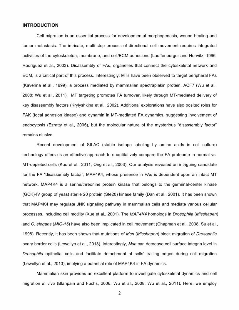
Abstract
Disassembly of focal adhesions (FAs) allows cell retraction and integrin detachment from the
ECM, processes critical for cell movement. Growth of MT (microtubule) can promote FA turnover by
serving as tracks to deliver proteins essential for FA disassembly. The molecular nature of this FA
“disassembly factor”, however, remains elusive. By quantitative proteomics, we identified MAP4K4
(mitogen-activated protein kinase kinase kinase kinase 4) as a FA regulator that associates with MTs.
Conditional knockout (cKO) of MAP4K4 in skin stabilizes FAs and impairs epidermal migration. By
exploring underlying mechanisms, we further show that MAP4K4 associates with EB2, a MT binding
protein, and IQSEC1, a guanine nucleotide exchange factor (GEF) specific for Arf6, whose activation
promotes integrin internalization. Together, our findings provide new insights into FA disassembly,
suggesting that MTs can deliver MAP4K4 toward FAs through EB2, where MAP4K4 can in turn
activate Arf6 via IQSEC1 and enhance FA dissolution.
2
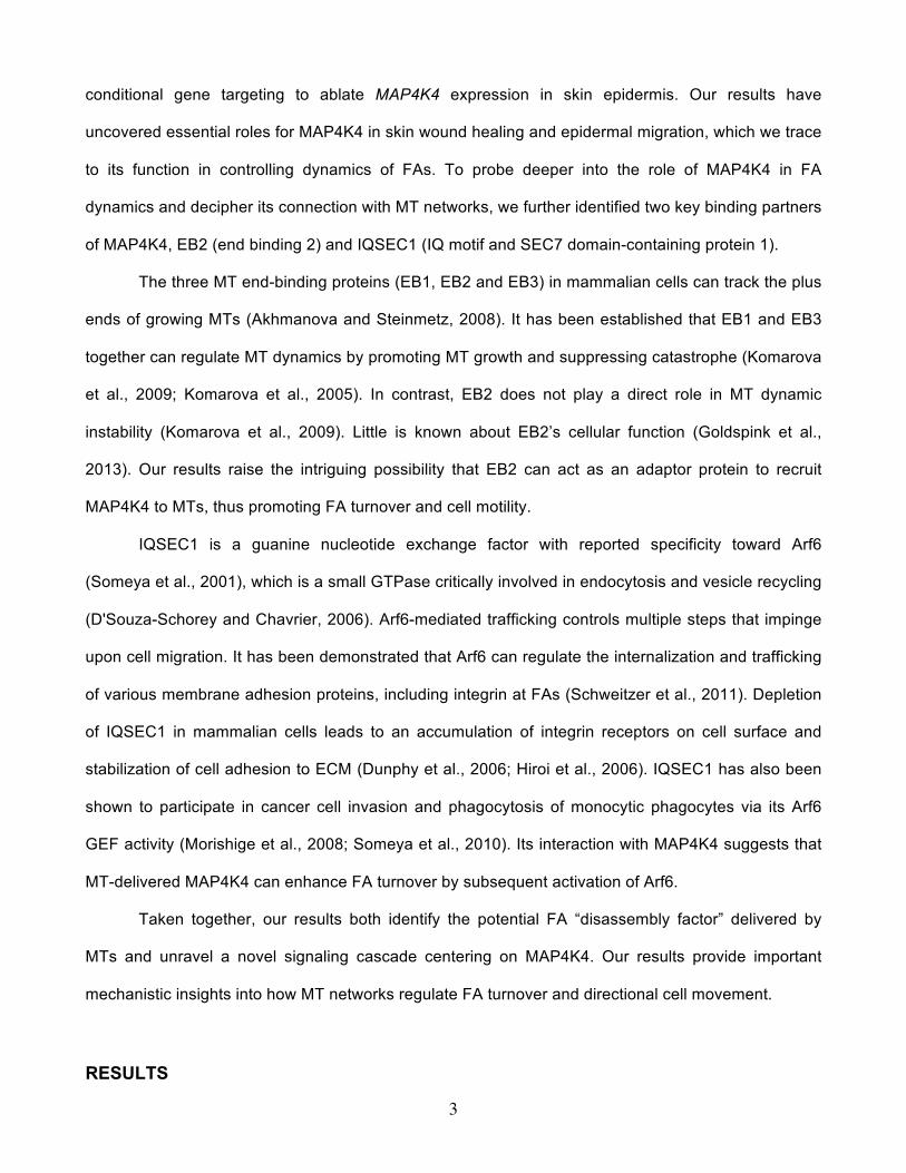
INTRODUCTION
Cell migration is an essential process for developmental morphogenesis, wound healing and
tumor metastasis. The intricate, multi-step process of directional cell movement requires integrated
activities of the cytoskeleton, membrane, and cell/ECM adhesions (Lauffenburger and Horwitz, 1996;
Rodriguez et al., 2003). Disassembly of FAs, organelles that connect the cytoskeletal network and
ECM, is a critical part of this process. Interestingly, MTs have been observed to target peripheral FAs
(Kaverina et al., 1999), a process mediated by mammalian spectraplakin protein, ACF7 (Wu et al.,
2008; Wu et al., 2011). MT targeting promotes FA turnover, likely through MT-mediated delivery of
key disassembly factors (Krylyshkina et al., 2002). Additional explorations have also posited roles for
FAK (focal adhesion kinase) and dynamin in MT-mediated FA dynamics, suggesting involvement of
endocytosis (Ezratty et al., 2005), but the molecular nature of the mysterious “disassembly factor”
remains elusive.
Recent development of SILAC (stable isotope labeling by amino acids in cell culture)
technology offers us an effective approach to quantitatively compare the FA proteome in normal vs.
MT-depleted cells (Kuo et al., 2011; Ong et al., 2003). Our analysis revealed an intriguing candidate
for the FA “disassembly factor”, MAP4K4, whose presence in FAs is dependent upon an intact MT
network. MAP4K4 is a serine/threonine protein kinase that belongs to the germinal-center kinase
(GCK)-IV group of yeast sterile 20 protein (Ste20) kinase family (Dan et al., 2001). It has been shown
that MAP4K4 may regulate JNK signaling pathway in mammalian cells and mediate various cellular
processes, including cell motility (Xue et al., 2001). The MAP4K4 homologs in Drosophila (Misshapen)
and C. elegans (MIG-15) have also been implicated in cell movement (Chapman et al., 2008; Su et al.,
1998). Recently, it has been shown that mutations of Msn (Misshapen) block migration of Drosophila
ovary border cells (Lewellyn et al., 2013). Interestingly, Msn can decrease cell surface integrin level in
Drosophila epithelial cells and facilitate detachment of cells’ trailing edges during cell migration
(Lewellyn et al., 2013), implying a potential role of MAP4K4 in FA dynamics.
Mammalian skin provides an excellent platform to investigate cytoskeletal dynamics and cell
migration in vivo (Blanpain and Fuchs, 2006; Wu et al., 2008; Wu et al., 2011). Here, we employ
3
conditional gene targeting to ablate MAP4K4 expression in skin epidermis. Our results have
uncovered essential roles for MAP4K4 in skin wound healing and epidermal migration, which we trace
to its function in controlling dynamics of FAs. To probe deeper into the role of MAP4K4 in FA
dynamics and decipher its connection with MT networks, we further identified two key binding partners
of MAP4K4, EB2 (end binding 2) and IQSEC1 (IQ motif and SEC7 domain-containing protein 1).
The three MT end-binding proteins (EB1, EB2 and EB3) in mammalian cells can track the plus
ends of growing MTs (Akhmanova and Steinmetz, 2008). It has been established that EB1 and EB3
together can regulate MT dynamics by promoting MT growth and suppressing catastrophe (Komarova
et al., 2009; Komarova et al., 2005). In contrast, EB2 does not play a direct role in MT dynamic
instability (Komarova et al., 2009). Little is known about EB2’s cellular function (Goldspink et al.,
2013). Our results raise the intriguing possibility that EB2 can act as an adaptor protein to recruit
MAP4K4 to MTs, thus promoting FA turnover and cell motility.
IQSEC1 is a guanine nucleotide exchange factor with reported specificity toward Arf6
(Someya et al., 2001), which is a small GTPase critically involved in endocytosis and vesicle recycling
(D'Souza-Schorey and Chavrier, 2006). Arf6-mediated trafficking controls multiple steps that impinge
upon cell migration. It has been demonstrated that Arf6 can regulate the internalization and trafficking
of various membrane adhesion proteins, including integrin at FAs (Schweitzer et al., 2011). Depletion
of IQSEC1 in mammalian cells leads to an accumulation of integrin receptors on cell surface and
stabilization of cell adhesion to ECM (Dunphy et al., 2006; Hiroi et al., 2006). IQSEC1 has also been
shown to participate in cancer cell invasion and phagocytosis of monocytic phagocytes via its Arf6
GEF activity (Morishige et al., 2008; Someya et al., 2010). Its interaction with MAP4K4 suggests that
MT-delivered MAP4K4 can enhance FA turnover by subsequent activation of Arf6.
Taken together, our results both identify the potential FA “disassembly factor” delivered by
MTs and unravel a novel signaling cascade centering on MAP4K4. Our results provide important
mechanistic insights into how MT networks regulate FA turnover and directional cell movement.
RESULTS
4
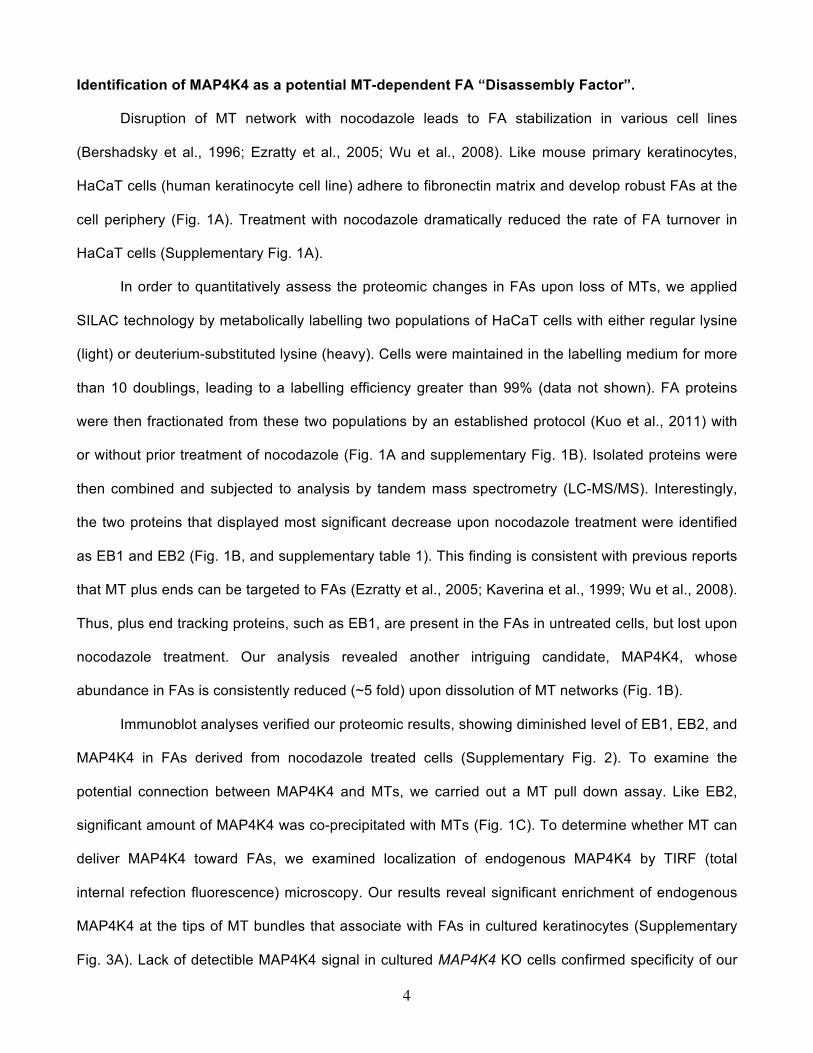
Identification of MAP4K4 as a potential MT-dependent FA “Disassembly Factor”.
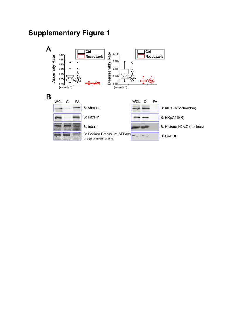
Disruption of MT network with nocodazole leads to FA stabilization in various cell lines
(Bershadsky et al., 1996; Ezratty et al., 2005; Wu et al., 2008). Like mouse primary keratinocytes,
HaCaT cells (human keratinocyte cell line) adhere to fibronectin matrix and develop robust FAs at the
cell periphery (Fig. 1A). Treatment with nocodazole dramatically reduced the rate of FA turnover in
HaCaT cells (Supplementary Fig. 1A).
In order to quantitatively assess the proteomic changes in FAs upon loss of MTs, we applied
SILAC technology by metabolically labelling two populations of HaCaT cells with either regular lysine
(light) or deuterium-substituted lysine (heavy). Cells were maintained in the labelling medium for more
than 10 doublings, leading to a labelling efficiency greater than 99% (data not shown). FA proteins
were then fractionated from these two populations by an established protocol (Kuo et al., 2011) with
or without prior treatment of nocodazole (Fig. 1A and supplementary Fig. 1B). Isolated proteins were
then combined and subjected to analysis by tandem mass spectrometry (LC-MS/MS). Interestingly,
the two proteins that displayed most significant decrease upon nocodazole treatment were identified
as EB1 and EB2 (Fig. 1B, and supplementary table 1). This finding is consistent with previous reports
that MT plus ends can be targeted to FAs (Ezratty et al., 2005; Kaverina et al., 1999; Wu et al., 2008).
Thus, plus end tracking proteins, such as EB1, are present in the FAs in untreated cells, but lost upon
nocodazole treatment. Our analysis revealed another intriguing candidate, MAP4K4, whose
abundance in FAs is consistently reduced (~5 fold) upon dissolution of MT networks (Fig. 1B).
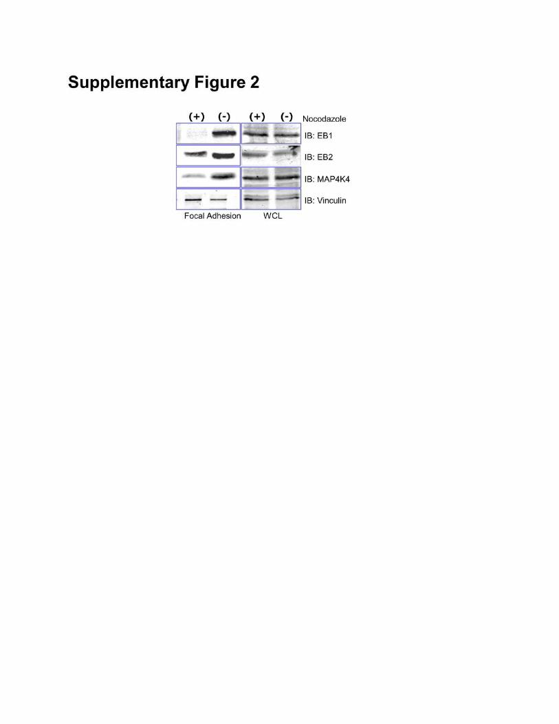
Immunoblot analyses verified our proteomic results, showing diminished level of EB1, EB2, and
MAP4K4 in FAs derived from nocodazole treated cells (Supplementary Fig. 2). To examine the
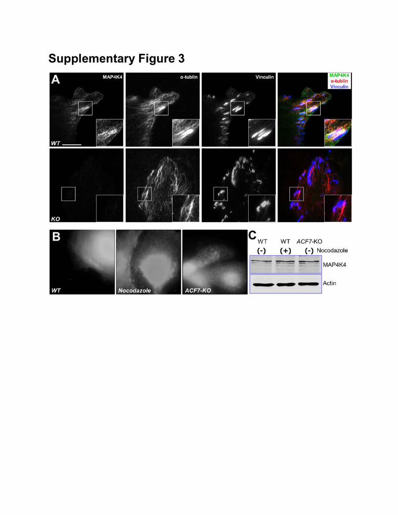
potential connection between MAP4K4 and MTs, we carried out a MT pull down assay. Like EB2,
significant amount of MAP4K4 was co-precipitated with MTs (Fig. 1C). To determine whether MT can
deliver MAP4K4 toward FAs, we examined localization of endogenous MAP4K4 by TIRF (total
internal refection fluorescence) microscopy. Our results reveal significant enrichment of endogenous
MAP4K4 at the tips of MT bundles that associate with FAs in cultured keratinocytes (Supplementary
Fig. 3A). Lack of detectible MAP4K4 signal in cultured MAP4K4 KO cells confirmed specificity of our
5
staining. Interestingly, co-localization between endogenous MAP4K4 and FA is markedly reduced in
nocodazole treated cells or ACF7 null cells (Fig. 1D-E). ACF7 has been demonstrated to play an
essential role to mediate targeting of MTs toward FAs (Wu et al., 2008). The reduction of MAP4K4
localization is specific to FA, as epi-fluorescence imaging of the same cells (Fig. 1D) show
comparable level of MAP4K4 (Supplementary Fig. 3B), and immunoblots confirmed that nocodazole
treatment or loss of ACF7 does not significantly change the level of endogenous MAP4K4
(Supplementary Fig. 3C). Together, these results suggest that MAP4K4 could serve as the potential
candidate that mediates crosstalk between MTs and FAs.
In support of this hypothesis, accumulating evidence suggests that MAP4K4 and its
homologues in other model organisms act as pro-migratory kinases (Chapman et al., 2008; Su et al.,
1998; Xue et al., 2001). Mutation of Msn, the Drosophila homolog of MAP4K4, increases surface
integrin level and impairs cell detachment (Lewellyn et al., 2013), strongly suggesting a role of
MAP4K4 in FA turnover. In line with previous findings, we observed that exogenous expression of
GFP-MAP4K4 but not GFP alone in mouse primary keratinocytes caused cell to round up and detach
from the matrix (data not shown). When exposed to fibronectin, cells expressing GFP-MAP4K4
exhibited significantly delayed adhesion and spreading comparing with GFP-expressing cells (Fig. 1F).
Taken together, our results identify MAP4K4 as a potential regulator of FA dynamics that associates
with MTs.
Conditional Knockout of MAP4K4 Leads to Aberrant Wound Repair and Defects in Epidermal
Cell Migration.
While adult mouse skin serves as an ideal system to study cell movement in vivo (Wu et al.,
2008), deletion of MAP4K4 in mice leads to early embryonic lethality (Xue et al., 2001). To circumvent
this issue, we developed skin cKO of MAP4K4. By homologous recombination, two loxP sites were
inserted in the 5’ UTR and the 2nd intron of MAP4K4 respectively. To conditionally target MAP4K4 in
skin, we bred MAP4K4fl/fl mice with K14-Cre recombinase transgenic mice, which efficiently excised
floxed exons by embryonic day E15.5 (Wu et al., 2008) (Fig. 2A). Neonatal mice genotypic for K14-
6
Cre and MAP4K4fl/fl alleles (cKO) were born in the expected Mendelian numbers and grew to
adulthood. Western blot confirmed the loss of MAP4K4 protein in cKO skin epidermis (Fig. 2A).
Although MAP4K4 skin cKO animals usually appeared smaller comparing with their littermates,
they manifested no gross morphological changes in skin or hair coat (Fig. 2B). Histologically,
epidermal homeostasis appeared normal and immunofluorescence staining with antibodies against β4
integrin (basal surface of the epidermis), keratin 5 (basal layer), keratin10 (spinous layer), and loricrin
(granular layer) all displayed localization patterns similar to WT (Wild Type) littermate’s skin (Fig. 2C).
Directional cell movement makes critical contribution to skin wound repair. When challenged to
respond to injury, MAP4K4 cKO skin exhibited a significant delay in repairing full-thickness wounds as
compared to WT skin. Histological analysis and quantification revealed that the area of
hyperproliferative epithelium (HE) that typically proliferates and migrates into the wound site was
diminished by more than 50% over 2-6 days following injury (Fig. 2D and E). Interestingly, despite the
delay in wound-closure, no significant alterations were found in cell proliferation upon loss of MAP4K4,
as judged by labeling for Ki67 (Fig. 2F). Thus, the delayed wound healing response in MAP4K4-
deficient skin seemed more likely to be rooted in potential defects in cell migration.
To examine this hypothesis, we isolated and analyzed primary mouse keratinocytes from WT
and cKO skin (Fig. 2H). Cells deficient for MAP4K4 exhibited a flatter and round morphology,
resembling ACF7 null cells that develop abnormally stable FA and migrate poorly in vitro (Wu et al.,
2008). We then monitored cell migration with isolated keratinocytes, during recovery of scratch
wounds introduced into cell monolayers. Whereas WT keratinocytes usually closed the gap usually
within 36 hours, KO keratinocytes moved only ~30% of this distance into the wound (Fig. 2G and
supplementary Fig. 4A). Videomicroscopy permitted image and monitoring the velocities and directed
migration of individual keratinocytes. Our results demonstrated that MAP4K4 KO cells moved
significantly more slowly compared to their WT counterparts. Quantification of cell motility revealed a
dramatic decrease in the average speed of KO keratinocytes (Figure 2H and supplementary Fig. 4B).
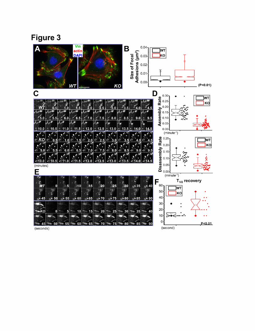
MAP4K4 Deficiency Leads to Aberrant Regulation of FA Dynamics
7
To determine the role of MAP4K4 in FA dynamics, we examined cell/ECM adhesions after
MAP4K4 deletion. Immunofluorescence microscopy showed significantly enhanced labeling of FAs in
MAP4K4 null keratinocytes relative to WT controls (Fig. 3A). Quantification of the presence of Vinculin
showed a significant increase in the size of FAs in MAP4K4 null cells (Fig. 3B).
We further employed confocal videomicroscopy to trace and examine the behavior of individual
FA (Wu et al., 2008). To visualize FAs, we transfected cells with plasmids encoding DsRed-Zyxin, a
fluorescently labeled FA marker protein (Wu et al., 2008). Representative examples of the
perturbations in FAs dynamics arising from MAP4K4-deficiency are shown in complete form in
Supplementary Video 1, and in montages in Figure 3C. During the interval of observation, FAs in
MAP4K4 null cells were often static, while many FAs in WT keratinocytes underwent continual bouts
of formation, maturation and disassembly. Quantification of the kinetics of individual FAs revealed
dramatic decrease in both the assembly and disassembly rates of FAs in KO cells (Fig. 3D).The
defects in FA dynamics were further substantiated by fluorescence recovery after photobleaching
(FRAP) experiments (Fig. 3E and supplementary Fig. 5). By comparison with GFP-Paxillin transfected
WT cells, we found that deletion of MAP4K4 resulted in a substantial increase in half-times of
fluorescence recovery after bleaching (Fig. 3F). Together, these data provide compelling evidence
that MAP4K4 regulates cell migration by promoting FA dynamics.
Identification of MAP4K4 Binding Partners
MT targeting promotes FA turnover (Ezratty et al., 2005; Kaverina et al., 1999; Rodriguez et al.,
2003; Wu et al., 2008). The phenotype of MAP4K4 deletion is highly analogous to what we observed
in ACF7 KO cells (Wu et al., 2008). However, unlike ACF7 null cells, MAP4K4 KO cells exhibit
bundled MT filaments that associate with peripheral FAs (Fig. 4A). Confocal videomicroscopy further
indicates comparable MT targeting frequency between WT and MAP4K4 KO cells (Supplementary
Fig. 6). Additionally, MT plus end dynamics are largely unaltered in MAP4K4 KO cells (Fig. 4B).
These results suggest that MAP4K4 does not regulate MT dynamics or FA targeting, but instead more
likely acts downstream of MTs to facilitate FA turnover.
8
Rho family small GTPases are known to play a pivotal in regulating FA dynamics and cell
migration (Jaffe and Hall, 2005). However, biochemical pull-down assays revealed comparable overall
levels of the GTP-bound state of RhoA, Rac1, and Cdc42 in WT and MAP4K4 KO cells
(Supplementary Fig. 7 A-C). Talin associates with integrin cytoplasmic tail and is involved in
mechanotransduction and activation of integrin (Calderwood et al., 2013). Proteolysis of talin by
calpain has been shown to be critical for FA dynamics (Franco et al., 2004). However, our immunoblot
assays demonstrate similar level of talin and cleaved talin in WT and MAP4K4 null cells
(Supplementary Fig. 7D). Together, these findings suggest that MAP4K4 regulates FA dynamics
through a distinct mechanism other than activation of Rho GTPases or talin proteolysis.
To probe deeply into the role of MAP4K4 in FA dynamics, we performed tandem affinity
purification to identify its binding partners. Purified MAP4K4 binding proteins were resolved by
electrophoresis and examined by silver staining (Fig. 4C). Interestingly, our analysis with LC-MS/MS
identified two MAP4K4 interacting proteins with potential relevance in cytoskeletal dynamics and FA
turnover, EB2 and IQSEC1 (Fig. 4D).
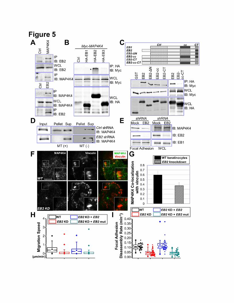
MAP4K4 Interaction with EB2
To verify MAP4K4 interaction with EB2, we carried out endogenous coimmunoprecipitation
assays. Consistent with MS results, we detected significant amount of EB2 in α-MAP4K4
immunoprecipitates and MAP4K4 in α-EB2 precipitates (Fig. 5A).
Mammalian cells including keratinocytes express three EB proteins that share substantial
sequence homology (Komarova et al., 2009). To test the potential interaction with other EBs, we
exogenously expressed EB1, EB2, and EB3 together with MAP4K4 in cells. Co-immunoprecipitation
showed that MAP4K4 specifically associates with only EB2, but not EB1 or EB3 (Fig. 5B). This
specificity is consistent with our SILAC analysis of FAs, which shows comparable fold changes of EB2
and MAP4K4 in FAs upon disruption of MTs (Fig. 1B). Sequence alignment of three EB family
members indicates three unique regions in EB2, the N terminus, the acidic C terminus (CT) and an
internal region close to the coiled coil domain (cc) (Fig. 5C). To examine their contribution to binding
9
with MAP4K4, we introduced deletion or swapping mutations to EB2 by either removing the N
terminus or replacing the unique regions in EB2 with corresponding sequences from EB1 (Fig. 5C).
We then carried out co-immunoprecipitation to determine their respective binding affinity to MAP4K4.
Our results show that, whereas the N terminus of EB2 is dispensable for MAP4K4 interaction, EB1
swapping mutation at the cc domain or CT diminished association with MAP4K4 at different degree
(Fig. 5C). Interaction with MAP4K4 was barely detectible when we used the EB2 mutant containing
swapping mutations at both cc domain and CT (Fig. 5C), suggesting that MAP4K4 associates with
EB2 at multiple locations.
MAP4K4 associates with MTs (Fig. 1C) but does not possess any recognizable MT binding
motif. As a MT binding protein, EB2 could function as an adaptor protein to recruit MAP4K4 to MTs.
To test this hypothesis, we prepared lentiviral vector encoding EB2 shRNA sequence as reported
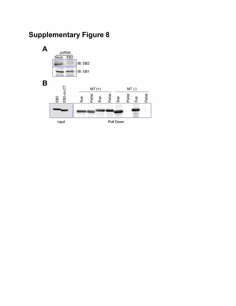
(Supplementary Fig. 8A) (Komarova et al., 2009; Komarova et al., 2005) and performed MT pull down
assay for MAP4K4. Interestingly, depletion of endogenous EB2 greatly reduces MT binding of
MAP4K4 (Fig. 5D). Additionally, knockdown of EB2 leads to a significant decrease of MAP4K4 level in
FAs (Fig. 5E-G). These results strongly suggest that EB2 acts as an essential adaptor, permitting MT-
delivery of MAP4K4 to FAs.
Consistent with our hypothesis, knockdown of EB2 in mouse primary keratinocytes led to
severe defects in cell motility (Fig. 5H). Additionally, FAs in EB2-depleted cells were stabilized, as
determined by confocal videomicroscopy (Fig. 5I). Exogenous expression of an EB2 mutant with
multiple silent mutations within the shRNA recognition sequence restored cell motility and FA
dynamics in knockdown cells (Fig. 5H and I). By contrast, expression of MAP4K4 non-binding mutant
of EB2 (cc-CT mutant, Fig. 5C) failed to rescue the defects (Fig. 5H and I), indicating an essential role
for MAP4K4 interaction in this process. The cc-CT mutation in EB2 does not alter its MT binding
affinity (Supplementary Fig. 8B), or change its ability to track MT plus ends (Supplementary video 2).
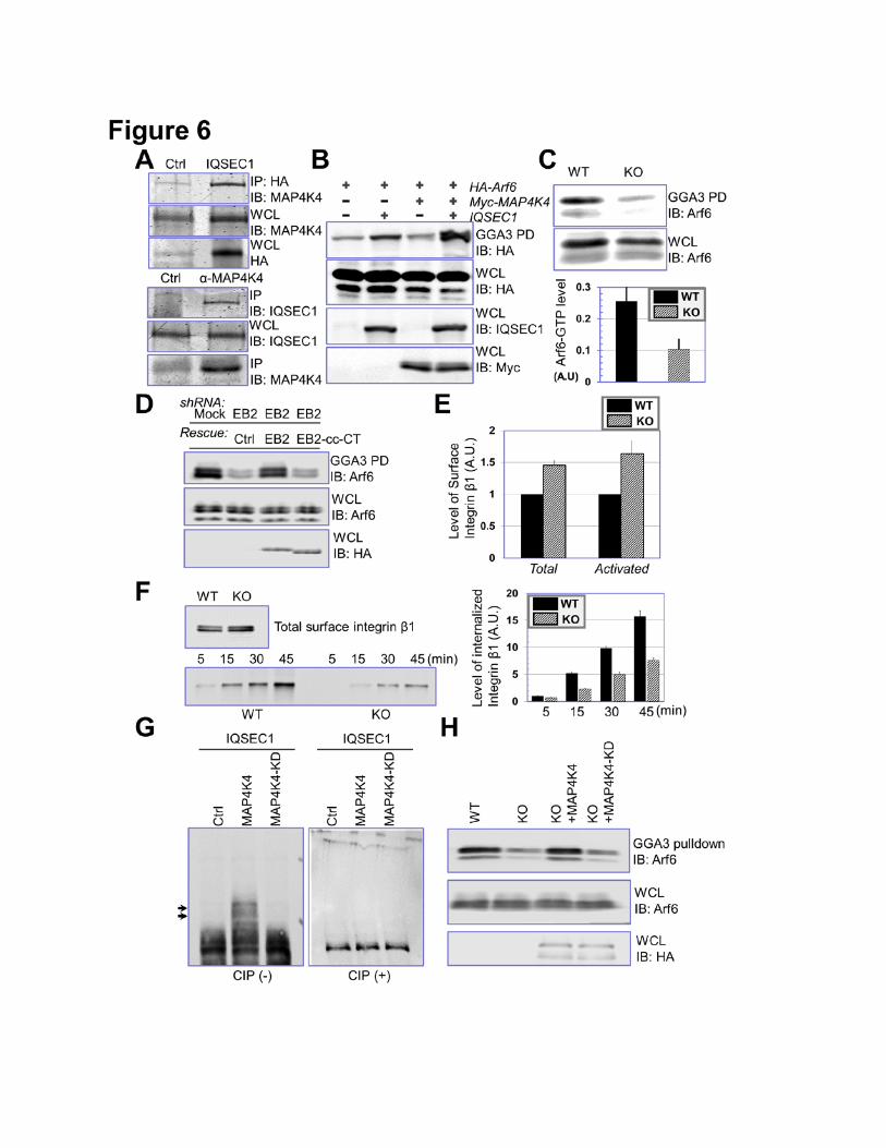
MAP4K4 Activates Arf6 through its Interaction with IQSEC1
10
Our MS analysis identified IQSEC1 as a MAP4K4 binding protein (Fig. 4C-D), and presence of
the N terminal peptide of IQSEC1 suggests that MAP4K4 associates with the longer splicing isoform
of IQSEC1 (Dunphy et al., 2006). Coimmunoprecipitation assay confirmed interaction between
IQSEC1 and MAP4K4 (Fig. 6A).
Mounting evidence suggests that IQSEC1, an Arf6 GEF protein, regulates cell adhesion to
matrix via Arf6 (Dunphy et al., 2006; Hiroi et al., 2006; Morishige et al., 2008; Someya et al., 2010).
We first examined the potential role of MAP4K4 in activation of Arf6. To this end, we exogenously
expressed IQSEC1 with or without MAP4K4 and performed GGA3 pull down assay to determine the
level of GTP-bound Arf6 (D'Souza-Schorey and Chavrier, 2006). Expression of IQSEC1 (the longer
isoform as BRAG2b) alone only modestly enhanced the Arf6 activity, whereas co-expression of both
IQSEC1 and MAP4K4 led to a significant increase in GTP-Arf6 level (Fig. 6B). Consistently, loss of
MAP4K4 in keratinocytes significantly reduced endogenous Arf6-GTP level as expected (Fig. 6C). To
test whether MAP4K4 binding with MTs is involved in Arf6 activation, we carried out GGA3 pull-down
analysis in EB2 knockdown cells. As expected, depletion of endogenous EB2 leads to dramatic
reduction of GTP-bound Arf6 level (Fig. 6D). Expression of WT EB2 but not EB2 mutant deficient for
MAP4K4 interaction (cc-CT mutant of EB2) can restore Arf6 activation, strongly suggesting that
MAP4K4 association with EB2 is essential for Arf6 activation in cells (Fig. 6D).
IQSEC1 and Arf6 control cell adhesion by regulation of integrin internalization and intracellular
trafficking (D'Souza-Schorey and Chavrier, 2006; Dunphy et al., 2006). To determine the role of
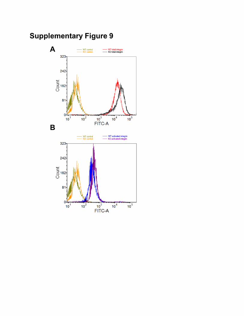
MAP4K4 in this process, we first quantitated the surface expression of endogenous β1 integrin by flow
cytometry. Our results indicate that KO of MAP4K4 in skin keratinocytes leads to an ~50% increase in
surface level of β1 integrin (total or active β1 integrin) relative to WT control cells (Fig. 6E and
supplementary Fig. 9). Immunoblotting of cell lysates demonstrated that overall expression of β1
integrin was not affected by loss of MAP4K4 (data not shown). As reported in other cell lines (Dunphy
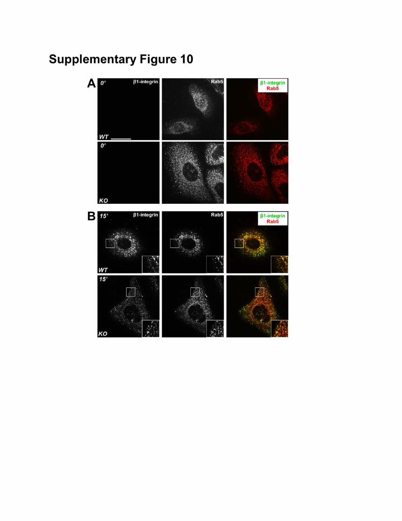
et al., 2006; Ezratty et al., 2005), β1 integrin in keratinocytes is undergoing active endocytosis and
intracellular trafficking. Internalized β1 integrin can be detected in intracellular vesicles and co-localize
with various endosomal markers, such as Rab5 (Supplementary Fig. 10). To quantitatively assess the
11
rate of β1 integrin internalization, we carried out reversible biotinylation assay (Wu et al., 2005). Our
results reveal that endocytosis of β1 integrin in MAP4K4 null cells is significantly reduced (Fig. 6F).
Together, our results provide compelling evidence that MAP4K4 promotes FA dynamics by regulating
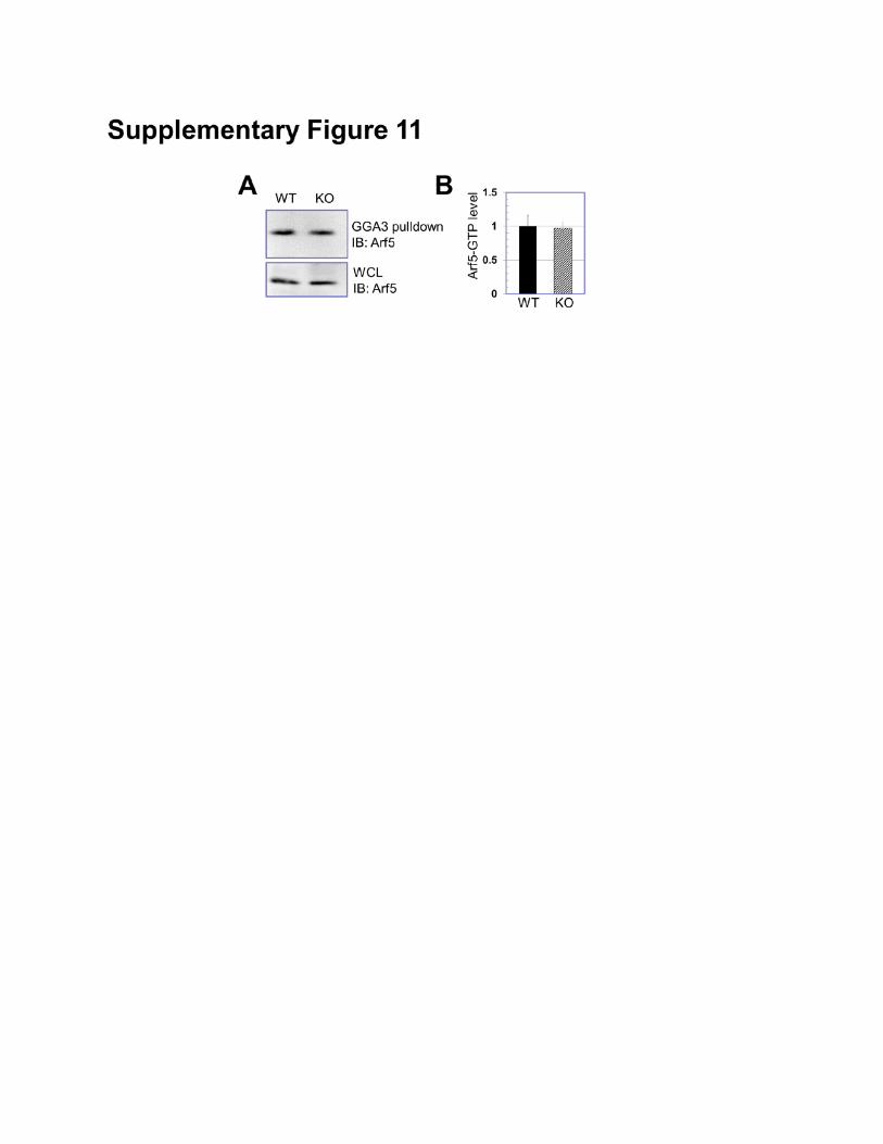
IQSEC1/Arf6 pathway and controlling endocytosis of integrin. Recent study suggested that IQSEC1
might regulate α5β1 integrin endocytosis via activation of Arf5 (Moravec et al., 2012). However, our
biochemical pull-down suggests that level of GTP-bound Arf5 is not significantly affected by deletion
of MAP4K4 (Supplementary Fig. 11).
Activity of GEF proteins can be regulated by post-translational modifications, such as
phosphorylation. Co-expression of IQSEC1 with WT MAP4K4 but not kinase defective (KD) mutant of
MAP4K4 leads to significant phosphorylation of IQSEC1 in vivo, which is sensitive to treatment of
alkaline phosphatase (Fig. 6G). To examine the potential relevance in IQSEC1 activity, we re-
expressed WT or KD mutant of MAP4K4 in MAP4K4 deficient cells. Pull-down results indicate that
WT but not KD mutant of MAP4K4 can rescue Arf6 activity in MAP4K4 KO cells, suggesting that
MAP4K4 might regulate Arf6 activation via protein phosphorylation (Fig. 6H). When expressed in
MAP4K4 KO cells, WT but not KD mutant of MAP4K4 can enhance FA disassembly and cell
movement, suggesting that the kinase activity of MAP4K4 is essential for its role in these processes
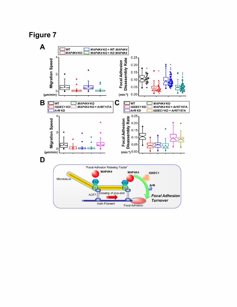
(Fig. 7A).
To determine the relevance of IQSEC1/Arf6 pathway in migration of keratinocytes, we
prepared shRNAs for both genes (Supplementary Fig. 12). Consistent with reports from other cell
lines (Dunphy et al., 2006; Morishige et al., 2008; Someya et al., 2001), depletion of endogenous
IQSEC1 or Arf6 in mouse keratinocytes markedly stabilized FAs and impaired cell motility (Fig. 7B
and C). To determine whether Arf6 acts downstream to MAP4K4 in these processes, we transfected
MAP4K4 KO cells with plasmid encoding Arf6 T157A, the fast cycling mutant of Arf6 (Santy, 2002).
Relative to mock transfection, expression of Arf6 T157A significantly rescued cell motility and FA
disassembly defects in MAP4K4 null cells (Fig. 7B and C). In addition, expression of Arf6 T157A
restored FA dynamics in cells depleted with IQSEC1 (Fig. 7C). Taken together, our results delineate a
12
new signaling cascade whereby MT dynamics regulate FA turnover through MAP4K4 interaction with
IQSEC1 and subsequent activation of Arf6 (Fig. 7D).
DISCUSSION
Accumulating evidence suggests that FAs can serve as the “hotspot” for crosstalk between MT
and actin networks (Rodriguez et al., 2003). Small’s group has shown that MTs can specifically grow
towards FAs, a process likely guided by underlying F-actin filaments (Kaverina et al., 1999;
Krylyshkina et al., 2002). Targeting of MTs toward FAs promotes dissolution of FAs at the cell
periphery, a feature in line with the long-standing observation that cells treated with MT
depolymerizing drugs contain enlarged and stabilized FAs (Bershadsky et al., 1996; Ezratty et al.,
2005; Wu et al., 2008). Further evidence has implicated dynamin and clathrin in this process, strongly
suggesting that FA dissolution is mediated by the endocytic pathway (Ezratty et al., 2009; Ezratty et
al., 2005). However, the molecular mechanisms by which MT targeting contributes to FA dynamics
have not been established. It has been speculated that MTs can serve as macromolecular tracks to
deliver an unknown “disassembly factor” to facilitate FA turnover (Krylyshkina et al., 2002). But the
nature of this “disassembly factor” and how this “disassembly factor” can activate endocytic machinery
to promote FA dissolution remain unclear.
Our study presents compelling evidence that MAP4K4 acts as a MT-dependent “disassembly
factor” to promote FA dynamics. Our model suggests that MAP4K4 associates with MT via its
interaction with EB2. Upon targeting to FAs via MTs, MAP4K4 can bind and phosphorylate IQSEC1,
which in turn activates Arf6 and endocytosis, leading to turnover of FAs. A schematic outlining this
model is presented in Figure 7D. Consistent with this model, ablation of MAP4K4 in skin epidermal
cells impairs wound healing in vivo. Furthermore, isolated MAP4K4 null cells display diminished Arf6
activity, stabilized FAs, and aberrant cell motility in vitro. Our results illuminate a hitherto unrecognized
signaling cascade underlying FA dynamics and cell migration.
Interestingly, our study suggests that loss of MAP4K4 in primary skin keratinocytes inhibits
both FA assembly and disassembly. This finding is consistent with our previous report on ACF7,
13
whose deletion abolishes targeting of MT plus ends to FAs and inhibits both FA assembly and
disassembly (Wu et al., 2008). In addition, treatment with nocodazole leads to stabilization of FAs,
and assembly and disassembly rate of FAs are decreased similarly (Supplementary Fig. 1A) (Wu et
al., 2008). Together, these results argue that in addition to FA dissolution, MT dynamics and targeting
of MTs to FAs may play an equally important role in FA assembly. The underlying molecular
mechanisms deserve further investigation.
Our study has also elucidated the role of EB2 in MT functionality. Growing MT ends
accumulate a special group of MT interacting proteins, often referred to as MT plus-end tracking
proteins (Akhmanova and Steinmetz, 2008). In addition to EB1, mammalian cells also express two
other EB proteins, EB2 and EB3, which are encoded by separate genes. It has been reported that
EB1 and EB3 together play a major role in mammalian cell to control MT dynamics by promoting
growth and suppressing catastrophe of MTs, whereas EB2 is dispensable in these processes
(Komarova et al., 2009; Komarova et al., 2005). In addition, unlike EB1 and EB3, enrichment of EB2
at MT plus ends is less prominent (Komarova et al., 2009). Recent evidence implicated EB2 in MT
reorganization during apico-basal differentiation of various epithelial cells (Goldspink et al., 2013). Our
study for the first time unravels a specific and essential role of EB2 in mediating the crosstalk between
MTs and FAs. Inactivation of EB2 in cultured keratinocytes leads to aberrant FA dynamics and
decreased motility, at least in part through its specific interaction with MAP4K4. Future studies will be
needed to fully understand how the three EB proteins act in different cellular processes in vivo. We
favor the hypothesis that unlike EB1 and EB3, EB2 might not be directly involved in regulation of MT
dynamic instability. Instead, via its unique sequence, EB2 may function as an adaptor molecule to
recruit signaling molecules to MTs, which is required for various cellular processes, including cell
motility.
Under different conditions, turnover of FAs can be mediated by clathrin-dependent endocytosis
or macropinocytosis (Ezratty et al., 2009; Gu et al., 2011). As a unique member of Arf protein family,
Arf6 localizes at the plasma membrane and endosomal compartments. Its role in endocytic
membrane trafficking is well established, but it also likely regulates macropinocytosis via its function in
14
remodeling F-actin network (Schweitzer et al., 2011). IQSEC1 preferentially activates Arf6 in cells
(Someya et al., 2001). Depletion of IQSEC1 by siRNA impairs integrin internalization as well as
phagocytosis, a process resembling macropinocytosis in epithelial cells (Dunphy et al., 2006; Hiroi et
al., 2006; Someya et al., 2010). Further investigation will be required to determine whether MAP4K4-
induced Arf6 activity promotes FA turnover via clathrin-dependent endocytosis or macropinocytosis,
or both. Nevertheless, identification of the interaction between MAP4K4 and IQSEC1 provides an
intriguing link between Arf6 signaling and MT network, fulfilling the gap in our understanding of MT-
induced turnover of FA.
In closing, our findings provide critical insights into the mechanics of MT-induced FA turnover.
In addition, our results suggest a novel signaling cascade centering on MAP4K4 in directional cell
movement and now pave the way for probing more deeply into the intricate signaling network
orchestrating the crosstalk between F-actin and MT cytoskeletal networks and localized sites of
membrane-associated activity.
15
EXPERIMENTAL PROCEDURES
SILAC (Stable Isotope Labeling) and FA fractionation
HaCaT cells were labeled with either “heavy” or “light” isotopic lysine using a SILAC protein
quantification kit (Thermo scientific, Rockford, IL, USA) according to manufacturer’s instruction. Briefly,
cells were grown in DMEM:F12 medium supplemented with 10% dialyzed fetal bovine serum and
either the “heavy” form of L-Lysine-2HCl (4, 4, 5, 5-D4) or “light” L-Lysine for more than six
generations to achieve more than 98% of labeling efficiency (Ong et al., 2003).
To isolate FA proteins, subconfluent HaCaT cells were treated with nocodazole (13 µm) or
vehicle control for 30 minutes. Fractionation was carried out essentially as described (Kuo et al.,
2011). Briefly, cells were hypotonically shocked with triethanolamine-containing solution. Cell bodies
were removed by hydrodynamic force with PBS repetitively. FA proteins were then collected and
denatured by scraping and sonication. Samples were analyzed with LC-MS/MS afterwards.
Generation of MAP4K4 cKO Mice
The MAP4K4 flox strain was kindly provided by Dr. Min Xie at University of Texas
Southwestern Medical Center and Prof. Michael Schneider at Imperial College London. The breeding
stock of MAP4K4 flox animals were provided by Dr. Edward Skolnik at New York University. MAP4K4
cKO animals were generated by breeding MAP4K4fl/fl mice to K14-Cre transgenic mice. All mice used
in this study were bred and maintained at the ARC (animal resource center) of University of Chicago
in accordance with institutional guidelines.
TIRF microscopy
TIRF imaging was done using Leica GSD/TIRFM Ground State Depletion Superresolution
Microscope. Solid state laser light (405nm, 488nm, 532nm, 642nm) was focused at the back focal
plane of a 160x NA 1.43 objective. Signals were recorded by Andor iXon3 897 high speed EMCCD
camera. For TIRF imaging, the stained cells were mounted in an open chamber filled with 1X PBS.
Pearson's coefficient for co-localization was calculated using NIH ImageJ software.
16
FRAP and FA Assembly/Disassembly Measurements
Kinetics of FA assembly and disassembly were performed essentially as previously described
(Wu et al., 2008). Keratinocytes were plated on fibronectin-coated dishes and transfected with plasmid
encoding DsRed-Zyxin. Time series of images were acquired on a 3i Marianas Yokogawa-type
spinning-disc confocal microscope equipped with a 100x α-plane (1.45 oil) lens and an EM charge-
coupled device camera. The rate constants for FA assembly and disassembly were obtained by
calculating the slope of relative fluorescence intensity increases or decreases of individual FA on a
semilogarithmic scale against time.
FRAP assays were performed essentially as described (Wu et al., 2008). Briefly, cells were
plated on fibronectin-coated 3.5-cm dishes and transfected with plasmid encoding GFP-paxillin. Three
or two prebleach events were performed followed by a 1-s bleach event. Fluorescence recovery was
recorded for 140 seconds after photobleaching events, and data from these photokinetic experiments
were analyzed using DeltaVision software.
ACKNOWLEDGMENTS
We are very grateful to Dr. Min Xie at University of Texas Southwestern Medical Center and
Prof. Michael Schneider at Imperial College London for sharing their MAP4K4 floxed mice in advance
of publication, and to Dr. Edward Skolnik at New York University for providing us the breeding stocks.
We thank Dr. Yulia Komarova at University of Illinois at Chicago for providing the EB2 construct. We
thank Dr. Martha Vaughan at National Institutes of Health for providing the IQSEC1 construct. We
thank Dr. Elaine Fuchs at the Rockefeller University for sharing reagents. We thank Dr. Don
Wolfgeher at University of Chicago Proteomics Core lab and Dr. Vytas Bindokas and Dr. Chrstine
Labno at University of Chicago Light Microscopy Core and Dr. Kathryn Fox at University of Chicago
for their excellent technical assistance. The animal studies were carried out in the ALAAC-accredited
animal research facility at the University of Chicago. This work was supported by a grant R01-

17
AR063630 from the National Institutes of Health, the Research Scholar Grant (RSG-13-198-01) from
the American Cancer Society, and the V scholar award from V foundation.
18
References:
Akhmanova, A., and Steinmetz, M.O. (2008). Tracking the ends: a dynamic protein network controls
the fate of microtubule tips. Nature reviews Molecular cell biology 9, 309-322.
Bershadsky, A., Chausovsky, A., Becker, E., Lyubimova, A., and Geiger, B. (1996). Involvement of
microtubules in the control of adhesion-dependent signal transduction. Current biology : CB 6, 1279-
1289.
Blanpain, C., and Fuchs, E. (2006). Epidermal stem cells of the skin. Annual review of cell and
developmental biology 22, 339-373.
Calderwood, D.A., Campbell, I.D., and Critchley, D.R. (2013). Talins and kindlins: partners in integrin-
mediated adhesion. Nature reviews Molecular cell biology 14, 503-517.
Chapman, J.O., Li, H., and Lundquist, E.A. (2008). The MIG-15 NIK kinase acts cell-autonomously in
neuroblast polarization and migration in C. elegans. Developmental biology 324, 245-257.
D'Souza-Schorey, C., and Chavrier, P. (2006). ARF proteins: roles in membrane traffic and beyond.
Nature reviews Molecular cell biology 7, 347-358.
Dan, I., Watanabe, N.M., and Kusumi, A. (2001). The Ste20 group kinases as regulators of MAP
kinase cascades. Trends in cell biology 11, 220-230.
Dunphy, J.L., Moravec, R., Ly, K., Lasell, T.K., Melancon, P., and Casanova, J.E. (2006). The Arf6
GEF GEP100/BRAG2 regulates cell adhesion by controlling endocytosis of beta1 integrins. Current
biology : CB 16, 315-320.
Ezratty, E.J., Bertaux, C., Marcantonio, E.E., and Gundersen, G.G. (2009). Clathrin mediates integrin
endocytosis for focal adhesion disassembly in migrating cells. The Journal of cell biology 187, 733-
747.
Ezratty, E.J., Partridge, M.A., and Gundersen, G.G. (2005). Microtubule-induced focal adhesion
disassembly is mediated by dynamin and focal adhesion kinase. Nature cell biology 7, 581-590.
Franco, S.J., Rodgers, M.A., Perrin, B.J., Han, J., Bennin, D.A., Critchley, D.R., and Huttenlocher, A.
(2004). Calpain-mediated proteolysis of talin regulates adhesion dynamics. Nature cell biology 6, 977-
983.
19
Goldspink, D.A., Gadsby, J.R., Bellett, G., Keynton, J., Tyrrell, B.J., Lund, E.K., Powell, P.P., Thomas,
P., and Mogensen, M.M. (2013). The microtubule end-binding protein EB2 is a central regulator of
microtubule reorganisation in apico-basal epithelial differentiation. Journal of cell science 126, 4000-
4014.
Gu, Z., Noss, E.H., Hsu, V.W., and Brenner, M.B. (2011). Integrins traffic rapidly via circular dorsal
ruffles and macropinocytosis during stimulated cell migration. The Journal of cell biology 193, 61-70.
Hiroi, T., Someya, A., Thompson, W., Moss, J., and Vaughan, M. (2006). GEP100/BRAG2: activator
of ADP-ribosylation factor 6 for regulation of cell adhesion and actin cytoskeleton via E-cadherin and
alpha-catenin. Proceedings of the National Academy of Sciences of the United States of America 103,
10672-10677.
Humke, E.W., Dorn, K.V., Milenkovic, L., Scott, M.P., and Rohatgi, R. (2010). The output of Hedgehog
signaling is controlled by the dynamic association between Suppressor of Fused and the Gli proteins.
Genes & development 24, 670-682.
Jaffe, A.B., and Hall, A. (2005). Rho GTPases: biochemistry and biology. Annual review of cell and
developmental biology 21, 247-269.
Kaverina, I., Krylyshkina, O., and Small, J.V. (1999). Microtubule targeting of substrate contacts
promotes their relaxation and dissociation. The Journal of cell biology 146, 1033-1044.
Komarova, Y., De Groot, C.O., Grigoriev, I., Gouveia, S.M., Munteanu, E.L., Schober, J.M.,
Honnappa, S., Buey, R.M., Hoogenraad, C.C., Dogterom, M., et al. (2009). Mammalian end binding
proteins control persistent microtubule growth. The Journal of cell biology 184, 691-706.
Komarova, Y., Lansbergen, G., Galjart, N., Grosveld, F., Borisy, G.G., and Akhmanova, A. (2005).
EB1 and EB3 control CLIP dissociation from the ends of growing microtubules. Molecular biology of
the cell 16, 5334-5345.
Krylyshkina, O., Kaverina, I., Kranewitter, W., Steffen, W., Alonso, M.C., Cross, R.A., and Small, J.V.
(2002). Modulation of substrate adhesion dynamics via microtubule targeting requires kinesin-1. The
Journal of cell biology 156, 349-359.
20
Kuo, J.C., Han, X., Hsiao, C.T., Yates, J.R., 3rd, and Waterman, C.M. (2011). Analysis of the myosin-
II-responsive focal adhesion proteome reveals a role for beta-Pix in negative regulation of focal
adhesion maturation. Nature cell biology 13, 383-393.
Lauffenburger, D.A., and Horwitz, A.F. (1996). Cell migration: a physically integrated molecular
process. Cell 84, 359-369.
Lewellyn, L., Cetera, M., and Horne-Badovinac, S. (2013). Misshapen decreases integrin levels to
promote epithelial motility and planar polarity in Drosophila. The Journal of cell biology 200, 721-729.
Matos, J., Lipp, J.J., Bogdanova, A., Guillot, S., Okaz, E., Junqueira, M., Shevchenko, A., and
Zachariae, W. (2008). Dbf4-dependent CDC7 kinase links DNA replication to the segregation of
homologous chromosomes in meiosis I. Cell 135, 662-678.
Moravec, R., Conger, K.K., D'Souza, R., Allison, A.B., and Casanova, J.E. (2012).
BRAG2/GEP100/IQSec1 interacts with clathrin and regulates alpha5beta1 integrin endocytosis
through activation of ADP ribosylation factor 5 (Arf5). The Journal of biological chemistry 287, 31138-
31147.
Morishige, M., Hashimoto, S., Ogawa, E., Toda, Y., Kotani, H., Hirose, M., Wei, S., Hashimoto, A.,
Yamada, A., Yano, H., et al. (2008). GEP100 links epidermal growth factor receptor signalling to Arf6
activation to induce breast cancer invasion. Nature cell biology 10, 85-92.
Ong, S.E., Foster, L.J., and Mann, M. (2003). Mass spectrometric-based approaches in quantitative
proteomics. Methods 29, 124-130.
Rodriguez, O.C., Schaefer, A.W., Mandato, C.A., Forscher, P., Bement, W.M., and Waterman-Storer,
C.M. (2003). Conserved microtubule-actin interactions in cell movement and morphogenesis. Nature
cell biology 5, 599-609.
Santy, L.C. (2002). Characterization of a fast cycling ADP-ribosylation factor 6 mutant. The Journal of
biological chemistry 277, 40185-40188.
Schweitzer, J.K., Sedgwick, A.E., and D'Souza-Schorey, C. (2011). ARF6-mediated endocytic
recycling impacts cell movement, cell division and lipid homeostasis. Seminars in cell &
developmental biology 22, 39-47.
21
Someya, A., Moss, J., and Nagaoka, I. (2010). The guanine nucleotide exchange protein for ADP-
ribosylation factor 6, ARF-GEP100/BRAG2, regulates phagocytosis of monocytic phagocytes in an
ARF6-dependent process. The Journal of biological chemistry 285, 30698-30707.
Someya, A., Sata, M., Takeda, K., Pacheco-Rodriguez, G., Ferrans, V.J., Moss, J., and Vaughan, M.
(2001). ARF-GEP(100), a guanine nucleotide-exchange protein for ADP-ribosylation factor 6.
Proceedings of the National Academy of Sciences of the United States of America 98, 2413-2418.
Su, Y.C., Treisman, J.E., and Skolnik, E.Y. (1998). The Drosophila Ste20-related kinase misshapen is
required for embryonic dorsal closure and acts through a JNK MAPK module on an evolutionarily
conserved signaling pathway. Genes & development 12, 2371-2380.
Wu, X., Gan, B., Yoo, Y., and Guan, J.L. (2005). FAK-mediated src phosphorylation of endophilin A2
inhibits endocytosis of MT1-MMP and promotes ECM degradation. Dev Cell 9, 185-196.
Wu, X., Kodama, A., and Fuchs, E. (2008). ACF7 regulates cytoskeletal-focal adhesion dynamics and
migration and has ATPase activity. Cell 135, 137-148.
Wu, X., Shen, Q.T., Oristian, D.S., Lu, C.P., Zheng, Q., Wang, H.W., and Fuchs, E. (2011). Skin stem
cells orchestrate directional migration by regulating microtubule-ACF7 connections through
GSK3beta. Cell 144, 341-352.
Wu, X., Suetsugu, S., Cooper, L.A., Takenawa, T., and Guan, J.L. (2004). Focal adhesion kinase
regulation of N-WASP subcellular localization and function. The Journal of biological chemistry 279,
9565-9576.
Wu, Y., Li, Q., and Chen, X.Z. (2007). Detecting protein-protein interactions by Far western blotting.
Nat Protoc 2, 3278-3284.
Xue, Y., Wang, X., Li, Z., Gotoh, N., Chapman, D., and Skolnik, E.Y. (2001). Mesodermal patterning
defect in mice lacking the Ste20 NCK interacting kinase (NIK). Development 128, 1559-1572.
Ydenberg, C.A., and Rose, M.D. (2009). Antagonistic regulation of Fus2p nuclear localization by
pheromone signaling and the cell cycle. The Journal of cell biology 184, 409-422.
22
FIGURE LEGENDS
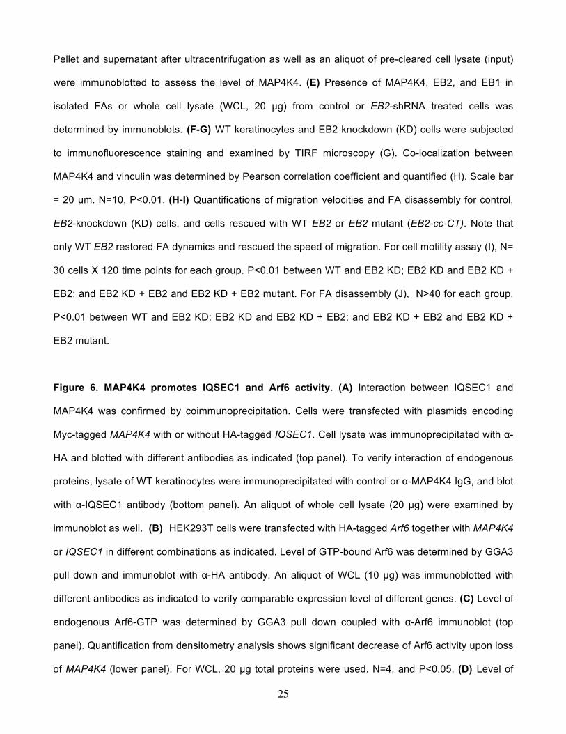
Figure 1. Identification of MAP4K4 as a MT-dependent FA protein. (A) Flow diagram of the
protocol used to isolate FA proteins for SILAC analysis. Representative HaCaT cells were stained
with α-vinculin antibody to visualize FAs in control of nocodazole treated cells (top two panels). Scale
bar = 20 µm. (B) Candidates that exhibit most significant changes in protein levels upon nocodazole
treatment. Cells labeled with light Lysine were treated with nocodozale, and heavy Lysine labeled
cells were not. Ratios of each candidate protein retrieved from heavy or light cells (H/L) were shown
together with the ratio variability. MAPRE1 or 2: microtubule associated protein, RP/EB family
member 1 or 2. The level change of vinculin is included as a control. (C) Pre-cleared cell lysate was
incubated with or without polymerized MTs. Interaction with MTs was examined by ultracentrifugation.
Presence of protein in the original lysate (input), pellet of centrifugation as well as supernatant was
determined by immunoblot with different antibodies as indicated. (D) WT mouse primary keratinocytes
treated with or without nocodazole and ACF7 KO keratinocytes were fixed and subjected to
immunofluorescence staining with different antibodies as indicated. Stained cells were examined by
TIRF microscopy. Scale bar = 20 µm. (E) Co-localization between MAP4K4 and FAs was determined
by Pearson correlation coefficient. N=10, and P<0.01. (F) GFP or GFP-MAP4K4 cells were harvested
and replated onto fibronectin-coated surface for 15 or 30 minutes. Cells were then fixed and stained.
Adhesion and spreading of cells (cell area) were quantified with Image J and shown as Box and
Whisker plots. Box and whisker plots indicates the mean (empty square within the box), 25th
percentile (bottom line of the box), median (middle line of the box), 75th percentile (top line of the
box), 5th and 95th percentile (whiskers), 1st and 99th percentile (solid diamonds) and minimum and
maximum measurements (solid squares), with actual data points shown at right. N>50 for each group,
and P<0.01 between GFP and MAP4K4 for each time point.
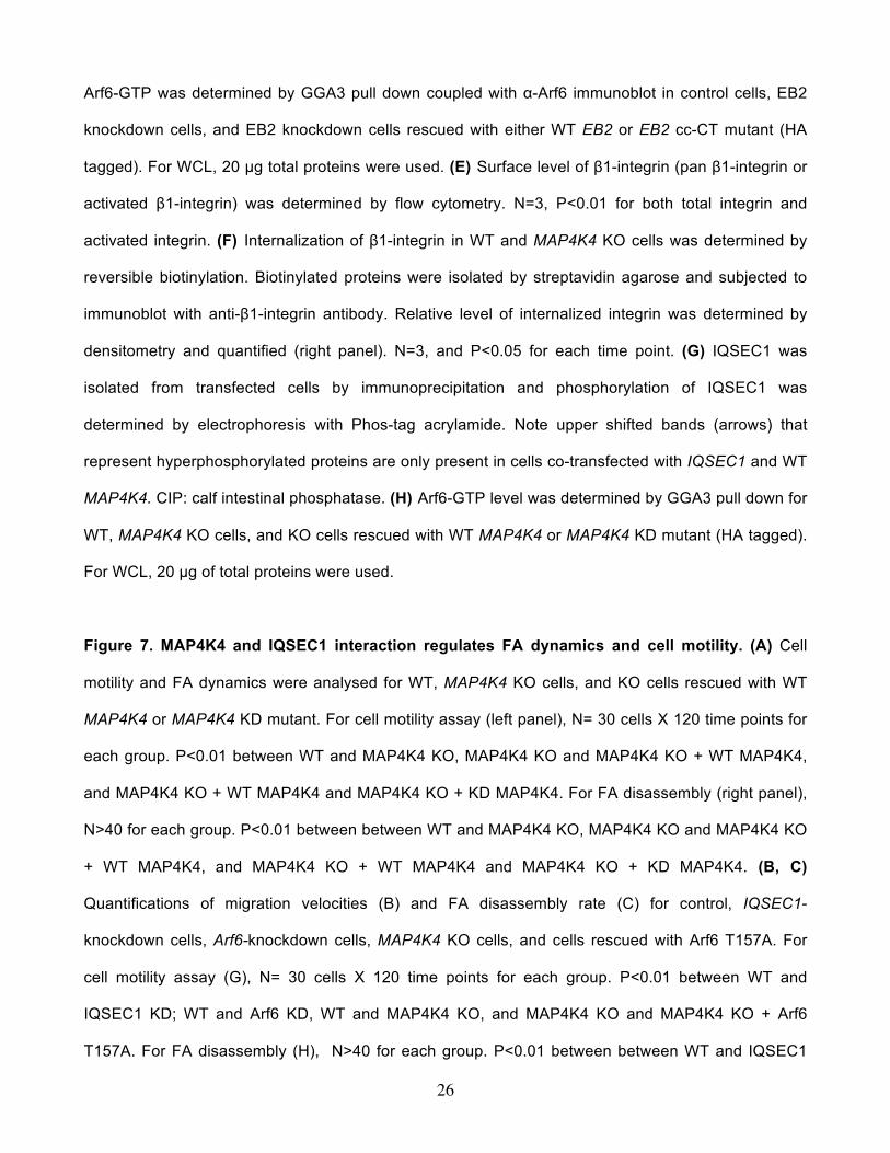
Figure 2. Cell migration defects in MAP4K4-deficient keratinocytes. (A) PCR genotyping of
MAP4K4 cKO mouse DNAs (top panel; WT: wildtype, Flox: MAP4K4 fl/fl; cKO: MAP4K4 fl/fl: K14-Cre).
Immunoblot analysis of P0 epidermal extracts (20 µg total protein) probed with different antibodies as
23
indicated. (B) MAP4K4 cKO mice can grow to adulthood (left panel). Histological analysis
(hematoxylin/eosin staining) confirms rather normal skin and hair follicles in cKO skin (right panel).
Scale bar = 50 µm. (C) Dorsal skins of neonatal mice were immunostained with different antibodies as
indicated (K5: keratin 5, K10: keratin 10, Lor: Loricrin, β4: β4-integrin, CD104). Scale bar=50 µm. (D)
Wound healing as monitored by histological staining of skin sections at the wound edges 4 days after
injury. Halves of wound sections are shown. Epi: epidermis; der: dermis; Es: eschar. Dotted lines
denote dermal–epidermal boundaries. Scale bar = 50 µm. (E) Quantification of the length of
hyperproliferative epidermis generated at times indicated after wounding. Number of biological
replicates (N) =30 for each group. P<0.01 for each time point. (F) Quantification of Ki67-positive cells
present in wound HE. Error bars represent standard deviations (SD). N=10, P=0.22. (H) Morphology
of primary keratinocytes isolated from WT or MAP4K4 cKO skin. Scale bar = 50 µm. (G) Migration of
confluent monolayers of mouse keratinocytes cultured from MAP4K4 cKO and WT littermates was
assessed by in vitro scratch-wound assays. The kinetics of in vitro wound healing was quantified. N=3,
P<0.01. (H) Movements of individual keratinocytes were traced by videomicroscopy. Migration tracks
of multiple cells for each group (WT or KO) are shown here as scatter plots. N=30.
Figure 3. MAP4K4 regulates FA dynamics. (A) Immunolabeling of WT and MAP4K4 null cells for F-
actin (red), nuclei (DAPI; blue), and FA marker vinculin (Vin; green). Scale bar = 20 µm. (B) Box and
whisker plot indicating the size distribution of FAs in WT and KO cells. N>300, and P<0.01. (C)
Representative time-lapse images (montages) of DsRed-Zyxin expressing keratinocytes. Note
formation and dissolution of FAs in WT cells and very static FAs in KO cells. Scale bar = 10 µm. (D)
Box and whisker plots revealing slow assembly and disassembly rates of FAs in MAP4K4 KO cells
relative to their WT counterparts. N>50 for each group, and P<0.01. (E) Fluorescence recovery after
photobleaching (FRAP) was used to visualize reduced dynamics of FAs in MAP4K4 KO vs WT cells
expressing GFP-Paxillin. Fluorescence recovery was recorded for 140 s after photobleaching.
Representative time-lapse images (montage) of FAs are shown. Scale bar = 5 µm. (F) Box-and-
24
whisker diagram quantifying the differences in half-time (T1/2) of FRAP between WT and KO cells.
N>10, and P<0.01.
Figure 4. Identification of MAP4K4 binding partners by tandem affinity purification. (A)
Immunofluorescence for MTs (red), FAs (vinculin, green), and nuclei (DAPI, blue) shows altered MT
organization in ACF7 KO cells but not in MAP4K4 deficient cells. Boxed areas are magnified as insets,
where only MT staining is shown. Note bundled MT filaments merge toward peripheral FAs in WT and
MAP4K4 KO cells. Scale bar = 20 µm. (B) Box and whisker plots for EB1 plus end dynamics in WT
and MAP4K4 KO cells. Three parameters of MT plus end dynamics were retrieved from the confocal
videos, plus end growth speed, growth lifetime, and growth displacement. Descriptive statistics of all
the results, including mean and standard deviation, were shown in the table below. (C) MAP4K4 and
its associated proteins were isolated by tandem affinity purification and resolved by SDS-PAGE. IgG
heavy chain and light chain are marked by arrowheads. The putative band for MAP4K4 is marked by
star. Putative band for EB2 is marked by an arrow. (D) Both EB2 and IQSEC1 were found as
MAP4K4 binding proteins. Identified peptides for each protein were highlighted (yellow).
Figure 5. EB2 recruits MAP4K4 to MTs and promotes FA dynamics. (A) Keratinocyte lysate was
immunoprecipitated with α-EB2 or α-MAP4K4 antibodies. Immunoprecipitate (IP) as well as whole cell
lysate (WCL, 10 µg total protein) was subjected to immunoblot with different antibodies as indicated.
(B) HEK293T cells were transfected with different plasmids as indicated. Cell lysates were
immunoprecipitated with α-HA antibody and immunoblotted with different antibodies as indicated. For
WCL, 10 µg total proteins were used. Note only EB2 specifically pulls down MAP4K4. (C) Schematic
representation of EB1, EB2, and various EB2 mutants used for coimmunoprecipitation assays (top
panel). CH: calponin homology domain; cc: coiled-coil domain; CT: acidic C terminus. Association of
EB2 or EB2 mutants with MAP4K4 was determined by coimmunoprecipitation as described above
(lower panel). For WCL, 10 µg total proteins were used. (D) Control or EB2 knockdown cells were
transfected with plasmid encoding MAP4K4. MT binding was examined by co-sedimentation assay.
25
Pellet and supernatant after ultracentrifugation as well as an aliquot of pre-cleared cell lysate (input)
were immunoblotted to assess the level of MAP4K4. (E) Presence of MAP4K4, EB2, and EB1 in
isolated FAs or whole cell lysate (WCL, 20 µg) from control or EB2-shRNA treated cells was
determined by immunoblots. (F-G) WT keratinocytes and EB2 knockdown (KD) cells were subjected
to immunofluorescence staining and examined by TIRF microscopy (G). Co-localization between
MAP4K4 and vinculin was determined by Pearson correlation coefficient and quantified (H). Scale bar
= 20 µm. N=10, P<0.01. (H-I) Quantifications of migration velocities and FA disassembly for control,
EB2-knockdown (KD) cells, and cells rescued with WT EB2 or EB2 mutant (EB2-cc-CT). Note that
only WT EB2 restored FA dynamics and rescued the speed of migration. For cell motility assay (I), N=
30 cells X 120 time points for each group. P<0.01 between WT and EB2 KD; EB2 KD and EB2 KD +
EB2; and EB2 KD + EB2 and EB2 KD + EB2 mutant. For FA disassembly (J), N>40 for each group.
P<0.01 between WT and EB2 KD; EB2 KD and EB2 KD + EB2; and EB2 KD + EB2 and EB2 KD +
EB2 mutant.
Figure 6. MAP4K4 promotes IQSEC1 and Arf6 activity. (A) Interaction between IQSEC1 and
MAP4K4 was confirmed by coimmunoprecipitation. Cells were transfected with plasmids encoding
Myc-tagged MAP4K4 with or without HA-tagged IQSEC1. Cell lysate was immunoprecipitated with α-
HA and blotted with different antibodies as indicated (top panel). To verify interaction of endogenous
proteins, lysate of WT keratinocytes were immunoprecipitated with control or α-MAP4K4 IgG, and blot
with α-IQSEC1 antibody (bottom panel). An aliquot of whole cell lysate (20 µg) were examined by
immunoblot as well. (B) HEK293T cells were transfected with HA-tagged Arf6 together with MAP4K4
or IQSEC1 in different combinations as indicated. Level of GTP-bound Arf6 was determined by GGA3
pull down and immunoblot with α-HA antibody. An aliquot of WCL (10 µg) was immunoblotted with
different antibodies as indicated to verify comparable expression level of different genes. (C) Level of
endogenous Arf6-GTP was determined by GGA3 pull down coupled with α-Arf6 immunoblot (top
panel). Quantification from densitometry analysis shows significant decrease of Arf6 activity upon loss
of MAP4K4 (lower panel). For WCL, 20 µg total proteins were used. N=4, and P<0.05. (D) Level of
26
Arf6-GTP was determined by GGA3 pull down coupled with α-Arf6 immunoblot in control cells, EB2
knockdown cells, and EB2 knockdown cells rescued with either WT EB2 or EB2 cc-CT mutant (HA
tagged). For WCL, 20 µg total proteins were used. (E) Surface level of β1-integrin (pan β1-integrin or
activated β1-integrin) was determined by flow cytometry. N=3, P<0.01 for both total integrin and
activated integrin. (F) Internalization of β1-integrin in WT and MAP4K4 KO cells was determined by
reversible biotinylation. Biotinylated proteins were isolated by streptavidin agarose and subjected to
immunoblot with anti-β1-integrin antibody. Relative level of internalized integrin was determined by
densitometry and quantified (right panel). N=3, and P<0.05 for each time point. (G) IQSEC1 was
isolated from transfected cells by immunoprecipitation and phosphorylation of IQSEC1 was
determined by electrophoresis with Phos-tag acrylamide. Note upper shifted bands (arrows) that
represent hyperphosphorylated proteins are only present in cells co-transfected with IQSEC1 and WT
MAP4K4. CIP: calf intestinal phosphatase. (H) Arf6-GTP level was determined by GGA3 pull down for
WT, MAP4K4 KO cells, and KO cells rescued with WT MAP4K4 or MAP4K4 KD mutant (HA tagged).
For WCL, 20 µg of total proteins were used.
Figure 7. MAP4K4 and IQSEC1 interaction regulates FA dynamics and cell motility. (A) Cell
motility and FA dynamics were analysed for WT, MAP4K4 KO cells, and KO cells rescued with WT
MAP4K4 or MAP4K4 KD mutant. For cell motility assay (left panel), N= 30 cells X 120 time points for
each group. P<0.01 between WT and MAP4K4 KO, MAP4K4 KO and MAP4K4 KO + WT MAP4K4,
and MAP4K4 KO + WT MAP4K4 and MAP4K4 KO + KD MAP4K4. For FA disassembly (right panel),
N>40 for each group. P<0.01 between between WT and MAP4K4 KO, MAP4K4 KO and MAP4K4 KO
+ WT MAP4K4, and MAP4K4 KO + WT MAP4K4 and MAP4K4 KO + KD MAP4K4. (B, C)
Quantifications of migration velocities (B) and FA disassembly rate (C) for control, IQSEC1-
knockdown cells, Arf6-knockdown cells, MAP4K4 KO cells, and cells rescued with Arf6 T157A. For
cell motility assay (G), N= 30 cells X 120 time points for each group. P<0.01 between WT and
IQSEC1 KD; WT and Arf6 KD, WT and MAP4K4 KO, and MAP4K4 KO and MAP4K4 KO + Arf6
T157A. For FA disassembly (H), N>40 for each group. P<0.01 between between WT and IQSEC1

27
KD; WT and Arf6 KD, WT and MAP4K4 KO, MAP4K4 KO and MAP4K4 KO + Arf6 T157A, and
IQSEC1 KD and IQSEC1 KD + Arf6 T157A. (D) A working model summarizing the role of MAP4K4 in
FA turnover and cell migration. We posit that MT dynamics are coordinated by cytoskeletal
crosslinkers, such as ACF7 that guides MT growth toward FAs. MT interacting protein EB2 can bind
and deliver MAP4K4 to FAs, where MAP4K4 can subsequently activate IQSEC1 and Arf6, leading to
FA turnover and efficient cell movement.
Supplementary Figure Legends
Supplementary Figure 1: Nocodazole treatment stabilizes focal adhesions in HaCaT cells. (A)
Box and whisker plots revealing slow assembly and disassembly rates of focal adhesions in HaCaT
cells treated with nocodazole. Box and whisker plots indicate the mean (empty square within the box),
25th percentile (bottom line of the box), median (middle line of the box), 75th percentile (top line of the
box), 5th and 95th percentile (whiskers), 1st and 99th percentile (solid diamonds) and minimum and
maximum measurements (solid squares), with actual data points shown at right. N>40 for each group,
and P<0.01. (B) Cell fractions and whole cells lysate (WCL) were subjected to immunoblot (IB) with
different antibodies as indicated. C: cytosolic fraction, FA: focal adhesion fraction. Note enrichment of
vinculin and paxillin (focal adhesion marker protein) and absence of GAPDH (cytosolic protein), AIF1
(mitochondria marker), ERp72 (ER marker protein), or H2A.Z (nuclear protein) in FA fraction.
Supplementary Figure 2: Changes of focal adhesion proteins upon treatment of nocodazole.
Protein abundance in isolated focal adhesions or whole cell lysate (WCL, 10 μg) from cells treated with
or without nocodazole was determined by immunoblot (IB) with different antibodies as indicated.
Supplementary Figure 3: Co-localization of MAP4K4 with MTs and focal adhesions. (A) WT and
MAP4K4 KO cells were fixed and stained with different antibodies as indicated. Stained cells were
examined by TIRF microscopy. Note enrichment of endogenous MAP4K4 at the ends of MT bundles
that associate with focal adhesions in WT cells, and loss of MAP4K4 staining in the KO cells. Scale bar
represents 20 µm. (B) Epi-fluorescence imaging of the same cells shown in Figure 1D. Note
comparable level of MAP4K4 in these cells. (C) Level of endogenous MAP4K4 in WT keratinocytes,
WT keratinocytes treated with nocodazole, and ACF7 KO keratinocytes was determined by
immunoblot. Cell lysates were also blotted with α-actin antibody to verify similar loading.
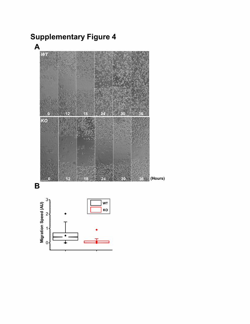
Supplementary Figure 4: Deletion of MAP4K4 inhibits cell motility in vitro. (A) Migration of
confluent monolayers of keratinocytes cultured from MAP4K4 cKO and WT littermates and subjected
to in vitro scratch-wound assays. Phase contrast images of the site were taken at hours indicated after
scratch-wounding. Scale bar represents 50 µm. (B) Box and whisker plots of WT and MAP4K4 KO cell
velocities in vitro. N= 30 cells X 120 time points. P<0.01.
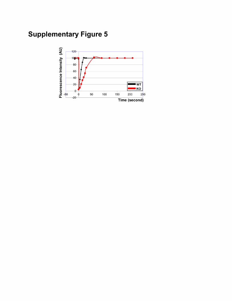
Supplementary Figure 5: FRAP analysis of WT and MAP4K4 KO cells. A representative example
of fluorescence recovery kinetics for WT and MAP4K4 KO cells after bleaching.
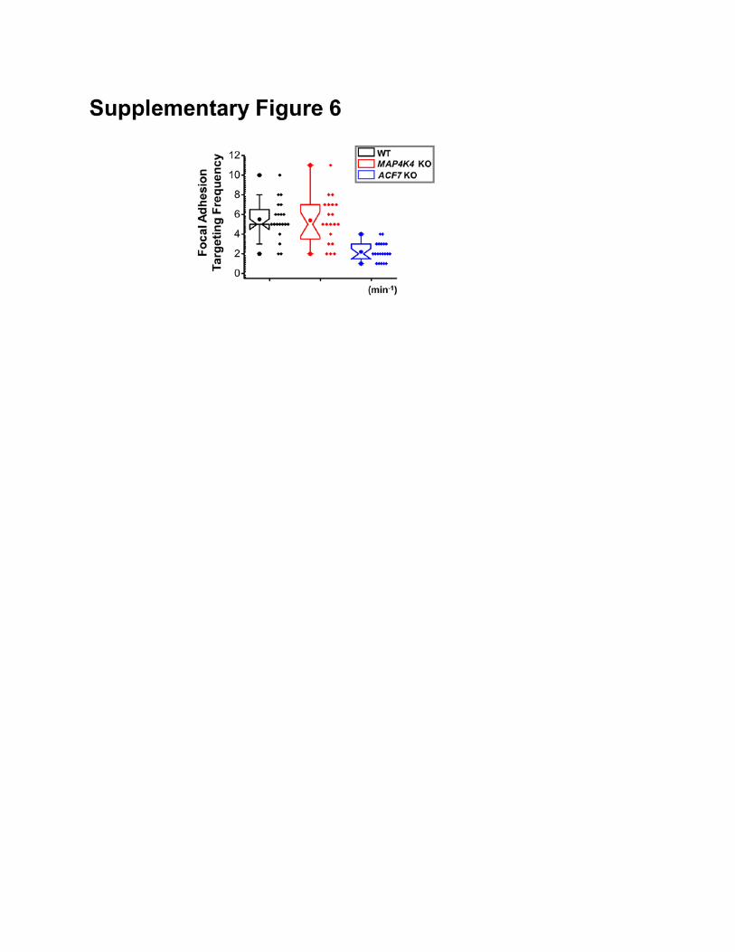
Supplementary Figure 6: MT targeting to focal adhesions in keratinocytes. MT targeting to focal
adhesions in WT, MAP4K4 KO, or ACF7 KO cells were quantified with confocal videomicroscopy and
shown as box-whisker plot. N=20 for each group. P<0.01 between WT and ACF7 KO, and P=0.88
between WT and MAP4K4 KO.
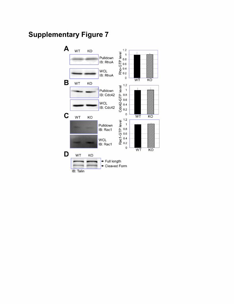
Supplementary Figure 7: Rho activity and talin activation. (A-C) Level of GTP-bound RhoA, Cdc42,
and Rac1 in WT and MAP4K4 KO cells were determined by pull-down assay and quantified by
densitometry (right panels). N=3, and P>0.5 for each group. (D) Level of talin and cleaved talin in WT
and MAP4K4 KO cells was examined by immunoblot.
Supplementary Figure 8: EB2 knockdown and binding of EB2-cc-CT mutation with MTs. (A)
Knockdown expression of endogenous EB2 was achieved by shRNA. Cells treated with control or EB2
shRNA were subjected to immunoblot with different antibodies as indicated. Note EB2 depletion does
not affect stability of other end binding proteins (EB1). (B) MT binding of EB2 or EB2-cc-CT mutant
was examined by co-sedimentation assay as described.
Supplementary Figure 9: FACS analysis of WT and MAP4K4 KO cells. Representative examples
of total β1-integrin (A) and active β1-integrin (B) on cell surface determined by flow cytometry for WT
and MAP4K4 KO cells.
Supplementary Figure 10: Endocytosis of β1-integrin. (A-B) Internalized surface β1-integrin was
visualized by immunofluorescence staining of WT and MAP4K4 KO cells. The cells were co-stained
with antibody against Rab5 (early endosome marker). Note internalized integrin after 15 minutes (B)
incubation in both WT and KO cells, but not present in the control cells (0 minute, A). Internalized β1
integrin localizes at Rab5-positve vesicles. Boxed areas are magnified as insets in (B). Note significant
co-localization but not identical localization pattern between internalized integrin and Rab5.
Supplementary Figure 11: Arf5 activity in WT and KO cells. (A-B) Arf5-GTP level was determined
by GGA3 pull-down in WT and MAP4K4 KO cells. For WCL, 20 μg of total proteins were used (A).
Level of Arf5 activation was determined by densitometry (B). N=3.
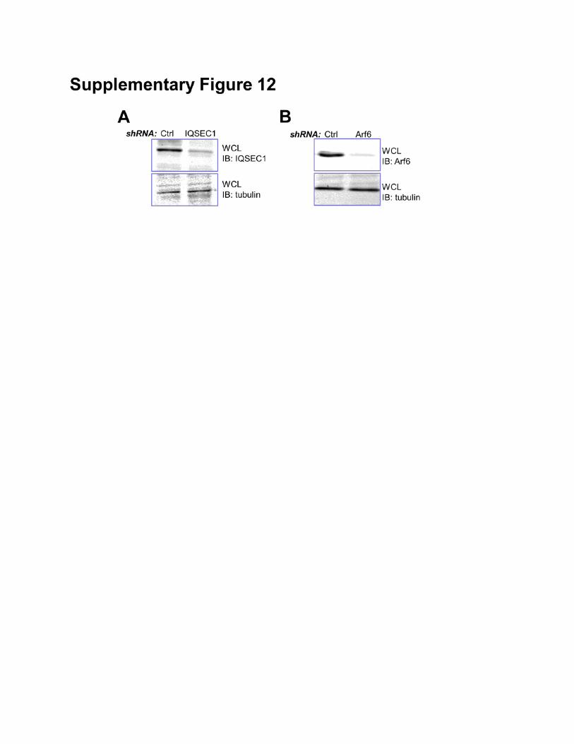
Supplementary Figure 12: Knockdown expression of Arf6 and IQSEC1 in mouse keratinocytes.
Keratinocytes treated with control or IQSEC1 shRNA (A) or Arf6 shRNA (B) were subjected to
immunoblot with different antibodies as indicated.
Supplementary Table (Spreadsheet): Quantitative analysis of focal adhesion proteome upon
nocodazole treatment by SILAC.
Focal adhesion proteins were isolated from control or nocodazole treated keratinocytes after
SILAC labeling (Fig. 1A and experimental procedures). All identified proteins with Heavy/Light ratio
greater than 1.5 or less than 0.5 are listed in the excel spreadsheet. Protein ID, name, together with
sequence information and H/L ratio are shown in the table.
Supplementary Video 1: Focal adhesion dynamics in WT and KO keratinocytes.
WT and KO keratinocytes expressing DsRed-Zyxin were plated on fibronectin (5 μg/ml)-coated
dishes. Dynamics of focal adhesion were recorded by confocal spinning-disc videomicroscopy. Images
were taken at 2 frames per minute.
Supplementary Video 2: Dynamic localization of EB2 cc-CT mutant.
WT keratinocytes transfected with TagRFP-EB2-cc-CT were plated on fibronectin (5
μg/ml)-coated dishes. Dynamics of EB2 mutant were recorded by confocal spinning-disc
videomicroscopy. Images were taken at 1 frame per second.
Supplementary Experimental Procedures
LC-MS/MS analysis
MS analysis was carried out by the proteomics core of University of Chicago. For both SILAC
samples and TAP (tandem affinity purification) samples, protein bands were excised by sterile razor
blade and chopped into ∼1 mm3 pieces. Each sample was washed in water and destained using
100 mM ammonium bicarbonate pH 7.5 in 50% acetonitrile. A reduction step was performed by addition
of 100 μl 50 mM ammonium bicarbonate pH 7.5 and 10 μl of 10 mM Tris(2-carboxyethyl)phosphine HCl
at 37°C for 30 min. The proteins were alkylated by adding 100 μl 50 mM iodoacetamide and allowed to
react in the dark at 20°C for 30 min. Gel samples were washed in water, then acetonitrile, and dried in a
SpeedVac. Trypsin digestion was carried out overnight at 37°C with 1:50 enzyme-protein ratio of
sequencing grade-modified trypsin (Promega) in 50 mM ammonium bicarbonate pH 7.5, and 20 mM
CaCl2. Peptides were extracted with 5% formic acid and vacuum dried.
The peptide samples were loaded to a 0.25 μl C8 OptiPak trapping cartridge custom-packed with
Michrom Magic C8 (Optimize Technologies), washed, then switched in-line with a 20 cm by 75 μm C18
packed spray tip nano column packed with Michrom Magic C18AQ, for a 2-step gradient. Mobile phase
A was water/acetonitrile/formic acid (98/2/0.2) and mobile phase B was
acetonitrile/isopropanol/water/formic acid (80/10/10/0.2). Using a flow rate of 350 nl/min, a 90 min,
2-step LC gradient was run from 5% B to 50% B in 60 min, followed by 50%–95% B over the next
10 min, hold 10 min at 95% B, back to starting conditions and re-equilibrated. The samples were
analyzed via electrospray tandem mass spectrometry (LC-MS/MS) on a Q-Exactive (Thermo scientific)
mass spetrometer, using a 60,000 RP survey scan, m/z 375-1950, with lockmasses, followed by 15
HCD CID scans on only doubly and triply charged precursors between 375 Da and 1950 Da. Inclusion
lists of expected acetylated or phosphorylated tryptic in-silico peptide ion masses were also used. Ions
selected for MS/MS were placed on an exclusion list for 60 seconds.
Tandem mass spectra were extracted by MSConvert (ProteoWizard 3.0.3768) All MS/MS
samples were analyzed using MaxQuant (Max Planck Institute of Biochemistry, Martinsried, Germany;
version 1.2.2.5. MaxQuant was set up to search the 140204_SPROT_HUMAN database (unknown
version, 47496 entries) also assuming strict trypsin. MaxQuant and X! Tandem were searched with a
fragment ion mass tolerance of 20 PPM and a parent ion tolerance of 20PPM. Carbamidomethyl of
cysteine was specified in MaxQuant as a fixed modification. Label:2H(4) of lysine, oxidation of
methionine, acetyl of the n-terminus, and phospho of serine, threonine and tyrosine were specified in
MaxQuant as variable modifications.
Skin Wound Healing
For skin wound healing assays, same sex littermates of ~ 12 wk old mice were anesthetized,
and two full-thickness excisional wounds were made on both sides of the dorsal midline (Wu et al.,
2008). Mice were housed separately, and no self-induced trauma was observed in control or cKO mice.
Tissue was collected 2-6 days after wounding, and wound reepithelialization was evaluated by
histological analyses. Hyperproliferative epidermis (HE) was identified by hematoxylin and eosin
staining, and the length of HE that extended into the wounds was measured and quantified.
Antibodies, Reagents, and Plasmid DNA Constructions
Rabbit polyclonal antibody against MAP4K4 was obtained from Bethyl Labs (Montgomery, TX).
Rat monoclonal antibody against EB1 and EB2 were obtained from Thermo (Waltham, MA). Guinea
pig anti K5, rabbit anti K10 and Loricrin antibodies were generous gifts from Dr. Elaine Fuchs at the
Rockefeller University. Rat monoclonal β4-integrin (CD104) was obtained from BD Pharmingen
(Franklin lakes, NJ). Mouse monoclonal antibodies against pan β1-integrin or activated β1-integrin
were obtained from Millipore (Billeria, MA). Mouse monoclonal antibody against IQSEC1 (GEP100)
was obtained from GeneScience (Tokyo, Japan). Human plasma fibronectin, nocodazole, taxol, ATP,
HA-conjugated Agarose, mouse monoclonal Vinculin, Paxillin, talin, acetylated tubulin, and β-tubulin
Abs were obtained from Sigma (St. Louis, MO). Texas Red-conjugated Phalloidin was obtained from
Invitrogen (Carlsbad, CA). Mouse monoclonal Abs against Myc, and rabbit polyclonal Abs against HA,
Rab5, AIF1, ERp72, sodium potassium ATPase, H2A.Z, and Ki67 were obtained from Santa Cruz
Biotechnology, Inc. (Santa Cruz, CA). Microtubule binding protein spindown kit and Arf6 Pull-down
activation assay kit were obtained from Cytoskeleton (Denver, CO). Monoclonal antibody against Arf5
was obtained from Abnova (Walnut, CA). Other chemicals or reagents were obtained from Sigma,
unless otherwise indicated. DNA miniprep and other extraction kits were obtained from Zymo
Research (Irvine, CA).
Plasmids encoding DsRed-Zyxin, GFP-Paxillin, and EB1 have been described (Kodama et al.,
2003; Schober et al., 2007). Full-length MAP4K4 cDNA was subcloned from Open Biosystem ORF
clones (Thermo, clone ID 100068356, encoding isoform 2 of mouse MAP4K4) into mammalian
expression vectors, pKH3S and pHANS (with N terminal HA or Myc tag). MAP4K4 kinase defective
mutant (D153N) was generated by overlapping PCR with primers: AAC TGC AGT TTC GGT GAA TAA
CGT GGT GAA TAT G , AAG GAA AAA AGC GGC CGC TCG AGA TGG CGA ACG ACT CTC C.
Plasmid encoding full-length EB2 cDNA was a generous gift from Dr. Yulia Komarova at University of
Illinois at Chicago. EB2 coding sequence was subcloned to other mammalian expression vectors,
including pKH3. Mutations in EB2 were created by over-lapping PCRs with primers: AAG GAA AAA
AGC GGC CGC TCA TGG CGG TCA ATG TGT ATT CTA C, GGA ATT CTT AAT ACT CTT CTT GTT
CCT CCT GTG GGC CCC CTT CAT CAG GTA TCA CAA AGC CTT CAT CGG AAG CGT AGA GCA
C, TCC TAA GGC TGG CCC CGG AAT GGT GCG AAA GAA TCC TGG TGT GGG CAA TGG AGA
TGA TGA AGC AGC TGA ACT CAA CGA GCA GGT ACA TTC, CCA CAG AGA CCC ATT GCA ACA
CAG AGG ACT ACT GCA GCT CCT AAG GCT GGC CCC GGA ATG, GAA GAT CTT CAA CCT GCC
CAA GAA GCC TCT CGG CTC CAG TAC TGC AGC CCC ACA GAG ACC CAT TGC AAC. ShRNA
vectors targeting EB2 and IQSEC1 were prepared from Decipher shRNA vector
(pRSI9-U6-UbiC-TagRFP-2A-Puro) with primers: ACC GGG ATG AAT GTT GAT AAG GTA GTT AAT
ATT CAT AGC TAC CTT ATC AAC ATT CAT CTT TTT T, CGA AAA AAA AGA TGA ATG TTG ATA
AGG TAG CTA TGA ATA TTA ACT ACC TTA TCA ACA TTC ATC C, ACC GGC CAG TGT TAC TGT
TGG CAA ATC TCG AGA TTT GCC AAC AGT AAC ACT GGT TTT TG, CGA ACA AAA ACC AGT
GTT ACT GTT GGC AAA TCT CGA GAT TTG CCA ACA GTA ACA CTG G, ACC GGC CTT TCA GAT
TGG AAG GAG TTC TCG AGA ACT CCT TCC AAT CTG AAA GGT TTT TG, CGA ACA AAA ACC
TTT CAG ATT GGA AGG AGT TCT CGA GAA CTC CTT CCA ATC TGA AAG GC', ACC GGG AAA
GAG AAC TGA TCA CCA TAC TCG AGT ATG GTG ATC AGT TCT CTT TCT TTT TG, CGA ACA
AAA AGA AAG AGA ACT GAT CAC CAT ACT CGA GTA TGG TGA TCA GTT CTC TTT CC, ACC
GGC TGT CAG CAT GGC TCA TCT TTC TCG AGA AAG ATG AGC CAT GCT GAC AGT TTT TG,
CGA ACA AAA ACT GTC AGC ATG GCT CAT CTT TCT CGA GAA AGA TGA GCC ATG CTG ACA
GC, ACC GGC CCT TTC AGA TTG GAA GGA GTC TCG AGA CTC CTT CCA ATC TGA AAG GGT
TTT TG, CGA ACA AAA ACC CTT TCA GAT TGG AAG GAG TCT CGA GAC TCC TTC CAA TCT
GAA AGG GC. Mutation at EB2 shRNA recognition site was created by overlapping PCR with primers:
AGC ATC CTT TAA ACG GAT GAA CGT CGA CAA AGT CAT CCC AGT GGA GAA G, CTT CTC
CAC TGG GAT GAC TTT GTC GAC GTT CAT CCG TTT AAA GGA TGC T. Plasmid encoding
IQSEC1 (Arf-GEP100) was a generous gift from Dr. Martha Vaughan at National Institutes of Health.
IQSEC1 coding sequence was transferred to other mammalian expression vectors, including pKH3S.
Plasmid encoding Arf6 T157A was generated by overlapping PCR with primers: CGC GGA TCC TCA
TGT TGG GCC TG, GAT TTG TAG TTA GAG GCT AAC CAT GTG AGC CC, GGG CTC ACA TGG
TTA GCC TCT AAC TAC AAA TC, CCG CTC GAG CGG CCG CCA GTG TG.
Histology and Immunofluorescence
Skin or wound samples were embedded in OCT, frozen, sectioned, and fixed in 4%
formaldehyde. For paraffin sections, samples were incubated in 4% formaldehyde at 4°C overnight,
dehydrated with a series of increasing concentrations of ethanol and xylene, and then embedded in
paraffin. Paraffin sections were rehydrated in decreasing concentrations of ethanol and subjected to
antigen unmasking in 10 mM Citrate, pH 6.0. Sections were subjected to hematoxylin and eosin
staining or immunofluorescence staining as described (Guasch et al., 2007). Antibodies were diluted
according to manufacturer’s instruction, unless indicated.
For immunofluorescence staining of cells, keratinocytes were fixed and stained with different
primary and secondary antibodies as indicated. Pictures were acquired on a fluorescence microscope
on a 16-bit scale. To examine β1-integrin endocytosis, we carried out treatment and
immunofluorescence staining essentially as described (Wu et al., 2005). Briefly, cells were incubated
with β1-integrin antibody at 4°C for 30 minutes and then cultured at 37°C for 15 minutes to allow
surface receptor internalization. Cells were then washed with ice-cold PBS and then with stripping
buffer (0.2M HAc, 0.5M NaCl, pH2.8) at 4°C for 30 minutess. After fixation, cells were stained with
antibodies against integrin and Rab5. Images were taken on Marianas spinning dish confocal
microscope.
Flow cytometry
Flow cytometry analysis of surface integrin was carried out essentially as described (Wu et al.,
2008).
Microtubule Pull Down Assay, Arf6 Pull Down Assay, and other biochemical analysis
Microtubule binding were examined using microtubule binding protein spin-down assay kit from
Cytoskeleton (Denver, CO), according to manufacturer’s instructions. Arf6 pull-down activation assay
(GGA3 pull down) was performed according to manufacturer’s instruction (Cytoskeleton, Denver, CO).
Rho family GTPases pulldown assay was done following the instructions of RhoA/Rac1/Cdc42
activation assay kit (Cytoskeleton, Denver, CO). Immunoprecipitation and western blotting were
performed as described previously (Wu et al., 2004). Tandem affinity purification was performed
essentially as described (Wu et al., 2007). Protein phosphorylation was determined by phos-tag
reagent (Wako pure Chemical Industries, Japan) according to manufacturer’s instruction.
Integrin endocytosis assay was performed as previously described (Wu et al., 2005). In brief,
cells were washed with ice-cold PBS. Cell membrane proteins were labelled with 0.2 mg/ml
Sulfo-NHS-SS Biotin in PBS. After incubation at 37°C for the indicated time points, cells were treated
with reduction buffer (42mM glutathione reduced form, 75mM NaCl, 1mM EDTA, 1% BSA, 75mM
NaOH) for 20mins on ice twice. Biotinylated proteins were pull-downed with streptavidin beads from
lysed cell lysates and examined by Western Blot. Transferrin receptor was used as an internal control
for the assay (Wu et al., 2005).
The acrylamide-pendant Phos-tag ligand provides a phosphate affinity SDS-PAGE for mobility
shift detection of protein phosphorylation. This methodology has now been well established and served
as an excellent alternative approach for radioisotope-free detection of protein phosphorylation (Humke
et al., 2010; Matos et al., 2008; Ydenberg and Rose, 2009).
Statistical analysis
Statistical analysis was performed using Excel or OriginLab software. Box plots are used to
describe the entire population without assumptions on the statistical distribution. A student t test was
used to assess the statistical significance (P value) of differences between two experimental
conditions.
Supplementary references
Guasch, G., Schober, M., Pasolli, H.A., Conn, E.B., Polak, L., and Fuchs, E. (2007). Loss of TGFbeta
signaling destabilizes homeostasis and promotes squamous cell carcinomas in stratified epithelia.
Cancer Cell 12, 313-327.
Humke, E.W., Dorn, K.V., Milenkovic, L., Scott, M.P., and Rohatgi, R. (2010). The output of Hedgehog
signaling is controlled by the dynamic association between Suppressor of Fused and the Gli proteins.
Genes & development 24, 670-682.
Kodama, A., Karakesisoglou, I., Wong, E., Vaezi, A., and Fuchs, E. (2003). ACF7: an essential
integrator of microtubule dynamics. Cell 115, 343-354.
Matos, J., Lipp, J.J., Bogdanova, A., Guillot, S., Okaz, E., Junqueira, M., Shevchenko, A., and
Zachariae, W. (2008). Dbf4-dependent CDC7 kinase links DNA replication to the segregation of
homologous chromosomes in meiosis I. Cell 135, 662-678.
Schober, M., Raghavan, S., Nikolova, M., Polak, L., Pasolli, H.A., Beggs, H.E., Reichardt, L.F., and
Fuchs, E. (2007). Focal adhesion kinase modulates tension signaling to control actin and focal
adhesion dynamics. J Cell Biol 176, 667-680.
Wu, X., Gan, B., Yoo, Y., and Guan, J.L. (2005). FAK-mediated src phosphorylation of endophilin A2
inhibits endocytosis of MT1-MMP and promotes ECM degradation. Dev Cell 9, 185-196.
Wu, X., Kodama, A., and Fuchs, E. (2008). ACF7 regulates cytoskeletal-focal adhesion dynamics and
migration and has ATPase activity. Cell 135, 137-148.
Wu, X., Suetsugu, S., Cooper, L.A., Takenawa, T., and Guan, J.L. (2004). Focal adhesion kinase
regulation of N-WASP subcellular localization and function. The Journal of biological chemistry 279,
9565-9576.
Wu, Y., Li, Q., and Chen, X.Z. (2007). Detecting protein-protein interactions by Far western blotting.
Nat Protoc 2, 3278-3284.
Ydenberg, C.A., and Rose, M.D. (2009). Antagonistic regulation of Fus2p nuclear localization by
pheromone signaling and the cell cycle. The Journal of cell biology 184, 409-422.
Related Documents