SZENT ISTVÁN EGYETEM MICROSPORE CULTURE AND GENETIC TRANSFORMATION STUDIES IN BARLEY AND TRITICALE DOKTORI ÉRTEKEZÉS MONOSTORI TAMÁS GÖDÖLLŐ 2003

Welcome message from author
This document is posted to help you gain knowledge. Please leave a comment to let me know what you think about it! Share it to your friends and learn new things together.
Transcript
SZENT ISTVÁN EGYETEM
MICROSPORE CULTURE AND GENETIC TRANSFORMATION STUDIES IN BARLEY AND TRITICALE
DOKTORI ÉRTEKEZÉS
MONOSTORI TAMÁS
GÖDÖLLŐ 2003
A doktori iskola: Növénytudományi Doktori Iskola
Tudományága: Növénytermesztési és kertészeti tudományok
Vezetője: Dr. Virányi Ferenc egyetemi tanár, az MTA Doktora Szent István Egyetem Növényvédelemtani Tanszék
Titkára Dr. Gyulai Gábor egyetemi docens, a biológiai tudomány kandidátusa Szenti István Eghgyetem Genetika és Növénynemesítés Tanszék
Program: Növénynemesítés Genetikai és Biotechnológiai Módszerekkel
Programvezető: Dr. Heszky László egyetemi tanár, akadémikus Szent István Egyetem Genetika és Növénynemesítési Tanszék
Témavezető: Dr. Pauk János tudományos főmunkatárs, a mezőgazdasági tudomány kandidátusa Gabonatermesztési Kutató Kht., Szeged
........................................................... ...........................................................
Az iskolavezető jóváhagyása A programvezető jóváhagyása

...........................................................
A témavezető jóváhagyása
Index
INDEX 1. INTRODUCTION ........................................................................................................................... 1 2. REVIEW OF LITERATURE .......................................................................................................... 5
2.1. INDUCTION OF HAPLOID EMBRYOGENESIS IN MICROSPORE CULTURE...............5 2.1.1. Haploid breeding in cereals............................................................................................... 5 2.1.1.1. Main factors of isolated microspore culture in cereals ............................................... 5 2.1.2. In vitro androgenesis in triticale........................................................................................ 9 2.1.3. The role of plant hormones in the induction of microspore embryogenesis ................... 10
2.2. PREPARATION OF NOVEL VECTOR CONSTRUCTS .....................................................10 2.2.1. Genetic transformation of barley..................................................................................... 11 2.2.2. Modification of biosynthetic pathways by genetic transformation................................. 13 2.2.3. Jasmonates....................................................................................................................... 14 2.2.4. Jasmonate-induced gene expression in barley ................................................................ 16
3. MATERIALS AND METHODS................................................................................................... 19 3.1. INDUCTION OF HAPLOID EMBRYOGENESIS IN MICROSPORE CULTURE.............19
3.1.1. Materials.......................................................................................................................... 19 3.1.1.1. Plant material............................................................................................................. 19 3.1.1.2. Culture media ............................................................................................................ 19 3.1.2. Methods ........................................................................................................................... 20 3.1.2.1. Determination of the developmental stage and the number of developing structures20 3.1.2.2. Isolation of microspores ............................................................................................ 20 3.1.2.3. Culture of microspores .............................................................................................. 21 3.1.2.4. Regeneration of plants............................................................................................... 21 3.1.2.5. Determination of ploidy level ................................................................................... 21 3.1.2.6. Analysis of data ......................................................................................................... 22
3.2. PREPARATION OF NOVEL VECTOR CONSTRUCTS .....................................................22 3.2.1. Materials.......................................................................................................................... 22 3.2.1.1. Plant material............................................................................................................. 22 3.2.1.2. Escherichia coli strain............................................................................................... 22 3.2.1.3. Plasmids, cDNAs and oligonucleotides .................................................................... 22 3.2.2. Molecular biological methods......................................................................................... 23 3.2.2.1. Transformation of E. coli cells .................................................................................. 23 3.2.2.2. Isolation and purification of plasmid DNA from E. coli........................................... 24 3.2.2.3. Restriction analysis ................................................................................................... 25 3.2.2.4. Gel electrophoresis and extraction of DNA from agarose gel .................................. 25 3.2.2.5. Dephosphorylation and ligation ................................................................................ 25 3.2.2.6. Colony hybridization................................................................................................. 26 3.2.2.7. Preparation of total plant DNA ................................................................................. 26 3.2.2.8. Polymerase chain reaction (PCR) ............................................................................. 27 3.2.2.9. Protein isolation and Western-blot analysis .............................................................. 27 3.2.2.10. PAT-assay ............................................................................................................... 28 3.2.3. Methods of plant cell and tissue culture.......................................................................... 28 3.2.3.1. Isolation and transformation of barley mesophyll protoplasts .................................. 28 3.2.3.2. Maize (Zea mays L.) suspension cultures ................................................................. 29 3.2.3.3. Barley callus cultures and plant regeneration via somatic embryogenesis ............... 29 3.2.3.4. Selection of the bombarded scutella.......................................................................... 29 3.2.4. Particle bombardment using the particle inflow gun ...................................................... 30 3.2.4.1. Coating of the gold particles ..................................................................................... 30 3.2.4.2. The setup of the particle inflow gun.......................................................................... 30 3.2.5. Assay for transient luc expression................................................................................... 31 3.2.6. Histochemical assay for transient β-glucuronidase expression....................................... 31
Index
4. RESULTS ...................................................................................................................................... 33 4.1. INDUCTION OF HAPLOID EMBRYOGENESIS IN MICROSPORE CULTURE.............33
4.1.1. The microspore culture of triticale .................................................................................. 33 4.1.1.1. Characteristic stages of triticale androgenesis in microspore culture ....................... 34 4.1.1.2. Influence of the hormone content of the induction medium on triticale androgenesis36 4.1.1.3. Ploidy level of the green plantlets ............................................................................. 38 4.1.2. Barley microspore culture ............................................................................................... 39 4.1.2.1. Isolation and culture of barley microspores .............................................................. 40 4.1.2.2. Effects of different induction media on barley androgenesis.................................... 41 4.1.2.3. Hormone-free induction of androgenesis in microspore cultures of barley.............. 42 4.1.2.4. Comparison of the nitrogen composition of N24-BA and 190-0 media ................... 43
4.2. PREPARATION OF NOVEL VECTOR CONSTRUCTS .....................................................45 4.2.1. Preparation of novel vector constructs ............................................................................ 45 4.2.2. Assays for transient gene expression in barley mesophyll protoplasts ........................... 48 4.2.2.1. Transient expression of the pat gene in barley mesophyll protoplasts ..................... 48 4.2.2.2. Transient expression of JIP23 cDNA in barley mesophyll protoplasts .................... 49 4.2.3. Somatic embryogenesis of barley.................................................................................... 49 4.2.4. Optimization of the bombardment parameters................................................................ 50 4.2.4.1. Transient assays with BMS suspension cells ............................................................ 51 4.2.4.2. Transient assays with isolated scutella...................................................................... 52 4.2.5. Stable transformation experiments with barley scutella ................................................. 53 4.2.5.1. Somatic embryogenesis in the bombarded scutella................................................... 54 4.2.5.2. Regeneration of putative transgenic plants ............................................................... 55 4.2.6. Analysis of the putative transgenic plants....................................................................... 56
4.3. NEW SCIENTIFIC RESULTS ...............................................................................................58 5. DISCUSSION ................................................................................................................................ 59
5.1. INDUCTION OF HAPLOID EMBRYOGENESIS IN MICROSPORE CULTURE.............59 5.1.1. Method of microspore isolation and culture in triticale .................................................. 59 5.1.2. Induction of androgenesis in isolated barley microspores using different culture media61 5.1.3. Hormone-free induction of androgenesis in microspore cultures of barley and triticale 62 5.1.4. Future prospects .............................................................................................................. 63
5.2. PREPARATION OF NOVEL VECTOR CONSTRUCTS .....................................................64 5.2.1. The structure of the novel plasmid vectors ..................................................................... 64 5.2.1.1. The promoters............................................................................................................ 64 5.2.1.2. The pat resistance gene ............................................................................................. 65 5.2.1.3. The AOS cDNA ........................................................................................................ 65 5.2.1.4. The JIP23 cDNA ....................................................................................................... 66 5.2.1.5. Transient expression of the transgenes in barley mesophyll protoplasts .................. 66 5.2.2. Plant regeneration through somatic embryogenesis from barley scutella....................... 66 5.2.3. Optimization of bombardment parameters for the use of PIG ........................................ 68 5.2.3.1. Bombardment method ............................................................................................... 68 5.2.3.2. Helium pressure and distance settings ...................................................................... 69 5.2.3.3. Osmotic treatment of the target tissues ..................................................................... 69 5.2.3.4. Changes in responsivity upon bombardment ............................................................ 69 5.2.4. Plant regeneration from bombarded scutella................................................................... 70 5.2.5. Future prospects .............................................................................................................. 71
6. SUMMARY................................................................................................................................... 73 7. ÖSSZEFOGLALÁS....................................................................................................................... 77 8. REFERENCES............................................................................................................................... 81 9. APPENDIX.................................................................................................................................... 99 ACKNOWLEDGEMENTS ............................................................................................................. 109
Abbreviations
ABBREVIATIONS α-LeA linolenic acid 2,4-D 2,4-dichlorophenoxyacetic acid ABA abscisic acid ACC 1-aminocyclopropane-1-carboxylic acid ANOVA analysis of variance AOC allene oxide cyclase AOS allene oxide synthase ATP adenosine triphosphate BAP 6-benzylaminopurine BSA bovine serum albumin CaMV Cauliflower Mosaic Virus cDNA copy DNA CIAP Calf Intestine Alkaline Phosphatase CV% coefficient of variation cv. cultivar DH doubled-haploid DMF dimethyl-formamide DMSO dimethyl sulfoxide DNA deoxyribonucleic acid DTT dithiothreitol E. coli Escherichia coli EDTA ethylenediaminetetraacetic acid ELS embryo-like structure = embryoid Exp. experiment FDA fluorescein diacetate Fig. figure GUS β-glucuronidase IAA indoleacetic acid JA jasmonic acid JAs jasmonates JIP jasmonate-induced protein JM methyl jasmonate; jasmonic acid methyl ester jrg jasmonate-regulated gene kDa kilo Dalton KIN kinetin LOX lipoxygenase LSD Least Significant Difference LUC luciferase mcs multiple cloning site MDE microspore-derived embryo ms microspore MS Murashige and Skoog medium or mean square NAA naphthalene acetic acid No. number OD optical density OPDA 12-oxo-phytodienoic acid PAA phenylacetic acid PAGE polyacrylamide-gelelectrophoresis PAT phosphinothricin-acetyltransferase
Abbreviations
PCR polymerase chain reaction PDS Particle Delivery System PEG polyethylene glycol PIG particle inflow gun PPT phosphinothricin PTGS post-transcriptional gene silencing PVP polyvinilpyrrolidone RNA ribonucleic acid rpm revolution per minute RuBPCase ribulose 1,5-bisphosphate carboxylase s standard deviation SDS sodium dodecyl sulfate SMC shoot meristematic culture TGS transcriptional gene silencing U unit vs. versus
1. Introduction 1
1. INTRODUCTION
Triticale (x Triticosecale Wittmack) and barley (Hordeum vulgare L.) are important food crops grown all over the world. Triticale is a synthetic amphiploid cereal which has been considerably improved since its first description in 1891 (Rimpau, 1891). Intensive research on this intergeneric hybrid, however, started only in the early 1950s in some countries, including Hungary (Kiss, 1955, 1966). During the last decade the harvested triticale area increased from 1.8 million ha in 1990 to 3 million ha in 2001 (FAO, 2001). Its increasing importance, however, is more obvious in Hungary: while the area was merely 2,280 ha in 1990, in 2001 it already reached 119,000 ha. In the same period, extensive studies on genetic problems (Lelley and Gimbel, 1989) and on molecular genetics (Balatero et al., 1995; Wang et al., 1996) of triticale have been performed. Furthermore, the methods of somatic (Stolarz and Lörz, 1986; Immonen, 1996) as well as haploid (Lukjanjuk and Ignatova, 1986; Immonen and Robinson, 2000) tissue cultures have been established and transgenic plants have also been produced (Zimny et al., 1995). Although recent reviews on breeding strategies of triticale focus mainly on traditional methods (Lelley, 1992; Baier and Gustafson, 1996), novel techniques of tissue culture can also contribute to the success of these programs. In contrast to triticale, the importance of barley has been well-known for centuries. With a harvested area of 54 million hectares and a production of 141 million tons, it is among the five most important cereals in the world (FAO, 2001). In Hungary, barley is at the third place with an area of 368,000 ha and a yield of 1,300,000 t (FAO, 2001). During the last century every possible aspects of barley breeding were investigated in detail. In addition to traditional methods, the studies in the main fields of yield, quality and resistance involved recent developments of biotechnology as well (for reviews, see Mannonen et al., 1994; Lemaux et al., 1999). Besides its use by the applied science of agriculture, barley is a preferred model species of basic research such as plant physiology and biochemistry, due to its diploid genome and autogamous nature. Thus, barley became the model organism in studies on the role of jasmonates and the analysis of function of the most abundant jasmonate-induced protein, JIP23, in monocots (for review, see Wasternack and Hause, 2002). In this thesis studies on two independent fields of research are reported and discussed:
1. Induction of haploid embryogenesis in isolated microspore cultures of triticale and barley under hormone-free conditions.
2. Preparation of novel vector constructs to alter endogenous levels of jasmonates and JIP23 via genetic transformation of barley.
These topics cover two important fields of current plant breeding research: (i) the involvement of doubled-haploid plants in the traditional breeding process and (ii) the improvement of agronomically useful traits via genetic transformation. The use of barley as a model organism in both studies represents a step towards the combination of androgenesis and genetic transformation in the same breeding program in the future. This can be performed through the introduction of foreign genes into microspores or microspore-derived embryoids. The preparation of novel vector constructs which are functionable in the genetic background of barley can broaden the choice of vectors currently available for barley transformation. Anther culture was early established in triticale (Wang et al., 1973) and improvements of its protocol have been usually studied parallel with wheat. As reports from other species such as rapeseed, barley and wheat suggest, an established method of microspore culture can provide further opportunities for the improvement of this alternative cereal crop through biotechnology. Effects of media conditioning on the in vitro development of isolated triticale microspores and pollen grains have already been described but plant regeneration has not been reported (Keller,
1. Introduction 2
1991). An efficient method for the isolation and culture of isolated triticale microspores has been developed only in our laboratory to date (Monostori et al., 1998; Pauk et al., 2000). The published results are integrated in this work as well. Our primary aim was to establish the method of isolated microspore culture for triticale. Here, our experiencies gained with barley and wheat microspore cultures were utilized (Puolimatka et al., 1996; Monostori and Pauk, unpublished). Thus, the objectives for this part of the thesis were:
• to establish the method of isolation and culture of triticale microspores and to regenerate fertile, green dihaploid plants,
• to describe in vitro development of isolated triticale microspores on the sporophytic pathway,
• to study the effects of one hormone-free and two media of various hormone composition on microspore embryogenesis, and
• to evaluate the ploidy level of regenerants from different genotypes. The results of the triticale experiments and our preliminary results from barley microspore culture raise the question, whether hormone-supplementation of induction media is essential for the induction of androgenesis and plant regeneration in microspore culture. In haploid tissue cultures of barley and other cereals induction media are routinely supplemented with hormones in order to promote embryogenesis. Induction of androgenesis in hormone-free media may confirm the proposed decisive role of stress signals in switching microspores from gametophytic to sporophytic development (Touraev et al., 1996a,b, 1997). On the other hand, the evaluation of regeneration capacity in cultures induced with or without exogenous growth regulators can elucidate the promoting role of hormones in haploid embryogenesis. In addition, the independence of embryogenesis and regeneration can be studied in terms of hormone-requirement. Information about species-specific hormone-requirement can be obtained if the hormone-free induction medium of triticale microspore cultures is tested in barley as well. Thus, the additional objectives of our work were:
• to study the induction of androgenesis and plant regeneration in barley microspore cultures without exogenous hormone supply, and
• to compare the effects of the hormone-free medium successfully used in triticale microspore cultures with those of a medium of optimized nitrogen-composition previously established exclusively for barley (Mordhorst and Lörz, 1993).
Transgenic plants offer new possibilities to manipulate the biosynthetic pathways and to analyze the mode of action in most plant hormone classes. Recent advances which have made this new approach possible are (i) the cloning of genes/cDNAs coding for enzymes involved in the biosynthesis of plant hormones and (ii) the gene-transfer methods established for a number of plant species (for reviews, see Hedden and Phillips, 2000). Depending on the orientation of the DNA fragment in the transformation vector (sense or antisense), the genes coding for the biosynthetic enzymes in the transgenic plants are overexpressed or down-regulated, respectively. These changes in the regulation of a biosynthetic pathway can lead to increased or reduced levels of the corresponding hormone. Moreover, modulated hormone levels may cause phenotypic changes in the transgenic plants. This way, data have been provided to understand better the mode of action of auxins (Ficcadenti et al., 1999), cytokinins (Hewelt et al., 1994), gibberellins (Coles et al., 1999) and ethylene (Hamilton et al., 1990). The endogenous level of jasmonates has been modified via homologous and heterologous transformations with AOS, coding for the key-enzyme of JA-biosynthesis, in dicotyledonous species (Harms et al., 1995; Wang et al., 1999a; Laudert et al., 2000). In monocots, however, roles of
1. Introduction 3
jasmonates have been studied via the exogenous application of jasmonates or in response to various stresses only. Alterations in endogeneous JA-levels of barley plants upon transformation with AOS in sense or antisense orientation can cause changes, among others, in tissue differentiation and in the process of senescence. Moreover, alterations in stress-response and in the expression of JA-responsive genes could help to get an insight into the function of jasmonates in barley. JIP23, the most abundant JA-inducible protein in barley, accumulates in tissues osmotically stressed by solute transport as well as in mature leaves exposed to osmotic stress. For this protein, however, no putative function could be drawn from data base searches. In barley, its role as stress-protective protein has been proposed (Hause et al., 1996, 1999). Furthermore, JIP23 may attribute to the well-known JA-induced down-regulation of photosynthetic genes as shown by heterologous overexpression in tobacco (Görschen et al., 1997b). The homologous overexpression or the antisense repression of JIP23 can allow us to elucidate its role in tissue-differentiation, in stress-response and in the mediation of JA-functions in different tissues and developmental stages of the barley plant. The second part of the current thesis represents studies performed in the frame of a two-year project on the role of jasmonates in the development of barley as well as on the analysis of function of JIP23 in this species. In these studies, a transgenic approach requires stable transformed plants – transient expression systems are not suitable to examine changes in gene-expression, hormone-level and other phenotypic traits in the course of development. For the given period of time, the primary goal of our work was to prepare the prerequisites for further stable transformation programs as well as for the biochemical and molecular biological studies in the future. Therefore, the objectives were:
• to prepare novel plasmid vectors carrying a resistance marker gene and one of barley AOS1 and JIP23 cDNAs in sense or antisense orientation, respectively,
• to test the functionality of the new constructs in the genetic background of barley via transient expression analysis of both the resistance and the important transgenes in PEG-transformed mesophyll protoplasts, and
• to establish a transformation protocol to be used in further stable transformation experiments (i) via optimization of bombardment parameters for a particle inflow gun and (ii) via preliminary particle bombardment studies using the new vectors and scutella of the cultivar ‘Salome’, the model genotype of jasmonate studies.

1. Introduction 4
2. Review of literature 5
2. REVIEW OF LITERATURE
2.1. INDUCTION OF HAPLOID EMBRYOGENESIS IN MICROSPORE CULTURE Since the description of the first haploid mutant in higher plants (Datura stramonium; Blakeslee et al., 1922) practice has proven the advantages of using haploids in plant breeding. Spontaneous or induced reduplication of the haploid genome results in homozygous lines in a single generation. Thus, the selection efficiency will increase, which leads to an accelerated breeding process (Snape, 1987; Morrison and Evans, 1988). Furthermore, recessive traits can be selected at plant level and gametoclonal variability can be utilized this way (for review, see Heszky, 2000). The first haploid plants of in vitro origin have been reported in Datura inoxia (Guha and Maheswari, 1964). During the last decades, however, hundreds of species have been successfully involved in anther culture and several varieties produced using this method have been released (for reviews, see Heszky, 1979; 2000; Foroughi-Wehr and Wenzel, 1989; Bajaj, 1990; Kush and Virmani, 1996; Forster, 2002). 2.1.1. Haploid breeding in cereals
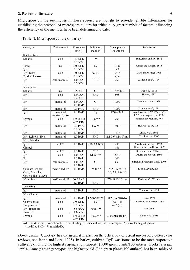
During the last 15 years, doubled-haploidy has been extensively used for the production of novel cultivars in cereals: 116 barley, 21 wheat, 8 rice and 3 triticale cultivars/lines originate from various techniques of haploid production (for review, see Forster, 2002). These procedures are based on chromosome-elimination (‘bulbosum-method’) and in vitro androgenesis (anther and microspore cultures). Gynogenesis through ovary culture could not become widespread practice due to its low efficiency (Castillo and Cistué, 1993). ‘Bulbosum-method’ is the traditional technique for haploid production in barley (Kasha and Kao, 1970; Devaux et al., 1990). Two-third of the doubled-haploid (DH) barley cultivars have been produced by this procedure (Forster, 2002). Advances in anther culture, however, have recently made this fully in vitro approach the most widely used method for the production of haploids both in barley (Kuhlmann and Foroughi-Wehr, 1989; Luckett and Smithard, 1995) and wheat (for review, see Barnabás et al., 2000). While anther culture is an established method for plant breeding, isolated microspores have remarkable features to be utilized in plant biotechnology. They offer a unicellular system of haploid cells which can be isolated in large quantities and synchronized in development. Thus, microspores are excellent targets for transformation methods and in vitro selection (for review, see Jähne and Lörz, 1995; Dunwell, 1996, for application in barley, see Table 1). Moreover, they can be used in studies on the biochemical and molecular background of embryogenesis (for reviews, see Reynolds, 1997; Touraev et al., 1997; in barley and wheat, see Reynolds and Kitto, 1992; Mordhorst et al., 1994; Vrinten et al., 1999). Microspore culture, however, also has an established role in the breeding of both mono- and dicotyledonous species: it has recently contributed to the production of five DH barley cultivars, and it is the method exclusively used for the production of DH rapeseed (for review, see Forster, 2002). The comparison of the efficiency of anther and microspore culture of barley shows a 100-to 200-fold higher regeneration rate in microspore culture (1170-2040 vs. 10.9 green plants/100 anthers) (Davies and Morton, 1998). In barley anther culture, the highest regeneration rate was 1300 green plants/100 anthers (Kao et al., 1991), while in microspore culture the maximum was 5000 green plants/100 anthers (Hoekstra et al., 1993). 2.1.1.1. Main factors of isolated microspore culture in cereals
Among cereals, plant regeneration from isolated microspore culture has been first reported for barley (Köhler and Wenzel, 1985) followed by wheat some years later (Datta and Wenzel, 1987).
2. Review of literature 6
Microspore culture techniques in these species are thought to provide reliable information for establishing the protocol of microspore culture for triticale. A great number of factors influencing the efficiency of the methods have been determined to date. Table 1. Microspore culture of barley
Donor plants. Genotype has the greatest impact on the efficiency of cereal microspore culture (for reviews, see Jähne and Lörz, 1995). In barley, cultivar ‘Igri’ was found to be the most responsive cultivar exhibiting the highest regeneration capacity (5000 green plants/100 anthers; Hoekstra et al., 1993). Among other genotypes, the highest yield (266 green plants/100 anthers) has been achieved
Genotype Pretreatment Hormones (mg/l)
Induction medium
Green plants/ 100 anthers
References
Shed culture Sabarlis cold 1.5 2,4-D
0.5 KIN P-9H - Sunderland and Xu, 1982
Dissa Igri
no 2.0 2,4-D 0.5 KIN
N6 0.08 0.0
Köhler and Wenzel, 1985
Igri; Dissa; F1; doublecross
cold 1.0 2,4-D 0.5 KIN
N6 1-2 17; 14; 4; 4
Datta and Wenzel, 1988
Igri cold/mannitol 1.0 IAA 0.2 KIN
FHG 266 Ziauddin et al., 1990
Maceration Sabarlis no 0.5 KIN C1 0.18/callus Wei et al., 1986 Igri cold 1.0 IAA
0.2 KIN FHG 608 Hunter, 1987
Igri mannitol 1.0 IAA 1.0 BAP
C1 1000 Kuhlmann et al., 1991
Igri mannitol 1.0 PAA FHG 1000 Ziauddin et al., 1992 Igri mannitol
(+ ABA, 2,4-D) 1.0 BAP L1 1240-5000 Hoekstra et al., 1992, 1993, 1996,
1997; van Bergen et al., 1999 Kymppi cold 1.75 2,4-D
0.25 KIN 108*** 266 Salmenkallio-Marttila, 1994
Igri mannitol 1.0 PAA 0.1 KIN
FW** 400 Harwood et al., 1995
Igri mannitol 1.0 BAP FHG 1738 Cistué et al., 1995 Igri; Reinette; Hop mannitol 1.0 BAP FHG 2.1-0.6-0.1/103 ms Castillo et al., 2000 Microblending Igri Gimpel
cold* 1.0 BAP N24A2.7G3 480 146
Mordhorst and Lörz, 1993; Jähne-Gärtner and Lörz, 1999
Igri cold* 1.0 BAP FHG - Scott and Lyne, 1994a,b Igri F1
cold 1.0 IAA 1.0 BAP
KFWC**
2040 140
Davies and Morton, 1998
Igri cold/mannitol 1.0 IAA 1.0 BAP
C1 n.d. Simon and Foroughi-Wehr, 2000
Celinka; Cooper; Cork; Douchka; Gotic; Nikel; Maeva
mann./medium 1.0 BAP FW1B** 24.5; 16.2; 0.2; 0.0; 3.8; 8.0; 4.2
Li and Devaux, 2001
30 cultivars cold/mannitol* 10.0 PAA 1.0 BAP FHG
720-1940/105 ms Kasha et al., 2001a,b
Vortexing Igri mannitol 1.0 BAP FHG - Vrinten et al., 1999 Miscallenous Igri mannitol 1.0 BAP LMS-60M** 282 (m), 940 (b) Olsen, 1991 Chernigovski; Dneprovski
cold 2.0 2,4-D 0.5 KIN
N6 62.7 (s) 49.3 (m)
Tiwari and Rahimbaev, 1992
Igri; Bonanza; Duke; F1
cold 0.5 NAA 1.0 KIN
mod. 49 - Kao, 1993
Kymppi cold 1.75 2,4-D 0.25 KIN
108C*** 300/spike (m/b*) Ritala et al., 2001
n.d. = no data; m = maceration; b = microblending; s = shed culture; ms = microspore; * microblending of spikes; ** modified FHG; *** modified N6
2. Review of literature 7
with ‘Kymppi’ (Salmenkallio-Marttila, 1994). A genotype independent microspore culture system of great efficiency has recently been described for barley, yielding up to 1940 green plants per 105 microspores (Kasha et al., 2001). In wheat, cultivar ‘Chris’ is preferred but filial generations of cross combinations are also frequently used (Hu et al., 1995; Hansen and Andersen, 1998). Due to the diversity in expressing results, however, regeneration rates are hard to compare here. Donor plants of appropriate quality can be obtained under controlled conditions in greenhouse or growth-chamber only. Low temperatures (12-18 ºC) are of advantage because plants grow more slowly, resulting in a more homogenously developed population of microspores (Jähne-Gärtner and Lörz, 1999). Developmental stage of microspores. The period around the first pollen mitosis was found to be the critical stage when microspores are the most susceptible to enter an alternative way of development (for reviews, see Reynolds, 1997). Microspores of the mid- to late-uninucleate (occasionally to early-binucleate) stages exhibit the greatest responsivity in isolated microspore cultures of both barley and wheat (Ziauddin et al., 1990; Hoekstra et al., 1992; Kao, 1993; Mejza et al., 1993; Hu et al., 1995; Kasha et al., 2001b). Pretreatments. To switch microspores from gametophytic to sporophytic development, a signal is necessary. This is provided by stress factors (N- or carbohydrate-deficiency, heat- or osmotic stress etc.) which microspores are subjected to during pretreatment (Touraev et al., 1996a,b, 1997). Stress-induced abscisic acid (ABA) inhibits further gametophytic development and apoptosis, thus maintaining the rate of viable microspores during pretreatment (van Bergen et al., 1996; Wang et al., 1999b). Pretreatments routinely used in barley and wheat microspore cultures include cold pretreatment of donor spikes and the incubation of anthers in mannitol. During incubation at 4-8 °C for 14-28 days, tapetum degenerates and the proportion of free microspores inside anther increases. This resembles the status observed prior to dehiscence under natural conditions, thus facilitating the isolation of microspores (Sunderland et al., 1984). During the incubation of anthers in 0.3 M mannitol for 3-5 days, the lack of metabolizable carbohydrates together with salt- and osmotic-stress act as signals (Touraev et al., 1996a,b; Hoekstra et al., 1997; van Bergen et al., 1999; Wang et al., 1999b). The combination of cold and starvation stresses is proposed to provide induction and
suspension of nuclear division independent of genotype (Kasha et al., 2001a). Isolation method. In barley, various methods of microspore isolation can be successfully used (Table 1). Shed-cultures, however, are more closely related to anther cultures because anthers floating in the induction medium presumably release conditioning factors into the medium (Köhler and Wenzel, 1985; Jähne and Lörz, 1995). Pestle maceration of isolated anthers is the most widely used isolation method and the highest number of green plants have also been achieved using this technique (Hoekstra et al., 1993). Upon homogenization in a microblendor, microspores are released from the anthers faster and are subjected to less mechanical stress, thus leading to an increase in the number of green regenerants (Olsen, 1991). Microblending of spikes offers a simple, less laboursome isolation method of great efficiency (Mejza et al., 1993; Mordhorst and Lörz, 1993; Kasha et al., 2001b). While wheat microspores are more sensitive to isolation procedure than barley microspores, maceration is not recommended there. The most effective methods are microblending and vortexing (Gustafson et al., 1995; Hu et al., 1995). Comparing the efficiency of the isolation methods in terms of microspore yield, microblending of anthers seems to be the most efficient method, although the yield of shed cultures can reach that of maceration (Table 2). No such data are available, however, for microblending of spikes neither in barley nor in wheat. Although, the highest number of green regenerants can be achieved by pestle maceration of anthers, the lower regeneration rate got by microblending of spikes is compensated by the relative ease and quickness of this method (Table 2).
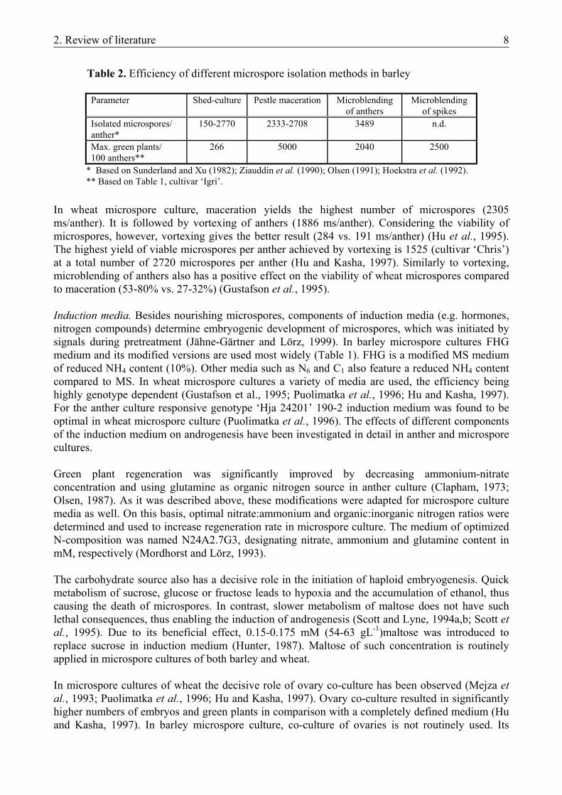
2. Review of literature 8
Table 2. Efficiency of different microspore isolation methods in barley
Parameter Shed-culture Pestle maceration Microblending
of anthers Microblending
of spikes Isolated microspores/ anther*
150-2770 2333-2708 3489 n.d.
Max. green plants/ 100 anthers**
266 5000 2040 2500
* Based on Sunderland and Xu (1982); Ziauddin et al. (1990); Olsen (1991); Hoekstra et al. (1992). ** Based on Table 1, cultivar ‘Igri’.
In wheat microspore culture, maceration yields the highest number of microspores (2305 ms/anther). It is followed by vortexing of anthers (1886 ms/anther). Considering the viability of microspores, however, vortexing gives the better result (284 vs. 191 ms/anther) (Hu et al., 1995). The highest yield of viable microspores per anther achieved by vortexing is 1525 (cultivar ‘Chris’) at a total number of 2720 microspores per anther (Hu and Kasha, 1997). Similarly to vortexing, microblending of anthers also has a positive effect on the viability of wheat microspores compared to maceration (53-80% vs. 27-32%) (Gustafson et al., 1995). Induction media. Besides nourishing microspores, components of induction media (e.g. hormones, nitrogen compounds) determine embryogenic development of microspores, which was initiated by signals during pretreatment (Jähne-Gärtner and Lörz, 1999). In barley microspore cultures FHG medium and its modified versions are used most widely (Table 1). FHG is a modified MS medium of reduced NH4 content (10%). Other media such as N6 and C1 also feature a reduced NH4 content compared to MS. In wheat microspore cultures a variety of media are used, the efficiency being highly genotype dependent (Gustafson et al., 1995; Puolimatka et al., 1996; Hu and Kasha, 1997). For the anther culture responsive genotype ‘Hja 24201’ 190-2 induction medium was found to be optimal in wheat microspore culture (Puolimatka et al., 1996). The effects of different components of the induction medium on androgenesis have been investigated in detail in anther and microspore cultures. Green plant regeneration was significantly improved by decreasing ammonium-nitrate concentration and using glutamine as organic nitrogen source in anther culture (Clapham, 1973; Olsen, 1987). As it was described above, these modifications were adapted for microspore culture media as well. On this basis, optimal nitrate:ammonium and organic:inorganic nitrogen ratios were determined and used to increase regeneration rate in microspore culture. The medium of optimized N-composition was named N24A2.7G3, designating nitrate, ammonium and glutamine content in mM, respectively (Mordhorst and Lörz, 1993). The carbohydrate source also has a decisive role in the initiation of haploid embryogenesis. Quick metabolism of sucrose, glucose or fructose leads to hypoxia and the accumulation of ethanol, thus causing the death of microspores. In contrast, slower metabolism of maltose does not have such lethal consequences, thus enabling the induction of androgenesis (Scott and Lyne, 1994a,b; Scott et al., 1995). Due to its beneficial effect, 0.15-0.175 mM (54-63 gL-1)maltose was introduced to replace sucrose in induction medium (Hunter, 1987). Maltose of such concentration is routinely applied in microspore cultures of both barley and wheat. In microspore cultures of wheat the decisive role of ovary co-culture has been observed (Mejza et al., 1993; Puolimatka et al., 1996; Hu and Kasha, 1997). Ovary co-culture resulted in significantly higher numbers of embryos and green plants in comparison with a completely defined medium (Hu and Kasha, 1997). In barley microspore culture, co-culture of ovaries is not routinely used. Its
2. Review of literature 9
application, however, was supposed to contribute to a decreased genotype-dependence (Li and Devaux, 2001). Microspore density in the induction medium. A minimal density of viable microspores (0.05 x 10⁵
/ml) is essential for their further development (Hoekstra et al., 1993). Optimal densities were determined on a wide range of 0.2-6.0 x 10⁵/ml, depending on genotype and culture protocol (Hoekstra et al., 1993; Gustafson et al., 1995; Davies and Morton, 1998; Castillo et al., 2000). In practice, a medium density of 0.5-1.0 x 10⁵ microspores/ml is used most frequently (Cistué et al., 1995; Hu et al., 1995; Puolimatka et al., 1996; Davies and Morton, 1998; Li and Devaux, 2001). The highest number of green ‘Igri’ regenerants was achieved in cultures of 0.2 x 10⁵/ml microspore density (Hoekstra et al., 1993). By microblending of spikes, best results were achieved when microspores of ‘Igri’ were cultured at densities of 1.0-2.5 x 10⁵/ml (Mordhorst and Lörz, 1993; Kasha et al., 2001b). In these cases the higher number of microspores should compensate the heterogeneity of microspore population caused by homogenization of the less developed florets of spikes as well. Pestle maceration and microblending of anthers provides a more homogeneous microspore population due to the processing of selected anthers. Regeneration media. Plants are usually regenerated on hormone-free MS-based (Murashige and Skoog, 1962) media using sucrose as carbohydrate source (Hunter, 1987, Hoekstra et al., 1992, Mejza et al., 1993, Hu et al., 1995). IAA or NAA added to regeneration medium, however, were found to result in more vigorous barley regenerants (Castillo et al., 2000). 2.1.2. In vitro androgenesis in triticale
For the production of haploid triticale plants both chromosome-elimination technique and in vitro androgenesis can be successfully used. Haploid/dihaploid plants were received from wide crosses with maize and pearl millet (Inagaki and Hash, 1998; Wedzony et al., 1998). Similarly to barley, however, anther culture is the established method for haploid production in triticale, resulting in three released DH cultivars to date (for reviews, see Lukjanjuk and Ignatova, 1986; Forster, 2002). Unlike in its two “ancestors”, wheat (Mejza et al., 1993; Puolimatka et al., 1996) and rye (Guo and Pulli, 1999), plant regeneration from isolated microspore culture has not been reported in triticale prior to our publications (Monostori et al., 1998; Pauk et al., 2000) the results of which are detailed in this thesis. Since the induction of the first anther culture-derived haploid plantlets (Wang et al., 1973), each step of the protocol has been investigated in detail. Triticale genotypes of various ploidy levels have been involved in this research. The majority of reports, however, are about hexaploid triticale, thus reflecting its importance in plant breeding. Besides triticale anther culture protocols, technical details of microspore cultures of other cereals, primarily wheat and barley, meant the starting point for establishing the protocol of microspore culture in triticale. Stress has an important role in the induction of androgenesis in triticale, too. Donor spikes are usually pretreated at 4 ºC for 7-21 days (Lukjanjuk and ignatova, 1986; Marciniak et al., 1998; González and Jouve, 2000). Prolonged cold stress as well as the combination of cold pretreatment with heat shock or mannitol starvation can improve plant regeneration at certain genotypes (Immonen and Robinson, 2000). As it is common in the haploid tissue cultures of cereals, however, genotype has the greatest impact on efficiency (Hassawi et al., 1990; Karsai and Bedő, 1997; Marciniak et al., 1998; González and Jouve, 2000). Highest green plant induction frequencies achieved with cultivars grown in Hungary were 1.5 green plant (‘Presto’), 10.1 plants (‘Moniko’) and 3.2 plants (‘Tewo’) per 100 anthers. ‘Moniko’ responded with strong changes to altered culture conditions, while regeneration rates of ‘Presto’ and ‘Tewo’ could not be improved by any changes in protocol (Karsai and Bedő, 1997). These three cultivars acted as crossing partners in the donor genotypes of our microspore culture experiments. Independent of genotype, anthers containing microspores at the mid- to late-uninucleate stages are the most suitable for culture.
2. Review of literature 10
A wide range of induction media have been found to be superior to others for different genotypes in different laboratories. These include B5 (Lukjanjuk and Ignatova, 1986), 85D12 (Hassawi et al., 1990), P2 (Marciniak et al., 1998), N6 (González and Jouve, 2000), C17 (Ponitka et al., 1999) and W14 (Immonen and Robinson, 2000). The best green plant regeneration results for ‘Presto’, ‘Moniko’ and ‘Tewo’have been achieved by using N6 medium supplemented with 0.26 M maltose and glutamine (Karsai and Bedő, 1997). This composition corresponds to the preferencies regarding nitrogen-composition and carbohydrate source of induction media detailed for barley (see 2.1.1.). The highest green plant regeneration frequency in triticale anther culture (15.36 green plants/100 anthers) has also been recorded with maltose-supplemented N6 medium (González and Jouve, 2000). As growth regulator, 2,4-D alone or in combination with kinetin is used in the induction medium (Marciniak et al., 1998; González and Jouve, 2000; Immonen and Robinson, 2000). Regeneration media are usually supplemented with hormones (IAA, NAA, kinetin), although plant regeneration of greatest efficiency has been reported on hormone-free medium (González and Jouve, 2000). MS-based media are usually used, however 190-2 regeneration medium has been found to be superior to MS for cultivar ‘Moniko’ (Karsai and Bedő, 1997). Previous to our work, isolated pollen/microspore culture without plant regeneration has been reported for triticale (Keller, 1991). First steps of androgenesis have been observed only after anther preculture or in anther-conditioned media. Furthermore, the positive effect of cold pre-treatment of spikes on pollen development was observed there. 2.1.3. The role of plant hormones in the induction of microspore embryogenesis
The role of hormones, if any, in the induction of androgenesis is less understood, while their role during embryo development has been found to be more evident (Sangwan and Sangwan-Norreel, 1990). In dicotyledonous plants like Nicotiana, Datura, and Brassica spp., exogenous hormone supply is not essential for the induction of pollen embryogenesis – the required signal is offered by stress factors (Nitsch, 1977; Sangwan and Sangwan-Norreel, 1990; Swanson, 1990; Touraev and Heberle-Bors, 1999). Gramineae, such as barley, triticale and wheat, are known to belong to the group of plants which require hormones in the induction medium of anther cultures. Anthers of this group, however, can also exhibit response if induced in hormone-free medium in the first stage of culture (for review, see Dunwell, 1985). In practice, however, auxins and/or cytokinins are routinely added to culture media in anther and microspore cultures of cereals to promote embryogenesis induced by stress signals. The role of these hormones, however, is still unknown (for reviews, see Clapham, 1977; Jähne and Lörz, 1995). In microspore cultures of barley, benzylaminopurine (BAP) is the most frequently used hormone (Table 1), while in microspore cultures of wheat auxins, occasionally in combination with cytokinins, are used the most widely (Datta and Wenzel, 1987; Gustafson et al., 1995; Hu and Kasha, 1997). Induction of androgenesis without exogenous hormone-supply has been reported only in anther cultures of barley (Cai et al., 1992) and oat (Kiviharju et al., 1997) as well as in isolated microspore cultures of wheat (Touraev et al., 1996). Our preliminary results in barley microspore culture suggested that hormone-free media could be used to promote androgenesis with great efficiency. Plant regeneration, however, resulted in albinos only, except for the genotype ‘Jokioinen 1490’ where a low number of green plants could be regenerated exclusively on hormone-free medium (unpublished data). 2.2. PREPARATION OF NOVEL VECTOR CONSTRUCTS
2. Review of literature 11
2.2.1. Genetic transformation of barley
Modification of the endogenous JA-level via manipulation of the biosynthetic pathway in transgenic plants has recently offered new possibilities to study the role of jasmonates in dicotyledonous species (see 2.2.3.). The most effective transformation method for dicots, the indirect gene-transfer mediated by Agrobacterium tumefaciens, was applied in these cases as well. Over a long period of time this transformation method was considered to be inapplicable in monocots, which were known to be out of the host range of the bacterium. Recently, transgenic plants produced via Agrobacterium-mediated transformation have been reported in cereals, too (barley: Tingay et al., 1997; maize: Ishida et al., 1996; rice: Hiei et al., 1994; wheat: Cheng et al., 1997). The potential advantages of this system include the introduction of low copy number of the transgene, its preferential integration into transcriptionally active regions of the chromosome and high co-expression of the introduced genes. These features have been described in barley transformation as well (Tingay et al., 1997; Horvath et al., 2000; Wang et al., 2001; Fang et al., 2002). In spite of these remarkable characteristics of Agrobacterium-mediated transformation, however, direct gene-transfer is still the most widely applied strategy for the genetic engineering of monocotyledonous species. Among these methods the electroporation- and PEG-mediated transformation of protoplasts has become an established method in the production of transgenic rice (Zhang et al., 1988). Its application for other species, however, was limited by the difficulties in plant regeneration as well as by the poor reproducibility of the protoplast-system. Among several promising attempts with various explants and techniques, the delivery of DNA-coated microprojectiles into regenerable cells or tissues through bombardment has recently proved the simplest and most effective transformation method in all cereal species (for reviews, see Datta, 1999; Gordon-Kamm et al., 1999; Lemaux et al., 1999; Vasil and Vasil, 1999b). Besides the commercially available and most widely used He-powered Particle Delivery System (Biolistic® PDS-1000/He by Bio-Rad; Kikkert, 1993) other particle bombardment devices are also in use (Iida et al., 1990; Christou et al., 1991; Jenes et al., 1996). The usually home-made models of particle inflow gun (PIG) are driven through the system developed by Finer et al. (1992). In barley also the biolistic gene-transfer was found to be the most effective method to perform transient expression studies (Mendel et al., 1989; 1992; Schledzewski and Mendel, 1994; Hänsch et al., 1995; Harwood et al., 2000) as well as to produce transgenic plants. Particle bombardments are usually carried out with PDS-1000/He, but PIGs have also been successfully applied in the production of transgenic barley plants (Koprek et al., 1996; Zhang et al., 1999) (Table 3). The most widely used target tissue is provided by the scutellar surface of immature embryos. Due to its high responsivity in regeneration systems based on somatic embryogenesis, cultivar ‘Golden Promise’ is the genotype comonly used. The highest transformation frequency reported in barley (15.2%) was also achieved by particle bombardment of immature embryos of ‘Golden Promise’ (Nuutila et al., 1999). Apart from this extra result, however, highest transformation frequency was approximately 4% (Wan and Lemaux, 1994; Jensen et al., 1995; Zhang et al., 2001). The highest transformation frequency achieved by Agrobacterium-mediated transformation was in the same range (4.2%), also with the same genotype and target tissue (Tingay et al., 1997). Biolistic transformation of microspores as well as protoplast transformation usually results in transgenic plants at 10-6-10-7 frequency (Table 3). Table 3. Reports about transgenic barley plants
Target material Genotype Resistance Transgene of TFj Reference
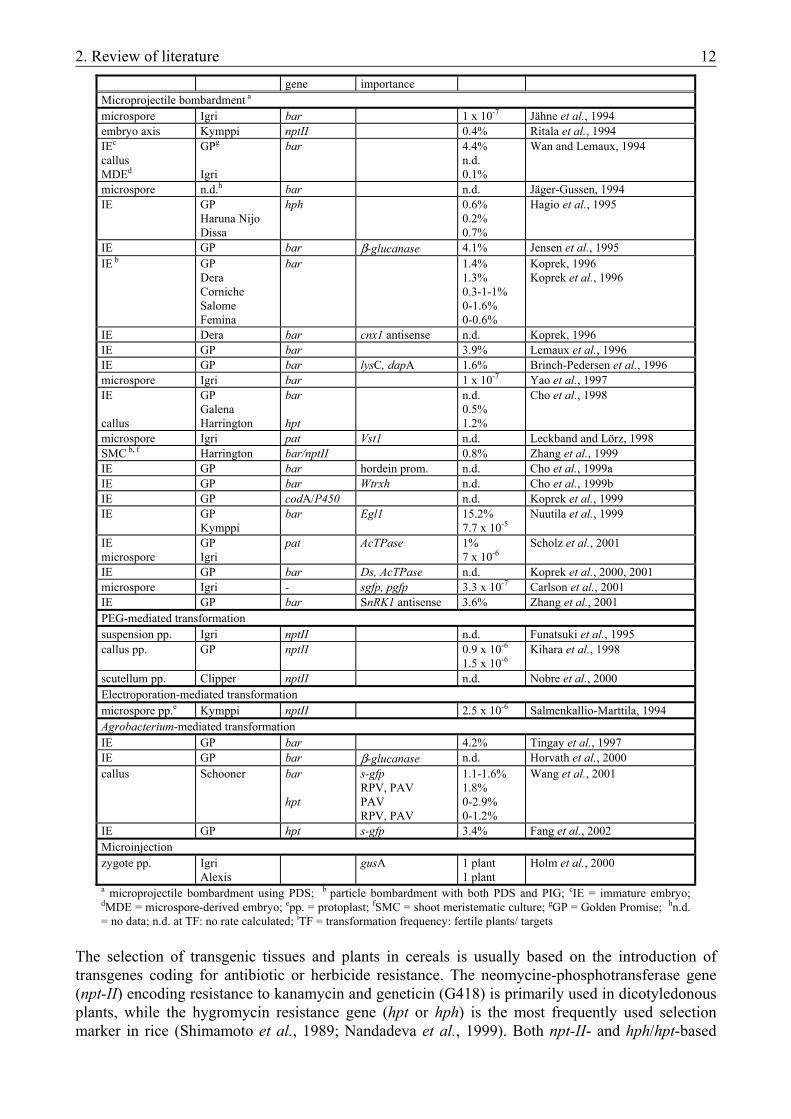
2. Review of literature 12
gene importance Microprojectile bombardment a microspore Igri bar 1 x 10⁻⁷ Jähne et al., 1994 embryo axis Kymppi nptII 0.4% Ritala et al., 1994 IEc callus MDEd
GPg
Igri
bar 4.4% n.d. 0.1%
Wan and Lemaux, 1994
microspore n.d.h bar n.d. Jäger-Gussen, 1994 IE GP
Haruna Nijo Dissa
hph 0.6% 0.2% 0.7%
Hagio et al., 1995
IE GP bar β-glucanase 4.1% Jensen et al., 1995 IE b GP
Dera Corniche Salome Femina
bar 1.4% 1.3% 0.3-1-1% 0-1.6% 0-0.6%
Koprek, 1996 Koprek et al., 1996
IE Dera bar cnx1 antisense n.d. Koprek, 1996 IE GP bar 3.9% Lemaux et al., 1996 IE GP bar lysC, dapA 1.6% Brinch-Pedersen et al., 1996 microspore Igri bar 1 x 10⁻⁷ Yao et al., 1997 IE callus
GP Galena Harrington
bar hpt
n.d. 0.5% 1.2%
Cho et al., 1998
microspore Igri pat Vst1 n.d. Leckband and Lörz, 1998 SMC b, f Harrington bar/nptII 0.8% Zhang et al., 1999 IE GP bar hordein prom. n.d. Cho et al., 1999a IE GP bar Wtrxh n.d. Cho et al., 1999b IE GP codA/P450 n.d. Koprek et al., 1999 IE GP
Kymppi bar Egl1 15.2%
7.7 x 10⁻⁵ Nuutila et al., 1999
IE microspore
GP Igri
pat AcTPase 1% 7 x 10⁻⁶
Scholz et al., 2001
IE GP bar Ds, AcTPase n.d. Koprek et al., 2000, 2001 microspore Igri - sgfp, pgfp 3.3 x 10⁻⁷ Carlson et al., 2001 IE GP bar SnRK1 antisense 3.6% Zhang et al., 2001 PEG-mediated transformation suspension pp. Igri nptII n.d. Funatsuki et al., 1995 callus pp. GP nptII 0.9 x 10⁻⁶
1.5 x 10⁻⁶ Kihara et al., 1998
scutellum pp. Clipper nptII n.d. Nobre et al., 2000 Electroporation-mediated transformation microspore pp.e Kymppi nptII 2.5 x 10⁻⁶ Salmenkallio-Marttila, 1994 Agrobacterium-mediated transformation IE GP bar 4.2% Tingay et al., 1997 IE GP bar β-glucanase n.d. Horvath et al., 2000 callus Schooner bar
hpt
s-gfp RPV, PAV PAV RPV, PAV
1.1-1.6% 1.8% 0-2.9% 0-1.2%
Wang et al., 2001
IE GP hpt s-gfp 3.4% Fang et al., 2002 Microinjection zygote pp. Igri
Alexis gusA 1 plant
1 plant Holm et al., 2000
a microprojectile bombardment using PDS; b particle bombardment with both PDS and PIG; cIE = immature embryo; dMDE = microspore-derived embryo; epp. = protoplast; fSMC = shoot meristematic culture; gGP = Golden Promise; hn.d. = no data; n.d. at TF: no rate calculated; iTF = transformation frequency: fertile plants/ targets
The selection of transgenic tissues and plants in cereals is usually based on the introduction of transgenes coding for antibiotic or herbicide resistance. The neomycine-phosphotransferase gene (npt-II) encoding resistance to kanamycin and geneticin (G418) is primarily used in dicotyledonous plants, while the hygromycin resistance gene (hpt or hph) is the most frequently used selection marker in rice (Shimamoto et al., 1989; Nandadeva et al., 1999). Both npt-II- and hph/hpt-based
2. Review of literature 13
selection have contributed to the production of transgenic barley plants, although the most widely used selection system in barley is based on the expression of phosphinothricin-acetyltransferase (PAT) encoded by the bar and the pat marker genes (Table 3). Phosphinothricin (PPT) acts as a competitive inhibitor of glutamine synthetase. The resulting ammonia accumulation and glutamine deficiency lead to the death of the plant cells. The pat gene of S. viridochromogenes origin (Wohlleben et al., 1988) and the bar gene from Streptomyces hygroscopicus (Thompson et al., 1987) confer resistance to the herbicides phosphinothricin (PPT), Basta® and bialaphos (L-PPT; Glufosinate-ammonium). PAT inactivates PPT by acetylation (de Block et al., 1987; Dröge et al., 1992). Besides the improvement of transformation efficiency, agronomically important traits such as malting and feeding quality (Brinch-Pedersen et al., 1996; Jensen et al., 1996; Nuutila et al., 1999; Cho et al., 1999b; Horvath et al., 2000) as well as resistance against pathogens (Leckband and Lörz, 1998; Wang et al., 2001) have also been improved via the transformation of barley. Further results were the alteration of nitrate-reductase activity (Koprek, 1996) and the introduction of transposable elements (Koprek et al., 2000, 2001; Scholz et al., 2001) (Table 3). In spite of the above-mentioned reports on successful production of transgenic plants, however, there are problems which hinder genetic transformation in becoming a routine method for the improvement of barley (Lemaux et al., 1999): (i) only few genotypes were found to be amenable to in vitro cultivation and to give high transformation frequency in the current transformation systems (Table 3), (ii) somatic mutation and stable epigenetic changes can arise during in vitro culture and may hinder various stages of the transformation process, (iii) using current transformation methods, transgene insertions occur randomly and their locations are not always optimal for gene expression; furthermore, the insertion of multiple copies, more common with direct gene4-transfer, can lead to both gene inactivation and genetic instability (for reviews, see Iyer et al., 2000). Considering these problems, the establishment of an in vitro plant regeneration system for the given genotype and the optimization of the bombardment parameters are essential for achieving the highest transformation frequency in the stable transformation experiments. 2.2.2. Modification of biosynthetic pathways by genetic transformation
The two ways to manipulate plant hormone levels through a transgenic approach are the expression of genes coding for enzymes involved in hormone biosynthesis or hormone degradation (for review, see Hedden and Phillips, 2000). To date, the biosynthesis, degradation and function of auxins (Tinland et al., 1991; Pandolfini et al., 2002), cytokinins (Smigocki, 1991; Hewelt et al., 1994; Faiss et al., 1997), gibberellins (Huang et al., 1998; Coles et al., 1999), ethylene (Hamilton et al., 1990; Oeller et al., 1991) and jasmonates (Bell et al., 1995; Harms et al., 1995; Wang et al., 1999a; Laudert et al., 2000) have been studied in transgenic plants. In the experiments with the “classical” plant hormones several parameters have been determined which could interfere with the transgenic strategy. These include: (i) the site of overproduction (Smigocki, 1991; Hewelt et al., 1994), (ii) metabolism of the hormone (Faiss et al., 1997), (iii) gene dosis (Hewelt et al., 1994), (iv) the type of the promoter controlling the transgene (Tinland et al., 1991; Hewelt et al., 1994), and (v) the genetic background (Pandolfini et al., 2002). Introduction of homologous or heterologous sequences into the plant genome is the usual way to get overexpression of the corresponding gene in the transgenic plants, although transgene copy number can be both positively and negatively correlated with the level of expression (Hobbs et al., 1992; Stöger et al., 1998). The insertion of the transgene, however is not always followed by its expression as expected. Gene silencing can occur e.g. after an initial high expression level attributed to the application of strong promoters such as CaMV 35S (Elmayan and Vaucheret, 1996). Transcriptional gene silencing (TGS) is resulted primarly by the methylation of the promoter
2. Review of literature 14
sequence, while post-transcriptional gene silencing (PTGS) acts, among others, via cosuppression of homologous endogenous genes by the transcribed sense transgene (for reviews, see Fagard and Vaucheret, 2000; Iyer et al., 2000). Insertion of multiple copies of the transgene, as the result of direct gene transfer methods, is considered to be one of the main sources of silencing, co-suppression, however, single-copy inserts can also exert the same effect (for review, see Iyer et al., 2000). Antisense RNA, as the transcript of an introduced antisense transgene sequence, can inhibit gene expression by binding to specific complementary reginons of the target RNA. Antisense strategies are usually used among others, (i) to produce mutants, (ii) to observe steps in metabolic pathways, (iii) to identify gene functions (iv) to determine sequence/promoter specifity and (v) transcript/protein relationships, (vi) to regulate plant development as well as (vii) to improve crops. As mechanisms of action both transcritional/posttranscritional and translational control can be supposed (for reviews, see Bourque, 1995). Among cereals, relatively few transgenic plants expressing antisense genes/cDNAs have been described. In barley, the antisense-method is mainly used in transient expression systems (Huntley and Hall, 1993; Schweizer et al., 2000). Transgenic barley plants expressing antisense transgenes have been reported in two cases only. Transgenic barley plants produced by heterologous transformation with antisense cnx-1 gene from Arabidopsis exhibited reduced nitrate-reductase activity (Koprek, 1996), and homologous transformation with an antisense SnRk1 protein kinase sequence resulted in abnormal pollen development and male sterility (Zhang et al., 2001). 2.2.3. Jasmonates
Since the first identification of jasmonic acid (JA) and its methyl ester (JM) in a fungal culture filtrate and in Jasminum grandiflorum L., respectively (Aldridge et al., 1971; Demole et al., 1962), several physiological roles of jasmonates in plants have been elucidated. First, promotion of senescence and inhibition of seedling growth were described (Ueda and Kato, 1980, 1982). Later on, inhibitory effects on root growth, pollen germination and photosynthetic activities have also been observed and exogenously applied jasmonates were found to promote fruit ripening, tuberization and accumulation of secondary metabolites (for reviews, see Sembdner and Parthier, 1993; Creelman and Mullet, 1997a,b; León and Sánchez-Serrano, 1999; Wasternack and Hause 2002). Jasmonates are ubiquitously occurring plant hormones in angiosperms, gymnosperms, ferns, algae and fungi (Ueda and Kato, 1980; Dathe et al., 1981; Meyer et al., 1984; Miersch et al., 1987; Ueda et al., 1991; Yamane et al., 1981). In higher plants the highest endogenous levels of jasmonates have been detected in young dividing tissues such as the stem apex, root tips, young leaves, flowers and immature fruits (Sembdner and Parthier, 1993; Creelman and Mullet, 1995; Hause et al., 1996). A rise in their endogenous levels could be observed in response to wounding (Peña-Cortés et al., 1995; Bergey et al., 1999; León et al., 2001), tendril coiling (Weiler et al., 1993), water deficit (Lehmann et al., 1995), elicitors of pathogen defense (Gundlach et al., 1992; Nojiri et al., 1996) and the pathogen itself (Penninckx et al., 1996). Usually, elevated JA levels during developmental processes or in response to biotic and abiotic stresses are accompanied with altered gene expression (see 2.2.4.).
2. Review of literature 15
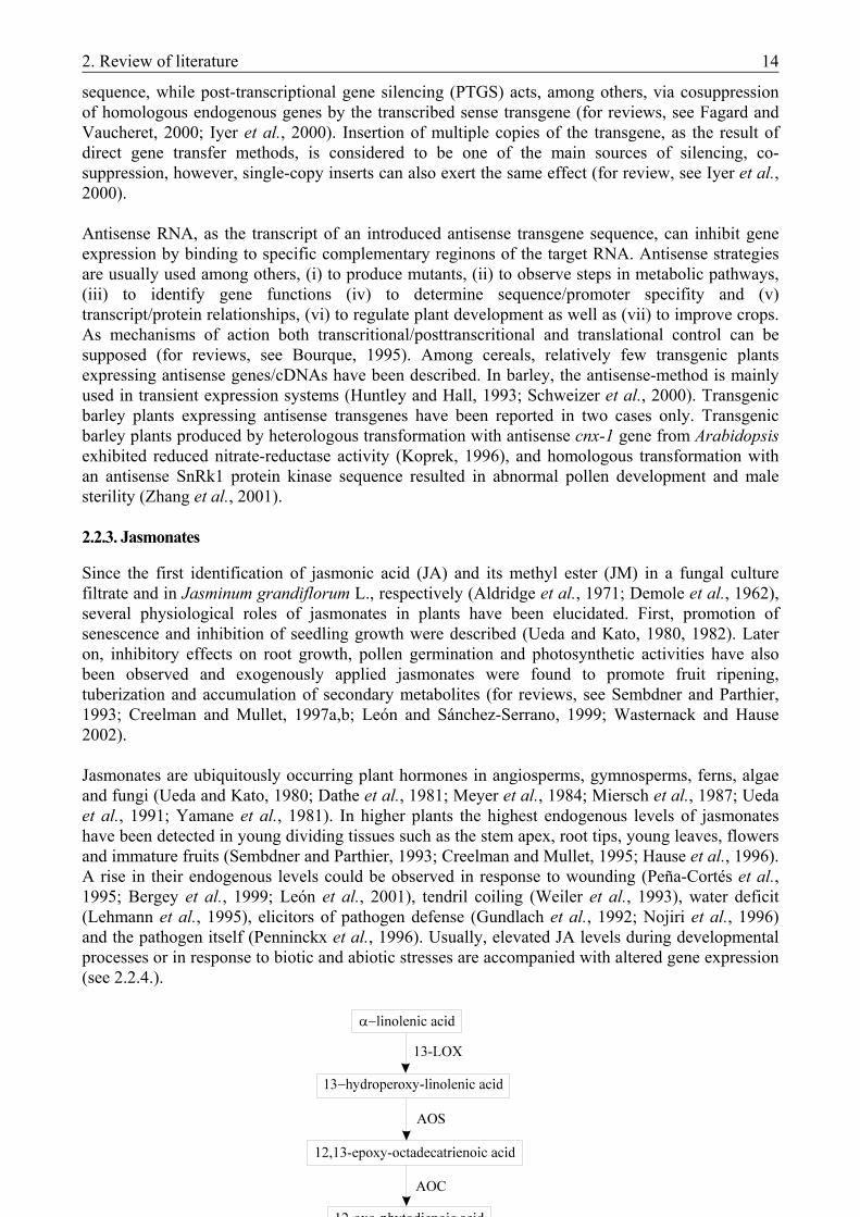
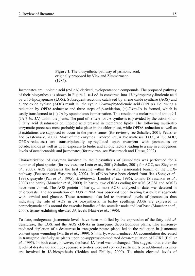
Figure 1. The biosynthetic pathway of jasmonic acid, originally proposed by Vick and Zimmermann (1984).
Jasmonates are linolenic acid (α-LeA)-derived, cyclopentanone compounds. The proposed pathway of their biosynthesis is shown in Figure 1. α-LeA is converted into 13-hydroperoxy-linolenic acid by a 13-lipoxygenase (LOX). Subsequent reactions catalyzed by allene oxide synthase (AOS) and allene oxide cyclase (AOC) result in the cyclic 12-oxo-phytodienoic acid (OPDA). Following a reduction by OPDA-reductase and three steps of β-oxidation, (+)-7-iso-JA is formed, which is easily transformed to (−)-JA by spontaneous isomerization. This results in a molar ratio of about 9:1 (JA:7-iso-JA) within the plants. The pool of α-LeA for JA synthesis is provided by the action of ω-3 fatty acid desaturases on linoleic acid present in membrane lipids. The following multi-step enzymatic processes most probably take place in the chloroplast, while OPDA-reduction as well as β-oxidations are supposed to occur in the peroxisomes (for reviews, see Schaller, 2001; Feussner and Wasternack, 2002). Most of the enzymes involved in JA biosynthesis (LOX, AOS, AOC, OPDA-reductase) are transcriptionally up-regulated upon treatment with jasmonates or octadecanoids as well as upon exposure to biotic and abiotic factors leading to a rise in endogenous levels of octadecanoids and jasmonates (for reviews, see Wasternack and Hause, 2002). Characterization of enzymes involved in the biosynthesis of jasmonates was performed for a number of plant species (for reviews, see León et al., 2001; Schaller, 2001; for AOC, see Ziegler et al., 2000). AOS represents the first enzyme within the AOS (jasmonate) branch of the LOX-pathway (Feussner and Wasternack, 2002). Its cDNAs have been cloned from flax (Song et al., 1993), guayule (Pan et al., 1995), Arabidopsis (Laudert et al., 1996), tomato (Sivasankar et al., 2000) and barley (Maucher et al., 2000). In barley, two cDNAs coding for AOS (AOS1 and AOS2) have been cloned. The AOS protein of barley, as most AOSs analyzed to date, was detected in chloroplasts. The accumulation of AOS mRNA was observed upon treating barley leaf segments with sorbitol and glucose. These treatments also led to increased levels of jasmonates thus indicating the role of AOS in JA biosynthesis. In barley seedlings AOSs are expressed in parenchymatic cells around the vascular bundles of the scutellar node and leaf base (Maucher et al., 2000), tissues exhibiting elevated JA levels (Hause et al., 1996). To date, endogenous jasmonate levels have been modified by the expression of the fatty acid 3 desaturase, the LOX and the AOS cDNAs in transgenic dicotyledonous plants. The antisense-mediated depletion of a desaturase in transgenic potato plants led to the reduction in jasmonate content upon wounding (Martín et al., 1999). Similarly, wound-induced JA accumulation decreased in transgenic Arabidopsis plants upon co-suppression-mediated down-regulation of LOX2 (Bell et al., 1995). In both cases, however, the basal JA-level was unchanged. This suggests that either the levels of desaturase and lipoxygenase activities were not reduced sufficiently or additional enzymes are involved in JA-biosynthesis (Hedden and Phillips, 2000). To obtain elevated levels of
2. Review of literature 16
jasmonates, AOS was constitutively overexpressed in different plants. Surprisingly, this did not alter the basic JA-levels of untreated tobacco and Arabidopsis plants (Wang et al., 1999a; Laudert et al., 2000), whereas in potato untreated leaves showed increased JA levels (Harms et al., 1995). In the latter case, however, JA-responsive gene expression appeared only upon wounding. This suggests that the elevated JA levels were sequestrated in the unwounded transgenic potato leaves (Harms et al., 1995).
2.2.4. Jasmonate-induced gene expression in barley
The level of jasmonates can rise endogenously upon various biotic and abiotic stresses. Such an endogenous change as well as the exogenous application of jasmonates are accompanied by alterations in the expression of various groups of genes (for reviews, see Creelman and Mullet, 1997a,b; Wasternack and Parthier, 1997; Wasternack and Hause, 2002). Jasmonates induce the synthesis of proteins involved in plant defense and signal transduction of stress responses, such as proteinase inhibitors, thionins, defensins, ribosome-inactivating proteins, chalcone synthase, lipoxygenase and calmodulin. Furthermore, genes coding for enzymes of jasmonate biosynthesis and secondary metabolism as well as seed and vegetative storage proteins are also JA-inducible. In contrast, the formation of some proteins mainly involved in photosynthesis is repressed in response to jasmonates (Reinbothe et al., 1994). The role of JA as a “master switch” has been illustrated in barley (Wasternack and Parthier, 1997). In leaf segments a thionin of 6 kDa (JIP6) (Andresen et al., 1992), a 23 kDa (JIP23) and a 37 kDa protein (JIP37) (Weidhase et al., 1987; Lehmann et al., 1995), a 60 kDa protein (JIP60) with ribosome-inactivating properties (Chaudry et al., 1994, Görschen et al., 1997a) and several LOX forms (Feussner et al., 1995; Vörös et al., 1998) are synthesized upon treatment with jasmonates. JIPs are inducible not only by exogenous application but by endogenous rise of jasmonates as well. In contrast, LOX2:Hv:1 (Vörös et al., 1998) and a group of jasmonate-regulated genes (jrg5, jrg10, jrg12; Lee et al., 1996) are exclusively inducible by exogenous JA. Whereas putative functions for thionin, JIP60 and LOX forms could be proposed, the possible functions of JIP23 and JIP37 are poorly understood (Andresen et al., 1992; Hause et al., 1996, 1999; Leopold et al., 1996). JIP37 shows partial homology to a phytase from maize (Maugenest et al., 1997), but up to date no similarities to published sequences have been found for JIP23 (Andresen et al., 1992). Genes coding for JIP23 were found in all cereals tested (Hause et al., 1999), but only two examples are known on homologous sequences in dicots: in Mesembryanthemum cristallinum (H. J. Bohnert and M. Ibdah, pers. comm.) and in Atriplex canescence (Cairney et al., 1995). In M. cristallinum the expression of jip23 is related to abiotic stress (UV and salt). Expression of genes coding for JIP23 can be induced by small amounts of exogenously applied jasmonates as well as by a low threshold of endogenous jasmonates exerted by various stress factors (Lehmann et al., 1995; Kramell et al., 2000). Usually up to 6 isoforms of JIP23 are synthesized upon jasmonate treatment, and they can be detected in barley seedlings as well. JIP23 and its mRNA occur specifically in cells and tissues exhibiting high osmolarity, e.g. the scutellum, scutellar node and the companion cells of phloem. This suggests that genes coding for JIP23 might be expressed in response to osmotic stresses as it appears during solute transport in developing seedlings (Hause et al., 1996). While all 80 barley cultivars exhibited jip23 expression during germination, some of them - lacking at least one JIP23 gene - failed to express jip23 upon treatment of primary leaves with jasmonates as well as upon treatment with 1 M sorbitol. This suggests that different genes might be responsible for JA-induced expression in differentiated leaves and for developmentally regulated expression (Hause et al., 1999).

2. Review of literature 17
Attempts have been made to obtain more information about the possible functions of JIP23 via heterologous expression of barley JIP23 cDNA in tobacco. One cDNA was sufficient to generate all the six JIP23 isoforms suggesting that JIP23 was modified posttranslationally. In transgenic tobacco plants overexpressing JIP23, several proteins such as the subunits of RuBPCase were down-regulated at the level of translation. The data suggest that discrimination among certain tobacco transcripts during translation initiation is caused by barley JIP23 (Görschen et al., 1997b).

2. Review of literature 18
3. Materials and methods 19
3. MATERIALS AND METHODS
3.1. INDUCTION OF HAPLOID EMBRYOGENESIS IN MICROSPORE CULTURE 3.1.1. Materials
3.1.1.1. Plant material
Barley. The experiments on the evaluation of different culture media in barley microspore culture were carried out using two genotypes: ‘Igri’ is a two-rowed winter-type, while ‘Kymppi’ is a two-rowed spring-type barley. Seeds were sown into a peat-soil mix and incubated in a greenhouse at room temperature. Two-three weeks after germination ‘Igri’ seedlings of 2-3 leaves were vernalized at 4 °C under continuous fluorescent light (40 µmolm-2s-1) for 8 weeks. Donor plants were grown in a controlled greenhouse in the years 1997-1998. From the tillering stage on, fertilizer (Volldünger) was applied weekly. Donor spikes were collected when the anthers in the most mature florets contained microspores of the mid- to late-uninucleate stage. During cold pretreatment a slow development of microspores was observed. Thus, late uninucleate to early binucleate stage was considered to be appropriate for isolation. Tillers being in the early booting stage with awns emerged about 0.5-0.8 cm from the flag leaf were cut between the 2nd and 3rd node and put into Erlenmeyer flasks containing fresh tap water. All leaves but the flag leaf were removed and the tillers were covered with a PVC bag to maintain high humidity (ca. 80% RH). Cold pretreatment of the donor spikes was performed under a dim fluorescent light at 4 °C for 14-21 days. Triticale. Five complete hexaploid (2n=6x=42, AABBRR) winter triticale genotypes were involved in the experiments: one cultivar (‘Presto’) and four F1 combinations (‘Tewo x Moniko’, ‘Presto x Moniko’, ‘Presto x Novisadi’, ‘Novisadi x Moniko’). The donor cultivars used in the crosses are of Polish (‘Presto’, ‘Moniko’, ‘Tewo’) and Yugoslavian (‘Novisadi’) origin and all are registered cultivars in Hungary. Donor plants were grown in the field nursery during the growing season of 1995-1996. Standard herbicides have been applied according to the weed control protocol of the institute (Cereal Research Non-profit Company, Szeged). Preliminary experiments on the methods of pretreatment and microspore isolation were performed with four genotypes (‘Tewo’; ‘Moniko x Tewo’, ‘Tewo x Moniko’, ‘Novisadi x Tewo’ F1). The collection and cold pretreatment of the donor spikes happened similarly to the method described above for barley. Spikes containing anthers with mid- to late-uninucleate microspores were collected when the tillers were in the late booting stage (sheath of the flag leaf open, upper spikelets emerged). 3.1.1.2. Culture media
Barley. In the evaluation of different induction media the following basic media were used (Appendix 1): - N24A2.7G3 (further referred to as N24-BA), an LA3 based medium with optimized N-composition (Mordhorst and Lörz, 1993) and - 190-2 medium (further referred to as 190-BA) originally invented in wheat anther culture (Zhuang and Jia, 1983). Both media were supplemented with 3 mM L-glutamine and 1 mgL-1 (4.4 µM) BAP (Mordhorst and Lörz, 1993). To study the necessity of hormones in the induction of androgenesis both media were prepared without BAP as well (N24-0 and 190-0, respectively).
3. Materials and methods 20
Each medium contained 175 mM maltose (Scott and Lyne, 1994) and pH was adjusted to 5.8 with 1 M KOH. Osmotic pressure was checked in each preparation using an osmometer. The media were filter-sterilized and stored at room temperature. Induced ELSs were incubated on induction media of reduced maltose content (80 mM) solidified with 0.2% Gelrite. Plants were regenerated on hormone-free LA3 medium supplemented with 80 mM maltose (Mordhorst and Lörz, 1992, 1993). Triticale. For the evaluation of the effects of different hormone compositions on the induction of androgenesis, 190-2 medium supplemented with 3 mM L-glutamine and the following growth regulator combinations were applied: - 1.5 mgL-1 (6.8 µM) 2.4-D and 0.5 mgL-1 (2.3 µM) kinetin (190-D/K), - 10 mgL-1 (73 µM) PAA (190-PAA), - no hormones (190-0). Further preparation details were the same as detailed at barley. Induced ELSs were cultured on solid induction medium as detailed at barley. Plants were regenerated on hormone-free 190-2 medium without glutamine supplement (Zhuang and Jia, 1983; Pauk et al., 1991). 3.1.2. Methods
3.1.2.1. Determination of the developmental stage and the number of developing structures
The developmental stage of microspores was determined prior to collecting spikes and/or prior to the isolation of microspores. Anthers from a floret in the central part of the spike were squashed in a drop of water and examined under an inverted microscope. The number of microspores was determined microscopically with a haemocytometer (Bürker). The different structures (dividing microspores, ELS etc.) developed in the cultures were counted in representative fields using an inverted microscope. 3.1.2.2. Isolation of microspores
In both triticale and barley, microspores were isolated via microblending segmented spikes based on the method described by Mordhorst and Lörz (1993). Pretreated spikes (1-10 pcs.) containing microspores of the late uninucleate to early binucleate stage (a slow progress in the development of microspores was observed during pretreatment) were removed from the leaf sheath and awns were cut down. Spikes were surface-sterilized in 2% sodium-hypochlorite for 20 min and rinsed three times with sterile water. Following sterilization they were cut into 1 cm segments and put into a 100 ml Waring Micro Blendor container (Eberbach Corp., Ann Arbour, Michigan, USA). Sixty ml of 0.3 M mannitol solution was added and microspores were isolated by blending twice for 5 sec at low speed. The quality of the maceration was visually monitored through the plastic cap of the vessel. The crude microspore suspension was filtered through 160 and 80 µm sterile nylon sieves to remove raw spike debris. The filtrate was divided between four centrifuge tubes (10 ml volume each) and centrifuged at 800 rpm for 5 min. The pellet was resuspended in 2 ml 0.3 M mannitol and the microspore suspension was carefully layered over a 0.58 M maltose solution. Following centrifugation at 600 rpm for 10 min, viable microspores were located in a band at the maltose/mannitol gradient interphase, while dead microspores and debris pelletted in the bottom of the tube. Viable cells were collected with a Pasteur pipette. They were resuspended (washed) in 0.3 M mannitol (8 ml/tube) and spun down at 600 rpm for 5 min.
3. Materials and methods 21
3.1.2.3. Culture of microspores
Following the last centrifugation, pelletted microspores were resuspended in 1 ml of the culture medium. The quantity of microspores was determined as detailed above. Viable microspores were identified in triticale by staining with fluorescein diacetate (Widholm, 1972). Culture density was adjusted to 0.7-1 x 10⁵ microspore/ml by adding induction medium. Two ml aliquots of the microspore suspension were cultured in Petri dishes (∅ 60 mm, Greiner). Four to seven replicates per medium type (barley) or per genotype (triticale) were prepared in the different experiments. Cultures were kept at 28 °C and 80% relative humidity in the dark. After 3-5 weeks of culture, embryo-like-structures (ELS) were plated on induction medium solidified with 0.25% Gelrite. Transfer happened using forceps in the case of slowly developing cultures yielding few ELSs. If high numbers of ELSs were formed, embryoids were transferred by pipette. ELSs were cultured at 28 °C under a 16 h photoperiod (20 µmolm-2s-1). 3.1.2.4. Regeneration of plants
Barley. To prove that the microspore cultures are capable of generating complete plants with shoots and roots, 5-10 plantlets (ca. 2 cm height) per type of medium were transferred from solid induction medium to LA3 regeneration medium and kept under unaltered conditions. Following tillering and rooting, plantlets were discarded without transplanting into soil, because growing up adult plants was not our aim in the barley microspore cultures. Triticale. ELSs reaching the bipolar stage were transferred to 190-2 regeneration medium in glass culture tubes (Pauk et al., 1991). The cultures were kept at 25 °C and a 16 h photoperiod. Four to five weeks after subculture the well-tillered and rooted plantlets were transplanted into non-sterilized peat/sand soil (1:1). During the following two weeks the plantlets were acclimatized in a growth cabinet (Conviron) at 25 °C, 80% relative humidity and a 16 h photoperiod (200 µmolm-2s-
1). Acclimatized plants were transferred to a conditioned greenhouse. Vernalization was carried out in a cold chamber at 2-4 °C under continuous fluorescent light (40 µmolm-2s-1) for six weeks. Prior to vernalization the ploidy level of the individual plants was determined and the haploid plants were treated with colchicine to double chromosome number (Henry and de Buyser, 1980). 3.1.2.5. Determination of ploidy level
Ploidy level was determined in all plants regenerated from triticale microspore cultures. The method based on the length of stomatal guard cells (Borrino and Powell, 1988) was applied to each plant (126 pcs.), while exact chromosome number was determined in randomly chosen samples (58 pcs.) only. The length of the stomatal guard cells was determined from a 10 mm distal leaf segment taken from microspore derived plants. Chlorophyll was extracted by incubating in 70 % ethanol overnight and the leaf segments were mounted in a drop of water on a glass slide with cover glass. The length of 10 stomatal guard cells per sample was measured (lengthwise of the guard cells) using an ocular micrometer. Plants exhibiting a stomatal guard cell length being 40-50% shorter than in control hexaploid plants, were considered to be haploids. Chromosome numbers were determined from root tip preparations. Root tips of the donor plants were incubated in a cold chamber at 4 ºC for 24 hours. The pretreated tips were collected and treated in saturated oxiquinolin suspension for 5 h and fixed in 3:1 (v:v) ethanol:glacial acetic acid. Prior to staining in acetocarmine, root tips were hydrolysed in 1 M HCl at 60 ºC for 10 min. Squash
3. Materials and methods 22
preparations were made in 45% acetic acid and chromosomes were counted in three well-spread cells per root tip. 3.1.2.6. Analysis of data
The evaluation of data started with the descriptive statistical analysis (mean, standard deviation, coefficient of variation) of the three androgenetic parameters (ELS/culture, albino plant % and green plant %) sorted by treatments (media). Standard deviation of data proved to be very high, therefore logarithmic transformation for data of ELS (given as non-proportional values) was performed prior to further analysis. Data of albino and green plants given as percentage were transformed using arcsin transformation. After transformation, data showed approximately normal distribution by using the paired two-sample t-test. Thus, further analysis was possible. Analysis of variance (ANOVA) was performed in order to analyze the effects of the three media on the three androgenic traits. In the case of the regeneration of albino and green plants, one-way multiple comparison of the means of different media was based on the LSD. The mean values in ELS production were compared in case of similar deviations by two sample t-test, and in case of different deviations by Welch-probe. ANOVA and other statistical tests (Fowler and Cohen, 1990) were computed using appropriate programmes from the MiniTab statistical package. 3.2. PREPARATION OF NOVEL VECTOR CONSTRUCTS
3.2.1. Materials
3.2.1.1. Plant material
For transformation studies the two-row spring-type barley (Hordeum vulgare L.) cultivar ‘Salome’ was used. Donor plants were grown in growth chambers (Heraeus Vötsch) under a 16 h photoperiod at 400 µmolm-2s-1 light level, 12 °C and 70% relative humidity (Wan and Lemaux, 1994). Barley seedlings used for protoplast isolation were grown on MS medium without hormones and sucrose at 25 °C under a 16 h photoperiod. Prior to laying on the medium mature caryopses were dehusked in 50% H2SO4 for 2 h on a rotary-shaker (120 rpm) and rinsed 10x in tap water. Sterilization in 3% Na-hypochlorite + 0.1% TWEEN 20 for 20 min in vacuum-exsiccator was followed by rinsing 5x in sterile distilled H2O. 3.2.1.2. Escherichia coli strain
In all experiments the E. coli strain XL1-Blue MRF’ (Bullock et al., 1987; Stratagene) was used. The original strain was stored as stock (700 µl bacterium culture/300 µl 50% glycerol) at -80 °C. 3.2.1.3. Plasmids, cDNAs and oligonucleotides
In the preparation of plasmid vectors for plant transformation the pUC18 (Norrander et. al., 1983; MBI Fermentas) cloning vector containing an extensive multiple cloning site (MCS) was applied. It was used for various purposes: (1) blue/white screening, (2) integrating new restriction sites to DNA fragments and (3) subcloning DNA fragments for sequencing. In the transient transformation experiments the following plasmids were used:
3. Materials and methods 23
pAHC18 (Christensen et al., 1992) contains the luciferase gene (luc) from the firefly, Photinus pyralis (Ow et al., 1986) under the control of the maize ubiquitin (Ubi-1) promoter and first intron and has the nos terminator from Agrobacterium tumefaciens. pAHC25 (Christensen et al., 1992) contains the phosphinothricin-acetyl-transferase gene (bar) from Streptomyces hygroscopicus (Thompson et al., 1987) as selection marker and the β-glucuronidase gene (uidA) (Jefferson et al., 1987). Both are under the control of the Ubi-1 promoter and first intron and have the nos terminator. Plasmids and cDNAs used for the preparation of vectors for stable transformation experiments: pAHC20 (Christensen et al., 1992) contains the bar gene as selection marker under the control of the Ubi-1 promoter and first intron and has the nos terminator. pWD26.41 (Dröge et al., 1992) contains the pat resistance gene from Streptomyces viridochromogenes (Wohlleben et al., 1988) as selection marker under the control of the 35S promoter from the cauliflower mosaic virus (Gardner et al., 1981) and has a nos terminator. Plasmids pAHC18, pAHC20, pAHC25 and pWD26.41 were a kind gift from Robert Hänsch (Botanical Institute, TU Braunschweig, Germany). AOS1 cDNA of 1819 bp (Accession: AJ250864) was prepared from barley leaves treated with JM (Maucher et al., 2000). JIP23-3 cDNA of 1111 bp was prepared from barley seedlings. Among the three JIP23 cDNAs prepared simultaneously, JIP23-3 exhibited the highest homology to the others (Hause, unpublished). Both cDNAs were cloned in the pBK-CMV vector (Stratagene). Nucleotide sequence and application of the oligonucleotides used are summarized in Table 4. Table 4. Nucleotide sequence and application of oligonucleotides
Name Direction Nucleotide sequence Application mcs-1 forward 5’-TGC CCG GGC ACT AGT ATC GAT CTA GAG
CGG CCG CAT GCA-3’ mcs-2 reverse 5’-TGC GGC CGC TCT AGA TCG ATA CTA GTG
CCC GGG CAT GCA-3’
Build multiple cloning site (MCS) in the new vectors with Ubi-1 promoter.
sb-85a forward 5’-AAT TTG CA-3’ EcoRI-PstI ligation. pat-12 forward 5’-CAA TCA CTA CAT CGA GAC GAG C-3’ pat-22 reverse 5’-AAG TCG CGC TGC CAG AAC-3’
Amplification of the pat gene fragment in PCR mapping.
Nucleotide sequences are shown from 5’ to 3’, regardless of the actual orientation. The applications are summarized briefly, detailed description can be found in the text. a sb-85 is used by the kind permission of Stephan Bau (unpublished). 3.2.2. Molecular biological methods
For the centrifugation of volumes up to 1.5 ml a Sigma 2K15 centrifuge, for the centrifugation of bigger volumes Eppendorf 5810R and Sorvall RC-28S (>250 ml) centrifuges were applied. Rpm values given at the different methods refer to these devices. 3.2.2.1. Transformation of E. coli cells
Competent E. coli cells were prepared from slowly saturated E. coli cultures (Inoue et al., 1990). The cells can be stored for several months before usage. Twelve colonies of the Tetracyclin-
3. Materials and methods 24
resistant E. coli strain (XL1-Blue MRF’) were cultured in 250 ml SOB medium without antibiotics at 18 °C, 200 rpm. At OD600 = 0.5 the culture was incubated on ice for 10 min followed by centrifugation for 10 min at 4 °C, 3,000 rpm. The pelletted cells were resuspended in 60 ml TFB-buffer followed by incubation on ice for 10 min and centrifugation for 10 min at 4 °C, 3,000 rpm. The pellet was resuspended in 20 ml TFB-buffer/1.5 ml DMSO and incubated on ice for 10 min. Aliquots of 600 µl were frozen in liquid N2 and stored at –80 °C. For transformation cells were thawn on ice. DNA (5-10 µl of the ligation-mix) was thoroughly mixed with 150-200 µl of the cell suspension. After incubation on ice for 30 min, cells were subjected to heat-shock at 42 °C for 30 sec and further incubated on ice for 5 min. After adding 800 µl SOC medium (SOB + 20 mM glucose) without antibiotics, the cell suspension was cultured for 30-60 min at 37 °C, 200 rpm. Transformed cells (100-300 µl of suspension) were selected on LB-plates containing antibiotics. SOB medium: TFB-buffer: 20 g/l tryptone 10 mM PIPES 5 g/l yeast extract 250 mM KCl 10 mM NaCl 55 mM MnCl2x4H20 2.5 mM KCl 15 mM CaCl2x2H2O 10 mM MgCl2 pH 6.7 10 mM MgSO4 pH 6.8-7.0 3.2.2.2. Isolation and purification of plasmid DNA from E. coli
Isolation of plasmid DNA. The mini- and maxiprep methods applied for the isolation of plasmid DNA are based on the rapid alkaline extraction procedure (Birnboim, 1983). For miniprep 1-1.5 ml of an overnight culture of a single E. coli colony in LB medium was centrifuged for 2 min at 4 °C, 15000 rpm. The pelletted cells were resuspended in 150 µl Resuspension Buffer. Equal volumes of Lysis Buffer and Neutralization Buffer were added subsequently by careful mixing. The incubation on ice for 20 min was followed by centrifugation for 10 min at 4 °C, 15000 rpm. The supernatant containing the isolated plasmid DNA was added to 1000 µl 96% ethanol (2.5 x buffer volume), incubated on ice for 10 min and centrifuged for 10 min at 4 °C, 15000 rpm. The pellet was washed with 500 µl 70% ethanol. After drying plasmid DNA was dissolved in 30-50 µl TE or H2O. For maxiprep 2 ml of an overnight culture was added to 30 ml LB medium with antibiotics and cultured at 37 °C, 200 rpm. At OD600 = 0.6 the cultures were diluted to 500 ml with LB medium and incubated under the same conditions. At OD600 = 0.4 chloramphenicol was added to get a final concentration of 1.7 % and the culturing was continued for 14-16 hours. Isolation steps and buffers used were the same as for miniprep with the following differences: (i) 5 ml of each buffers were used, (ii) DNA was precipitated by using 10.5 ml iso-propanol (0.7 x buffer volumes) instead of 96% ethanol and (iii) plasmid DNA was dissolved in 100-500 µl TE. Resuspension Buffer: Lysis Buffer: Neutralization Buffer: 50 mM Tris-HCl pH 8.0 200 mM NaOH 3.0 M K-acetate pH 5.5 10 mM EDTA pH 8.0 1% SDS 100 µg/ml RNase A Purification of plasmid DNA with the Bio-Rad Quantum Prep® Plasmid Miniprep Kit and with the QIAGEN Plasmid Midi Kit. Plasmid DNA for nucleotide sequencing was prepared from overnight cultures according to the manufacturer’s Instruction Manual of the Bio-Rad Quantum Prep® Plasmid Miniprep Kit. The purification of plasmid DNA isolated by maxiprep was performed according to the QIAGEN Plasmid Handbook (1997).
3. Materials and methods 25
Purification of DNA using the phenol extraction method. In order to remove protein contaminants from prepared DNA or to stop enzymatic reactions, DNA solutions or reaction mixes were mixed with an equal volume of phenol/chloroform/isoamyl-alcohol (25:24:1, v:v:v) and centrifuged for 2 min at 15000 rpm. In the case of low DNA concentration the extraction was repeated with the lower (phenol-containing) phase, in the case of high protein content with the upper (DNA-containing) phase. Phenol residues were removed by mixing and centrifuging the upper phase with equal volume of chloroform/isoamyl-alcohol (24:1, v:v). Purified DNA was precipitated with 0.1 x vol. 3 M Na-acetate (pH 5.2) and 2.5 x vol. 96% ethanol at -20 °C for 30 min. After centrifugation for 30 min at 4 °C and 15 000 rpm, DNA pellet was washed with 70% ethanol for 5 min at 4 °C, 15 000 rpm. Dry pellet was dissolved in TE. 3.2.2.3. Restriction analysis
The optimal conditions (salt concentration and composition of the buffer, incubation temperature) for digestion with one or two restriction enzymes were set according to the recommendations of Boehringer (Mannheim) and/or Fermentas (Vilnius, Lithuania). In general, ca. 1 U enzyme per 1 µg plasmid-DNA was used in a total volume of 15 µl of the appropriate digestion buffer (SuRE/Cut Buffers from Boehringer or Five Buffer Plus System from Fermentas). The time of the digestion varied between 1 hour and overnight. To determine the optimal conditions needed for partial digestion two methods were used: (i) the time of reaction was varied while the other conditions remained constant or (ii) a set of serial dilutions of the enzyme were prepared (Ausubel et al., 1999). The enzymatic reaction was stopped by heat inactivation at 65 °C or by chelating Mg2+ with 12.5 mM EDTA. 3.2.2.4. Gel electrophoresis and extraction of DNA from agarose gel
DNA fragments were separated by electrophoresis in 0.7-1.0% agarose gels in 1x TAE buffer supplemented with 0.05% (w/v) ethidium bromide (Sambrook et al., 1989). Smart Ladder (Eurogentec) or λDNA EcoRI/HindIII (Fermentas) were used as DNA molecular length markers according to the manufacturers’ instructions. DNA samples were loaded onto the gel in 1x Loading Dye Solution (Fermentas). Voltage and running time were set according to the size of the gel (Sambrook et al., 1989). The DNA fragments were visualized and photographed in a UV transilluminator. DNA fragments were cut out of the agarose gel and extracted by using the QIAquick Gel Extraction Kit protocol (QIAquick Spin Handbook, 1997). 3.2.2.5. Dephosphorylation and ligation
Prior to ligation vector plasmid fragments were treated with Calf Intestine Alkaline Phosphatase (CIAP) in order to prevent self-ligation of termini. Up to 1 µg fragment DNA, 10 U CIAP (Fermentas) in 1x CIAP Buffer was used independently of the size of the fragments. The treatment was performed at 37 °C for 30 min and stopped by phenol extraction followed by precipitation with ethanol (see 3.2.2.2.). Optimal transformation efficiency was achieved when the insert:vector molar ratio was 3:1 in the ligation mix. 1U T4 DNA ligase (Fermentas) per 1 µg DNA was used in a final volume of 20 µl of the 1x ligase buffer. The ligation was performed at 14 °C for 16-20 h.
3. Materials and methods 26
3.2.2.6. Colony hybridization
The method is based on that described by Hanahan and Meselson (1980, 1983) for plasmid screening at high colony density adapted by Sambrook et al. (1989). Colony-lifting. Freshly transformed E. coli cells were plated on a moderately-charged nylon membrane (Schleicher & Schuell Nitrane® N) placed on selective LB medium. After 2-3 hours of incubation at 37 °C (diameter of colonies ≤0.5 mm) the membrane was peeled off the medium and two copies were prepared successively by evenly pressing the replicas to the master-membrane. The replicas were laid on selective LB medium and incubated for 1.5-3 hours. After that they were removed from the medium and prepared for hybridization as follows (3 ml solution/membrane): - lysis of cells in 10% SDS for 3 min, - denaturation of DNA in 1.5 M NaCl/0.5 M NaOH for 3 min, - neutralization in 1.5 M NaCl/0.5 M Tris-HCl pH 8.0 for 5 min, - rinsing in 0.2 M Tris-HCl pH 7.5/2 x SSC-buffer for 30 sec, - air drying on filter paper, - immobilizing DNAs on the membrane by baking at 80 °C for 2 h. Preparation of radioactively labeled DNA probes. Dephosphorylated single-stranded oligonucleotide (mcs-2) (Table 4) of 39 bases was used as probe. Via forward reaction catalyzed by T4 polynucleotide kinase the terminal phosphate of [γ-32P]ATP was transferred to the 5’-hydroxyl termini of the probe. The 25 µl reaction mix contained 1x reaction buffer, 20 pmol oligonucleotide, 16 pmol (50 µCi) [γ-32P]ATP (3000 Ci/mmol) and 1.3 U T4 PNK. It was incubated at 37 °C for 60 min and stopped by heating to 75 °C for 10 min. Before adding to the hybridizing solution unincorporated [γ-32P]ATPs were removed and the labeled oligonucleotides were desalted in a NAP™-5 Column (Pharmacia Biotech) containing Sephadex® G-25 Medium according to the manufacturer’s instructions. Prehybridization, hybridization and autoradiography. The membranes were prehybridized in Prehybridization Solution at 60 °C for 1 h. After adding 25 µl of the 32P-labelled DNA probe to the prehybridizing solution the hybridization was carried out at 60 °C overnight. The membranes were washed in 6x SSC/0.1% SDS for 2x15 min at room temperature and for 2 x 20 min at 60 °C. Membranes were exposed to X-ray film at -80 °C overnight. In the case of repeated hybridization the filters were washed in boiling 0.05x SSC/0.1% SDS for 2 x 3 min after autoradiography to remove the previously hybridized probes. Following autoradiography, colonies on the master membrane respecting to signals on both of the replicas were cultured and tested as described in 3.2.2.2.-3.2.2.4. Prehybridization Solution: Denhardt’s Reagent (100x): 6x SSC 2% Ficoll 400 100x Denhardt’s Reagent 2% PVP 100 µg/ml boiled herring sperm DNA 2% BSA fraction V 0.5% SDS 3.2.2.7. Preparation of total plant DNA
Total DNA of putative transgenic barley plants was isolated using the quick miniprep method (Dellaporta et al., 1983). Young leaves (100 mg) of in vitro regenerated plantlets were collected and frozen in liquid N2. Following thorough grinding with a glass rod the samples were incubated in 1 ml Extraction Buffer at 65 °C for 10 min. After adding 300 µl 5.0 M K-acetate (pH 4.8) the extracts were incubated on ice for 10 min and centrifuged for 10 min at 4 °C, 15 000 rpm. Proteins were
3. Materials and methods 27
removed from the supernatant by mixing with 800 µl phenol-chloroform-isoamylalcohol followed by centrifuging for 5 min at 12000 rpm. DNA was precipitated by adding 1 ml iso-propanol to the upper phase followed by centrifugation for 5 min at 15000 rpm. After washing with 70% ethanol and air-drying the DNA pellet was dissolved in 50-100 µl TE. Extraction Buffer for plant DNA: 100 mM Tris-HCl pH 8.0 50 mM EDTA pH 8.0 500 mM NaCl 1.5% SDS 10 mM mercapto-ethanol 3.2.2.8. Polymerase chain reaction (PCR)
PCR reactions were set up according to Sambrook et al. (1989). Fifty µl reaction mix contained 0.1 µg template DNA, downstream and upstream primers 300 nM each, 0.2 mM dNTP, 1.25 U Taq DNA Polymerase (Life Technologies) and 1.5 mM MgCl2 in 1x PCR buffer. Following an initial denaturing step at 94 °C for 3 min, the amplification cycle (94 °C for 15 sec, 52 °C for 30 sec and 72 °C for 2 min) was repeated 35 times. The reaction was finished with a final elongation step at 72 °C for 10 min. PCR reactions were performed by using an Eppendorf Mastercycler. 3.2.2.9. Protein isolation and Western-blot analysis
Extraction of proteins. Proteins were extracted from the protoplasts via the method of Meyer et al. (1988). Two ml 10 mM Tris-HCl (pH 8.0) and 3 ml phenol (saturated with 0.1 M Tris-HCl pH 8.0) were added to 5 ml protoplast culture and stirred for 30 min at room temperature. Following centrifugation at 5000 rpm for 10 min, proteins were precipitated by the addition of 4x volumes of 0.1 M NH4-acetate in methanol to the phenol phase and by incubation at -20 °C for 4 h. After sedimentation of proteins by centrifugation at 5000 rpm for 10 min the protein pellet was washed 3 times with 0.1 M NH4-acetate in methanol. The air-dried pellet was dissolved in SDS Sample Buffer facilitated by heating to 90-100 °C for 3 min. Protein concentration was determined by the method of Esen (1978) using SDS Sample Buffer (68 mM Tris-HCl pH 8.0, 10% glycerol, 2.3% SDS, 5% β-mercaptoethanol) and standard BSA solution (2 mg/ml) for calibration. 5 µl protein samples were pipetted to filter paper discs and incubated in the Staining Solution (25% iso-propanol, 10% acetic acid, 0.5 g/l Coomassie Brilliant Blue R-250) for 20 min. After washing 3 times with dd. H2O (1 x cold, 2 x boiling) and drying, stained proteins were eluted from the filter discs with 3 ml 0.5% SDS solution at 55 °C for 20 min. Protein concentration was determined spectrophotometrically at 578 nm. SDS Sample Buffer: Staining Solution: 68 mM Tris-HCl pH 8.0 25% iso-propanol 10% glycerol 10% acetic acid 2.3% SDS 0.5 g/l Coomassie Brilliant Blue R-250 5% β-mercaptoethanol SDS-polyacrylamide-gelelectrophoresis (PAGE) and Western-blot. Total protein (1 µg) was separated in a 12% polyacrylamide gel (Laemmli, 1970). As molecular weight marker 5 µl Prestained Protein Molecular Weight Marker (Fermentas) was used. Electrophoretic transfer of the proteins onto BA 85 nitrocellulose membrane (Schleicher and Schuell, Darmstadt) was performed by semi-dry electroblotting (Kyhse-Andersen, 1984) using Blot Buffer.
3. Materials and methods 28
For immunostaining blots were equilibrated in TBST and blocked with 1% BSA in TBST for 1 h. Blots were incubated for 2 h with the rabbit monospecific polyclonal antibody raised against JIP23 (diluted 1:10000 in 1% BSA/TBST). After washing (4 x 10 min) incubation with the anti-rabbit IgG conjugated with alkaline phosphatase (Boehringer, Mannheim; diluted 1:2000 in 1% BSA/TBST) was performed for 1 h. Blots were equilibrated in Reaction Buffer for 5 min and stained with p-nitroblue tetrazolium chloride/5-bromo-4-chloro-3-indolylphosphate (NBT/BCIP) substrate solution (45 µl NBT stock/35 µl BCIP stock in 10 ml Reaction Buffer; stock solutions: 50 mg/ml of NBT or BCIP, dissolved in 70% and 100% DMF, respectively). The staining reaction was stopped by washing the filters with 10 mM Tris-HCl pH 8.0/1 mM EDTA. Blot Buffer: TBST: Reaction Buffer: 25 mM Tris 20 mM Tris-HCl pH 7.5 100 mM Tris-HCl pH 9.5 0.1 M glycine 150 mM NaCl 100 mM NaCl 20% methanol 0.05% TWEEN 20 50 mM MgCl2 0.075% SDS 3.2.2.10. PAT-assay
Protein extracts of transformed mesophyll protoplasts (3.2.3.1.) and those of putative transgenic plants (3.2.3.4.) were incubated with [14C] acetyl-CoA and PPT followed by thin-layer chromatography (de Block et al., 1987, modified by Schulze, pers. comm.). The PAT-assays were performed at the Botanical Institute of the TU Braunschweig by Jutta Schulze. 3.2.3. Methods of plant cell and tissue culture
The composition of media and buffers used is given in Appendix 4 and 5. 3.2.3.1. Isolation and transformation of barley mesophyll protoplasts
The method of protoplast isolation based on that described by Schulze (unpublished). The recommended enzyme composition (4% cellulase and 0.02% pectinase), however, did not result in suitable amounts of protoplasts for our transformation experiments. In a set of trials combinations of different concentrations of cellulase (2%, 4%) and pectinase (0.01%, 0.02%, 0.06%) were tested. We found that the only combination resulting in protoplasts of suitable amount and quality was that of 2% cellulase (1 U/mg; Serva) and 0.02% pectinase (1 U/mg; Merck), therefore this was used in the transformation experiments. Fully developed leaves of 7-10 days old seedlings were cut into pieces of ca. 2 mm. Approximately 400 mg of the leaf material was incubated in 5 ml enzyme solution (EW) on a rotary shaker (50-60 rpm) at 25 °C in the dark for 15-16 h. Protoplasts were separated from the leaf debris by filtering through a nylon mesh (56 µm) and rinsed with 2x5 ml washing solution (WLW). Following centrifugation at 600 rpm for 5 min the pelletted protoplasts were gently resuspended in 1 ml WLW. The protoplast suspension was layered on 10 ml 0.6 M sucrose and centrifuged for 10 min at 600 rpm. Viable protoplasts could be collected from the green band located in the upper (WLW) phase, whereas dead protoplasts and debris were pelletted. The protoplast suspension was diluted with 10x volume of WLW and centrifuged for 5 min at 600 rpm. To the pelletted protoplasts 1 ml WLW was added and the protoplast concentration was determined in a haemocytometer (Thoma). Following centrifugation at 600 rpm for 5 min the supernatant was thoroughly removed and the protoplast density was set to 1x10⁶/ml with MaMg/G solution. 500 µl aliquots of the protoplast suspension were used for transformation as follows (Negrutiu et al., 1987): - heat shock at 45 °C for 5 min in water bath, - 30 sec on ice, - adding 10 µg DNA/5x 105 protoplasts,
3. Materials and methods 29
- adding 500 µl Ca/PEG solution, - incubation at room temperature for 20 min (with occasional shaking). Increasing amounts of WLW (1, 2, 3 and 4 ml) were added to the suspension at intervals of 2 min. Protoplasts were pelletted by centrifuging at 600 rpm for 5 min and resuspended in the culture medium (T/G) at a density of 2x10⁵/ml. Proteins for PAT-assay (3.2.2.10.) and Western-blotting (3.2.2.9.) were extracted from the protoplasts after 2, 3 and 6 days of cultivation at 25 °C in the dark. 3.2.3.2. Maize (Zea mays L.) suspension cultures
Suspension cultures of the maize genotype ‘Black Mexican Sweet’ (BMS) were cultured in 50 ml of L32 liquid medium under continuous shaking (120 rpm) at 25 °C in the dark. The medium was exchanged weekly retaining 1/3 of the original culture. For transient transformation experiments ca. 3.5 ml of the suspension was evenly spread over a sterile filter paper disc (∅ 5 cm) and placed on solid L32 medium. The bombardment was performed one day after plating. 3.2.3.3. Barley callus cultures and plant regeneration via somatic embryogenesis
Donor spikes were collected ca. 14 days after anthesis, wrapped in moist paper towels and stored at 4 °C for up to 5 days. After dehusking, the caryopses were surface-sterilized with 70% ethanol for 1 min, followed by stirring in 3% sodium hypochlorite + 0.1% Tween 20 for 15 min and rinsing 5x with sterile distilled water. Immature embryos of 1.8-2.5 mm were aseptically excised from the caryopses under a stereomicroscope. After removing the embryo axis the isolated scutella were placed on 91/4 (2 mgL-1 2,4-D) or 91/4m3 medium (3 mgL-1 2,4-D) (Schulze, pers. comm.), with the scutellum exposed and the adaxial side in contact with the medium. Prior to bombardment 25 scutella close together in the middle of a 6 cm diameter Petri dish were cultured at 25 °C in the dark for 1 day. When cultured for 4-6 days under the same conditions, 25 or 45 scutella were placed apart from each other in Petri dishes of 6 or 9 cm diameter, respectively. Calli formed on the surface of the scutella were transferred to fresh medium after about 2 weeks. Embryogenic structures formed were transferred to 91/4m1 medium of lower hormone concentration (1 mgL-1 2,4-D). Appearing shoot primordia were transferred to MS0.5 medium containing 0.5 mgL-1 2,4-D and lacking casein-hydrolysate, and cultured in diffuse light. Plant regeneration was performed on hormone-free MS0 medium containing 30 g/l sucrose instead of maltose under 6 h photoperiod at 160 µmolm-2s-1 light level and 23 °C. Osmotic treatment of the scutella. To decrease tissue damage upon bombardment, the osmotic treatment of scutella (Altpeter et al., 1996a; Vasil and Vasil, 1999a) was applied in one set of experiments. Scutella were incubated on 91/4m medium for 5-7 days. Four to six hours prior to bombardment, scutella exhibiting initial steps of callus development were transferred to an identical medium supplemented with osmotica (0.2 M sorbitol + 0.2 M mannitol). After bombardment the scutella stayed still 16-20 hours on this medium then they were exposed to selection as described in 3.2.3.4. 3.2.3.4. Selection of the bombarded scutella
The pat and bar resistance genes incorporated into the vectors used for stable transformation (pUAOSsense, pUAOSantisense, pUJIPsense, pUJIPantisense, pAHC25) confer resistance against phosphinothricin (PPT). The herbicide ‘Bialaphos’ (a PPT-derivative containing two L-alanine
3. Materials and methods 30
residues, produced by fermentation of Streptomyces hygroscopicus) was used as selective agent in a concentration of 3 mg/ml. Bombarded scutella were transferred to 91/4m medium supplemented with Bialaphos 2 days after bombardment. The transfer to fresh medium was repeated 1-2 times every 14 days. The third selection step was performed, however with scutella showing embryogenic callus formation. Further transfer of calli to media of reduced hormone content happened like described in 3.2.3.3. The only difference was the addition of 3 mg/l Bialaphos to the media (91/4m1bi and MS0.5bi). Plant regeneration was performed as described in 3.2.3.3., at a Bialaphos concentration of 1 mg/l (MS0bi).
3.2.4. Particle bombardment using the particle inflow gun
3.2.4.1. Coating of the gold particles
The coating of the gold particles with plasmid DNA was based on the method of Klein et al. (1988a,b) which was modified and established for the PIG by Koprek (1996). For 12 bombardments 6 mg gold particles (0.3-3 µm, Chempur or 1.0 µm, Bio-Rad) were suspended in 200 µl 70% ethanol by vortexing and sterilized for 10 min. After centrifugation at 4000 rpm for 30 sec, the pelleted gold particles were rinsed by vortexing in 100 µl sterile distilled water followed by centrifugation at 800 rpm for 15 sec. Gold particles were resuspended in 100 µl 50% sterile glycerol by sonication and put on ice for ca. 15 min. Ten µg DNA (ca. 1 µg/µl) was added to the gold suspension by careful mixing followed by 15 min incubation on ice. One hundred µl 2.5 M CaCl2 and 40 µl 0.1 M spermidine were added in subsequent steps and carefully mixed by finger vortexing. After ca. 10 min sedimentation 200 µl of the supernatant was carefully removed. Prior to bombardment 4 µl gold suspension was pipetted into the center of an aluminum foil disc fitting in the syringe filter unit of the particle inflow gun (Koprek, 1996). During the optimization of the bombardment parameters (see 4.2.4.1. and 4.2.4.2.), two further methods were applied. The coating protocol was supplemented with additional steps based on a method established for the PDS-1000 (Koprek, 1996). The coated gold particles in glycerol were centrifuged for 15 sec at 800 rpm and washed subsequently with 200 µl 70% ethanol and 100 µl 100% ethanol. Gold particles were resuspended in 45 µl or 150 µl 100% ethanol by 3 sec sonication. According to the bombardment protocol, (i) 3 µl suspension was placed in the center of an aluminum foil disc and let dry (Pauk, pers. comm.) or (ii) 10 µl suspension was pipetted onto a metal filter disc fitting in the filter unit (Stenzel, 1998). Reduction of the amount of gold to decrease tissue damage. To decrease tissue damage caused by bombardment, the amount of gold was reduced from 6 mg (500 µg/shot) to 3 mg (250 µg/shot). This resulted in a reduced number of blue spots/scutellum in the histochemical GUS-assay (data not shown). Altogether 14725 scutella were bombarded with 250 µg gold/shot in the stable transformation experiments. 3.2.4.2. The setup of the particle inflow gun
The particle inflow gun (PIG) used in the experiments is based on the model described by Finer et al. (1992). It was built in the workshop of the Leiden University, The Netherlands. The vacuum chamber of the PIG is equipped with separate vents for the vacuum outlet and the air inlet and a vacuum gauge, too. The helium pressure can be set directly on the flask. The helium is driven to the compression unit through a solenoid controlled by an electric switch. After the activation of the solenoid, helium streams into the filter unit connected to the compression unit. Here it accelerates the gold particles placed either on the lower surface of an aluminum foil disc or on the upper surface of a wire screen fitted into the filter unit (see 3.2.4.1.). The accelerated particles are
3. Materials and methods 31
dispersed by a wire mesh (∅ 5 cm) placed in the center of the upper shelf of the vacuum chamber. Besides dispersion, this mesh protects the target tissues placed on the lower shelf against direct helium pressure. Shelves can be adjusted at distances of 0, 4, 7, 10 and 13 cm from the tip of the filter unit (Fig. 14). 3.2.5. Assay for transient luc expression
Luciferase catalyzes the ATP-dependent oxidative decarboxylation of luciferin. The luc activity is measured by the amount of emitted light during the reaction (Luehrsen et al., 1992). Two days after bombardment ca. 300 mg BMS cells or 1-5 scutella were collected from the entire surface of the plate. Six hundred µl 0.1 M NaH2PO4 (pH 7.8) was added to the BMS cells followed by vortexing for 5 sec and centrifugation for 2 min at 10000 rpm, 4 °C. The pellet was resuspended in 200 µl extraction buffer and the cells were homogenized by sonication for 2 x 10 sec (1 min break). After centrifugation for 15 min at 10,000 rpm, 4 °C, 450 µl of the supernatant (protein extract) was put on ice. Barley scutella and leaves of tobacco plants expressing luc constitutively were homogenized in 200 µl extraction buffer with a pre-cooled thorn followed by sonication for 10 sec. To measure luciferase activity, 100 µl extract was given to 350 µl reaction buffer in a 10 ml tube and placed into the luminometer (Lumat LB 9501, Berthold). One hundred µl luciferin solution was automatically injected into the probe and the light emission was measured for 10 sec. The protein concentration of the samples was determined according to Bradford (1976). Five µl extract was mixed with 1000 µl Bio-Rad protein reagent (diluted 4:1, v/v, with H2O). After 10 min incubation at room temperature the extinction was measured at 595 nm against a clear sample. The extinction value of 0.1 corresponds to 2 µg protein in the assay. Results were expressed in terms of relative light units (RLU) per milligram protein. Extraction buffer: Luciferin stock-solution: 100 mM NaH2PO4 10 mg D-Luciferin 5 mM DTT 3.57 ml 25 mM Glycylglycine pH 7.8
aliquoted by 40 µl, stored at -20 °C
Luciferin solution: Reaction buffer: 40 µl Luciferin stock-.solution 15 mM MgSO4 2 ml 25 mM Glycylglycine pH 7.8 25 mM Glycylglycine pH 7.8 5 mM ATP 3.2.6. Histochemical assay for transient β-glucuronidase expression
The histochemical staining is based on the release of a water-insoluble indigo blue dye following the addition of 5-bromo-4-chloro-3-indolyl-β-D-glucuronic acid (X-GlcA), a substrate cleaved by β-glucuronidase at the β1 glucuronic bond between its glucuronic acid and 5-bromo-4-chloro-3-indolyl parts. The number and the distribution of the blue spots can be used as parameter for transformation efficiency. The staining of the bombarded BMS suspension cells was carried out according to Jefferson et al. (1987). Two days after the bombardment 500 µl staining solution containing X-GlcA was pipetted onto the plated suspensions followed by overnight incubation at 37 °C in the dark. For the assay of scutella the improved protocol for eliminating endogenous β-glucuronidase background in barley was used (Hänsch et al., 1995). Two days after the bombardment the scutella and the staining solution were pretreated for 60 min at 55 °C before pipetting 400-500 µl of the solution onto the scutella on the plate. The evaluation by counting the blue spots on the scutella’s surface was carried out after overnight incubation at 55 °C in the dark.

3. Materials and methods 32
Staining solutions for histochemical GUS-assays from BMS suspensions (Jefferson et al., 1987): from scutella (Hänsch et al., 1995): 50 mM Na-phosphate buffer pH 7.4 100 mM Na-phosphate buffer pH 7.4 10 mM Na2-EDTA 10 mM Na2-EDTA 0.1% Triton X-100 0.5 mM K3Fe(CN)6 0.4% sarcosyl 0.5 mM K4Fe(CN)6 2 mM X-GlcA 0.4 M sorbitol 2% DMSO 2 mM X-GlcA 10 mM β-mercaptoethanol 2% DMSO 10 mM β-mercaptoethanol
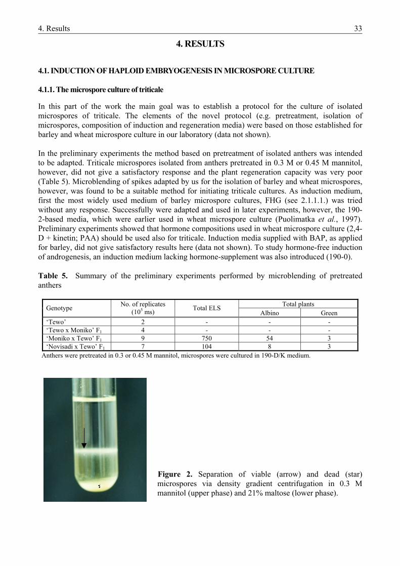
4. Results 33
4. RESULTS
4.1. INDUCTION OF HAPLOID EMBRYOGENESIS IN MICROSPORE CULTURE 4.1.1. The microspore culture of triticale
In this part of the work the main goal was to establish a protocol for the culture of isolated microspores of triticale. The elements of the novel protocol (e.g. pretreatment, isolation of microspores, composition of induction and regeneration media) were based on those established for barley and wheat microspore culture in our laboratory (data not shown). In the preliminary experiments the method based on pretreatment of isolated anthers was intended to be adapted. Triticale microspores isolated from anthers pretreated in 0.3 M or 0.45 M mannitol, however, did not give a satisfactory response and the plant regeneration capacity was very poor (Table 5). Microblending of spikes adapted by us for the isolation of barley and wheat microspores, however, was found to be a suitable method for initiating triticale cultures. As induction medium, first the most widely used medium of barley microspore cultures, FHG (see 2.1.1.1.) was tried without any response. Successfully were adapted and used in later experiments, however, the 190-2-based media, which were earlier used in wheat microspore culture (Puolimatka et al., 1997). Preliminary experiments showed that hormone compositions used in wheat microspore culture (2,4-D + kinetin; PAA) should be used also for triticale. Induction media supplied with BAP, as applied for barley, did not give satisfactory results here (data not shown). To study hormone-free induction of androgenesis, an induction medium lacking hormone-supplement was also introduced (190-0). Table 5. Summary of the preliminary experiments performed by microblending of pretreated anthers
Total plants Genotype No. of replicates (105 ms) Total ELS
Albino Green ‘Tewo’ 2 - - - ‘Tewo x Moniko’ F1 4 - - - ‘Moniko x Tewo’ F1 9 750 54 3 ‘Novisadi x Tewo’ F1 7 104 8 3
Anthers were pretreated in 0.3 or 0.45 M mannitol, microspores were cultured in 190-D/K medium.
Figure 2. Separation of viable (arrow) and dead (star) microspores via density gradient centrifugation in 0.3 M mannitol (upper phase) and 21% maltose (lower phase).
*
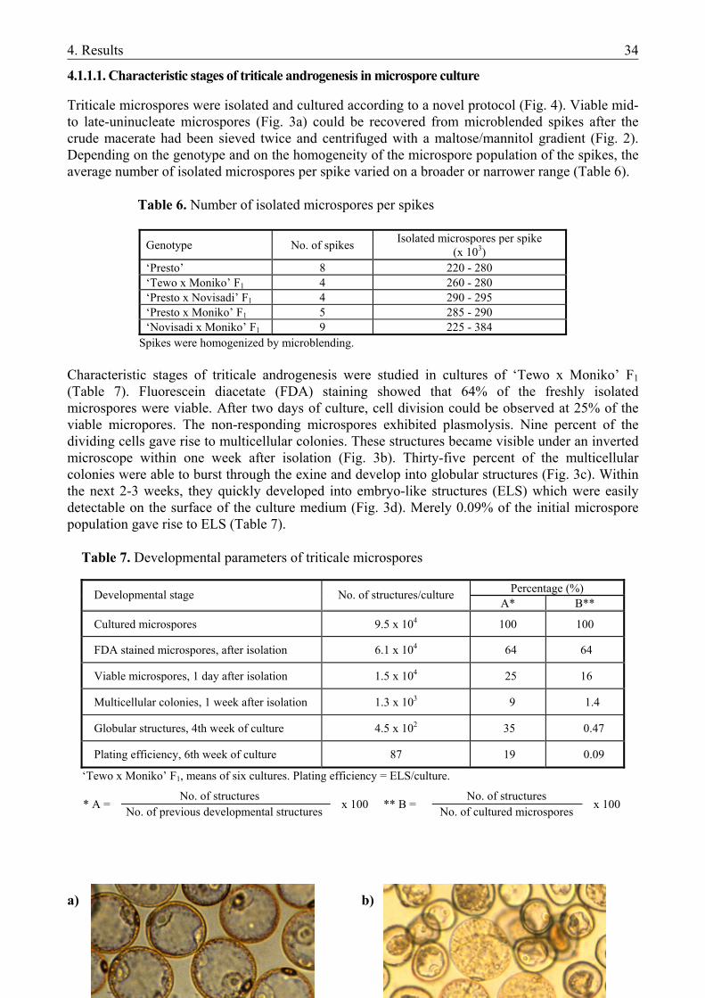
4. Results 34
4.1.1.1. Characteristic stages of triticale androgenesis in microspore culture
Triticale microspores were isolated and cultured according to a novel protocol (Fig. 4). Viable mid- to late-uninucleate microspores (Fig. 3a) could be recovered from microblended spikes after the crude macerate had been sieved twice and centrifuged with a maltose/mannitol gradient (Fig. 2). Depending on the genotype and on the homogeneity of the microspore population of the spikes, the average number of isolated microspores per spike varied on a broader or narrower range (Table 6).
Table 6. Number of isolated microspores per spikes
Genotype No. of spikes Isolated microspores per spike (x 103)
‘Presto’ 8 220 - 280 ‘Tewo x Moniko’ F1 4 260 - 280 ‘Presto x Novisadi’ F1 4 290 - 295 ‘Presto x Moniko’ F1 5 285 - 290 ‘Novisadi x Moniko’ F1 9 225 - 384
Spikes were homogenized by microblending. Characteristic stages of triticale androgenesis were studied in cultures of ‘Tewo x Moniko’ F1 (Table 7). Fluorescein diacetate (FDA) staining showed that 64% of the freshly isolated microspores were viable. After two days of culture, cell division could be observed at 25% of the viable micropores. The non-responding microspores exhibited plasmolysis. Nine percent of the dividing cells gave rise to multicellular colonies. These structures became visible under an inverted microscope within one week after isolation (Fig. 3b). Thirty-five percent of the multicellular colonies were able to burst through the exine and develop into globular structures (Fig. 3c). Within the next 2-3 weeks, they quickly developed into embryo-like structures (ELS) which were easily detectable on the surface of the culture medium (Fig. 3d). Merely 0.09% of the initial microspore population gave rise to ELS (Table 7). Table 7. Developmental parameters of triticale microspores
Percentage (%) Developmental stage No. of structures/culture A* B**
Cultured microspores 9.5 x 10⁴ 100 100
FDA stained microspores, after isolation 6.1 x 10⁴ 64 64
Viable microspores, 1 day after isolation 1.5 x 10⁴ 25 16
Multicellular colonies, 1 week after isolation 1.3 x 10³ 9 1.4
Globular structures, 4th week of culture 4.5 x 10² 35 0.47
Plating efficiency, 6th week of culture 87 19 0.09
‘Tewo x Moniko’ F1, means of six cultures. Plating efficiency = ELS/culture. * A =
x 100
** B =
x 100
No. of structures No. of previous developmental structures
No. of structures No. of cultured microspores
a) b)
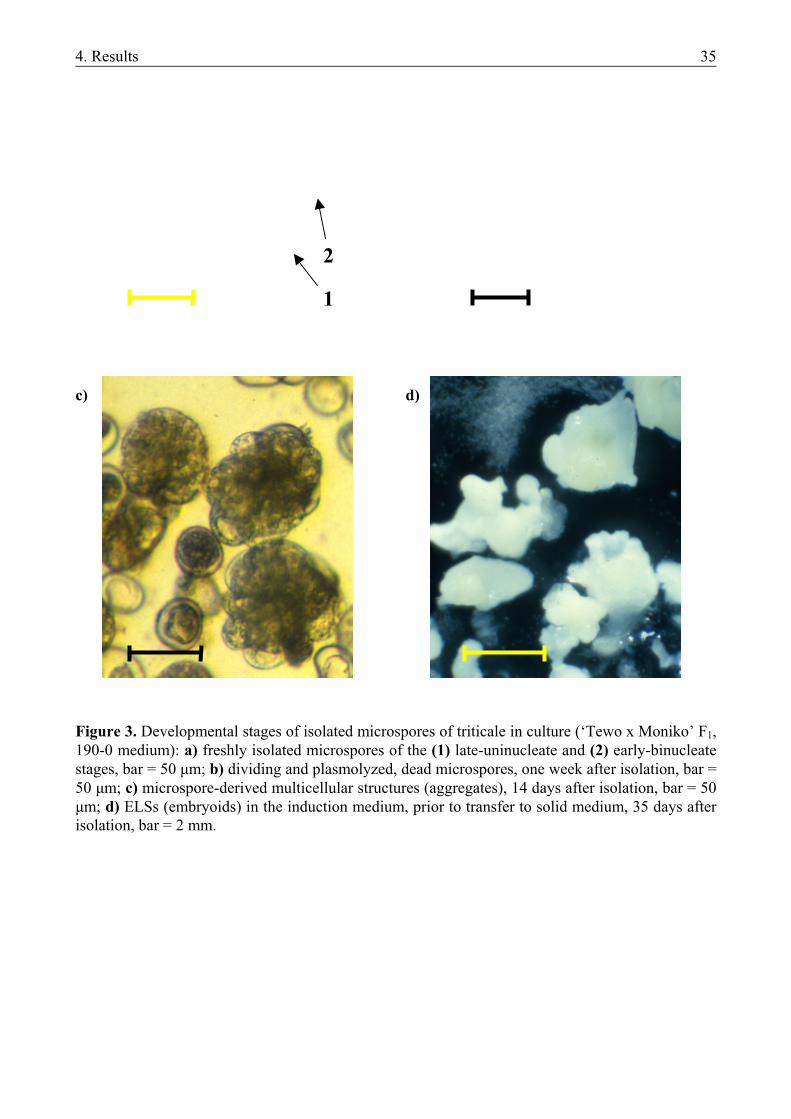
4. Results 35
2 1 c) d) Figure 3. Developmental stages of isolated microspores of triticale in culture (‘Tewo x Moniko’ F1, 190-0 medium): a) freshly isolated microspores of the (1) late-uninucleate and (2) early-binucleate stages, bar = 50 µm; b) dividing and plasmolyzed, dead microspores, one week after isolation, bar = 50 µm; c) microspore-derived multicellular structures (aggregates), 14 days after isolation, bar = 50 µm; d) ELSs (embryoids) in the induction medium, prior to transfer to solid medium, 35 days after isolation, bar = 2 mm.
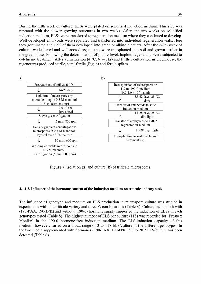
4. Results 36
During the fifth week of culture, ELSs were plated on solidified induction medium. This step was repeated with the slower growing structures in two weeks. After one-two weeks on solidified induction medium, ELSs were transferred to regeneration medium where they continued to develop. Well-developed embryoids were separated and transferred into individual regeneration vials. Here they germinated and 19% of them developed into green or albino plantlets. After the 8-9th week of culture, well-tillered and well-rooted regenerants were transplanted into soil and grown further in the greenhouse. Following the determination of ploidy-level, haploid regenerants were subjected to colchicine treatment. After vernalization (4 ºC, 6 weeks) and further cultivation in greenhouse, the regenerants produced sterile, semi-fertile (Fig. 6) and fertile spikes. a) b)
Figure 4. Isolation (a) and culture (b) of triticale microspores.
4.1.1.2. Influence of the hormone content of the induction medium on triticale androgenesis
The influence of genotype and medium on ELS production in microspore culture was studied in experiments with one triticale variety and three F1 combinations (Table 8). Culture media both with (190-PAA, 190-D/K) and without (190-0) hormone supply supported the induction of ELSs in each genotypes tested (Table 8). The highest number of ELS per culture (118) was recorded for ‘Presto x Moniko’ in the 190-0 hormone-free induction medium. The ELS-induction capacity of this medium, however, varied on a broad range of 3 to 118 ELS/culture in the different genotypes. In the two media supplemented with hormones (190-PAA, 190-D/K) 5.8 to 20.7 ELS/culture has been detected (Table 8).
Pretreatment of spikes at 4 ºC 14-21 days
Isolation of microspores by microblending in 0.3 M mannitol
(1-5 spikes/blending) 2 x 10 sec,
low speed Sieving, centrifugation 5 min, 800 rpm
Density gradient centrifugation: microspores in 0.3 M mannitol,
layered over 21% maltose 10 min, 600 rpm
Washing of viable microspores in 0.3 M mannitol,
centrifugation (5 min, 600 rpm)
Resuspension of microspores in 1-2 ml 190-0 medium (0.9-1.0 x 10⁵ ms/ml)
35-42 days, 28 ºC,
dark Transfer of embryoids to solid
induction medium
14-28 days, 28 ºC,
dim light Transfer of embryoids to 190-2
regeneration medium 21-28 days, light
Transplanting to soil, colchicine treatment etc.
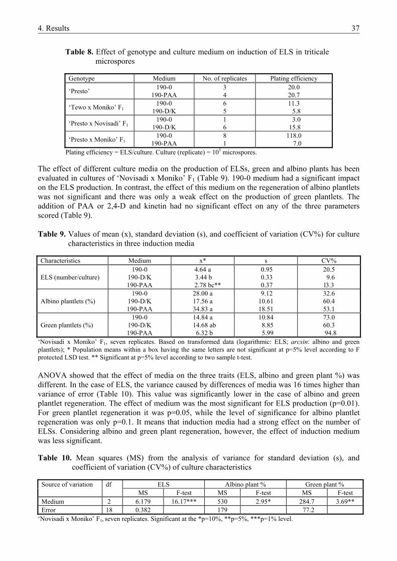
4. Results 37
Table 8. Effect of genotype and culture medium on induction of ELS in triticale
microspores
Genotype Medium No. of replicates Plating efficiency
‘Presto’ 190-0 190-PAA
3 4
20.0 20.7
‘Tewo x Moniko’ F1 190-0
190-D/K 6 5
11.3 5.8
‘Presto x Novisadi’ F1 190-0
190-D/K 1 6
3.0 15.8
‘Presto x Moniko’ F1 190-0
190-PAA 8 1
118.0 7.0
Plating efficiency = ELS/culture. Culture (replicate) = 105 microspores. The effect of different culture media on the production of ELSs, green and albino plants has been evaluated in cultures of ‘Novisadi x Moniko’ F1 (Table 9). 190-0 medium had a significant impact on the ELS production. In contrast, the effect of this medium on the regeneration of albino plantlets was not significant and there was only a weak effect on the production of green plantlets. The addition of PAA or 2,4-D and kinetin had no significant effect on any of the three parameters scored (Table 9). Table 9. Values of mean (x), standard deviation (s), and coefficient of variation (CV%) for culture
characteristics in three induction media Characteristics Medium x* s CV% ELS (number/culture)
190-0 190-D/K 190-PAA
4.64 a 3.44 b
2.78 bc**
0.95 0.33 0.37
20.5 9.6 l3.3
Albino plantlets (%)
190-0 190-D/K 190-PAA
28.00 a 17.56 a 34.83 a
9.12 10.61 18.51
32.6 60.4 53.1
Green plantlets (%)
190-0 190-D/K 190-PAA
14.84 a 14.68 ab 6.32 b
10.84 8.85 5.99
73.0 60.3
94.8 ‘Novisadi x Moniko’ F1, seven replicates. Based on transformed data (logarithmic: ELS; arcsin: albino and green plantlets); * Population means within a box having the same letters are not significant at p=5% level according to F protected LSD test. ** Significant at p=5% level according to two sample t-test.
ANOVA showed that the effect of media on the three traits (ELS, albino and green plant %) was different. In the case of ELS, the variance caused by differences of media was 16 times higher than variance of error (Table 10). This value was significantly lower in the case of albino and green plantlet regeneration. The effect of medium was the most significant for ELS production (p=0.01). For green plantlet regeneration it was p=0.05, while the level of significance for albino plantlet regeneration was only p=0.1. It means that induction media had a strong effect on the number of ELSs. Considering albino and green plant regeneration, however, the effect of induction medium was less significant. Table 10. Mean squares (MS) from the analysis of variance for standard deviation (s), and
coefficient of variation (CV%) of culture characteristics
ELS Albino plant % Green plant % Source of variation df MS F-test MS F-test MS F-test
Medium 2 6.179 16.17*** 530 2.95* 284.7 3.69** Error 18 0.382 179 77.2
‘Novisadi x Moniko’ F1, seven replicates. Significant at the *p=10%, **p=5%, ***p=1% level.
4. Results 38
The effect of different media on embryo production (ELS) was statistically confirmed. Values measured on 190-0 medium are higher than those on the other two media. 190-PAA medium gave the poorest result, although it cannot be proven on 95% confidence level by ANOVA. Because standard deviation of data measured on 190-0 medium is many times higher than those measured on the two other media, means of the different media were compared using Welch-test (Table 9). The results confirmed that 190-0 medium caused a higher level of embryogenesis and its effect is significantly stronger than those of both the 190-2,4D/K (p=5%) and the 190-PAA (p=1%) media. Two sample t-test showed the differences between 190-2,4D/K and 190-PAA media to be significant. As there was no significant difference between the standard deviations of regenerated albino and green plants on the three different media, one way ANOVA was sufficient for the evaluation of the effect of media (Table 10). Different media caused no significant difference in the number of albino plants, but there was a significant difference on the 95% confidence level between 190-0 and 190-PAA media with respect to green plant production (Table 9).
a) b)
4.1.1.3. Ploidy level of the green plantlets
A total amount of 126 green plants were regenerated and transferred to soil in the different experiments (Table 11). Regenerants were derived primarly from F1 combinations and were therefore of potential breeding value. Before vernalization, the ploidy level of plants was determined by measuring the stomatal guard cell size and by counting chromosome number in root tip cells. Stomatal guard cells of the putative haploids were 40-50% shorter than those of the spontaneously diploidized or natural control hexaploids. Average stomatal guard cell sizes ranged 21-32 µm for haploid and 35-39 µm for spontaneous diploid plants (Figure 5; Appendix 1). Mixoploids had either guard cells of intermediate length or guard cells of both haploid and diploid size were identified in different parts of the same leaf. After this relatively quick test, 113 plants were found to be haploid, 7 were mixoploids and 6 were spontaneous diploids. For 45 haploids for all mixoploids and diploids, chromosome numbers were exactly determined from root tip preparates. A significant correlation was determined between the results of the two methods for assessing ploidy level. Fifty-seven (45%) of the regenerants set seed after the colchicine-treatment of haploid plants (Table 11; Figure 6). From the ‘Presto x Novisadi’ F1 no fertile plants were obtained. The average number of seeds per spike ranged between 13 and 20 (Table 11). Except for one ‘Presto’ plant, all of the spontaneously rediploidized plants were fertile. There was no significant difference in the seed set
Figure 5. Stomatal guard cells of haploid (a) and spontaneously rediploidized (b) triticale plants. ‘Novisadi x Moniko’ F1, bar = 10 µm.
4. Results 39
of spontaneously rediploidized plants and of those rediploidized by colchicine treatment (Appendix 1).
Figure 6. Partially fertile spike of a triticale plant derived from microspore culture. ‘Novisadi x Moniko’ F1.
Table 11. Ploidy level of regenerants from triticale microspore culture
Transplanted plantlets
Fertile plants* Seed/spike Ploidy level ** Genotype
n (3x) mixoploid 2n (6x)
Presto 18 9 (50%) 20 15 (10) 1 (1) 2 (2) ‘Tewo x Moniko’ F1 12 8 (67%) 18 11 1 (1) - ‘Presto x Novisadi’ F1 6 - - 6 - - ‘Presto x Moniko’ F1 19 9 (47%) 13 19 (7) - - ‘Novisadi x Moniko’ F1 71 31 (43%) 20 62 (28) 5 (5) 4 (4) Total 126 57 (45%) 113 (45) 7 (7) 6 (6)
* In parenthesis are the fertile plant per total plant values. ** Numbers of root tips evaluated are in parentheses. Fertility of plants determined after colchicine treatment, on the basis of seed set. The results of the experiments performed with microspore cultures of triticale can be summarized as follows:
i. An effective method for regeneration of haploid/dihaploid plants from isolated microspores has been developed for triticale.
ii. Hormone-free induction medium had a significant impact on ELS production. Its effect on
green and plant regeneration, however, was not strong. Media supplemented with hormones had no significant effect on any of the three parameters.
4.1.2. Barley microspore culture
In this part of the work the primary goal was to study the induction of androgenesis and plant regeneration in barley microspore cultures without exogenous hormone-supplement. Furthermore, the hormone-free induction medium (190-0) efficiently used in triticale microspore culture (see 4.1.1.) was also tested in barley, comparing its effect on plant regeneration with those of an induction medium optimized for barley microspore culture (N24-BA). The studies were performed in two steps:
4. Results 40
i. Isolated microspores of cultivar ‘Igri’, the model genotype of barley haploid tissue cultures, were induced in N24-BA medium as well as in modified 190-2 medium, both with and without hormone supplement (Table 12, Fig. 7). N24-BA is a medium of optimized nitrogen composition. It was established exclusively for microspore culture of barley and it contains 1 mgL-1 BAP (Mordhorst and Lörz, 1993). Hormone-free N24 is designated as N24-0. 190-BA and 190-0 media - with and without BAP-supplement, respectively - are based on 190-2 medium invented for wheat anther culture (Zhuang and Jia, 1983, Pauk et al., 1991). Furthermore, 190-0 and its variants supplemented with other hormones (2,4-D, kinetin or PAA) were successfully used in microspore culture of triticale (see 4.1.1.2.).
ii. Isolated microspores of the cultivars ‘Igri’ and ‘Kymppi’, a genotype successfully involved
in microspore culture (Salmenkallio-Marttila, 1995; Ritala et al., 2001), were induced in N24-BA medium as well as in 190-0 medium. N24-BA is an optimized induction medium for barley microspore culture (see above), while 190-0 was found to induce the highest number of embryoids in triticale microspore culture (Tables 8 and 9).
4.1.2.1. Isolation and culture of barley microspores
Microspores of the late-uninucleate to early-binucleate stage were isolated by microblending of cold-pretreated spikes. It was observed that blending of up to 30 spikes at the same time and repeated blending of the sample (as suggested by Mordhorst and Lörz, 1993) did not result in a higher yield of isolated microspores (data not shown). If the spikes contained microspores of the required stage of development, blending of a single spike could result in 0.7-0.9 x 10⁵ microspores (cv. ‘Kymppi’), a sufficient amount for one culture plate. To offer an amount of microspores, possibly sufficient for more plates, five spikes were blended two times for 5 sec at low speed. Following cleaning of microspore suspension by sieving and density-gradient centrifugation, microspores were cultured at a density of 0.7-1.0 x 10⁵/ml. Non-responding microspores exhibited plasmolysis in the first four days of culture. Cell aggregates originating from dividing microspores could be observed after 7-14 days of culture, depending on genotype. In spite of this early proliferation, however, at several genotypes studied in the preliminary experiments, the aggregates arrested in development and died showing a necrotic symptom. No plasmolysis could be observed in these cases (data not shown). At cultivars ‘Igri’ and ‘Kymppi’, the first embryo-like structures (ELS) could be observed after 21-28 days of culture, irrespective of genotype and medium. These embryoids were transferred to solid induction medium after 32-38 days of culture. Slower developing structures were transferred 7-14 days later. In the case of a lower number of embryoids (up to 150), the transfer happened by using a forceps. At a higher number of ELSs, a Pasteur-pipette made of plastic was used for transfer. The highest number of embryoids was detected in 190-0 medium (76 ELS) at the first transfer. It was followed by N24-BA (35 ELS) and N24-0 (22 ELS). The lowest number of embryoids was registered in 190-BA (11 ELS). This order has been determined in two experiments. Further transfers were performed with pipette, thus total embryo production could not be calculated. Germination of embryoids on solid induction media could be observed from the seventh day following transfer, irrespective of cultivar and medium. These plantlets usually had no or very poor root system on the induction medium (Fig. 7). To check whether they can develop into complete plantlets, randomly chosen plantlets of up to 2 cm height were transferred to hormone-free LA3 medium. The numbers of plantlets involved were 95 and 22 for cv. ‘Kymppi’ (from N24-BA and 190-0 medium, respectively) and 10, 20, 20 and 13 for cv. ‘Igri’ (from N24-BA, 190-BA, N24-0 and 190-0 media, respectively). Each case, the development of plantlets continued on LA3, reaching a well-rooted state ready to be transplanted into soil.
4. Results 41
4.1.2.2. Effects of different induction media on barley androgenesis
Table 12 and Figure 7 show green plant production from microspore cultures of ‘Igri’ induced in four different media. Due to the high values of CV% (>100%), statistical analysis of data was not possible (data not shown). Values of mean (x) are given for information only (Table 12).
Induction on N24-BA medium resulted in the highest number of green plants in each experiment except for No. 3. Except for the result in this experiment, 190-0 medium resulted in the lowest number of green plants. Although mean values suggest N24-0 being the second best medium, this result is based on one peak result (52 green plants/culture) achieved in Exp. 4. 190-BA medium gave better result than N24-0 in three experiments (Exp. 2, 3, 5), it was inferior in two cases (Exp. 4, 6) and the results were equal in the first experiment.
a) N24-BA b) N24-0
c) 190-BA d) 190-0
Figure 7. Regeneration of barley plantlets (cv. ‘Igri’) on (a) N24-BA, (b) N24-0, (c) 190-BA and (d) 190-0 medium.
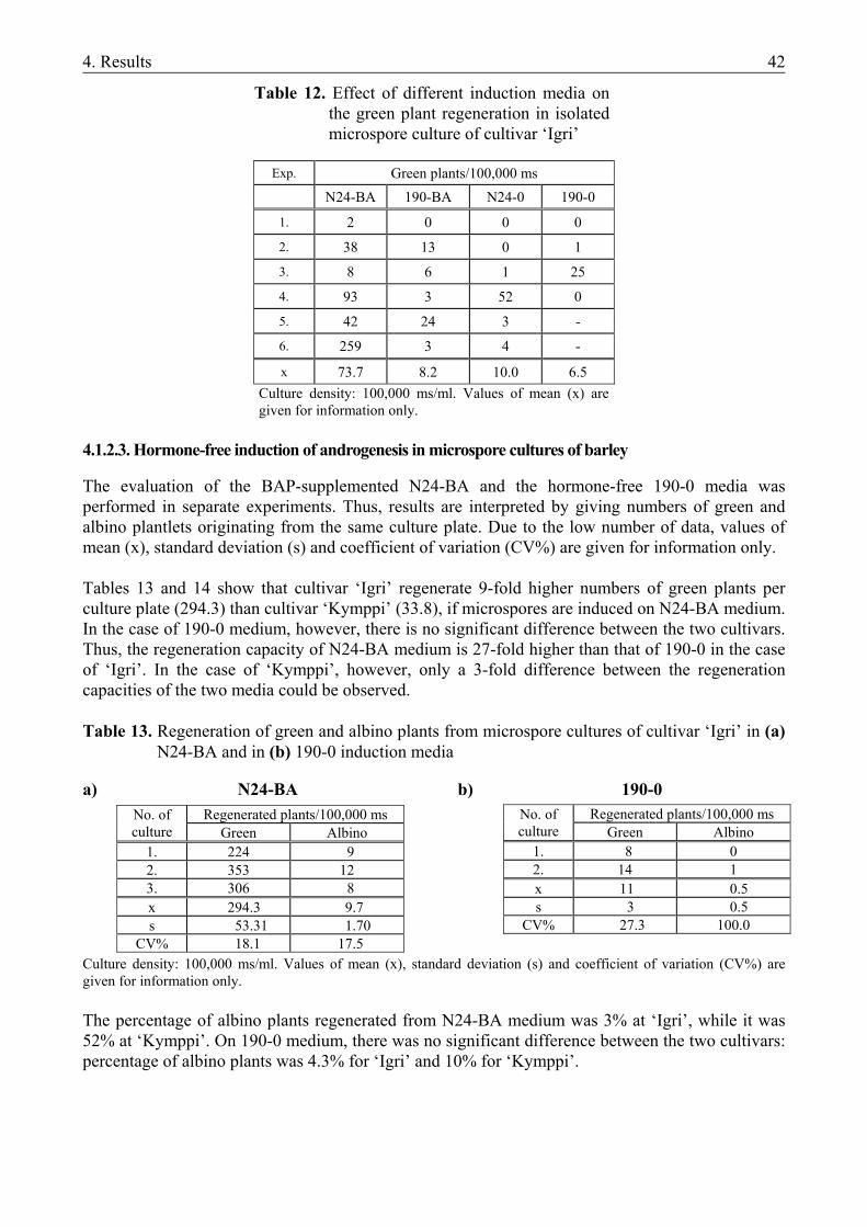
4. Results 42
Table 12. Effect of different induction media on the green plant regeneration in isolated microspore culture of cultivar ‘Igri’
Exp. Green plants/100,000 ms
N24-BA 190-BA N24-0 190-0
1. 2 0 0 0
2. 38 13 0 1
3. 8 6 1 25
4. 93 3 52 0
5. 42 24 3 -
6. 259 3 4 -
x 73.7 8.2 10.0 6.5 Culture density: 100,000 ms/ml. Values of mean (x) are given for information only.
4.1.2.3. Hormone-free induction of androgenesis in microspore cultures of barley
The evaluation of the BAP-supplemented N24-BA and the hormone-free 190-0 media was performed in separate experiments. Thus, results are interpreted by giving numbers of green and albino plantlets originating from the same culture plate. Due to the low number of data, values of mean (x), standard deviation (s) and coefficient of variation (CV%) are given for information only. Tables 13 and 14 show that cultivar ‘Igri’ regenerate 9-fold higher numbers of green plants per culture plate (294.3) than cultivar ‘Kymppi’ (33.8), if microspores are induced on N24-BA medium. In the case of 190-0 medium, however, there is no significant difference between the two cultivars. Thus, the regeneration capacity of N24-BA medium is 27-fold higher than that of 190-0 in the case of ‘Igri’. In the case of ‘Kymppi’, however, only a 3-fold difference between the regeneration capacities of the two media could be observed.
Table 13. Regeneration of green and albino plants from microspore cultures of cultivar ‘Igri’ in (a)
N24-BA and in (b) 190-0 induction media a) N24-BA b) 190-0
Culture density: 100,000 ms/ml. Values of mean (x), standard deviation (s) and coefficient of variation (CV%) are given for information only. The percentage of albino plants regenerated from N24-BA medium was 3% at ‘Igri’, while it was 52% at ‘Kymppi’. On 190-0 medium, there was no significant difference between the two cultivars: percentage of albino plants was 4.3% for ‘Igri’ and 10% for ‘Kymppi’.
Regenerated plants/100,000 ms No. of culture Green Albino
1. 8 0 2. 14 1 x 11 0.5 s 3 0.5
CV% 27.3 100.0
Regenerated plants/100,000 ms No. of culture Green Albino
1. 224 9 2. 353 12 3. 306 8 x 294.3 9.7 s 53.31 1.70
CV% 18.1 17.5
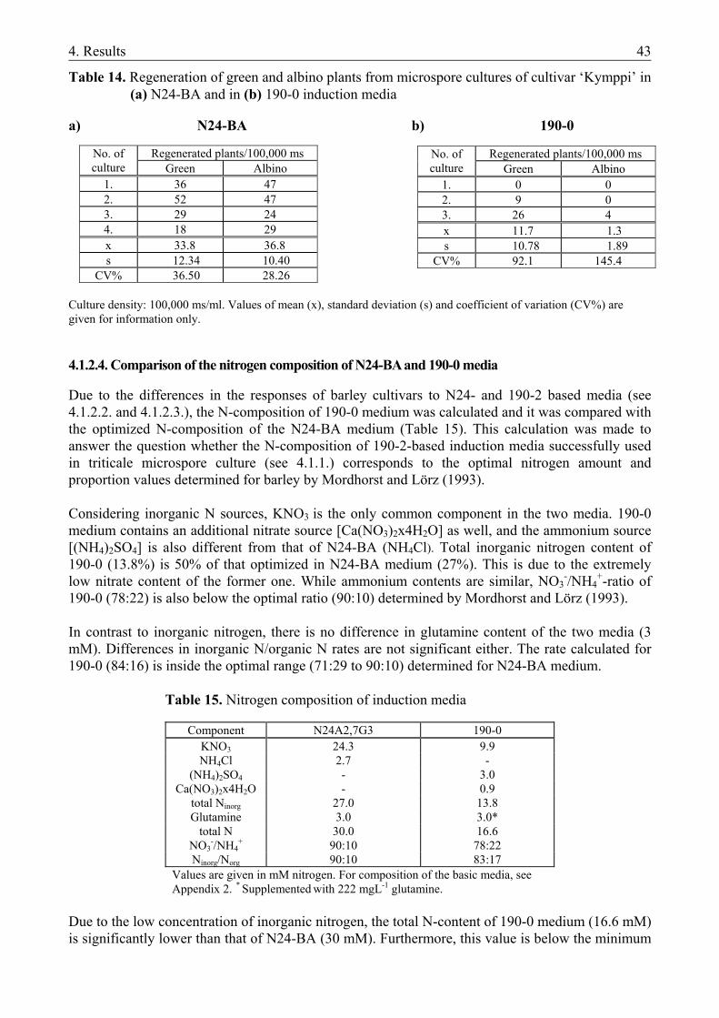
4. Results 43
Table 14. Regeneration of green and albino plants from microspore cultures of cultivar ‘Kymppi’ in (a) N24-BA and in (b) 190-0 induction media
a) N24-BA b) 190-0
Culture density: 100,000 ms/ml. Values of mean (x), standard deviation (s) and coefficient of variation (CV%) are given for information only.
4.1.2.4. Comparison of the nitrogen composition of N24-BA and 190-0 media
Due to the differences in the responses of barley cultivars to N24- and 190-2 based media (see 4.1.2.2. and 4.1.2.3.), the N-composition of 190-0 medium was calculated and it was compared with the optimized N-composition of the N24-BA medium (Table 15). This calculation was made to answer the question whether the N-composition of 190-2-based induction media successfully used in triticale microspore culture (see 4.1.1.) corresponds to the optimal nitrogen amount and proportion values determined for barley by Mordhorst and Lörz (1993). Considering inorganic N sources, KNO3 is the only common component in the two media. 190-0 medium contains an additional nitrate source [Ca(NO3)2x4H2O] as well, and the ammonium source [(NH4)2SO4] is also different from that of N24-BA (NH4Cl). Total inorganic nitrogen content of 190-0 (13.8%) is 50% of that optimized in N24-BA medium (27%). This is due to the extremely low nitrate content of the former one. While ammonium contents are similar, NO3
-/NH4+-ratio of
190-0 (78:22) is also below the optimal ratio (90:10) determined by Mordhorst and Lörz (1993). In contrast to inorganic nitrogen, there is no difference in glutamine content of the two media (3 mM). Differences in inorganic N/organic N rates are not significant either. The rate calculated for 190-0 (84:16) is inside the optimal range (71:29 to 90:10) determined for N24-BA medium.
Table 15. Nitrogen composition of induction media
Component N24A2,7G3 190-0 KNO3 24.3 9.9 NH4Cl 2.7 -
(NH4)2SO4 - 3.0 Ca(NO3)2x4H2O - 0.9
total Ninorg 27.0 13.8 Glutamine 3.0 3.0*
total N 30.0 16.6 NO3
-/NH4+ 90:10 78:22
Ninorg/Norg 90:10 83:17 Values are given in mM nitrogen. For composition of the basic media, see Appendix 2. * Supplemented with 222 mgL-1 glutamine.
Due to the low concentration of inorganic nitrogen, the total N-content of 190-0 medium (16.6 mM) is significantly lower than that of N24-BA (30 mM). Furthermore, this value is below the minimum
Regenerated plants/100,000 ms No. of culture Green Albino
1. 36 47 2. 52 47 3. 29 24 4. 18 29 x 33.8 36.8 s 12.34 10.40
CV% 36.50 28.26
Regenerated plants/100,000 ms No. of culture Green Albino
1. 0 0 2. 9 0 3. 26 4 x 11.7 1.3 s 10.78 1.89
CV% 92.1 145.4
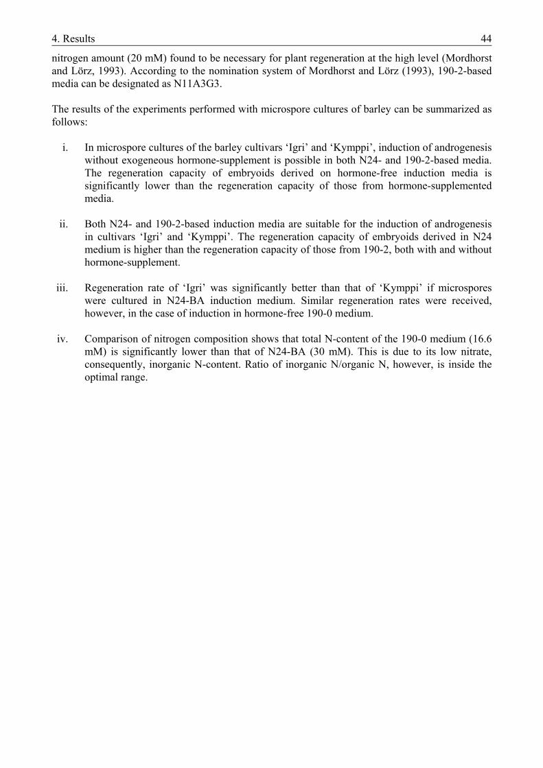
4. Results 44
nitrogen amount (20 mM) found to be necessary for plant regeneration at the high level (Mordhorst and Lörz, 1993). According to the nomination system of Mordhorst and Lörz (1993), 190-2-based media can be designated as N11A3G3. The results of the experiments performed with microspore cultures of barley can be summarized as follows:
i. In microspore cultures of the barley cultivars ‘Igri’ and ‘Kymppi’, induction of androgenesis without exogeneous hormone-supplement is possible in both N24- and 190-2-based media. The regeneration capacity of embryoids derived on hormone-free induction media is significantly lower than the regeneration capacity of those from hormone-supplemented media.
ii. Both N24- and 190-2-based induction media are suitable for the induction of androgenesis
in cultivars ‘Igri’ and ‘Kymppi’. The regeneration capacity of embryoids derived in N24 medium is higher than the regeneration capacity of those from 190-2, both with and without hormone-supplement.
iii. Regeneration rate of ‘Igri’ was significantly better than that of ‘Kymppi’ if microspores
were cultured in N24-BA induction medium. Similar regeneration rates were received, however, in the case of induction in hormone-free 190-0 medium.
iv. Comparison of nitrogen composition shows that total N-content of the 190-0 medium (16.6
mM) is significantly lower than that of N24-BA (30 mM). This is due to its low nitrate, consequently, inorganic N-content. Ratio of inorganic N/organic N, however, is inside the optimal range.
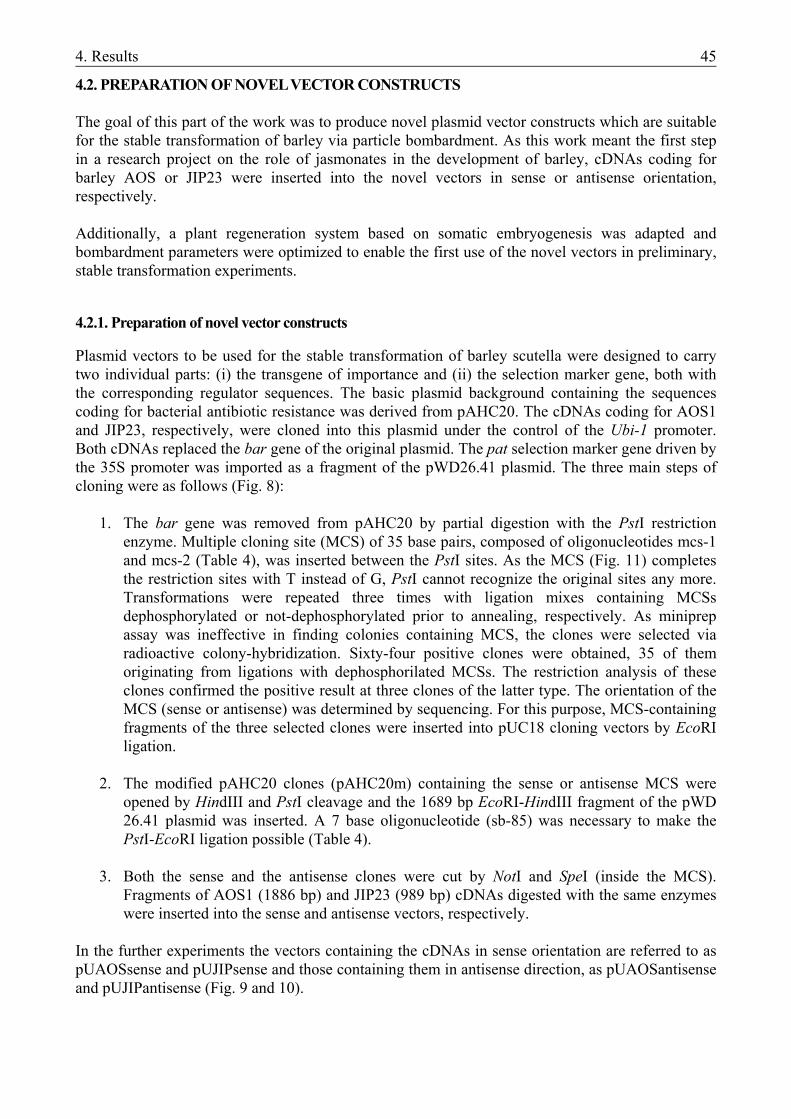
4. Results 45
4.2. PREPARATION OF NOVEL VECTOR CONSTRUCTS The goal of this part of the work was to produce novel plasmid vector constructs which are suitable for the stable transformation of barley via particle bombardment. As this work meant the first step in a research project on the role of jasmonates in the development of barley, cDNAs coding for barley AOS or JIP23 were inserted into the novel vectors in sense or antisense orientation, respectively. Additionally, a plant regeneration system based on somatic embryogenesis was adapted and bombardment parameters were optimized to enable the first use of the novel vectors in preliminary, stable transformation experiments.
4.2.1. Preparation of novel vector constructs
Plasmid vectors to be used for the stable transformation of barley scutella were designed to carry two individual parts: (i) the transgene of importance and (ii) the selection marker gene, both with the corresponding regulator sequences. The basic plasmid background containing the sequences coding for bacterial antibiotic resistance was derived from pAHC20. The cDNAs coding for AOS1 and JIP23, respectively, were cloned into this plasmid under the control of the Ubi-1 promoter. Both cDNAs replaced the bar gene of the original plasmid. The pat selection marker gene driven by the 35S promoter was imported as a fragment of the pWD26.41 plasmid. The three main steps of cloning were as follows (Fig. 8):
1. The bar gene was removed from pAHC20 by partial digestion with the PstI restriction
enzyme. Multiple cloning site (MCS) of 35 base pairs, composed of oligonucleotides mcs-1 and mcs-2 (Table 4), was inserted between the PstI sites. As the MCS (Fig. 11) completes the restriction sites with T instead of G, PstI cannot recognize the original sites any more. Transformations were repeated three times with ligation mixes containing MCSs dephosphorylated or not-dephosphorylated prior to annealing, respectively. As miniprep assay was ineffective in finding colonies containing MCS, the clones were selected via radioactive colony-hybridization. Sixty-four positive clones were obtained, 35 of them originating from ligations with dephosphorilated MCSs. The restriction analysis of these clones confirmed the positive result at three clones of the latter type. The orientation of the MCS (sense or antisense) was determined by sequencing. For this purpose, MCS-containing fragments of the three selected clones were inserted into pUC18 cloning vectors by EcoRI ligation.
2. The modified pAHC20 clones (pAHC20m) containing the sense or antisense MCS were
opened by HindIII and PstI cleavage and the 1689 bp EcoRI-HindIII fragment of the pWD 26.41 plasmid was inserted. A 7 base oligonucleotide (sb-85) was necessary to make the PstI-EcoRI ligation possible (Table 4).
3. Both the sense and the antisense clones were cut by NotI and SpeI (inside the MCS).
Fragments of AOS1 (1886 bp) and JIP23 (989 bp) cDNAs digested with the same enzymes were inserted into the sense and antisense vectors, respectively.
In the further experiments the vectors containing the cDNAs in sense orientation are referred to as pUAOSsense and pUJIPsense and those containing them in antisense direction, as pUAOSantisense and pUJIPantisense (Fig. 9 and 10).
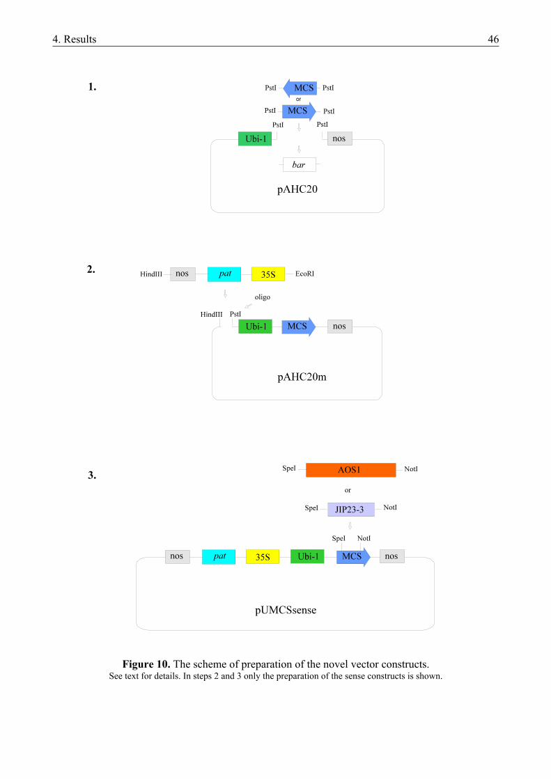
4. Results 46
Figure 10. The scheme of preparation of the novel vector constructs. See text for details. In steps 2 and 3 only the preparation of the sense constructs is shown.
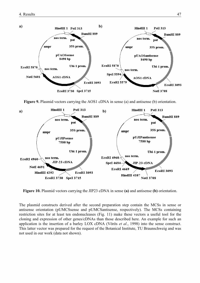
4. Results 47
a) b)
Figure 9. Plasmid vectors carrying the AOS1 cDNA in sense (a) and antisense (b) orientation. a) b)
Figure 10. Plasmid vectors carrying the JIP23 cDNA in sense (a) and antisense (b) orientation. The plasmid constructs derived after the second preparation step contain the MCSs in sense or antisense orientation (pUMCSsense and pUMCSantisense, respectively). The MCSs containing restriction sites for at least ten endonucleases (Fig. 11) make these vectors a useful tool for the cloning and expression of other genes/cDNAs than those described here. An example for such an application is the insertion of a barley LOX cDNA (Vörös et al., 1998) into the sense construct. This latter vector was prepared for the request of the Botanical Institute, TU Braunschweig and was not used in our work (data not shown).
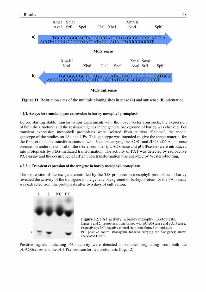
4. Results 48
XmaI SmaI XmaIII AvaI SrfI SpeI ClaI XbaI NotI SphI
a)
MCS sense
XmaIII XmaI SmaI NotI XbaI ClaI SpeI AvaI SrfI SphI
b)
MCS antisense Figure 11. Restriction sites of the multiple cloning sites in sense (a) and antisense (b) orientation.
4.2.2. Assays for transient gene expression in barley mesophyll protoplasts
Before starting stable transformation experiments with the novel vector constructs, the expression of both the structural and the resistance genes in the genetic background of barley was checked. For transient expression mesophyll protoplasts were isolated from cultivar ‘Salome’, the model genotype of the studies on JAs and JIPs. This genotype was intended to give the target material for the first set of stable transformations as well. Vectors carrying the AOS1 and JIP23 cDNAs in sense orientation under the control of the Ubi-1 promoter (pUAOSsense and pUJIPsense) were introduced into protoplasts by PEG-mediated transformation. The activity of PAT was detected by radioactive PAT-assay and the occurrence of JIP23 upon transformation was analyzed by Western-blotting. 4.2.2.1. Transient expression of the pat gene in barley mesophyll protoplasts
The expression of the pat gene controlled by the 35S promoter in mesophyll protoplasts of barley revealed the activity of the transgene in the genetic background of barley. Protein for the PAT-assay was extracted from the protoplasts after two days of cultivation.
1 2 NC PC
Figure 12. PAT activity in barley mesophyll protoplasts. Lanes 1 and 2: protoplasts transformed with pUAOSsense and pUJIPsense, respectively; NC: negative control (non-transformed protoplasts);
→ PC: positive control (transgenic tobacco carrying the bar gene); arrow: acetylated L-PPT.
Positive signals indicating PAT-activity were detected in samples originating from both the pUAOSsense- and the pUJIPsense-transformed protoplasts (Fig. 12).
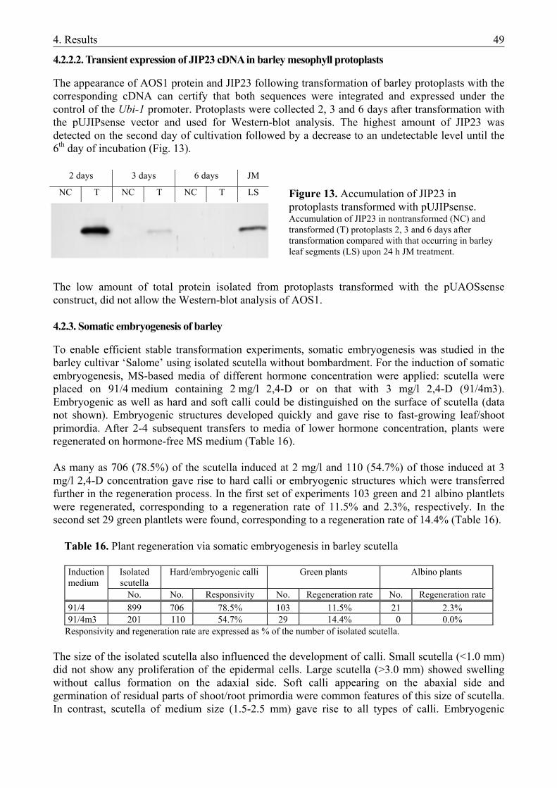
4. Results 49
4.2.2.2. Transient expression of JIP23 cDNA in barley mesophyll protoplasts
The appearance of AOS1 protein and JIP23 following transformation of barley protoplasts with the corresponding cDNA can certify that both sequences were integrated and expressed under the control of the Ubi-1 promoter. Protoplasts were collected 2, 3 and 6 days after transformation with the pUJIPsense vector and used for Western-blot analysis. The highest amount of JIP23 was detected on the second day of cultivation followed by a decrease to an undetectable level until the 6th day of incubation (Fig. 13).
The low amount of total protein isolated from protoplasts transformed with the pUAOSsense construct, did not allow the Western-blot analysis of AOS1. 4.2.3. Somatic embryogenesis of barley
To enable efficient stable transformation experiments, somatic embryogenesis was studied in the barley cultivar ‘Salome’ using isolated scutella without bombardment. For the induction of somatic embryogenesis, MS-based media of different hormone concentration were applied: scutella were placed on 91/4 medium containing 2 mg/l 2,4-D or on that with 3 mg/l 2,4-D (91/4m3). Embryogenic as well as hard and soft calli could be distinguished on the surface of scutella (data not shown). Embryogenic structures developed quickly and gave rise to fast-growing leaf/shoot primordia. After 2-4 subsequent transfers to media of lower hormone concentration, plants were regenerated on hormone-free MS medium (Table 16). As many as 706 (78.5%) of the scutella induced at 2 mg/l and 110 (54.7%) of those induced at 3 mg/l 2,4-D concentration gave rise to hard calli or embryogenic structures which were transferred further in the regeneration process. In the first set of experiments 103 green and 21 albino plantlets were regenerated, corresponding to a regeneration rate of 11.5% and 2.3%, respectively. In the second set 29 green plantlets were found, corresponding to a regeneration rate of 14.4% (Table 16). Table 16. Plant regeneration via somatic embryogenesis in barley scutella
Induction medium
Isolated scutella
Hard/embryogenic calli Green plants Albino plants
No. No. Responsivity No. Regeneration rate No. Regeneration rate 91/4 899 706 78.5% 103 11.5% 21 2.3% 91/4m3 201 110 54.7% 29 14.4% 0 0.0%
Responsivity and regeneration rate are expressed as % of the number of isolated scutella. The size of the isolated scutella also influenced the development of calli. Small scutella (<1.0 mm) did not show any proliferation of the epidermal cells. Large scutella (>3.0 mm) showed swelling without callus formation on the adaxial side. Soft calli appearing on the abaxial side and germination of residual parts of shoot/root primordia were common features of this size of scutella. In contrast, scutella of medium size (1.5-2.5 mm) gave rise to all types of calli. Embryogenic
2 days 3 days 6 days JM
NC T NC T NC T LS Figure 13. Accumulation of JIP23 in protoplasts transformed with pUJIPsense. Accumulation of JIP23 in nontransformed (NC) and transformed (T) protoplasts 2, 3 and 6 days after transformation compared with that occurring in barley leaf segments (LS) upon 24 h JM treatment.
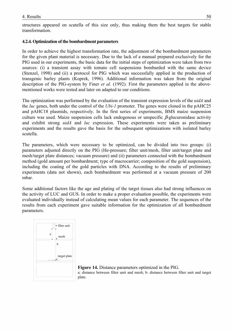
4. Results 50
structures appeared on scutella of this size only, thus making them the best targets for stable transformation. 4.2.4. Optimization of the bombardment parameters
In order to achieve the highest transformation rate, the adjustment of the bombardment parameters for the given plant material is necessary. Due to the lack of a manual prepared exclusively for the PIG used in our experiments, the basic data for the initial steps of optimization were taken from two sources: (i) a transient assay with tomato cell suspensions bombarded with the same device (Stenzel, 1998) and (ii) a protocol for PIG which was successfully applied in the production of transgenic barley plants (Koprek, 1996). Additional information was taken from the original description of the PIG-system by Finer et al. (1992). First the parameters applied in the above-mentioned works were tested and later on adapted to our conditions. The optimization was performed by the evaluation of the transient expression levels of the uidA and the luc genes, both under the control of the Ubi-1 promoter. The genes were cloned in the pAHC25 and pAHC18 plasmids, respectively. In the first series of experiments, BMS maize suspension culture was used. Maize suspension cells lack endogenous or unspecific β-glucuronidase activity and exhibit strong uidA and luc expression. These experiments were taken as preliminary experiments and the results gave the basis for the subsequent optimizations with isolated barley scutella. The parameters, which were necessary to be optimized, can be divided into two groups: (i) parameters adjusted directly on the PIG (He-pressure; filter unit/mesh, filter unit/target plate and mesh/target plate distances; vacuum pressure) and (ii) parameters connected with the bombardment method (gold amount per bombardment; type of macrocarrier; composition of the gold suspension), including the coating of the gold particles with DNA. According to the results of preliminary experiments (data not shown), each bombardment was performed at a vacuum pressure of 200 mbar. Some additional factors like the age and plating of the target tissues also had strong influences on the activity of LUC and GUS. In order to make a proper evaluation possible, the experiments were evaluated individually instead of calculating mean values for each parameter. The sequences of the results from each experiment gave suitable information for the optimization of all bombardment parameters.
Figure 14. Distance parameters optimized in the PIG. a: distance between filter unit and mesh; b: distance between filter unit and target plate.
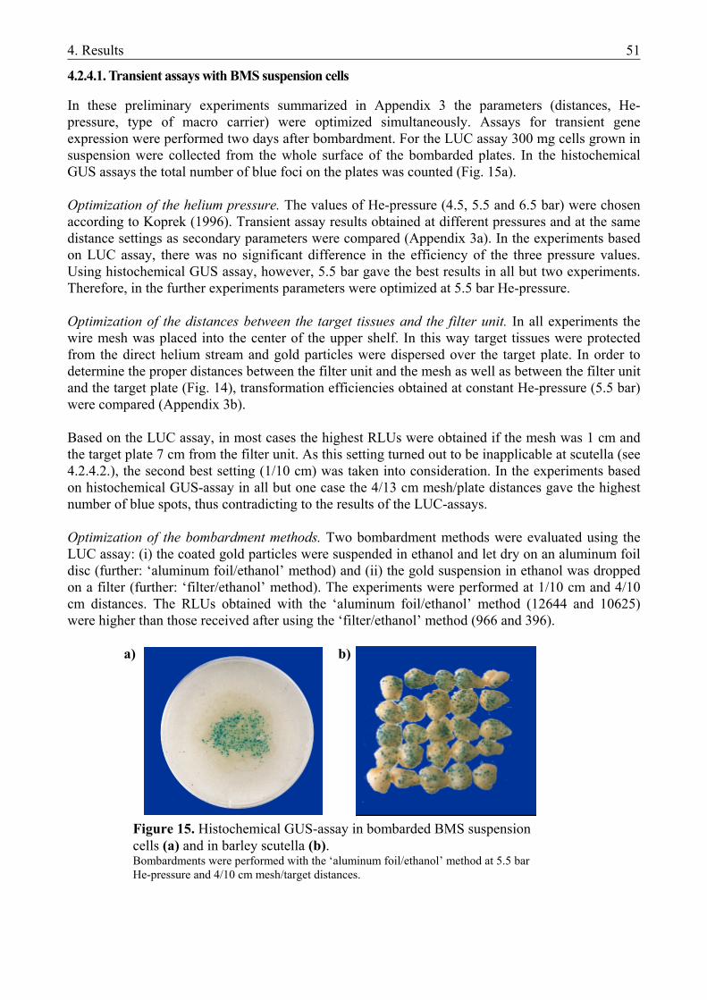
4. Results 51
4.2.4.1. Transient assays with BMS suspension cells
In these preliminary experiments summarized in Appendix 3 the parameters (distances, He-pressure, type of macro carrier) were optimized simultaneously. Assays for transient gene expression were performed two days after bombardment. For the LUC assay 300 mg cells grown in suspension were collected from the whole surface of the bombarded plates. In the histochemical GUS assays the total number of blue foci on the plates was counted (Fig. 15a). Optimization of the helium pressure. The values of He-pressure (4.5, 5.5 and 6.5 bar) were chosen according to Koprek (1996). Transient assay results obtained at different pressures and at the same distance settings as secondary parameters were compared (Appendix 3a). In the experiments based on LUC assay, there was no significant difference in the efficiency of the three pressure values. Using histochemical GUS assay, however, 5.5 bar gave the best results in all but two experiments. Therefore, in the further experiments parameters were optimized at 5.5 bar He-pressure. Optimization of the distances between the target tissues and the filter unit. In all experiments the wire mesh was placed into the center of the upper shelf. In this way target tissues were protected from the direct helium stream and gold particles were dispersed over the target plate. In order to determine the proper distances between the filter unit and the mesh as well as between the filter unit and the target plate (Fig. 14), transformation efficiencies obtained at constant He-pressure (5.5 bar) were compared (Appendix 3b). Based on the LUC assay, in most cases the highest RLUs were obtained if the mesh was 1 cm and the target plate 7 cm from the filter unit. As this setting turned out to be inapplicable at scutella (see 4.2.4.2.), the second best setting (1/10 cm) was taken into consideration. In the experiments based on histochemical GUS-assay in all but one case the 4/13 cm mesh/plate distances gave the highest number of blue spots, thus contradicting to the results of the LUC-assays. Optimization of the bombardment methods. Two bombardment methods were evaluated using the LUC assay: (i) the coated gold particles were suspended in ethanol and let dry on an aluminum foil disc (further: ‘aluminum foil/ethanol’ method) and (ii) the gold suspension in ethanol was dropped on a filter (further: ‘filter/ethanol’ method). The experiments were performed at 1/10 cm and 4/10 cm distances. The RLUs obtained with the ‘aluminum foil/ethanol’ method (12644 and 10625) were higher than those received after using the ‘filter/ethanol’ method (966 and 396).
a) b)
Figure 15. Histochemical GUS-assay in bombarded BMS suspension cells (a) and in barley scutella (b). Bombardments were performed with the ‘aluminum foil/ethanol’ method at 5.5 bar He-pressure and 4/10 cm mesh/target distances.
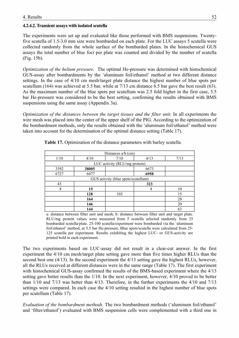
4. Results 52
4.2.4.2. Transient assays with isolated scutella
The experiments were set up and evaluated like those performed with BMS suspensions. Twenty-five scutella of 1.5-3.0 mm size were bombarded on each plate. For the LUC assays 5 scutella were collected randomly from the whole surface of the bombarded plates. In the histochemical GUS assays the total number of blue foci per plate was counted and divided by the number of scutella (Fig. 15b). Optimization of the helium pressure. The optimal He-pressure was determined with histochemical GUS-assay after bombardments by the ‘aluminum foil/ethanol’ method at two different distance settings. In the case of 4/10 cm mesh/target plate distance the highest number of blue spots per scutellum (164) was achieved at 5.5 bar, while at 7/13 cm distance 6.5 bar gave the best result (63). As the maximum number of the blue spots per scutellum was 2.5 fold higher in the first case, 5.5 bar He-pressure was considered to be the best setting, confirming the results obtained with BMS suspensions using the same assay (Appendix 3a). Optimization of the distances between the target tissues and the filter unit. In all experiments the wire mesh was placed into the center of the upper shelf of the PIG. According to the optimization of the bombardment methods, only the results obtained with the ‘aluminum foil/ethanol’ method were taken into account for the determination of the optimal distance setting (Table 17). Table 17. Optimization of the distance parameters with barley scutella
Distances a/b (cm)
1/10 4/10 7/10 4/13 7/13 LUC activity (RLU/mg protein)
3392 38005 6673 6727 6477 6958
GUS activity (blue spots/scutellum) 43 323 8 15 4 10
128 103 15 164 28 146 20 144 63
a: distance between filter unit and mesh; b: distance between filter unit and target plate. RLU/mg protein values were measured from 5 scutella selected randomly from 25 bombarded scutella/plate. 25-100 scutella/experiment were bombarded via the ‘aluminum foil/ethanol’ method, at 5.5 bar He-pressure. Blue spots/scutella were calculated from 25-125 scutella per experiment. Results exhibiting the highest LUC- or GUS-activity are printed bold in each experiment.
The two experiments based on LUC-assay did not result in a clear-cut answer. In the first experiment the 4/10 cm mesh/target plate setting gave more than five times higher RLUs than the second best one (4/13). In the second experiment the 4/13 setting gave the highest RLUs, however, all the RLUs received at different distances were in the same range (Table 17). The first experiment with histochemical GUS-assay confirmed the results of the BMS-based experiment where the 4/13 setting gave better results than the 1/10. In the next experiment, however, 4/10 proved to be better than 1/10 and 7/13 was better than 4/13. Therefore, in the further experiments the 4/10 and 7/13 settings were compared. In each case the 4/10 setting resulted in the highest number of blue spots per scutellum (Table 17). Evaluation of the bombardment methods. The two bombardment methods (‘aluminum foil/ethanol’ and ‘filter/ethanol’) evaluated with BMS suspension cells were complemented with a third one in
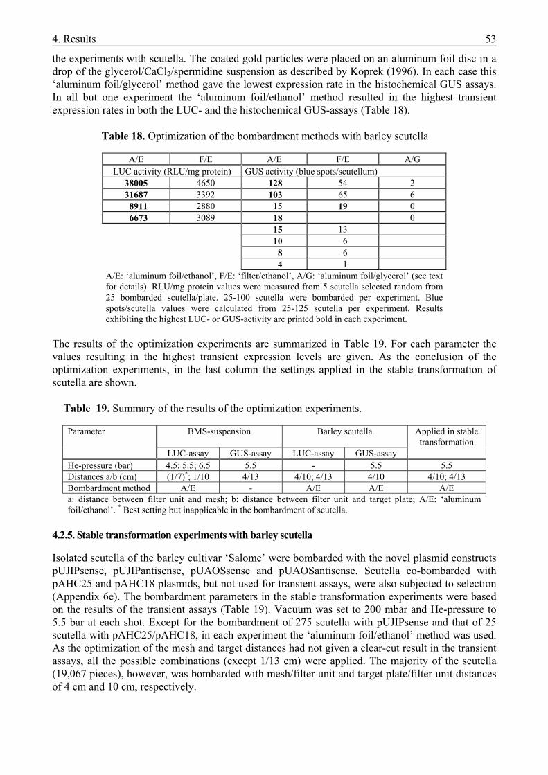
4. Results 53
the experiments with scutella. The coated gold particles were placed on an aluminum foil disc in a drop of the glycerol/CaCl2/spermidine suspension as described by Koprek (1996). In each case this ‘aluminum foil/glycerol’ method gave the lowest expression rate in the histochemical GUS assays. In all but one experiment the ‘aluminum foil/ethanol’ method resulted in the highest transient expression rates in both the LUC- and the histochemical GUS-assays (Table 18).
Table 18. Optimization of the bombardment methods with barley scutella
A/E F/E A/E F/E A/G LUC activity (RLU/mg protein) GUS activity (blue spots/scutellum)
38005 4650 128 54 2 31687 3392 103 65 6 8911 2880 15 19 0 6673 3089 18 0
15 13 10 6 8 6 4 1
A/E: ‘aluminum foil/ethanol’, F/E: ‘filter/ethanol’, A/G: ‘aluminum foil/glycerol’ (see text for details). RLU/mg protein values were measured from 5 scutella selected random from 25 bombarded scutella/plate. 25-100 scutella were bombarded per experiment. Blue spots/scutella values were calculated from 25-125 scutella per experiment. Results exhibiting the highest LUC- or GUS-activity are printed bold in each experiment.
The results of the optimization experiments are summarized in Table 19. For each parameter the values resulting in the highest transient expression levels are given. As the conclusion of the optimization experiments, in the last column the settings applied in the stable transformation of scutella are shown. Table 19. Summary of the results of the optimization experiments.
Parameter BMS-suspension Barley scutella Applied in stable transformation
LUC-assay GUS-assay LUC-assay GUS-assay He-pressure (bar) 4.5; 5.5; 6.5 5.5 - 5.5 5.5 Distances a/b (cm) (1/7)*; 1/10 4/13 4/10; 4/13 4/10 4/10; 4/13 Bombardment method A/E - A/E A/E A/E a: distance between filter unit and mesh; b: distance between filter unit and target plate; A/E: ‘aluminum foil/ethanol’. * Best setting but inapplicable in the bombardment of scutella.
4.2.5. Stable transformation experiments with barley scutella
Isolated scutella of the barley cultivar ‘Salome’ were bombarded with the novel plasmid constructs pUJIPsense, pUJIPantisense, pUAOSsense and pUAOSantisense. Scutella co-bombarded with pAHC25 and pAHC18 plasmids, but not used for transient assays, were also subjected to selection (Appendix 6e). The bombardment parameters in the stable transformation experiments were based on the results of the transient assays (Table 19). Vacuum was set to 200 mbar and He-pressure to 5.5 bar at each shot. Except for the bombardment of 275 scutella with pUJIPsense and that of 25 scutella with pAHC25/pAHC18, in each experiment the ‘aluminum foil/ethanol’ method was used. As the optimization of the mesh and target distances had not given a clear-cut result in the transient assays, all the possible combinations (except 1/13 cm) were applied. The majority of the scutella (19,067 pieces), however, was bombarded with mesh/filter unit and target plate/filter unit distances of 4 cm and 10 cm, respectively.
4. Results 54
4.2.5.1. Somatic embryogenesis in the bombarded scutella
In the first set of experiments performed without osmotic treatment, the scutella were incubated on 91/4 or 91/4m3 medium for one day before bombardment. During this time intensive proliferation of the epidermal cells could be observed. At the transfer to the first selection medium three days after bombardment, early callus formation was visible on the edges of the most responsive scutella. These calli turned out to be highly embryogenic. They could be kept in culture for longer time and developed shoot/leaf primordia at higher frequency than other callus lines (data not shown). The development of the non-responsive scutella was arrested in the swollen stage. The color of these tissues turned to brown till the end of the first two weeks showing deleterious effect of the selection agent. a) b)
Embryo culture 3 mg/l 2,4-D
1 day
Bombardment 3 day
Selection 3 mg/l 2,4-D
3 mg/l Bialaphos
14 days Selection
3 mg/l 2,4-D 3 mg/l Bialaphos (all structures) 1-2 x 14 days
Selection 1 mg/l 2,4-D
3 mg/l Bialaphos (embryoids, hard calli)
1-2 x 14 days
14 days 14 days
Selection 1 mg/l 2,4-D
3 mg/l Bialaphos (embryoids, hard calli)
Selection 0.5 mg/l 2,4-D
3 mg/l Bialaphos (only embryoids)
14 days 14 days
Selection 0.5 mg/l 2,4-D
3 mg/l Bialaphos (only embryoids)
Plant regeneration without 2,4-D
1 mg/l Bialaphos
14 days Plant regeneration
without 2,4-D 1 mg/l Bialaphos
Figure 16. Selection strategies of bombarded barley scutella.
a) without osmotic treatment; b) with osmotic treatment. See text for details.
Appendix 6 and Figure 16 show the selection strategies applied. The developmental status and the number of structures (scutella, calli, embryoids) transferred to the next selection step varied among the experiments. Modified versions of two basic selection strategies were used: after the first selection step (i) only the developing hard and embryogenic calli were transferred to a medium of lower 2,4-D concentration or (ii) all the isolated scutella and the growing calli were transferred to a medium identical with the first one. In the case of (ii), the number of the transferred structures was reduced in a later step. Scutella not showing any response in the first two weeks, however, did not
Embryo culture 3 mg/l 2,4-D
5-7 days
Osmotic treatment 4-6 hours
Bombardment
Osmotic treatment 16-20 hours
Selection 3 mg/l 2,4-D
3 mg/l Bialaphos (embryoids, hard calli)
14-16 hours
Selection 1 mg/l 2,4-D
3 mg/l Bialaphos (embryoids, hard calli)
14 days
Selection 0.5 mg/l 2,4-D
3 mg/l Bialaphos (only embryoids)
14 days
Plant regeneration without 2,4-D
1 mg/l Bialaphos
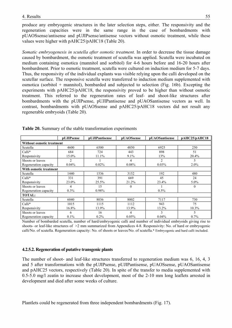
4. Results 55
produce any embryogenic structures in the later selection steps, either. The responsivity and the regeneration capacities were in the same range in the case of bombardments with pUAOSsense/antisense and pUJIPsense/antisense vectors without osmotic treatment, while these values were higher with pAHC25/pAHC18 (Table 20). Somatic embryogenesis in scutella after osmotic treatment. In order to decrease the tissue damage caused by bombardment, the osmotic treatment of scutella was applied. Scutella were incubated on medium containing osmotica (mannitol and sorbitol) for 4-6 hours before and 16-20 hours after bombardment. Prior to osmotic treatment, scutella were cultured on induction medium for 5-7 days. Thus, the responsivity of the individual explants was visible relying upon the calli developed on the scutellar surface. The responsive scutella were transferred to induction medium supplemented with osmotica (sorbitol + mannitol), bombarded and subjected to selection (Fig. 16b). Excepting the experiments with pAHC25/pAHC18, the responsivity proved to be higher than without osmotic treatment. This referred to the regeneration rates of leaf- and shoot-like structures after bombardments with the pUJIPsense, pUJIPantisense and pUAOSantisense vectors as well. In contrast, bombardments with pUAOSsense and pAHC25/pAHC18 vectors did not result any regenerable embryoids (Table 20). Table 20. Summary of the stable transformation experiments pUJIPsense pUJIPantisense pUAOSsense pUAOSantisense pAHC25/pAHC18 Without osmotic treatment Scutella 4600 6500 4850 6925 250 Calli* Responsivity
684 15.0%
724 11.1%
443 9.1%
898 13%
51 20.4%
Shoots or leaves Regeneration capacity
2 0.04%
1 0.02%
4 0.08%
2 0.03%
5 2.0%
With osmotic treatment Scutella 1440 1536 3152 192 480 Calli* Responsivity
331 23.0%
391 25.5%
669 21.2%
45 23.4%
24 5.0%
Shoots or leaves Regeneration capacity
4 0.3%
15 0.98%
0 1 0.5%
0
TOTAL: Scutella 6040 8036 8002 7117 730 Calli* Responsivity
1015 16.8%
1115 13.9%
1112 13.9%
943 13.2%
75 10.3%
Shoots or leaves Regeneration capacity
6 0.1%
16 0.2%
4 0.05%
3 0.04%
5 0.7%
Number of bombarded scutella, number of hard/embryogenic calli and number of individual embryoids giving rise to shoots- or leaf-like structures of >2 mm summarized from Appendices 4-8. Responsivity: No. of hard or embryogenic calli/No. of scutella. Regeneration capacity: No. of shoots or leaves/No. of scutella.* Embryogenic and hard calli included.
4.2.5.2. Regeneration of putative transgenic plants
The number of shoot- and leaf-like structures transferred to regeneration medium was 6, 16, 4, 3 and 5 after transformations with the pUJIPsense, pUJIPantisense, pUAOSsense, pUAOSantisense and pAHC25 vectors, respectively (Table 20). In spite of the transfer to media supplemented with 0.5-5.0 mg/l zeatin to increase shoot development, most of the 2-10 mm long leaflets arrested in development and died after some weeks of culture. Plantlets could be regenerated from three independent bombardments (Fig. 17).
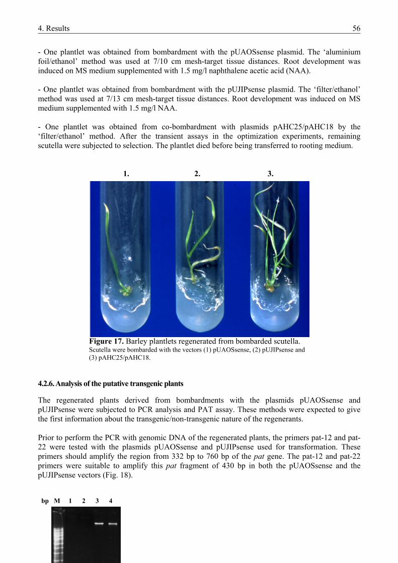
4. Results 56
- One plantlet was obtained from bombardment with the pUAOSsense plasmid. The ‘aluminium foil/ethanol’ method was used at 7/10 cm mesh-target tissue distances. Root development was induced on MS medium supplemented with 1.5 mg/l naphthalene acetic acid (NAA). - One plantlet was obtained from bombardment with the pUJIPsense plasmid. The ‘filter/ethanol’ method was used at 7/13 cm mesh-target tissue distances. Root development was induced on MS medium supplemented with 1.5 mg/l NAA. - One plantlet was obtained from co-bombardment with plasmids pAHC25/pAHC18 by the ‘filter/ethanol’ method. After the transient assays in the optimization experiments, remaining scutella were subjected to selection. The plantlet died before being transferred to rooting medium.
1. 2. 3. .
Figure 17. Barley plantlets regenerated from bombarded scutella. Scutella were bombarded with the vectors (1) pUAOSsense, (2) pUJIPsense and (3) pAHC25/pAHC18.
4.2.6. Analysis of the putative transgenic plants
The regenerated plants derived from bombardments with the plasmids pUAOSsense and pUJIPsense were subjected to PCR analysis and PAT assay. These methods were expected to give the first information about the transgenic/non-transgenic nature of the regenerants. Prior to perform the PCR with genomic DNA of the regenerated plants, the primers pat-12 and pat-22 were tested with the plasmids pUAOSsense and pUJIPsense used for transformation. These primers should amplify the region from 332 bp to 760 bp of the pat gene. The pat-12 and pat-22 primers were suitable to amplify this pat fragment of 430 bp in both the pUAOSsense and the pUJIPsense vectors (Fig. 18). bp M 1 2 3 4
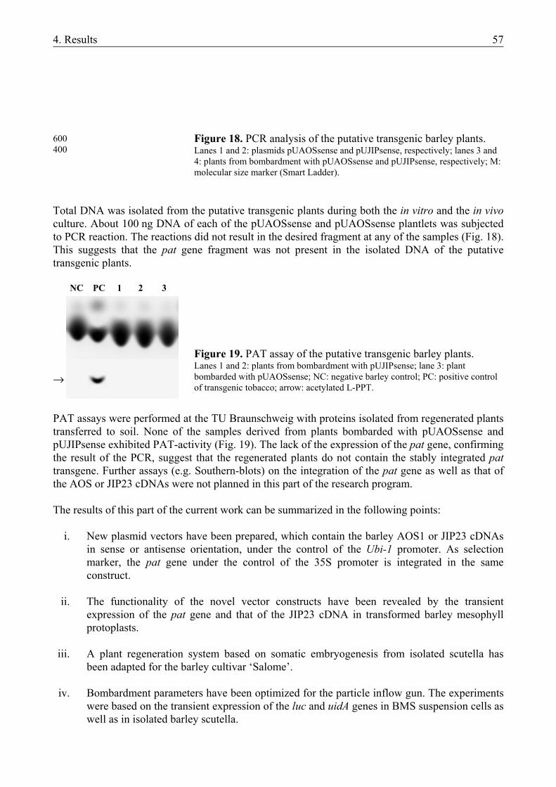
4. Results 57
Total DNA was isolated from the putative transgenic plants during both the in vitro and the in vivo culture. About 100 ng DNA of each of the pUAOSsense and pUAOSsense plantlets was subjected to PCR reaction. The reactions did not result in the desired fragment at any of the samples (Fig. 18). This suggests that the pat gene fragment was not present in the isolated DNA of the putative transgenic plants. NC PC 1 2 3 → PAT assays were performed at the TU Braunschweig with proteins isolated from regenerated plants transferred to soil. None of the samples derived from plants bombarded with pUAOSsense and pUJIPsense exhibited PAT-activity (Fig. 19). The lack of the expression of the pat gene, confirming the result of the PCR, suggest that the regenerated plants do not contain the stably integrated pat transgene. Further assays (e.g. Southern-blots) on the integration of the pat gene as well as that of the AOS or JIP23 cDNAs were not planned in this part of the research program. The results of this part of the current work can be summarized in the following points:
i. New plasmid vectors have been prepared, which contain the barley AOS1 or JIP23 cDNAs
in sense or antisense orientation, under the control of the Ubi-1 promoter. As selection marker, the pat gene under the control of the 35S promoter is integrated in the same construct.
ii. The functionality of the novel vector constructs have been revealed by the transient
expression of the pat gene and that of the JIP23 cDNA in transformed barley mesophyll protoplasts.
iii. A plant regeneration system based on somatic embryogenesis from isolated scutella has
been adapted for the barley cultivar ‘Salome’.
iv. Bombardment parameters have been optimized for the particle inflow gun. The experiments were based on the transient expression of the luc and uidA genes in BMS suspension cells as well as in isolated barley scutella.
600 400
Figure 18. PCR analysis of the putative transgenic barley plants. Lanes 1 and 2: plasmids pUAOSsense and pUJIPsense, respectively; lanes 3 and 4: plants from bombardment with pUAOSsense and pUJIPsense, respectively; M: molecular size marker (Smart Ladder).
Figure 19. PAT assay of the putative transgenic barley plants. Lanes 1 and 2: plants from bombardment with pUJIPsense; lane 3: plant bombarded with pUAOSsense; NC: negative barley control; PC: positive control of transgenic tobacco; arrow: acetylated L-PPT.
4. Results 58
v. Isolated scutella of the barley cv. ‘Salome’ have been bombarded with the plasmids
described in (i) and according to the settings determined in (iv). Bombarded scutella have been selected and putative transgenic plants have been grown up using the regeneration system of (iii). Pre- and post-bombardment incubation of scutella on osmoticum-containing medium significantly improved responsivity and regeneration capacity. PCR and PAT-assays have been performed with the putative transgenic plants. Both tests gave negative results.
4.3. NEW SCIENTIFIC RESULTS
Based on the results of the microspore culture experiments, the following can be considered to be new scientific results:
i. The protocol for the regeneration of haploid/dihaploid plants from isolated microspores of triticale has been developed. The method was established in experiments with five genotypes and it is suitable for the application in plant breeding. Its further practical advantage is that through the microspore isolation method used here, the laboursome process of anther isolation can be avoided.
ii. The induction of haploid embryogenesis does not require exogenous hormone-supplement
neither in triticale nor in barley microspore culture. It is the first description in unicellular systems of these two monocot species, that hormones are not essential exogenous factors required for the induction of haploid embryogenesis.
Based on the results of the vector preparation experiments, the following can be considered to be new scientific results:
i. Novel plasmid vector constructs have been prepared, which contain the barley AOS1 or JIP23 cDNAs in sense or antisense orientation, respectively, under the control of the Ubi-1 promoter. Based on the same construct, vectors have been prepared, which contain a multiple cloning site (MCS) in sense or antisense orientation, respectively, downstream of the Ubi-1 promoter. The cloning site includes restriction sites for at least ten enzymes. Thus, these vectors can be used for the cloning (and expression) of further genes/cDNAs.
ii. The functionality of the novel vector constructs in the genetic background of barley has
been revealed by transient assays in transformed barley mesophyll protoplasts. Thus, it is suggested that the AOS and JIP23 sense and antisense vector constructs are suitable for the modification of endogenous jasmonate and JIP23 levels in barley an other cereals via genetic transformation.
5. Discussion 59
5. DISCUSSION
5.1. INDUCTION OF HAPLOID EMBRYOGENESIS IN MICROSPORE CULTURE
The primary goals of this part of the thesis were (i) to establish the method of isolated microspore culture for triticale and (ii) to study the induction of embryogenesis and plant regeneration without exogenous hormone-supply in microspore cultures of triticale and barley. An identical microspore isolation protocol (microblending of spikes) and induction media of the same salt and vitamin composition (190-2) have been applied for both species.
5.1.1. Method of microspore isolation and culture in triticale
Up to date, haploid triticale plants have been generated via chromosome elimination technique (Inagaki and Hash, 1998, Wedzony et al., 1998) and via anther culture (Wang et al., 1973; Bernard, 1980; Lukjanjuk and Ignatova, 1986; Martinez Garcia et al., 1992; Karsai et al., 1994; González and Jouve, 2000; Immonen and Robinson, 2000). Based upon preliminary studies in barley and wheat, the protocol of microspore culture has been established for triticale as well. Microspore embryogenesis could be induced and plants could be regenerated in cultures of directly isolated triticale microspores as well. Thus, microspore culture represents a novel way to induce triticale haploids and offers a comparable alternative to the previously established anther culture.
In triticale microspore culture, the induction of androgenesis was successful when microspores were in similar developmental stage as in anther cultures. This means the mid- to late-uninucleate stages which was optimal at the collection of donor spikes. During the pretreatment of spikes, however, microspores continued development and they were in the late-uninucleate to early-binucleate stages at isolation. Isolated microspores of this stage gave highly regenerable cultures in our experiments. In comparison, responsive cultures of microspores of the early-binucleate stage were not reported from anther cultures, moreover, no embryoid development was observed from microspores of this stage compared to the optimal late-uninucleate stage in triticale anther culture (Lukjanjuk and Ignatova, 1986). Microspores of the mid-uninucleate stage also give good response (Karsai and Bedő, 1997; Karsai et al., 1994; González and Jouve, 2000) but microspores of earlier stages did not show any response in triticale anther culture (Lukjanjuk and Ignatova, 1986; Hassawi and Liang, 1990). A difference in the optimal microspore developmental stages has been observed in anther and microspore cultures of barley and wheat as well (Hoekstra et al., 1992; Mordhorst and Lörz, 1993; Puolimatka et al., 1996). Inside intact anthers, the prolonged development of microspores is supposed. This can explain that in anther culture an earlier microspore developmental stage is optimal compared to microspore culture (Hoekstra et al., 1992). The amount, viability and embryogenic capacity of microspores isolated via microblending of spikes were sufficient for the initiation of cultures. This isolation protocol is easy to perform due to the omission of the laboursome process of anther isolation. Microblending of spikes was earlier established for barley and wheat with great efficiency (Mejza et al., 1993; Mordhorst and Lörz, 1993; Puolimatka et al., 1996; Kasha et al., 2001b). For detailed discussion about microblending, see 5.1.2. Separation of viable cells from dead ones by density gradient centrifugation was first suggested for protoplast preparation (Harms and Potrykus, 1978). In the present work, the simplified version of the method has been used. This protocol is based on mannitol and maltose gradient centrifugation (‘maltose-cushion’). It has been successfully adapted also for barley and wheat (Mordhorst and Lörz, 1993; Puolimatka et al., 1996; Kasha et al., 2001b). Using this method the ratio of viable microspores can be increased to 70-96%. This protocol can increase the number of viable microspores, and is recommended if their ratio is under 50% in the crude microspore suspension (Mordhorst and Lörz, 1993; Kasha et al., 2001b).
5. Discussion 60
Viable microspores of dense cytoplasm were successfully cultured in induction media of various hormone composition (hormone-free; 2,4-D + kinetin; PAA). Macro- and microelements as well as vitamins were those of the 190-2 medium. In triticale anther culture, 190-2 medium has been applied as regeneration medium so far (Wang and Hu, 1984; Karsai and Bedő, 1997). This corresponds to its original invention for regeneration in wheat anther culture (Zhuang and Jia, 1983). In wheat microspore culture, 190-2 has been successfully used as induction medium at one genotype: both embryoid and green plant regeneration rates exceeded those received with other media. In the other genotype, however, 190-2 proved to be inferior, thus suggesting a cultivar-specific response to different media (Puolimatka et al., 1996). In triticale microspore cultures, high levels of embryogenesis could be achieved in induction media without ovary-conditioning. Isolated triticale microspores induced in ovary-conditioned media did not give regenerable embryoids (data not shown). Ovary-conditioning is not necessary for the induction but for the maintenance of embryogenesis (Datta and Wenzel, 1987; Puolimatka et al., 1996). Nevertheless, it has been found to be essential in wheat microspore cultures (Mejza et al., 1993; Puolimatka et al., 1996; Hu and Kasha, 1997). In contrast, microspore cultures of barley result in plants at the same frequency without ovary-conditioning (Mordhorst and Lörz, 1993; Kasha et al., 2001b). Ovaries are supposed to release “conditioning factors” such as PAA into the induction medium, thus promoting microspore embryogenesis (Ziauddin et al., 1992; Mejza et al., 1993; Hu and Kasha, 1997). Similarly, in non-regenerable microspore cultures of triticale the conditioning factors released by floating anthers were supposed to be required for the induction of androgenesis (Keller, 1991). Each triticale genotype involved in microspore culture responded at a satisfactory level in the regeneration experiment. Most cultivars acting in the F1 combinations (‘Presto’, ‘Moniko’, ‘Tewo’) have been previously tested in anther culture and were found to be responsive (Karsai et al., 1994). These genotypes maintained their good tissue culture response in cross-combinations under microspore culture conditions. This corresponds to the highly inheritable nature of embryo induction and, independently from the previous one, of plant regeneration which was analysed in triticale anther culture (Balatero et al., 1995). Haploid embryogenesis was observed in the isolated microspore culture of triticale. Early steps of androgenesis (until the multicellular structure stage) have been earlier described in non-regenerable microspore cultures of triticale (Keller, 1991). This type of development has been observed in microspore culture of other cereals, such as barley, wheat and rye (Hoekstra et al., 1992; Mejza et al., 1993; Guo and Pulli, 1999). In haploid cultures of monocots, embryogenesis is the preferred way of development, pretending the symmetrical division of the microspores (Szakács and Barnabás, 1995). Embryoids can develop directly from dividing microspores (Guha and Maheshwari, 1966) or indirectly through a preceding callus-phase (Niizeki and Oono, 1968). A decisive role of the type and concentration of auxins on the developmental pathway has been observed in both somatic and haploid tissue cultures (Dudits et al., 1991; Hoekstra et al., 1996). For a detailed discussion of the roles of plant hormones, see 5.1.3. From the ELSs of different genotypes, altogether 126 green plants were regenerated. The rate of albino regenerants, however, was high (>50%) in triticale microspore culture. In triticale anther culture high rates of albinism (up to 100%) can be usually observed, however this phenomenon is highly dependent on genotype and culture conditions (e.g. González et al., 1997; Immonen and Robinson, 2000). The influence of the same factors on the rate of albinism was observed in barley microspore cultures (Kasha et al., 2001 and 5.1.2. of this work). Albinism is a typical problem in anther and microspore cultures of cereals and it has been explained with several possible reasons, effects of genotype and culture conditions being among them (for reviews, see Lukjanjuk and
5. Discussion 61
Ignatova, 1986; Jähne and Lörz, 1995). Deletions in or degradation of the plastid genome are general phenomena in albino pollen plants (Day and Ellis, 1985; Carreda et al., 2000). The stomatal guard cell length and cytological examination of the 126 microspore-derived green plants indicated that the wast majority (90%) of the regenerants is haploid in triticale microspore culture. This high frequency of haploid regenerants is unique among haploid tissue cultures of cereals. In anther culture of triticale high frequencies (>60%) of spontaneous dihaploids could be achieved, but this phenomenon was strongly genotype-dependent (Immonen and Robinson, 2000). Microspore cultures of barley and wheat usually regenerate spontaneous dihaploid plants at a high frequency (>40%) as well (Ziauddin et al., 1992; Mordhorst and Lörz, 1993; Gustafson et al., 1995; Puolimatka et al., 1996). Aneuploidy also frequently occurs in microspore cultures (Hu and Kasha, 1997) and is especially common in anther cultures of triticale, due to its amphidiploid nature (Lukjanjuk and Ignatova, 1986; Immonen and Robinson, 2000). 5.1.2. Induction of androgenesis in isolated barley microspores using different culture media
Induction media of various composition have been used in barley microspore culture to date. Media were supplemented with plant hormones in each case (Table 1). Neither the induction media previously established for triticale nor the hormone-free induction of androgenesis has been studied in barley microspore culture to date. The microblending-based isolation and culture method, originally invented by Mordhorst and Lörz (1993), has been successfully adapted to our conditions with the cultivars ‘Igri’ and ‘Kymppi’. Direct microblending of spikes is an effective method for initiating microspore cultures in barley, although the regeneration rates achieved are slightly lower than those received after maceration or microblending of anthers (Table 1). Recently, at cultivar ‘Kymppi’ microblending of spikes resulted in a poorer regeneration capacity than maceration by a teflon rod (Ritala et al., 2001). This difference in the efficiency can be explained with the fact that microblended spikes contain anthers from the less-developed florets as well, while in other methods, anthers from selected parts of the spikes are processed (Mordhorst and Lörz, 1993). Nevertheless, the microblending of spikes offers a less laboursome method as the isolation of anthers is avoided. The protocol was originally invented for the model cultivar ‘Igri’ but it has also been adapted for other genotypes (Kasha et al., 2001b). Microspores cultures produced by this method have been successfully used as target tissues for transformation (Jähne et al., 1994; Leckband and Lörz, 1998; Scholz et al., 2001). Both N24- and 190-2-based media were capable of inducing androgenesis in isolated microspores of both barley cultivars tested. N24 medium has a nitrogen-composition optimized exclusively for microspore cultures of the cultivar ‘Igri’ (Mordhorst and Lörz, 1993). The original 190-2 medium, however, was invented for regeneration in wheat anther culture (Zhuang and Jia, 1983). It has never been used in tissue cultures of barley. The green plant regeneration capacity of microspores was higher on the N24- than on the 190-2-based media. A possible reason for this difference can be the different nitrogen composition of these media (Table 15). While induction of androgenesis requires a stress signal provided by pretreatment, nitrogen content of the medium is responsible for embryogenic development and regeneration capacity of the microspores (Mordhorst and Lörz, 1993). This can explain the lower number of green plants regenerated from 190-0, in spite of the higher numbers of ELSs recorded on this medium compared to N24. The nitrate- and inorganic N-content, consequently the total N-content, of 190-2 medium is half of that calculated for N24. Ammonium content and inorganic/organic N-ratio, however, are within the optimal range. Optimized NO3
-/NH4+-ratio has a significant impact on plant regeneration capacity of both haploid
and somatic tissue cultures (Grimes and Hodges, 1990; Mordhorst and Lörz, 1993). A high level of nitrate and a small amount of ammonium or other forms of reduced nitrogen are essential for normal growth and development in haploid and somatic tissue cultures of both mono- and
5. Discussion 62
dicotyledonous species (Halperin and Wetherell, 1965; Gamborg, 1970; Grimes and Hodges, 1990; Schmitz and Lörz, 1990). In the case of a NO3
-/NH4+-ratio under 30:70, however, poor development
of microspores was caused by the toxic effect of ammonium (Mordhorst and Lörz, 1993). High levels of ammonium-nitrate suppress embryoid formation and promote callus formation instead, leading to a low response and to a high percentage of albino regenerants (Olsen, 1987). The reduction of ammonium–nitrate content to 1/10 in MS medium led to an increased response of the cultured anthers (Clapham, 1973; Olsen, 1987). 190-2 medium was supplemented with 3 mM glutamine to increase the extremely low organic N-content (0.047 mM) of the original medium. Thus, organic N-content and inorganic/organic N-ratio were within the optimum range of the N24 medium (Mordhorst and Lörz, 1993). The role of glutamine in the promotion of embryogenesis has been early shown in pollen/microspore cultures of tobacco (Nitsch, 1977; Wernicke and Kohlenbach, 1977). Studies on its role in anther and microspore cultures of barley, however, gave contradictory results. In most cases, glutamine had a positive effect on embryogenesis and plant regeneration (Olsen, 1987; Hoekstra et al., 1992; Mordhorst and Lörz, 1993). In contrast, no or inhibitory effect of glutamine have also been observed (Xu and Sunderland, 1981; Jähne et al., 1994; Ritala et al., 2001). 5.1.3. Hormone-free induction of androgenesis in microspore cultures of barley and triticale
Haploid embryoids were induced and green plantlets were regenerated from microspore cultures of both barley and triticale, without exogenous hormone-supplement in the induction and regeneration media. In barley, the regeneration rate achieved this way was significantly lower compared to the media containing hormones, for both cultivars tested. In contrast, a positive effect of hormone-free induction medium on embryoid yield has been detected in microspore culture of several triticale genotypes. Its impact on green and albino plant production, however, was not significant. Barley anthers responded similarly to induction media with or without hormone-supplement, but the hormone-free medium gave rise to calli of lower regeneration capacity (Cai et al., 1992). This is in full concordance with the results reported here, and suggests that induction of androgenesis does not require hormones, but hormones are essential for efficient plant regeneration. In the microspore culture of some triticale genotypes, however, this theory was not confirmed, because regeneration capacity did not decrease in the absence of hormones. Moreover, in microspore cultures of the barley genotype ‘Jokioinen 1490’, green plants could be regenerated exclusively from microspores induced on hormone-free medium. The regeneration rate, however, was very low (<1%) (unpublished data). Similarly, the beneficial effect of hormone-free induction medium both on anther response and regeneration has been detected in oat anther culture (Kiviharju et al., 1997). The theory of hormone-free induction of androgenesis is further supported by the recently suggested role of stress as the single factor required for switching microspores from gametophytic to sporophytic pathway (for review, see Heberle-Bors, 1998). This proposal is based on the identical conditions required for the pretreatment of tobacco and wheat anthers/spikes prior to isolation of microspores (Touraev et al., 1996a,b). Because the pretreatments applied in these species (starvation + heat shock) are partially different from those applied for barley and triticale in our studies (starvation + cold), the species-specifity of the required signal is suggested. This is, however, in contradiction with the above-mentioned general validity of the ‘starvation + heat shock signal’ model, although this pretreatment protocol has not been tested in our experiments. Hormones commonly used in haploid cultures of barley are basically different from those used in triticale cultures. In barley, the cytokinin BAP is used most frequently, while in triticale the synthetic auxin 2,4-D, occasionally supplemented with the cytokinin-type kinetin, is the main source of hormones. In this sense, triticale cultures resemble those of wheat, also based on auxin-supplement (Puolimatka et al., 1996, Hu and Kasha, 1997). The role of exogenously applied
5. Discussion 63
hormones in haploid tissue cultures of cereals and the reasons for the species-specific preferences in their use are less understood. The preferential use of cytokinins has been confirmed in our preliminary experiments. Here, auxin-based induction media (190-D/K and 190-PAA) did not result in plant regeneration from barley microspore cultures (data not shown). This preference for cytokinins is not confirmed by some other reports. In anther culture, high endogenous levels of auxins in barley anthers are supposed to lead to no or inhibitory effects of auxins added to the media. The removal of anther tissues, however, improved auxin (PAA)-sensibility of barley microspores (Ziauddin et al., 1992). Moreover, 2,4-D has been found to be as effective as BAP in both anther and microspore cultures of barley (Hoekstra et al., 1996). To achieve maximum embryogenesis and regeneration rates, however, the higher the contrentation of 2,4-D is the shorter the application time must be (Dudits et al., 1991; Hoekstra et al., 1996). The negative effect of high 2,4-D concentration for an extended period has been observed in anther cultures of barley, wheat and triticale (Marsolais and Kasha, 1985; Hassawi et al., 1990). According to these reports, the preference for cytokinins in the promotion of embryogeneis in barley haploid cultures seems to be overcome and the promoting role of 2,4-D can be extended over this species as well. Thus, the failure in the induction of regenerable embryoids in barley microspore cultures can be explained, most probably, with the unsuitable concentration of auxins added. The same concentrations, however, were successfully applied for wheat and triticale microspores. In triticale microspore cultures, the combination of 2,4-D + kinetin as hormone-supplement resulted in higher rates of green regenerants than PAA. Thus, the increased regeneration capacity reported as a result of PAA-addition to microspore cultures of barley and wheat (Ziauddin et al., 1992; Hu et al., 1995; Hu and Kasha, 1997), has not been confirmed here. Androgenesis and plant regeneration are considered to be independent events of different genetic background (Knudsen et al., 1989; Mordhorst and Lörz, 1993). Our results suggest that androgenesis and plant regeneration are independent events regarding hormone-requirement as well. Induction of androgenesis does not need exogenous hormone-supplement neither in barley nor in triticale microspore culture. The regeneration of plants, however, exhibits differences between genotypes and/or species regarding exogenous hormone-requirement: regeneration is hormone-dependent in barley and in some of the triticale genotypes tested, while it is independent of hormone-supplement in other triticale genotypes. 5.1.4. Future prospects
The easy-to-perform method for isolation and culture of isolated microspores of triticale could be used for generation of haploid plants from different genotypes, as described in this work. The doubled haploid lines received from different F1 combinations were readily introduced into the traditional breeding process. This shows that the protocol can be involved in the production of new triticale cultivars. The avoiding of anther isolation offers a less laboursome alternative to the traditional anther culture. Although transgenic triticale plants have been produced only by particle bombardment of scutellar tissues to date, promising results in barley suggest that isolated microspores of triticale could also be used as target tissues for transformation. Hormone-free induction of embryogenesis in isolated microspores of two cereal species confirm that stress signals are necessary and sufficient for the induction of androgenesis as described earlier in wheat. The differences in regeneration capacity between species and genotypes, however, propose further studies on various pretreatments such as stress signals and on various hormone
5. Discussion 64
combinations, involving a broader range of genotypes in the experiments. These results could clarify the need for a species- or genotype-specific stress signal which can have a promoting effect on regeneration capacity as well. Supposing, however, that embryogenesis and plant regeneration are independent events, studies on a specific hormone-requirement for regeneration seem to be also necessary. 5.2. PREPARATION OF NOVEL VECTOR CONSTRUCTS The goal of the current work was to prepare novel plasmid vectors carrying cDNAs coding for AOS, the key-enzyme of JA-biosynthesis, or JIP23 in sense or antisense orientation, respectively. In addition, bombardment parameters were optimized for a particle inflow gun and the new constructs were used in a set of stable transformation experiments performed with ‘Salome’, the model cultivar of jasmonate studies. The plasmids and the results of the optimization experiments can be utilized in the production of transgenic barley plants which can mean the subject of further studies on the role of jasmonates in the development of barley and on the analysis of the function of JIP23 in this species. 5.2.1. The structure of the novel plasmid vectors
For the stable transformation of barley via particle bombardment, new vectors were prepared which carry (i) a cDNA coding for the gene of interest in sense or antisense orientation under the control of the Ubi-1 promoter and (ii) the pat resistance gene under the control of the 35S promoter. Our novel constructs (pUAOSsense/antisense, pUJIPsense/antisense) are based on plasmids (pAHC20 and pWD26.41) which have already been used in barley transformation. In addition to the vectors for the modification of AOS and JIP23 level, intermediate products of the vector preparation can also be utilized in the transformation of barley and other (primarly monocot) species. These vectors are derived from the penultimate stage of vector preparation when the AOS or JIP23 cDNA is still not inserted into the multiple cloning site (pUMCSsense and pUMCSantisense). The complete MCS contains restriction sites for at least ten enzymes thus enabling the cloning of a broad range of genes/cDNAs. Its utilization can be further broadened by the feature that fully identical constructs are available for the preparation of sense and antisense constructs. Ubi-1 promoter-based expression vectors of similar structure have not been reported yet. From the basic set of Ubi-1 promoter-based vectors prepared by Christensen and Quail (1996), pAHC17 contains a polylinker. This sequence offers restriction site for six enzymes, but no expression cassette containing a selection marker gene is included in the plasmid. The expression vectors pRT100 to 104 based on the 35S promoter also contain restriction sites in the same range (5-8 sites) as pAHC17 and our MCS (Töpfer et al., 1987). 5.2.1.1. The promoters
The use of two different promoters to control a gene of interest and a resistance gene is a repeatedly performed approach to avoid or minimize transcriptional gene silencing (de Wilde et al., 2000). The co-transformation of the expression cassettes (i) 35S::resistance gene and (ii) promoter of various origin::transgene of interest has already been successfully used for the generation of transgenic barley plants (Jähne et al., 1994; Ritala et al., 1994; Leckband and Lörz, 1998; Fang et al., 2002). To date, the combination of 35S and Ubi-1 promoters controlling different genes in the same plasmid has not been used in vectors for the particle bombardment of barley. Binary vectors of such construction, however, are efficiently used in the Agrobacterium-mediated transformation of barley (Horvath et al., 2000; Wang et al., 2001; Fang et al., 2002).
5. Discussion 65
In a transient expression system of barley, promoters of monocotyledonous origin (maize Ubi-1, maize-derived Emu, rice Act-1) exhibited higher promoter strength than the CaMV 35S promoter (Schledzewski and Mendel, 1994). Such a difference, however, has not been detected in stable transformation: transgenes controlled by the 35S or by the Ubi-1 promoter were expressed with similar efficiency in the transgenic plants (Wan and Lemaux, 1994). In wheat biolistic transformation, the transformation efficiency was 0.8-1.1% if the resistance gene was under the control of the 35S promoter (Becker et al., 1994, Nehra et al., 1994) and it was in the same range (0.1-1.5%) in the case of a control by the Ubi-1 promoter (Weeks et al., 1993, Altpeter et al., 1996a,b). Immature embryos/scutella of barley are usually co-bombarded with plasmids which integrate 35S and Ubi-1 promoters separately. These results suggest that the combination of expression cassettes controlled by the Ubi-1 and 35S promoters, respectively, in the same plasmid vector, like applied in our novel constructs, can be used for the generation of transgenic barley plants. 5.2.1.2. The pat resistance gene
The successful regeneration of plants containing stably integrated transgenes requires an appropriate selection system. Selection systems based on PAT are widely used in cereal transformation. From the genes encoding PAT, bar is the most widely used resistance gene for barley transformation (Table 3). The pat gene, which shows significant homology to bar (Wohlleben et al., 1998), has also been successfully involved in the production of transgenic barley plants (Leckband and Lörz, 1998; Scholz et al., 2001). Selection of transgenic tissues expressing PAT in barley is usually performed on media supplemented with 3-5 mg/l bialaphos (for reviews, see Lemaux et al., 1999). In our experiments a bialaphos concentration of 3 mg/l concentration was sufficient to select non-responsive scutella. These scutella exhibited necrosis after a few days on selective medium and usually died within two weeks of selection, similarly to an earlier report about the selection of non-responsive scutella of the cultivar ‘Salome’ (Koprek, 1996). The responsive scutella produced calli and embryogenic structures, thus confirming the suitability of the selection system. 5.2.1.3. The AOS cDNA
Constitutive overexpression or down-regulation of AOS by genetic transformation means one possible way to modify endogenous jasmonate level, as it was shown in dycotelodonous species (Harms et al., 1995; Wang et al., 1999a; Laudert et al., 2000). The AOS cDNAs isolated from barley have not been used for homologous transformation so far, and no attempts have been made to analyze JA-function in transgenic monocotyledonous plants. Several features of jasmonates, however, support a transgenic approach for studying their role in monocots: (i) in barley high levels of JAs can be detected localized in organs and tissues stressed by water deficit (scutella, nodes, senescent flag leaf, husks) as well as in dividing tissues such as root tips and leaf bases (Hause, unpublished); (ii) products of JA-degradation have not been detected to date (Miersch, pers. comm.); (iii) JM, JA and its amino acid-conjugates can be involved in the signalling pathway simultaneously (Wasternack et al., 1998). Thus, problems such as those caused by the influence of several factors (e.g. transport, metabolism, gene dosis) on the phenotypic expression of cytokinin-function do not occur here (Hewelt et al., 1994; Faiss et al., 1997). Furthermore, unlike in the transgenic manipulation of cytokinin- and auxin-biosynthesis performed to date (Tinland et al., 1991; Hewelt et al., 1994), the control of transgenes by constitutive promoters should not inhibit hormone-synthesis or the regeneration of specific organs. To study the effects of jasmonate deficiency, a vector was constructed which contains the barley AOS1 cDNA in antisense orientation (pUAOSantisense). Down-regulation of endogenous hormone
5. Discussion 66
levels by transformation with antisense genes/cDNAs coding for enzymes involved in their biosynthesis, is a well-established technique in several hormone classes (for review, see Hedden and Phillips, 2000). Besides the numerous examples for ACC antisense plants (Hamilton et al., 1990; Oeller et al., 1991), Arabidopsis plants with down-regulated IAA (Grsic et al., 1998; Ouyang et al., 2000) and gibberellin (Coles et al., 1999) production also confirm the functionality of the antisense strategy. Similarly to our pUAOSantisense construct, the vectors applied in these studies contain homologous antisense sequences. 5.2.1.4. The JIP23 cDNA
In barley, putative functions of the most abundant jasmonate-induced protein, JIP23, have been described in early stages of development and in excised leaf segments (Lehmann et al., 1995; Hause et al., 1996). Because of the high tissue- and cell specificity of its expression and its occurrence in all barley cultivars (Hause et al., 1999), this protein may have an essential function. The constitutive overexpression of JIP23 in barley should give detailed insights into functions of JIP23 in each developmental stage. Such an overexpression may override other effects of JIP23 formed during barley development. Constitutive overexpression may lead to altered photosynthetic capacity of leaves, altered responses upon abiotic stresses (drought, osmotic stress), and altered developmental processes such as germination. In a similar way, the suppression of JIP23 gene expression by an antisense approach should also lead to phenotypical alterations giving hints to the function of JIP23 during germination and stress response. This is suggested by effects described for constitutive overexpression of barley JIP23 in tobacco (Görschen et al., 1997b). The amount of JIP23 correlated proportionally with the number of transgene integrations and was inversely correlated with the amounts of certain house-keeping proteins, mainly proteins involved in photosynthesis (e.g. subunits of RuBPCase). Transformation with antisense JIP23 cDNA, however, did not alter the protein pattern. 5.2.1.5. Transient expression of the transgenes in barley mesophyll protoplasts
To date, our novel vectors containing the AOS1 and JIP23 cDNAs have not been used in the homologous stable transformation of barley. To test their functionality within a transient gene expression system, barley mesophyll protoplasts were transformed with the sense constructs (pUAOSsense, pUJIPsense) by PEG-mediated gene-transfer. Barley protoplasts of various origin (cell suspensions, mesophyll, aleurone and endosperm cells) offer ideal target cells for transient expression systems. Except for some attempts with electroporation and microinjection, PEG-mediated gene-transfer is the usual way of introducing foreign genes into barley protoplasts (Table 3). The PAT activity and the immunological detection of JIP23 indicated that both the resistance gene (pat) and the transgene (JIP23 cDNA) could be transiently expressed in barley. In the case of AOS1 Western-blot has not been prepared because of technical reasons, see above. Therefore, these vectors were regarded to be suitable for the stable transformation of barley as well.
5.2.2. Plant regeneration through somatic embryogenesis from barley scutella
Tissue cultures of high regeneration capacity are prerequisites for the generation of transgenic plants at high efficiency. Before starting the transformation we adapted an in vitro plant regeneration system based on somatic embryogenesis from isolated scutella of the barley cultivar ‘Salome’. In barley transformation the most frequently used target explants are immature embryos. Stably transformed plants could be generated after the particle bombardment of or Agrobacterium-mediated gene transfer into the adaxial (scutellar) side of excised immature embryos (Table 3). The
5. Discussion 67
scutellum is of advantage compared to other explants due to its high regeneration capacity. This can also be seen in the relatively high regeneration rate of transgenic plants derived from scutella which can exceed 4% (Wan and Lemaux, 1994; Jensen et al., 1995; Tingay et al., 1997). Embryo axis disturbs the formation of embryogenic calli because of precocious germination and the production of non-embryogenic soft calli (Nonohay et al., 1999). This could be avoided by complete removal of the embryo axis from the abaxial side of the scutella. Since any residual tissue of the axis can generate undesired structures, this laboursome method was essential for the preparation of scutella of the recalcitrant cultivar ‘Salome’ in our experiments. In barley transformation based on the highly responsive genotype ‘Golden Promise’, a simpler method, the longitudal bisection of embryos, is used (Wan and Lemaux, 1994). Somatic embryoids were generated, without a callus-phase, on ‘Salome’ scutella. The regeneration rate of green plants, however, was very low (11.5-14.4%) in our experiments. This corresponds to earlier data where the induction frequency of embryogenic structures was 10% and it decreased to 9% in bombarded scutella (Koprek, 1996). In contrast, in the cultivar ‘Golden Promise’, the most frequently used genotype in barley transformation, induction frequencies of 83% and 80%, respectively, could be achieved in the same experiment. Besides the effects of the genotype, somatic embryogenesis in cultured scutella can be influenced by (i) the growing conditions of the donor plants, (ii) the developmental state of the immature embryos and (iii) the in vitro culture parameters. Donor ‘Salome’ plants were grown in a fully controlled growth chamber and donor spikes were collected about 14 days post-anthesis. In general, higher and less variable green plant regeneration rates can be achieved from growth chamber-grown plants. These consistently high rates are prerequisites for an efficient transformation program (Dahleen, 1999). Somatic embryos and embryogenic calli could be observed only at immature embryos of 1.5-2.5 mm in size. Under growing conditions similar to ours, immature embryos of both narrower (1.5-1.8 mm; Koprek et al., 1996) and broader (1.0-2.5 mm; Cho et al., 1998) size ranges can be capable of plant regeneration through somatic embryogenesis, depending on genotype. Somatic embryogenesis in isolated barley scutella was induced on MS-based media supplemented with casein-hydrolysate, maltose and 2 or 3 mg/l 2,4-D (91/4 and 91/4m3 medium, respectively). In scutellum cultures of ‘Salome’, 3 mg/l 2,4-D resulted in a lower number of hard and embryogenic calli. The plant regeneration rate, however, improved indicating a better (probably more embryogenic) callus quality (Table 16). Such an improved embryogenic capacity has also been observed by Schulze (pers. comm.), suggesting 3 mg/l 2,4-D as the optimal concentration in scutellum cultures of ‘Salome’. In scutellum cultures of ‘Salome’ a very low regeneration rate could be achieved, representing a remarkable disandvantage for stable transformation. The preferential use of this cultivar, however, was supported by the following facts:
i. There is a cultivar-specific difference in JIP patterns and in the JA- and stress-induced
accumulation of JIP23 in barley (Herrmann et al., 1989; Hause et al., 1999). Additionally, JIP23 is supposed to be a reporter for a rise of endogenous jasmonates (Lehmann et al., 1995; Hause et al., 1996). In previous studies on the response of barley leaf segments upon JA-treatment, ‘Salome’ responded the most quickly and exhibited the strongest changes in comparison with other cultivars (Hause and Wasternack, pers. comm.). The cDNAs coding for AOS (Maucher et al., 2000) and JIP23 (Hause, pers. comm.), respectively, were initially isolated from this cultivar, thus providing the proper probes for homologous transformation.
5. Discussion 68
ii. Via bombardment of scutella from the cultivar ‘Salome’ with PIG, one fertile transgenic plant exhibiting PAT- and GUS-activity has been produced (Koprek, 1996). This suggests the suitability of this cultivar for stable genetic transformation.
5.2.3. Optimization of bombardment parameters for the use of PIG
The transformation protocol used here was based on the bombardment of isolated scutella by a particle inflow gun (PIG). The device had been previously used only for transient assays, among others, in barley leaves, onion epidermal cells and tomato suspension cells. To our knowledge, no stable transformation experiments had been performed with this PIG previously. The conditions optimal for a high transient gene expression rate do not necessarily result in a high number of transgenic plants (Becker et al., 1994; Altpeter et al., 1996a; Koprek, 1996). Therefore, in generating stable transformants, the optimal ratio of maximal DNA transfer and minimal cell death caused by bombardment has to be find out (Klein and Jones, 1999). Transient transformation rates can be converted into stable transformation rates with a frequency of 1-9% (Klein et al., 1988b; Russell et al., 1992; Ritala et al., 1993). Therefore, in the case of genotypes of low embryogenic potential, such as ‘Salome’ in our experiments, optimization must fulfill different requirements. On the one hand, optimized parameters have to lead to a transient expression strong enough to offer a reasonable regeneration potential of stable transformants. On the other hand, however, the few cells possessing regeneration potential must stay viable after bombardment. Transgenic barley plants have already been generated from isolated scutella of recalcitrant cultivars by bombardment with PIG. In the case of cultivars ‘Salome’ and ‘Femina’ the bombardment conditions offered by the PIG - especially the moderate gas-pressure - seemed to be essential (Koprek, 1996). 5.2.3.1. Bombardment method
Using PIG, DNA-coated microcarriers are usually accelerated directly in the He-stream (Finer et al., 1992). This bombardment method was modified by the introduction of a macrocarrier (aluminium foil disc) to carry the particle-suspension in glycerol (Koprek, 1996). Bombardments with this ‘aluminium foil/glycerol’ method gave very low transient expression rates in our experiments. This might be be caused, at least partially, by the mesh which can dramatically decrease the velocity of gold particles suspended in the viscosic solution. The mesh, however, was essential to save scutella from being removed from the plate by the He-stream. In a different model of PIG without mesh, ‘aluminium foil/glycerol’ method was successfully used to generate transgenic barley plants from genotypes (e.g. ‘Salome’) exhibiting lower potential for the production of embryogenic calli (Koprek, 1996). Due to the lower gas blast and acoustic shock caused by the PIG, the upper layers of scutellar tissues were less injured than by PDS, the most widely used particle delivery system. Thus, their embryogenic capacity could be maintained. The novel ‘aluminium foil/ethanol’ method invented by Pauk (pers. comm.) has been used with the greatest efficiency here. It resulted in higher levels of transient GUS and LUC expression than the ‘filter/ethanol’ method. The ‘aluminium foil/ethanol’ method resembles the working principle of PDS (see 3.2.4.2.) which enables a more effective acceleration of the microcarriers. At the same time, however, the target tissues are subjected to a moderate He-blast and acoustic shock, keeping these beneficial characteristics of the original PIG system. This method has already resulted in transgenic wheat plants (Pauk, unpublished) while the ‘filter/ethanol’ method was used only for transient expression assays (Hunold et al., 1994; Stenzel, 1998).
5. Discussion 69
5.2.3.2. Helium pressure and distance settings
The influence of helium pressure on transformation efficiency must be evaluated at a given distance between the microcarrier outlet (on filter unit) and the target tissue. Additionally, other factors such as bombardment method, the amount of particles per shot etc. have to be taken into consideration as well. The optimal helium pressure of 5.5 bar determined in our experiments is the same as reported by Koprek (1996) by using a different bombardment method (‘aluminium foil/glycerol’ instead of ‘aluminium foil/ethanol’) and a different model of PIG. This pressure is in the range (4-7 bar) usually applied for PIGs (Hunold et al., 1993; Vain et al., 1993). We had to use, however, a longer filter unit/target plate distance as usual. The generally recommended 6 cm was completely inapplicable in our PIG because scutella were blown out of the plate by the gas stream, even if a mesh was used. Target plate/filter unit distances of 10 and 13 cm combined with 4 cm mesh/filter unit distance gave the highest transient expression efficiency. Mesh has always been inserted between the particle outlet and the target tissue (i) to reduce the size of the microprojectile aggregates, (ii) to help to spread particles over the target plate and (iii) to moderate the gas blast and the acoustic shock at the same time. These beneficial effects, however, were not tested in our experiments. Morover, they were not always visible as a significant increase in the number of transformation events in other bombardment studies (Finer et al., 1992; Russell et al., 1992; Sanford et al., 1993; Hunold et al., 1994). 5.2.3.3. Osmotic treatment of the target tissues
In a set of experiments scutella were incubated pre- and post-bombardment on medium containing osmotica (0.2 M mannitol + 0.2 M sorbitol). With the introduction of this osmotic treatment, the responsivity of the bombarded scutella of ‘Salome’ increased from 9-15% to 21-23%. There was also a significant improvement in the regeneration capacity which was 0-0.98% with and 0.02-0.08% without osmotic treatment (Table 20). In wheat, pre- and post bombardment treatment of embryos on media of 0.4 M osmoticum (mannitol + sorbitol) had a significant impact on the improvement of transformation efficiency (Altpeter et al., 1996a). The osmotic treatment of the target tissues has been recently introduced in the biolistic transformation of embryogenic calli, shoot meristematic cultures and immature embryos of barley (Ahlandsberg et al., 1999; Zhang et al., 1999; Scholz et al., 2001). Experiments to prove its positive effects, however, have not been performed. Osmotica cause plasmolysis which reduces cell damage by preventing extrusion of the protoplasm from the bombarded cells, thus leading to increase in transient gene expression as well as in the recovery of stably transformed clones (Vain et al., 1993). Osmotic treatment may also act through inducing stress response that protects the cells from apoptotic reaction to particle penetration (Klein and Jones, 1999). 5.2.3.4. Changes in responsivity upon bombardment
Bombardment parameters resulting in the highest transient GUS and LUC activities have been determined with BMS suspension cells and barley scutella. The changes in the regeneration capacity caused by bombardment were checked after the application of these settings in stable transformation experiments. Without bombardment the induction rate of hard and embryogenic calli (responsivity) was 54.7-78.5% (Table 16). Upon bombardment with the novel vector constructs it decreased to 21.2-25.5% if osmotic treatment was used. The lack of osmotic treatment led to a more significant decrease to 9.1-15.0% (Table 20). In contrast, in earlier studies with ‘Salome’ the induction frequency of embryogenic structures (10-11%) did not decrease upon bombarded with PIG mand one transgenic plant could be regenerated, in spite of the low responsivity (Koprek,
5. Discussion 70
1996). Since the responsivity reported here was in the same range (9.1-25.5%), our bombardment strategy was supposed to be capable of generating transgenic plants of ‘Salome’. 5.2.4. Plant regeneration from bombarded scutella
In this thesis the results of the first application of the novel plasmid constructs in stable transformation experiments are reported. The AOS and JIP23 cDNAs cloned in the vector constructs were isolated from the barley cultivar ‘Salome’, the model genotype of jasmonate studies, thus, ‘Salome’ was chosen to be involved in the first transformation studies performed with the new vectors. Due to the very poor responsivity and regeneration capacity of this genotype, however, these experiments were taken as preliminary ones – preceding further bombardments using genotypes of high regeneration capacity. Hard and embryogenic calli induced from bombarded scutella became necrotic after 4-6 weeks upon selection and usually died 5-6 weeks after bombardment. Surviving shoot primordia and green, potentially regenerative structures were incubated on regeneration medium in the light. However, their development was arrested and they did not show any response to neither hormones stimulating shoot development (zeatin, BAP) nor to NAA stimulating root development. The only exceptions from this were two putative transformants regenerated from scutella bombarded with pUAOSsense and pUJIPsense plasmids. These plantlets developed slowly and failed to generate roots. Although the lack of roots could indicate the non-transformed, ‘escape’ status (Altpeter, pers. comm.; Schulze, pers. comm.), the regeneration process was continued anyway. After several weeks of incubation on NAA-containing medium, roots had been generated and the plants were transferred to soil. PCR and PAT-assays were found to be representative enough to show the presence of the pat sequence and the activity of the PAT enzyme in the putative transgenic regenerants. Both tests gave negative results. The detection of the integrated AOS1 and JIP23-cDNAs should have been performed only with the PAT-positive plants. This is the strategy generally used in stable transformation systems: co-transformation/co-expression frequency is determined in resistant transgenic plants only. The co-transformation frequency of transgenes was 100% in most transformation studies with barley, wheat, maize and oat (Becker et al., 1994; Hagio et al., 1995; Ishida et al., 1996; Gless et al., 1998). For barley lower co-transformation frequency of 84% and even lower co-expression frequencies of 33-48% have also been reported. In each case, however, the number of the resistant transgenic plants was taken 100% (Jähne et al., 1994; Wan and Lemaux, 1994). The failure in the generation of transgenic plants may be attributed to several factors such as (i) the low number of scutella giving rise to totipotent cells, (ii) the limited number of totipotent cells into which DNA can be introduced and (iii) the small number of totipotent cells being capable of generating fertile, green plants (Lemaux et al., 1999). Moderate He-pressure, osmotic treatment and the incubation of bombarded scutella in an ‘intermediate step’, however, can significantly enhance the transformation efficiency in recalcitrant genotypes (Cho et al., 1998). In our experiments, low He-pressure was provided by the PIG. This factor alone should be sufficient to improve transformation efficiency in ‘Salome’ (Koprek, 1996). Osmotic treatment improved the responsivity in our experiments too, but failed to increase transformation efficiency (see 4.2.5.1.). Bombarded scutella of the ‘intermediate step’ are incubated on a medium of modified composition (2,4-D + BAP, high cc. of CuSO4) and are exposed to dim light. Under these conditions calli of higher embryogenic capacity are produced. Morover, the proliferation of small numbers of green, totipotent cells is also promoted. This way, regenerability may be improved because sufficient amounts of transformed tissues are available to give rise to plants (Cho et al., 1998). In our experiments, green, potentially regenerative tissues were incubated on media containing cytokinin
5. Discussion 71
(zeatin) and exposed to light. The green structures could be kept alive for several weeks, but failed to develop further. 5.2.5. Future prospects
The novel vector constructs of proven functionality are ready for use in transformation. The constructs carrying the AOS or JIP23 cDNAs can be used to produce transgenic plants for the studies on the role of jasmonates in the development of barley as well as the analysis of function of JIP23. According to the results of the preliminary stable transformation studies, however, the use of a barley genotype of better regeneration capacity, such as cultivar ‘Golden Promise’, seems to be essential. This cultivar has established protocols for the PDS-1000/He particle bombardment system which could be adapted for the current transformation studies. Recently, for the transformation of barley Agrobacterium-mediated gene-transfer has been found to give better results than the traditional biolistic protocol (Altpeter, pers. comm.). Supported by published results as well, this method could also be introduced. In this case, however, new vector constructs should be designed, maintaining the order of promoters and transgenes of the current ones. The novel vector constructs containing the full-length multiple cloning site offer a broad range of usage in the future. The various genes/cDNAs cloned in the MCS enable the involvement of the constructs in different transformation studies based on particle bombardment.

5. Discussion 72
6. Summary 73
6. SUMMARY
I. Induction of haploid embryogenesis in isolated microspore cultures of triticale and barley under hormone-free conditions. This part of the thesis is dealing with different aspects of microspore embryogenesis. Anther culture, one of the techniques based on androgenesis, is commonly used to accelerate the breeding process of triticale and other cereals today. Isolated microspore culture, however, has some additional advantages: it offers a unicellular system of synchronized haploid cells, which are excellent targets for transformation and for studies on the biochemical and molecular background of embryogenesis. In contrast to anther culture, microspore culture of triticale has not been reported to date. 1. We established the protocol for isolated microspore culture of triticale introducing five genotypes (‘Presto’ and ‘Tewo x Moniko’, ‘Presto x Moniko’, ‘Presto x Novisadi’, ‘Novisadi x Moniko’ F1) and three induction media of different hormone-composition (hormone-free; 2,4-D + kinetin; PAA) into the experiments. The microblending of cold-pretreated spikes could be efficiently used for the isolation of microspores. The concentration of viable microspores was increased by density-gradient centrifugation. Microspores of the late-uninucleate to early-binucleate stages exhibited the capacity for division and gave rise to embryoids in induction media both with and without hormone-supplement. Plant regeneration was performed on hormone-free regeneration medium, regardless of the induction medium applied. Altogether 126 plants have been regenerated and transferred into soil. 2. In microspore cultures of triticale 90% of the regenerants were haploid which is in contrast with microspore cultures of other cereals, where usually spontaneous dihaploid plants can be regenerated at high percentage. Following colchicine-treatment, the proportion of fully or partially fertile triticale plants varied between 43% and 67%, excepting ’Presto x Novisadi’ F1 where no fertile plants were obtained. The fertile plants offered a proper material to be introduced into the traditional breeding process. 3. Hormone-free induction medium had a positive impact on embryoid production from ‘Novisadi x Moniko’ F1 triticale microspores. Its effect on green and albino plant regeneration, however, was not significant. Induction media supplemented with hormones had no significant effect on any of the three parameters (embryoid, green and albino plant production). In microspore cultures of the barley cultivars ‘Igri’ and ‘Kymppi’ androgenesis could be induced in media both with and without exogenous hormone-supplement. Here, however, the regeneration capacity of embryoids was significantly lower in hormone-free media. These data suggest that androgenesis and plant regeneration are independent events with respect to hormone-requirement as well. Cold pretreatment of spikes provides the signal which initiates and promotes embryogenesis in isolated microspores of both triticale and barley. The regeneration of plants, however, exhibits differences between genotypes and/or species regarding exogenous hormone-requirement. 4. The induction medium successfully used in triticale microspore culture has been tested also in barley micropore culture. Here, this medium gave a significantly lower regeneration rate in comparison with a medium of optimized nitrogen-composition. This difference was irrespective of the hormone-content of media and might be caused by the significantly lower (50%) inorganic N-content of the triticale-medium. Barley cultivars responded similarly to the suboptimal medium, while there was a significant difference in their response to a medium of optimized composition.
6. Summary 74
Our results suggest that some critical factors of androgenesis (e.g. stress required for the induction of embryogenesis) are of general validity irrespective of species and genotype, while others (e.g. hormone-requirement of plant regeneration, optimal composition of media) are specific for species and genotype. II. Preparation of novel vector constructs to alter endogenous levels of jasmonates and JIP23 via genetic transformation of barley. Barley can be regarded as a model system for studying the role of the jasmonates and jasmonate-induced proteins in monocots. While studies on jasmonate modulation by transgenic approaches have already been performed in dicotyledonous species, no corresponding data are available for monocots. Most of our current knowledge on the role of jasmonates in monocots is based on treatment with JAs or on stress-induced JA-formation. The comprehensive goal of our research program was to apply a transgenic approach for the studies on the role of jasmonates in the development of barley. 1. As one part of the main program and the goal of the current work, novel plasmid vectors have been prepared. Two of them carry the cDNA coding for AOS1 from barley in sense and in antisense orientation, respectively, under the control of the Ubi-1 promoter. Additionally, the vector contains the pat selection marker gene under the control of the 35S promoter. Since AOS has regulatory role in JA-biosynthesis, modulation of endogenous jasmonate levels can be expected in AOS-transformants. 2. JIP23 is the most abundant jasmonate-induced protein of barley. Therefore, the analysis of its function was also attempted by a transgenic approach. Corresponding vectors were constructed by inserting a cDNA coding for JIP23 of barley in sense or antisense orientation, respectively, into the basic vector construct identical to that established for AOS. 3. As an intermediate product of vector preparation, two vector constructs were produced which carry a multiple cloning site (MCS) in sense or antisense orientation, downstream of the Ubi-1 promoter. Additionally, the vector contains the pat selection marker gene under the control of the 35S promoter. The MCS includes restriction sites for at least 10 enzymes, thus enabling the cloning (and expression) of a wide range of genes/cDNAs . 4. The novel vector constructs were shown to be functionally active on the basis of the transient expression of the pat gene and that of the JIP23 cDNA in transformed barley mesophyll protoplasts. 5. Preliminary stable transformation experiments were planned with the barley cultivar ‘Salome’, the model genotype of jasmonate studies. Previously, a plant regeneration system based on somatic embryogenesis had been adapted for the barley cultivar ‘Salome’. Scutella of 1.5-2.5 mm diameter were optimal for the generation of embryogenic structures on media containing 2,4-D. The plant regeneration capacity was very low (10-15%). 6. Prior to stable transformation, bombardment parameters were optimized by transient assays in maize suspension cells and barley scutella bombarded by a particle inflow gun. A He-pressure of 5.5 bar and a target distance of 10-13 cm gave the highest transient β-glucuronidase and luciferase expression rates. Above the target plate a wire mesh has been applied to reduce the size of the microprojectile aggregates and to moderate gas blast and acoustic shock. The best transient expression results were received if coated gold particles were placed on the surface of an aluminium foil disc upon bombardment.
6. Summary 75
7. Scutella of cultivar ‘Salome’ were bombarded with the novel vector constructs, as their first use in stable transformation experiments. Bombarded scutella were exposed to selection on media containing 3 mg/l bialaphos. This concentration was sufficient to select non-responsive scutella and allowed the responsive ones to produce calli or embryogenic structures. Responsivity and regeneration capacity of ‘Salome’ scutella could be significantly increased by pre- and post-bombardment incubation of scutella on osmotica-containing medium. 8. Two putative transgenic lines have been received from the preliminary stable transformation experiments. These originated from bombardments with plasmids carrying the AOS1 and the JIP23 cDNAs in sense orientation, respectively. PCR-analysis and PAT-assay, however, revealed that the pat sequence was not present and was not expressed in these plants. According to the results detailed above, functionable vector constructs are available for the production of transgenic barley plants exhibiting altered jasmonate or JIP23 levels. Similarly, a wide range of vectors and subsequently transgenic plants can be generated by the use of the vector construct containing an extensive MCS. In addition, bombardment parameters were optimized for a particle inflow gun, thus facilitating the usage of the device in further studies based on the particle bombardment of scutella. Similarly, the first set of stable transformation experiments with the new vector constructs give useful information for further experiments, regarding the choice of donor genotype, bombardment and selection method.

6. Summary 76
7. Összefoglalás 77
7. ÖSSZEFOGLALÁS
Dolgozatomban két, egymástól független kutatási programban elért, a növénybiotechnológia különböző területeit érintő eredményeimről számolok be.
I. Haploid embriogenezis indukciója tritikále és árpa mikrospóra-tenyészetében, hormonmentes körülmények között.
E program célja a mikrospóra-embriogenezis különböző kérdéseinek vizsgálata volt. Az androgenezisen alapuló módszerek közül a portoktenyésztés napjainkban általánosan alkalmazott eljárás mind a tritikále, mind az árpa nemesítésében. A fajta-előállítás folyamatának lerövidítése által szerzett előny mellett az izolált mikrospórák tenyészetei a növénybiotechnológia szempontjából figyelemreméltó jellemzőkkel is rendelkeznek. Szinkronizált fejlődésű sejtekből álló egysejtes rendszer lévén a mikrospóra-tenyészetek kitűnő alapanyagot jelentenek a transzformáció, valamint az embriogenezis biokémiai és molekuláris hátterének tanulmányozása számára. Míg a tritikále portoktenyésztése egy, napjainkban a fajtaelőállítás gyakorlatában is alkalmazott technológia, tritikále mikrospórák eredményes tenyésztéséről az első beszámolók a jelen dolgozatban ismertetett eredményeink alapján készültek.
1. Munkánk során kidolgoztuk a tritikále mikrospóra tenyésztésének módszerét. A kísérleteket öt genotípussal (Presto valamint Tewo x Moniko, Presto x Moniko, Presto x Novisadi, Novisadi x Moniko F1) és három, különböző hormon-összetételű (hormonmentes; 2,4-D + kinetin; PAA) indukciós tápoldattal végeztük. A tápoldatok makro-, mikroelem- és vitamin-összetétele megegyezett a 190-2 táptalajéval. A hidegben (4 °C, 14-21 nap) előkezelt kalászok homogenizátorban történő feldolgozása hatékony eljárásnak bizonyult a mikrospórák izolálására. Az életképes mikrospórák arányát sűrűség-gradiens („maltóz párna”) centrifugálással növeltük meg. A késői egy-sejtmagvas és korai két-sejtmagvas fejlődési állapotú mikrospórák osztódása mind hormonmentes, mind hormontartalmú tápoldatban embrioidok fejlődését eredményezte. Az embrioidok további fejlődését szilárd indukciós táptalajon biztosítotuk. A növényregenerálás hormonmentes regeneráló táptalajon (190-2) történt. Összesen 126 növényt regeneráltunk és ültettünk talajba.
2. A tritikále mikrospóra-tenyészetekből kapott regeneránsok 90%-a haploid volt, ami ellentétben áll az egyéb gabonafélék mikrospóra-tenyészeteiben kapott spontán dihaploid növények nagy arányával. A kolchicin-kezelést követően, az egyes genotípusoktól függően, a teljesen illetve részlegesen fertilis tritikále növények aránya 43% és 57% közöt változott, kivéve a Presto x Novisadi F1-et, melyből nem kaptunk fertilis növényt. A felnevelt növények a hagyományos nemesítés számára megfelelő alapanyagot jelenthetnek.
3. A hormonmentes indukciós tápoldat pozitív hatással volt a Novisadi x Moniko F1 tritikále mikrospóra-tenyészetek embrioid-kihozatalára, a zöld és albinó növények regenerációját, azonban nem befolyásolta szignifikánsan. A hormonokkal (2,4-D + kinetin illetve PAA) kiegészített indukciós táptalajok sem az embrioid-indukcióra, sem az albinó és zöld növények regenerációjára nem voltak szignifikáns hatással.
Az Igri és Kymppi árpafajták mikrospóra-tenyészetében az androgenezis indukciója mind hormontartalmú, mind hormonmentes N24A2,7G3 illetve 190-2 alapú tápoldatokban lehetséges volt. A hormonmentes tápoldatban kapott embrioidok regenerációs kapacitása, azonban szignifikánsan alacsonyabb volt, függetlenül a tápoldat só- és vitamin-összetételétől. Adataink alapján, az embriogenezis és a növényregeneráció egymástól független eseményként való értékelése az eltérő hormonszükséglettel is igazolható. A kalászok hideg előkezelése jelenti az embriogenezis indukciójához szükséges szignált mind a tritikále, mind az árpa mikrospóra-tenyészetében. A
7. Összefoglalás 78
növényregenerációban, azonban, már különbségek tapasztalhatók az egyes fajok és/vagy genotípusok hormon-szükségletét illetően.
4. A tritikáléban sikerrel alkalmazott hormonmentes indukciós tápoldatot (190-0) árpa mikrospóra-tenyészetben is teszteltük. Itt, azonban, ez a tápoldat szignifikánsan alacsonyabb szintű regenerációt eredményezett, mint egy optimalizált nitrogén-összetételű tápoldat (N24A2,7G3). Ez a különbség független volt a tápoldatok hormontartalmától, és feltételezhetően a tritikále-táptalaj szignifikánsan (50%) alacsonyabb szervetlen nitrogén-tartalmával áll összefüggésben. Az Igri fajta válaszadása szignifikánsan jobb volt a Kymppi-énél az optimalizált összetételű tápoldatban, míg a hormonmentes tápoldatban a két genotípus válaszadása hasonló szinten volt.
Eredményeink, tehát, azt mutatják, hogy az androgenezis egyes meghatározó tényezői (pl. az indukciót kiváltó stressz) fajtól és genotípustól függetlenül általános érvényűek, míg mások (pl. a növényregeneráció hormon-igénye és a táptalajok optimális összetétele) fajra illetve genotípusra specifikusak.
II. Új vektorok építése az endogén jazmonát és JIP23 szint megváltoztatására az árpa genetikai transzformációja útján.
Az árpa a jazmonátok valamint a jazmonátok által indukált fehérjék egyszikűekben betöltött szerepének tanulmányozására használt modellnövény. Míg a jazmonátok endogén szintjének transzgénikus úton való módosítását kétszikűekben többször is leírták, egyszikűekben hasonló eredményekről nem számoltak be. A jazmonátok egyszikűekben játszott szerepéről szerzett információink jazmonát-kezeléseken illetve stressz által indukált jazmonát-szintézisen alapulnak. Kutatási programunk átfogó céljaként, a jazmonátok árpában betöltött szerepét transzgénikus növényekben kívántuk vizsgálni.
1. A jelen dolgozathoz kapcsolódó munkák fő céljaként új plazmid vektorokat állítottunk elő. A két vektor az árpa AOS1 cDNS-ét tartalmazza szensz illetve antiszensz helyzetben, az Ubi-1 promoter szabályozása alatt. A pat szelekciós markergén a 35S promóter irányítása alatt található a vektorokban. Az AOS jazmonát-szintézisben betöltött szabályozó szerepéből adódóan a plazmid felhasználásával előállított transzgénikus növényekben az endogén jazmonát szint megváltozását vártuk.
2. A JIP23 a legnagyobb mennyiségben előforduló jazmonátok által indukált fehérje árpában. Hatásmechanizmusának tanulmányozására szintén a transzformáción alapuló megoldást választottuk. A szükséges vektorok alapjául az AOS-transzformációnál is használt konstrukció szolgált, melybe az árpa JIP23 cDNS-t szensz illetve antiszensz helyzetben építettük be.
3. A vektorépítés köztes termékeként létrehoztunk egy olyan konstrukciót, mely egy többszörös klónozóhelyet (multiple cloning site, MCS) tartalmaz szensz vagy antiszensz helyzetben, az Ubi-1 promótertől 3’ irányban. A konstrukció szintén magában foglalja a 35S promóter irányítása alatt lévő pat gént tartalmazó expressziós kazettát. A MCS legalább tíz restrikciós endonukleáz hasítási helyét tartalmazza, így a vektor különböző gének/cDNS-ek klónozására (és kifejeztetésére) alkalmas.
4. Az új vektorok funkcionálisan aktívnak bizonyultak a pat gén és a JIP23 cDNS árpa mezofillum protoplasztokban mutatott tranziens expressziója alapján.
5. Az új vektorok stabil transzformációban történő felhasználására előkísérleteket terveztünk a jazmonát-kísérletek modell genotípusa, a Salome árpafajta felhasználásával. Ezt megelőzően, az éretlen embriókból izolált scutellumokból szomatikus embriogenezis útján történő növényregenerálás rendszerét alkalmaztuk erre a genotípusra. 2,4-D-tartalmú MS táptalajon a 1,5-
7. Összefoglalás 79
2,5 mm átmérőjű scutellumokat voltak legalkalmasabbak embriogén kalluszok indukciójára. A Salome fajta ebben a rendszerben igen alacsony regenerációs hányadost (10-15%) mutatott.
6. A stabil transzformációs kísérletek megkezdése előtt a génbelövés paramétereit génpuskával (particle inflow gun) belőtt kukorica szuszpenziós sejtekben és árpa szkutellumokban, a bar és a luc riportergének tranziens expressziója alapján optimalizáltuk. 5,5 bar He-nyomás valamint a célszövet és gyorsító egység közti 10-13 cm távolság eredményezte a legmagasabb szintű expressziót. A célszövet fölé helyezett drótszövet a mikrohordozókból képződött aggregátumok méretét, valamint a gáznyomás által okozott akusztikus sokkot csökkentette. A legmagasabb szintű tranziens expressziót eredményező belövési módszernél a DNS-sel bevont aranyrészecskéket egy alufólia-korong felületére etanolban vittük fel.
7. Az új vektorok stabil transzformációs programban történő első felhasználásaként, a Salome árpafajta szkutellumait lőttük be a korábban optimalizált paraméterek figyelembe vételével. A belőtt szkutellumokat 3 mg/l bialaphos-t tartalmazó táptalajon szelektáltuk. Ez a herbicid koncentráció elpusztította a nem-reagáló szkutellumokat, ugyanakkor lehetővé tette kalluszok illetve embriogén struktúrák fejlődését a reszponzív szöveteken. A Salome fajta szkutellumainak belövés előtt és után ozmotikumokat tartalmazó táptalajon való inkubálása jelentős mértékben javította a reszponzivitást és a regenerálóképességet.
8. A stabil transzformációs előkísérletekből összesen kettő, feltételezetten transzgénikus vonalat állítottunk elő. Ezek az AOS1 illetve a JIP23 cDNS-t szensz helyzetben tartalmazó plazmidokkal történt belövésekből származtak. PCR- és PAT-analízis bizonyította, hogy a pat gén nem volt jelen, illetve nem fejeződött ki a regenerált növényekben.
A fenti eredmények alapján, tehát, működőképes vektorok állnak rendelkezésre módosított jazmonát illetve JIP23 szintet mutató transzgénikus árpa növények előállítására. Emellett, vektorok - és velük transzgénikus növények - széles köre állítható elő a többszörös klónozóhelyet tartalmazó vektorok felhasználásával. A fentiek mellett, a belövési paraméterek optimalizálása megkönnyíti a kísérletekben használt génpuska alkalmazását további, szkutellumok belövésén alapuló kísérletekben. Hasonlóképpen, az új vektorokkal végzett első stabil transzformációs kísérletek eredményei hasznos információkkal szolgálhatnak további kísérletekben a donor genotípus, valamint a belövési és szelekciós módszerek megválasztásához.

7. Összefoglalás 80
8. References 81
8. REFERENCES
Ahlandsberg, S., Sathish, P., Sun, C., Jansson, C. (1999): Green fluorescent protein as a reporter system in the transformation of barley cultivars. Physiol. Plant. 107: 194-200.
Aldridge, D.C., Galt, S., Giles, D., Turner, W.B. (1971): Metabolites of Lasiodiplodia theombromae. J. Chem. Soc. Chem. Commun. Pp. 1623-27.
Altpeter, F., Vasil, V., Srivastava, V., Stöger, E., Vasil, I.K. (1996a): Accelerated production of transgenic wheat (Triticum aestivum L.) plants. Plant Cell Rep. 16: 12-17.
Altpeter, F., Vasil, V., Srivastava, V., Stöger, E., Vasil, I.K. (1996b): Integration and expression of the high-molecular-weight glutenin subunit 1Ax1 gene into wheat. Nature Biotechnol. 14: 1155-1159.
Andresen, I., Becker, W., Schlüter, K., Burges, J., Parthier, B., Apel, K. (1992): The identification of leaf thionin as one of the main jasmonate-induced proteins of barley (Hordeum vulgare). Plant Mol. Biol. 19: 193-204.
Ausubel, F.M., Brent, R., Kingston, R.E., Moore, D.D., Seidman, J.G., Struhl, K. (Eds.): Current Protocols in Molecular Biology. John Wiley & Sons Inc., USA.
Baier, A.C., Gustafson, J.P. (1996): Breeding strategies for triticale. In: Guedes-Pinto, H., Darvey, N., Carnide, V.P. (Eds.): Developments in Plant Breeding, Vol. 5. Triticale : Today and Tomorrow. Kluwer Academic Publishers, Dordrecht, Boston, London, Pp. 563-569.
Bajaj, Y.P.S. (1990): In vitro production of haploids and their use in cell genetics and plant breeding. In: Bajaj, Y.P.S. (Ed.): Haploids in Crop Improvement I. Biotechnology in Agriculture and Forestry. Springer Verlag. Pp. 3-44.
Balatero, C.H., Darvey, N.L., Lucket, D.J. (1995): Genetic analysis of anther culture response in 6x triticale. Theor. Appl. Genet. 90: 279-284.
Barnabás, B., Szakács, É., Karsai, I., Bedő, Z. (2000): In vitro androgenesis of wheat from fundamentals to practical application. In: Bedő, Z., Láng, L. (eds.): Wheat in a Global Environment. Developments in Plant Breeding, Vol. 9. Kluwer Academic Publishers, Dordrecht, Boston, London. Pp.: 517-525.
Becker, D., Brettschneider, R., Lörz, H. (1994): Fertile transgenic wheat from microprojectile bombardment of scutellar tissue. Plant J. 5: 299-307.
Bell, E., Creelman, R.A., Mullet, J.A. (1995): A chloroplast lipoxygenase is required for wound-induced jasmonic acid accumulation in Arabidopsis. Proc. Natl. Acad. Sci. USA 92: 8675-8679.
Bergey, D.R., Ryan, C.A. (1999): Wound- and systemin-inducible calmodulin gene expression in tomato leaves. Plant Mol. Biol. 40: 815-823.
Bernard, S. (1980): In vitro androgenesis in hexaploid triticale: determination of physical conditions increasing embryoid and green plant production. Z. Pflanzenzüchtg. 85: 308-321.
Birnboim, H.C. (1983): A rapid alkaline extraction method for the isolation of plasmid DNA. Methods Enzymol. 100: 243-255.
Blakeslee, A., Belling, J., Farnham, M.E., Bergner, A.D. (1922): A haploid mutant in Datura stramonium. Science 55: 646-647.
Borrino, E.M., Powell, W. (1988): Stomatal guard cell length as an indicator of ploidy in microspore-derived plants of barley. Genome 30: 158-160.
8. References 82
Bourque, J.E. (1995): Antisense strategies for genetic manipulations in plants. Plant Sci. 105: 125-149.
Bradford, M.M. (1976): A rapid and sensitive method for the quantitation of microgram quantities of protein utilizing the principle of protein-dye binding. Anal Biochem 72: 248-254.
Brinch-Pedersen, H., Galili, G., Knudsen, S., Holm, P.B. (1996): Engineering of the aspartate family biosynthetic pathway in barley (Hordeum vulgare L.) by transformation with heterologous genes encoding feed-back-insensitive aspartate kinase and dihydrodipicolinate synthase. Plant Mol. Biol. 32: 611-620.
Bullock, W.O., Fernandez, J.M., Short, J.M. (1987). XL1-Blue: A high efficiency plasmid transforming recA Escherichia coli strain with beta-galactosidase selection. BioTechniques 5: 376-379.
Cai, Q., Szarejko, I., Polok, K., Maluszynski, M. (1992): The effect of sugars and growth regulators on embryoid formation and plant regeneration from barley anther culture. Plant Breed. 109: 218-226.
Cairney, J., Newton, R.J., Funkhouser, E.A., Chang, S. (1995): Nucleotide sequence of a cDNA from Atriplex canescens (Pursh.) Nutt. A homolog of a jasmonate-induced protein from barley. Plant Physiol. 108: 1289-1290.
Carlson, A.R., Letarte, J., Chen, J., Kasha, K.J. (2001): Visual screening of microspore-derived transgenic barley (Hordeum vulgare L.) with green-fluorescent protein. Plant Cell Rep. 20: 331-337.
Carreda, S., Doncoeur, C., Devaux, P., Sangwan, R.S., Clément, C. (2000): Plastid differentiation during androgenesis in albino and non-albino producing cultivars in barley (Hordeum vulgare L.). Sex. Plant Reprod. 13: 95-104.
Castillo, A.M., Cistué, L. (1993): Production of gynogenic haploids of Hordeum vulgare L. Plant Cell Rep. 12: 139-143.
Castillo, A.M., Vallés, M.P., Cistué, L. (2000): Comparison of anther and isolated microspore cultures in barley. Effects of culture density and regeneration medium. Euphytica 113: 1-8.
Chaudry, B., Müller-Uri, F., Cameron-Mills, V., Gough, S., Simpson, D., Skriver, K., Mundy, J. (1994): The barley 60kDa jasmonate-induced protein (JIP60) is a novel ribosome-inactivating protein. Plant J. 6. 815-824.
Cheng, M., Fry, J.E., Pang, S., Zhou, H., Hironaka, C.M., Duncan, D.R., Conner, T.W., Wan, Y. (1997): Genetic transformation of wheat mediated by Agrobacterium tumefaciens. Plant Physiol. 115: 971-980.
Cho, M.J., Wen, J., Lemaux, P.G. (1998): Transformation of recalcitrant barley cultivars through improvement of regenerative ability and decreased albinism. Plant Sci. 138: 229-244.
Cho, M.J., Choi, H.W., Buchanan, B.B., Lemaux, P.G. (1999a): Inheritance of tissue-specific expression of barley hordein promoter-uidA fusions in transgenic barley plants. Theor. Appl. Genet. 98: 1253-1262.
Cho, M.J., Wong, J.H., Marx, C., Jiang, W., Lemaux, P.G., Buchanan, B.B. (1999b): Overexpression of thioredoxin h leads to enhanced activity of starch debranching enzyme (pullulanase) in barley grain. Proc. Natl. Acad. Sci. USA 96: 14641-14646.
Christensen, A.H., Quail, P.H. (1996): Ubiquitin promoter-based vectors for high-level expression of selectable and/or screenable marker genes in monocotyledonous plants. Transgenic Res. 5: 213-218.
8. References 83
Christensen, A.H., Sharrock, R.A., Quail, P.H. (1992): Maize polyubiquitin genes: structure, thermal perturbation of expression and transcript splicing, and promoter activity following transfer to protoplasts by electroporation. Plant Mol. Biol. 18: 675-689.
Christou, P., Ford, T.L., Kofron, M. (1991): Production of transgenic rice (Oryza sativa) plants from agronomically important indica and japonica varieties via electric discharge particle acceleration of exogenous DNA into immature zygotic embryos. Bio/Technology 9: 957-962.
Cistué, L., Ziauddin, A., Simion, E., Kasha, K.J. (1995): Effects of culture conditions on isolated microspore response of barley cultivar Igri. Plant Cell Tiss. Org. Cult. 42: 163-169.
Clapham, D. (1973): Haploid Hordeum plants from anthers in vitro. Z. Pflanzenzüchtg. 69: 142-155.
Clapham, D.H. (1977): Haploid induction in cereals. In: Reinert, J., Bajaj, Y.P.S. (Eds.): Applied and Fundamental Aspects of Plant Cell, Tissue, and Organ Culture. Springer Verlag. Pp. 279-298.
Coles, J.P., Phillips, A.L., Croker, S.J., Garcia-Lepe, R., Lewis, M.J., Hedden, P. (1999): Modification of gibberellin production and plant development in Arabidopsis by sense and antisense expression of gibberellin 20-oxidase genes. Plant J. 17: 547-556.
Creelman, R.A., Mullet, J.E. (1995): Jasmonic acid distribution and action in plants: Regulation during development and response to biotic and abiotic stress. Proc. Natl. Acad. Sci. USA 92: 4114-4119.
Creelman, R.A., Mullet, J.E. (1997a): Biosynthesis and action of jasmonates in plants. Annu. Rev. Plant Physiol. Plant Mol. Biol. 48: 355-381.
Creelman, R.A., Mullet, J.E. (1997b): Oligosaccharins, brassinolides, and jasmonates: nontraditional regulators of plant growth, development, and gene expression. Plant Cell 9: 1211-1223.
Dahleen, L.S. (1999): Donor-plant environment effects on regeneration from barley embryo-derived callus. Crop Sci. 39: 682-685.
Dathe, W., Rönsch, H., Preiss, A., Schade, W., Sembdner, G., Schreiber, K. (1981): Endogenous plant hormones of the broad bean, Vicia faba L. (−)-jasmonic acid, a plant growth inhibitor in pericarp. Planta 153: 530-535.
Datta, S.K. (1999): Transgenic cereals: Oryza sativa (rice). In: Vasil, I.K. (ed.): Molecular improvement of cereal crops. Advances in Cellular and Molecular Biology of Plants, Vol. 5. Kluwer Academic Publishers, Dordrecht, Boston, London. Pp.: 149-187.
Datta, S.K., Wenzel, G. (1987): Isolated micropsore derived plant formation via embryogenesis in Triticum aestivum L. Plant Sci. 48: 49-54.
Datta, S.K., Wenzel, G. (1988): Single microspore derived embryogenesis and plant formation in barley (Hordeum vulgare L.). Arch. Züchtungsforschung 18: 125-131.
Davies, P.A., Morton, S. (1998): A comparison of barley isolated microspore and anther culture and the influence of cell culture density. Plant Cell Rep. 17: 206-210.
Day, A., Ellis, T.H.N. (1985): Deleted forms of plastid DNA in albino pplants from cereal anther culture. Curr. Genet. 9: 671-678.
de Block, M., Botterman, J., Vandewiele, M., Dockx, J., Thoen, C., Gosselé, V., Movva, N.R., Thompson, C., Van Montagu, M., Leemans, J. (1987): Engineering herbicide resistance in plants by expression of a detoxifying enzyme. EMBO J. 6: 2513-2518.
8. References 84
Dellaporta, S.L., Wood, J., Hicks, J.B. (1983): A plant DNA minipreparation version II. Plant Mol. Biol. Rep. 1: 19-21.
Demole, E., Lederer, E., Mercier, D. (1962): Isolement et détermination de la structure du jasmonate de méthyle, constituant odorant charactéristique de l’essence de jasmin. Helv. Chim. Acta 45: 675-685.
Devaux, P., Adamski, T., Surma, M. (1990): Studies on low crossabilities encountered with the Hordeum bulbosum method for haploid production of barley, Hordeum vulgare L. Plant Breeding 104: 305-311.
de Wilde, C., Van Houdt, H., De Buck, S., Angenon, G., De Jaeger, G., Depicker, A. (2000): Plants as bioreactors for protein production: avoiding the problem of transgene silencing. Plant Mol. Biol. 43: 347-359.
Dröge, W., Broer, I., Pühler, A. (1992): Transgenic plants containing the phosphinothricin-N-acetyltransferase gene metabolize the herbicide L-phosphinothricin (glufosinate) differently from untransformed plants. Planta 187: 142-151.
Dudits, D., Bögre, L., Györgyey, J. (1991): Molecular and cellular approaches to the analysis of plant embryo development from somatic cells in vitro. J. Cell Sci. 99: 475-484.
Dunwell, J.M. (1985): Haploid cell cultures. In: Dixon, R.A. (Ed.): Plant Cell Culture: A Practical Approach. IRL Press Ltd., Oxford. Pp. 21-36.
Dunwell, J.M. (1996): Microspore culture. In: Jain, S.M., Sopory, S.K., Veilleux, R.E. (Eds.): In vitro haploid production in higher plants. Vol. 1. Kluver Academic Publishers. Pp. 205-216.
Elmayan, T., Vaucheret, H. (1996): Expression of single copies of a strongly expressed 35S transgene can be silenced post-transcriptionally. Plant J. 9: 787-797.
Esen, A. (1978): A simple method for quantitative, semiquantitative and qualitative assay of protein. Anal. Biochem. 89: 264-273.
Fagard, M., Vaucheret, H. (2000): (Trans)Gene silencing in plants: How many mechanisms? Annu. Rev. Plant Physiol. Plant Mol. Biol. 51:167-194.
Faiss, M., Zalubilova, J., Strnad, M., Schmülling, T. (1997): Conditional transgenic expression of the ipt gene indicates a function for cytokinins in paracrine signaling in whole tobacco plants. Plant J. 12: 401-415.
Fang, Y.-D., Akula, C., Altpeter, F. (2002): Agrobacterium-mediated barley (Hordeum vulgare L.) transformation using green fluorescent protein as a visual marker and sequence analysis of the T-DNA::barley genomic DNA junctions. J. Plant Physiol. 159: 1131-1138.
FAO (2001): FAO Database. http://www.fao.org
Feussner, I., Hause, B., Vörös, K., Parthier, B., Wasternack, C. (1995): Jasmonate-induced lipoxygenase forms are localizedin chloroplasts of barley leaves (Hordeum vulgare cv. Salome). Plant J. 7: 949-957.
Feussner, I., Wasternack, C. (2002): The lipoxygenase pathway. Ann. Rev. Plant Physiol. 53: 275-297.
Ficcadenti, N., Sestili, S., Pandolfini, T., Cirillo, C., Rotino, G.L., Spena, A. (1999): Genetic engineering of parthenocarpic fruit development in tomato. Mol. Breed. 5: 463-470.
Finer, J.J., Vain, P., Jones, M.W:, McMullen, M.D. (1992): Development of the particle inflow gun for DNA delivery to lant cells. Plant Cell Rep. 11: 323-328.
Foroughi-Wehr, B., Wenzel, G. (1989): Androgenic haploid production. IAPTC Newsletter 58: 11-18.
8. References 85
Forster, B. (2002): Table of cultivars/lines produced by doubled haploidy. http://www.scri.sari.ac.uk/assoc/COST851/Default.htm
Fowler, J., Cohen, L. (1990): Practical statistics for field biology. Open Univ. Press., Milton Keynes, Philadelphia. Pp. 1-227.
Funatsuki, H., Kuroda, M., Lazzeri, P.A., Müller, E., Lörz, H., Kishinami, I. (1995): Fertile transgenic barley generated by direct DNA transfer to protoplasts. Theor. Appl. Genet. 91: 707-712.
Gamborg, O.L., Miller, R.A:, Ojima, K. (1968): Nutrient requirement of suspension cultures of soybean root cells. Exp. Cell Res. 50: 151-158.
Gamborg, O.L. (1970): The effects of amino acids and ammonium on the growth of plant cells in suspension culture. Plant Physiol. 45: 372-375.
Gardner, R.C., Howarth, A.J., Hahn, P., Brow-Luedi, M., Shepherd, R.J., Messing, J.C. (1981): The complete nucleotide sequence of an infectious clone of cauliflower mosaic virus by M13mp7 shotgun sequencing. Nucl. Acid Res. 9: 2871-2888.
Gless, C., Lörz, H., Jähne-Gärtner, A. (1998): Transgenic oat plants obtained at high efficiency by microprojectile bombardment of leaf base segments. J. Plant Physiol. 152: 151-157.
González, J.M., Jouve, N. (2000): Improvement of anther culture media for haploid production in triticale. Cereal Res. Comm. 28: 65-72.
Gordon-Kamm, W.J., Baszczynski, C.L., Bruce, W.B., Tomes, D.T. (1999): Transgenic cereals: Zea mays (maize). In: Vasil, I.K. (ed.): Molecular Improvement of Cereal Crops. Advances in Cellular and Molecular Biology of Plants, Vol. 5. Kluwer Academic Publishers, Dordrecht, Boston, London. Pp.: 189-253.
Görschen, E., Dunaeva, M., Hause, B., Reeh, I., Wasternack, C., Parthier, B. (1997a): Expression of the ribosome-inactivating protein JIP-60 from barley in transgenic tobacco leads to an abnormal phenotype and alterations on the level of translation. Planta 202: 470-478.
Görschen, E., Dunaeva, M., Reeh, I., Wasternack, C. (1997b): Overexpression of the jasmonate-inducible 23 kDa protein (JIP 23) from barley in transgenic tobacco leads to the repression of leaf proteins. FEBS Letters 419: 58-62.
Grimes, H.D., Hodges, T.K. (1990): The inorganic NO3:NH4 ratio influences plant regeneration and auxin sensitivity in primary callus derived from immature embryos of indica rice (Oryza sativa L.) J. Plant Physiol. 136: 362-367.
Grsic, S., Sauerteig, S., Neuhaus, K., Albrecht, M., Rossiter, J., Ludwig-Müller, J. (1998): Physiological analysis of transgenic Arabidopsis thaliana plants expressing one nitrilase isoform in sense or antisense direction. J. Plant Physiol. 153: 446-456.
Guha, S., Maheshwari, S.C. (1964): In vitro production of embryos from anthers of Datura. Nature, 204: 497.
Guha, S., Maheshwari, S.C. (1966): Cell division and differentiation of embryos in the pollen grains of Datura in vitro. Nature 212: 97-98.
Gundlach, H., Müller, M.J., Kutchan, T.M., Zenk, M.H. (1992): Jasmonic acid is a signal transducer in elicitor-induced plant cell cultures. Proc. Natl. Acad. Sci. USA 89: 2389-2393.
Guo, Y.-D., Pulli, S. (1999): Isolated microspore culture and plant regeneration in rye (Secale cereale L.). Plant Cell Rep. 19: 875-880.
Gustafson, V.D., Baenziger, P.S., Wright, M.S., Stroup, W.W., Yen, Y. (1995): Isolated wheat microspore culture. Plant Cell Tiss. Org. Cult. 42: 207-213.
8. References 86
Hagio, T., Hirabayashi, T., Machii, H., Tomotsune, H. (1995): Production of fertile transgenic barley (Hordeum vulgare L.) plant using the hygromycin-resistance marker. Plant Cell Rep. 14: 329-334.
Halperin, W., Wetherell, D.F. (1965): Ammonium requirement for embryogenesis in vitro. Nature 203: 519-520.
Hamilton, A.J., Lycett, G.W. , Grierson, D. (1990): Antisense gene that inhibits synthesis of the hormone ethylene in transgenic plants. Nature 346: 284-287.
Hanahan, D., Meselson, M. (1980): Plasmid screening at high colony density. Gene 10: 63.
Hanahan, D., Meselson, M. (1983): Plasmid screening at high colony density. Meth. Enzymol. 100: 333.
Hänsch, R., Koprek, T., Mendel. R.R., Schulze, J. (1995): An improved protocol for eliminating endogenous β-glucuronidase background in barley. Plant Sci. 105: 63-69.
Hansen, N.J.P., Andersen, S.B. (1998): In vitro chromosome doubling with colchicine during microspore culture in wheat (Triticum aestivum L.). Euphytica 102: 101-108.
Harms, C.T., Potrykus, I. (1978): Fractionation of plant protoplast types iso-osmotic density gradient centrifugation. Theor. Appl. Genet. 53: 57-63.
Harms, K., Atzorn, R., Brash, A., Kühn, H., Wasternack, C., Willmitzer, L., Peña-Cortés, H. (1995): Expression of a flax allene oxide synthase cDNA leads to increased endogenous jasmonic acid (JA) levels in transgenic potato plants but not to a corresponding activation of JA-responding genes. Plant Cell 7: 1645-1654.
Harwood, W.A., Bean, S.J., Chen, D.F., Mullineaux, P.M., Snape, J.W. (1995):. Transformation studies in Hordeum vulgare using a highly regenerable microspore system. Euphytica 85: 113-118.
Harwood, W.A., Ross, S.M., Cilento, P., Snape, J.W. (2000): The effect of DNA/gold particle preparation technique, and particle bombardment device, on the transformation of barley (Hordeum vulgare). Euphytica 111: 67-76.
Hassawi, D.S., Liang, G.H. (1990): Effect of cultivar, incubation temperature and stage of development on anther culture in wheat and triticale. Plant Breed. 105: 332-336.
Hassawi, D.S., Qi, J., Liang, G.H. (1990): Effects of growth regulator and genotype on production of wheat and triticale polyhaploids from anther culture. Plant Breed. 104: 40-45.
Hause, B., Demus, U., Teichmann, C., Parthier, B., Wasternack, C. (1996): Developmental and tissue-specific expression of JIP23, a jasmonate-inducible protein of barley. Plant Cell Physiol. 37: 641-649.
Hause, B., Hertel, S.C., Klaus, D., Wasternack, C. (1999): Cultivar-specific expression of the jasmonate-induced-protein of 23 kDa (JIP23) occurs in Hordeum vulgare L. by jasmonates but not during seed germination. Plant Biol. 1: 83-89.
Heberle-Bors, E. (1998): Experimental control of pollen development. In: Chupeau, Y., Caboche, M., Henry, Y. (Eds.): Androgenesis and Haploid Plants. Springer Verlag, Berlin, Heidelberg; INRA, Paris. Pp. 38-53.
Hedden, P., Phillips, A.L. (2000): Manipulation of hormone biosynthetic genes in transgenic plants. Curr. Opin. Biotechn. 11: 130-137.
Heller R. (1953): Recherches sur la nutrition minérale des tissus végétaux cultivés in vitro. Ann. Sci. Nat. Bot. Biol. Veg. 14: 1-223.
8. References 87
Henry, Y., de Buyser, J. (1980): Androgenese sur des bles tendres en cours de selection. 2. L’obtention des grains. Z. Pflanzenzüchtg. 84: 9-17.
Herrmann, G., Lehmann, J., Peterson, A., Sembdner, G., Weidhase, R.A., Parthier, B. (1989): Species and tissue specificity of jasmonate-induced abundant proteins. J. Plant Physiol. 134: 703-709.
Heszky, L. (1979): Haploid növények felnevelése portoktenyészetben. In: Dudits, D., Maliga, P. Farkas, G. (Eds.): Növényi Sejtgenetikai és Szövettenyésztési Módszerek Alkalmazása. Akadémiai Kiadó, Budapest. Pp. 118-135.
Heszky, L. (2000): Az ivaros szaporodás biotechnológiája. In: Dudits, D., Heszky, L. (Eds.): Növényi Biotechnológia és Géntechnológia. Agroinform Kiadó, Budapest. Pp. 57-96.
Hewelt, A., Prinsen, E., Schell, J., van Onckelen, H., Schmülling, T. (1994): Promoter tagging with a promoterless ipt gene leads to cytokinin-induced phenotypic variability in transgenic tobacco plants: implications of gene dosage effects. Plant J. 6: 879-891.
Hiei, Y., Ohta, S., Komari, T., Kumasho, T. (1994): Efficient transformation of rice (Oryza sativa L.) mediated by Agrobacterium and sequence analysis of the boundaries of the T-DNA. Plant J. 6. 271-282.
Hobbs, S.L.A., Warkentin, T.D., deLong, C.M.O. (1993): Transgenic copy number can be positively or negatively associated with transgene expression. Plant Mol. Biol. 21: 17-26.
Hoekstra, S., van Bergen, S., van Brouwershaven, I.R., Schilperoort, R.A., Wang, M. (1997): Androgenesis in Hordeum vulgare L.: Effects of mannitol, calcium and abscisic acid on the anther pretreatment. Plant Sci. 126: 211-218.
Hoekstra, S., van Bergen, S., van Brouwershaven, R., Schilperoort, R.A., Heidekamp, F. (1996): The interaction of 2,4-D application and mannitol pretreatment in anther and microspore culture of Hordeum vulgare L. cv. Igri. J. Plant Physiol. 148: 696-700.
Hoekstra, S., van Zijderveld, M.H., Heidekamp, F., van der Mark, F. (1993): Microspore culture of Hordeum vulgare L.: the influence of density and osmolality. Plant Cell Rep. 12: 661-665.
Hoekstra, S., van Zijderveld, M.H., Louwerse, J.D., Heidekamp, F., van der Mark, F. (1992): Anther and microspore culture of Hordeum vulgare L. cv. Igri. Plant Sci. 86: 89-96.
Holm, P.B., Olsen, O., Schnorf, M., Brinch-Pedersen, H., Knudsen, S. (2000): Transformation of barley by microinjection into isolated zygote protoplasts. Transgenic Res. 9: 21-32.
Horvath, H., Huang, J., Wong, O, Kohl, E., Okita, T., Kannangara, C.G., von Wettstein, D. (2000): The production of recombinant proteins in transgenic barley grains. Proc. Natl. Acad. Sci. USA 97: 1914-1919.
Hu, T., Kasha, K.J. (1997): Improvement of isolated microspore culture of wheat (Triticum aestivum L.) through ovary co-culture. Plant Cell Rep. 16: 520-525.
Hu, T.C., Ziauddin, A., Simion, E., Kasha, K.J. (1995): Isolated microspore culture of wheat (Triticum aestivum L.) in a defined medium. I. Effects of pretreatment, isolation methods and hormones. In Vitro Cell Dev. Biol. 31: 79-83.
Huang, S., Raman, A.S., Ream, J.E., Fujiwara, H., Cerny, R.E., Brown, S.M. (1998): Overexpression of 20-oxidase confers a gibberellin-overproduction phenotype in Arabidopsis. Plant Physiol. 118: 773-781.
Hunold, R., Bronner, R., Hahne, G. (1994): Early events in microprojectile bombardment: cell viability and particle location. Plant J. 5: 593-604.
8. References 88
Hunter, C.P. (1987): European Patent Application. No. 0245898 A2 Shell International Research Maatschappij B.V.
Huntley, C.C., Hall, T.C. (1993): Interference with brome mosaic virus replication by targeting the minus strand promoter. J. Gen. Virol. 74 :2445-2452.
Iida, A., Seki, M., Kamad, M., Yamada, Y., Morikawa, H. (1990): Gene delivery into cultured plant cells by DNA-coated gold particles accelerated by a pneumatic gun. Theor. Appl. Genet. 80: 813-816.
Immonen, A.S.T. (1996): Triticale breeding and synthesis - applications of tissue culture. Acad. dissert. in plant breeding, Helsinki. Pp. 1-61.
Immonen, S., Robinson, J. (2000): Stress treatments and ficoll for improving green plant regeneration in triticale anther culture. Plant Sci. 150: 77-84.
Inagaki, M.N., Hash, C.T. (1998): Production of haploids in bread wheat, durum wheat and hexaploid triticale crossed with pearl millet. Plant Breed. 117: 485-487.
Inoue, H., Nojima, H., Okayama, H. (1990): High efficiency transformation of Escherichia coli with plasmids. Gene 96: 23-28.
Ishida, Y., Saito, H., Ohta, S., Hiei, Y., Komari, T., Kumashiro, T. (1996): High efficiency transformation of maize (Zea mays L.) mediated by Agrobacterium tumefaciens. Nature Biotechnol. 14: 745-750.
Iyer, L.M., Kumpatla, S.P., Chandrasekharan, M.B., Hall, T.C. (2000): Transgene silencing in monocots. Plant Mol. Biol. 43: 323-346.
Jäger-Gussen, M. (1994): Antheren- bzw. Mikrosporenkultur zur Erzeugung von Getreidezuchtstämmen mit speziellen Qualitäts- bzw. Resistenzeigenschaften. Biotechnologie in der Pflanzenzüchtung. Projetträger Biologie, Energie, Ökologie des Bundesministeriums für Forschung und Technologie, Forschungszentrum Jülich GmbH. Pp. 51-61.
Jähne, A., Becker, D., Brettschneider, R., Lörz, H. (1994): Regeneration of transgenic, microspore-derived, fertile barley. Theor. Appl. Genet. 89: 525-533.
Jähne, A., Lörz, H. (1995): Cereal microspore culture. Plant Sci. 109: 1-12.
Jähne-Gärtner, A., Lörz, H. (1999): Protocols for anther and microspore culture of barley. In: Hall, R.D. (Ed.): Plant Cell Culture Protocols. Methods in Molecular Biology, Vol. 111. Humana Press Inc. Pp. 269-279.
Jefferson, R.A., Kavanagh, T.A., Bevan, M.W. (1987): GUS fusions: β-glucuronidase as a sensitive and versatile gene fusion marker. EMBO J. 6: 3901-3907.
Jenes, B., Bittencourt, P.A.L., Csányi, A., Pauk, J., Nagy, I., Toldi, O., Balázs, E. (1996): The GENEBOOSTER - a new microparticle bombardment device - for genetic transformation of plants. Plant Tiss. Cult. Biotech. 2: 42-51.
Jensen, L.G., Olsen, O., Kops, O., Wolf, N., Thomsen, K.K., von Wettstein, D. (1996): Transgenic barley expressing a protein-engineered, thermostable (1,3-1,4)-β-glucanase during germination. Proc. Natl. Acad. Sci. USA 93: 3487-3491.
Kao, K.N., Saleem, M., Abrams, S., Pedras, M., Horn, D., Mallard, C. (1991): Culture conditions for induction of green plants from barley microspores by anther culture methods. Plant Cell Rep. 9: 595-601.
Kao, K.N. (1993): Viability, cell division and microcallus formation of barley microspores in culture. Plant Cell Reports 12: 366-369.
8. References 89
Karsai, I., Bedő, Z. (1997): Effect of carbohydrate content on the embryoid and plant production in triticale anther culture. Cereal Res. Comm. 25: 109-116.
Karsai, I., Bedő, Z., Hayes, P.M. (1994): Effect of induction medium pH and maltose concentration on in vitro androgenesis of hexaploid winter triticale and wheat. Plant Cell Tiss. Org. Cult. 39: 49-53.
Kasha, K.J., Hu, T.C., Oro, R., Simion, E., Shim, Y.S. (2001a): Nuclear fusion leads to chromosome doubling during mannitol pretreatment of barley (Hordeum vulgare L.) microspores. J. Exp. Bot. 52: 1227-1238.
Kasha, K.J., Kao, K.N. (1970): High frequency haploid production in barley (Hordeum vulgare). Nature 225: 874-875.
Kasha, K.J., Simion, E., Oro, R., Yao, Q.A., Hu, T.C., Carlson, A.R. (2001b): An improved in vitro technique for isolated microspore culture of barley. Euphytica 120: 379-385.
Keller, J. (1991): Influence of media conditioning on in vitro development in pollen suspensions of triticale. Biol. Zent.bl. 110: 207-214.
Kihara, M., Saeki, K., Ito, K. (1998): Rapid production of fertile transgenic barley (Hordeum vulgare L.) by direct gene transfer to primary callus-derived protoplasts. Plant Cell Rep. 17: 937-940.
Kikkert, J.R. (1993): The Biolistic® PDS-1000/He device. Plant Cell Tiss. Org. Cult. 33: 221-226.
Kiss, A. (1955): Wheat-rye hybrids and the genetic analysis of Triticale No.1. MTA Agrároszt. Közl. 6: 85-119.
Kiss, A. (1966): Neue Richtung in der Triticale-Züchtung. Z. Pflanzenzüchtg. 55: 309-329.
Kiviharju, E., Puolimatka, M., Pehu, E. (1997): Regeneration of anther-derived plants of Avena sterilis. Plant Cell Tiss. Org. Cult. 48: 147-152.
Klein, T.M., Gradziel, T., Fromm, M.E., Sanford, J.C. (1988a): Factors influencing gene delivery into Zea mays cells by high-velocity microprojectiles. Bio/Technology 6: 559-563.
Klein, T.M., Harper, E.C., Svab, Z., Sanford, J.C., Fromm, M.E., Maliga, P. (1988b): Stable genetic transformation of intact Nicotiana cells by the particle bombardment process. Proc. Natl. Acad. Sci. USA 85: 8502-8505.
Klein, T.M., Jones, T. (1999): Methods of genetic transformation: The gene gun. In: Vasil, I.K. (ed.): Molecular Improvement of Cereal Crops. Advances in Cellular and Molecular Biology of Plants, Vol. 5. Kluwer Academic Publishers. Pp. 21-42.
Knudsen, S., Due, I.K., Andersen, S.B. (1989): Components of response in barley anther culture. Plant Breed. 103: 241-246.
Köhler, F., Wenzel, G. (1985): Regeneration of isolated barley microspores in conditioned media and trials to characterize the responsible factor. J. Plant Physiol. 121: 181-191.
Koprek, T. (1996): Fertile transgene Pflanzen verschiedener Genotypen von Hordeum vulgare L. nach Transformation mittels Mikroprojektilbeschuß. Dr. rer. nat. Dissertation. Naturwissenschaftliche Fakultät der Technischen Universität Carolo-Wilhelmina zu Braunschweig. Pp. 1-97.
Koprek, T., Hänsch, R., Nerlich, A., Mendel, R.R., Schulze, J. (1996): Fertile transgenic barley of different cultivars obtained by adjusment of bombardment conditions to tissue response. Plant Sci. 119: 79-91.
8. References 90
Koprek, T., McElroy, D., Louwerse, J., Williams-Carrier, R., Lemaux, P. (1999): Negative selection systems for transgenic barley (Hordeum vulgare L.): comparison of bacterial codA- and cytochrome P450 gene-mediated selection. Plant J. 19: 719-726.
Koprek, T., McElroy, D., Louwerse, J., Williams-Carrier, R., Lemaux, P.G. (2000): An efficient method for dispersing Ds elements in the barley genome as a tool for determining gene function. Plant J. 24: 253-264.
Koprek, T., Rangel, S., McElroy, D., Louwerse, J.D., Williams-Carrier, R.E., Lemaux, P.G. (2001): Transposon-mediated single-copy gene delivery leads to increased transgene expression stability in barley. Plant Physiol. 125: 1354-1362.
Kramell, R., Miersch, O., Atzorn, R., Parthier, B., Wasternack, C. (2000): Octadecanoid-derived alteration of gene expression and the 'oxylipin signature' in stressed barley leaves. Implications for different signaling pathways. Plant Physiol. 123: 177-188.
Kuhlmann, U., Foroughi-Wehr, B. (1989): Production of doubled haploid lines in frequencies sufficient for barley breeding programs. Plant Cell Rep. 8: 78-81.
Kuhlmann, U., Foroughi-Wehr, B., Graner, A., Wenzel, G. (1991): Improved culture system for microspores of barley to become a target for DNA uptake. Plant Breed. 107: 165-168.
Kush, G.S., Virmani, S.S. (1996): Haploids in plant breeding. In: Jain, S.M., Sopora, S.K., Veilleux, R.E. (Eds.): In Vitro Haploid Production in Higher Plants, Vol.1. Kluwer Academic Publishers, Dordrecht, Boston, London. Pp.: 11-34.
Kyhse-Andersen, J. (1984): Electroblotting of multiple gels: a simple apparatus without buffer tank for rapid transfer of proteins from polyacrylamide to nitrocellulose. J. Biochem. Biophys. Methods 10: 203-209.
Laemmli, U.K. (1970): Cleavage of structural proteins during the assembly of the head of bacteriophage T4. Nature 227: 680-685.
Laudert, D., Pfannschmidt, U., Lottspeich, F., Holländer-Czytko, H., Weiler, E.W. (1996): Cloning, molecular and functional characterization of Arabidopsis thaliana allene oxide synthase (CYP74), the first enzyme of the octadecanoid pathway to jasmonates. Plant Mol. Biol. 31: 323-335.
Laudert, D., Schaller, F., Weiler, E.W. (2000): Transgenic Nicotiana tabacum and Arabidopsis thaliana plants overexpressing allene oxide synthase. Planta 211: 163-165.
Leckband, G., Lörz, H. (1998): Transformation and expression of a stilbene synthase gene of Vitis vinifera L. in barley and wheat for increased fungal resistance. Theor. Appl. Genet. 96: 1004-1012.
Lee, J.E., Parthier, B., Löbler, M. (1996): Jasmonate signalling can be uncoupled from abscisic acid signalling in barley: identification of jasmonate-regulated transcripts which are not induced by abscisic acid. Planta 199: 625-632.
Lehmann, J., Atzorn, R., Brückner, C., Reinbothe, S., Leopold, J., Wasternack, C., Parthier, B. (1995): Accumulation of jasmonate, abscisic acid, specific transcripts and proteins in osmotically stressed barley leaf segments. Planta 197: 156-162.
Lelley, T. (1992): Triticale, still a promise? Plant Breed. 109: 1-17.
Lelley, T., Gimbel, E.M. (1989): "Genome combining ability" of wheat and rye in triticale. Plant Breeding 102: 273-280.
Lemaux, P.G., Cho, M., Louwerse, J., Williams, R., Wan, Y. (1995): Bombardment-mediated transformation methods for barley. BioRad US/EG Bulletin 2007: 1-6.
8. References 91
Lemaux, P.G., Cho, M., Zhang, S., Bregitzer, P. (1999): Transgenic cereals: Hordeum vulgare L. (barley). In: Vasil, I.K. (Ed.): Molecular Improvement of Cereal Crops. Advances in Cellular and Molecular Biology of Plants, Vol. 5. Kluwer Academic Publishers, Dordrecht, Boston, London. Pp.: 255-316.
León, J., Rojo, E., Sanchez-Serrano, J.J. (2001): Wound signalling in plants. J. Exp. Bot. 52: 1-9.
León, J., Sánchez-Serrano, J.J. (1999): Molecular biology of jasmonic acid biosynthesis in plants. Plant Physiol. Biochem. 37: 373-380.
Leopold, J., Hause, B., Lehmann, J., Graner, A., Parthier, B., Wasternack, C. (1996): Isolation, characterization and expression of a cDNA coding for a jasmonate-inducible protein of 37 kDa in barley leaves. Plant Cell Environ. 19: 675-684.
Li, H., Devaux, P. (2001): Enhancement of microspore culture efficiency of recalcitrant barley genotypes. Plant Cell Rep. 20: 475-481.
Luckett, D.J., Smithard, R.A. (1995): A comparison of several published methods for barley anther culture. Plant Cell Rep. 14: 763-767.
Luehrsen, K.R., de Wet, J.R., Walbot, V. (1992): Transient expression analysis in plants using firefly luciferase reporter gene. Meth. Enzymol. 216: 397-414.
Lukjanjuk, S.F., Ignatova, S.A. (1986): Triticale: production of haploid and homozygous plants. In: Bajaj, Y.P.S. (ed): Biotechnology in Agriculture and Forestry, Vol. 2. Crops. Springer Verlag, Berlin/Heidelberg/New York/Tokyo. Pp. 530-543.
Mannonen, L., Kauppinen, V., Enari, T. (1994): Recent developments in the genetic engineering of barley. Crit. Rev. Biotechn. 14: 287-310.
Marciniak, K., Banaszak, Z., Wedzony, M. (1998): Effect of genotype, medium and sugar on triticale (x Triticosecale Wittm.) anther culture response. Cereal Res. Comm. 26: 145-151.
Marsolais, A.A., Kasha, K.J. (1985): Callus induction from barley microspores. The role of sucrose and auxin in a barley anther culture medium. Can. J. Bot. 63: 2209-2212.
Martín, M., León, J., Dammann, C., Albar, J.-P., Griffiths, G., Sánchez-Serrano, J.J. (1999): Antisense-mediated depletion of potato leaf ω3 fatty acid desaturase lowers linolenic acid content and reduces gene activation in response to wounding. Eur. J. Biochem. 262: 283-290.
Martinez-Garcia, C., Martin Sanchez, J.A., Sin Casas, E. (1992): Plant regeneration from anther culture in six hexaploid triticale varieties and their F1 hybrids. Proc. 18th EUCARPIA Congress, July 06-11th, Angers-France. Pp. 189-190.
Maucher, H., Hause, B., Feussner, I., Ziegler, J., Wasternack, C. (2000): Allene oxide synthases of barley (Hordeum vulgare cv. Salome): tissue specific regulation in seedling development. Plant J. 21: 199-213.
Maugenest, S., Martinez, I., Lescure, A.-M. (1997): Cloning and characterization of a cDNA encoding a maize seedling phytase. Biochem. J. 322: 511-517.
Mejza, S.J., Morgant, V., DiBona, D.E., Wong, J.R. (1993): Plant regeneration from isolated microspores of Triticum aestivum. Plant Cell Rep. 12: 149-153.
Mendel, R.R., Clauss, E., Schulze, J., Steinbiß, H.H., Nerlich, A. (1992): Gene transfer to barley. In: Gresshoff, P.M. (ed.): Current Topics in Plant Molecular Biology, Vol. 1. Chapman and Hall, New York. Pp. 117-121.
Mendel, R.R., Müller, B., Schulze, J., Kolesnikov, V., Zelenin, A. (1989): Delivery of foreign genes to intact barley cells by high-velocity microprojectiles. Theor. Appl. Genet. 78: 31-34.
8. References 92
Meyer, A., Miersch, O., Büttner, C., Dathe, W., Sembdner, G. (1984): Occurence of the plant growth regulator jasmonic acid in plants. J. Plant Growth Regul. 3: 1-8.
Meyer, Y., Grosset, J., Chartier, Y., Cleyet-Marel, J.C. (1988): Preparation by two-dimensional electrophoresis of proteins for antibody production: antibodies against proteins whose synthesis is reduced by auxin in tobacco mesophyll protoplasts. Electrophoresis 9: 704-712.
Miersch, O., Press, A., Sembdner, G., Schreiber, K. (1987): (+)-7-iso-jasmonic acid and related compounds from Botryodiplodia theobromae. Phytochemistry 26: 1037-1039.
Monostori, T., Puolimatka, M., Pauk, J. (1998): Triticale (x Triticosecale Wittmack) in vitro androgenesis in isolated microspore culture (in Hungarian with abstract in English). Növénytermelés 47: 241-251.
Mordhorst, A.P., Lörz, H. (1992): Electrostimulated regeneration of plantlets from protoplasts derived from cell suspensions of barley (Hordeum vulgare). Physiol. Plant. 85: 289-294.
Mordhorst, A.P., Lörz, H. (1993): Embryogenesis and development of isolated barley (Hordeum vulgare L.) microspores are influenced by the amount and composition of nitrogen sources in culture media. J. Plant Physiol. 142: 485-492.
Mordhorst, A.P., Stirn, S., Dresselhaus, T., Lörz, H. (1994): Exogeneous and endogeneous factors controlling regeneration in microspore cultures of barley (Hordeum vulgare L.). Proc. of the 8th Int. Congress on Plant Tissue and Cell Culture, Firenze, Italy. Kluwer Academic Publishers, Dordrecht, Boston, London.
Morel, G., Wetmore, R.H. (1951): Tissue culture of monocotyledons. Amer. J. Bot. 38:138-140. Morrison, R.A., Evans, D.A. (1988): Haploid plants from tissue culture: new plant varieties in a
shortened time frame. Bio/Technology 6: 684-690.
Murashige, T., Skoog, F. (1962): A revised medium for rapid growth and bioassays with tobacco tissue cultures. Physiol. Plant. 15: 473-497.
Nandadeva, Y.L., Lupi, C.G., Meyer, C.S., Devi, P.S., Potrykus, I., Bilang, R. (1999): Microprojectile-mediated transient and integrative transformation of rice embryogenic suspension cells: effects of osmotic cell conditioning and of the physical configuration of plasmid DNA. Plant Cell Rep. 18: 500-504.
Negrutiu, I., Shillito, R., Potrykus, I., Biasini, G., Sala, F. (1987): Hybrid genes in the analysis of transformation conditions. I. Setting up a simple method for direct gene transfer in plant protoplasts. Plant Mol. Biol. 8: 363-373.
Nehra, N.S., Chibbar, R:N., Leung, N., Caswell, K., Mallard, C., Steinhauer, L., Baga, M., Kartha, K.K. (1994): Self-fertile transgenic wheat plants regenerated from isolated scutellar tissues following microprojectile bombardment with two distinct gene constructs. Plant J. 5: 285-297.
Niizeki, H., Oono, K. (1968): Induction of haploid rice plant from anther culture. Proc. Jap. Acad. 44: 554-557.
Nitsch, C. (1977): Culture of isolated microspores. In: Reinert, J., Bajaj, Y.P.S. (Eds.): Applied and Fundamental Aspects of Plant Cell, Tissue, and Organ Culture. Springer Verlag. Pp. 269-278.
Nobre, J., Davey, M. R., Lazzeri, P. A., Cannell, M. E. (2000): Transformation of barley scutellum protoplasts: regeneration of fertile transgenic plants. Plant Cell Rep. 19: 1000-1005.
Nojiri, H., Sugimori, M., Yamane, H., Nishimura, Y., Yamada, A., Shibuya, N., Kodama, O., Murofushi, N., Omori, T. (1996): Involvement of jasmonic acid in elicitor-induced phytoalexin production in suspension-cultured rice cells. Plant Physiol. 110: 387-392.
8. References 93
Nonohay, J.S., Mariath, J.E.A:, Winge, H. (1999): Histological analysis of somatic embryogenesis in Brazilian cultivars of barley, Hordeum vulgare vulgare, Poaceae. Plant Cell Rep. 18: 929-934.
Norrander, J., Kempe, T., Messing, J. (1983). Construction of improved M13 vectors using oligodeoxynucleotide-directed mutagenesis. Gene 26: 101-106.
Nuutila, A.M., Ritala, A., Skadsen, R.W., Mannonen, L., Kauppinen, V. (1999): Expression of fungal thermotolerant endo-1,4-β-glucanase in transgenic barley seeds during germination. Plant Mol. Biol. 41: 777-783.
Oeller, P.W., Lu, M.W., Taylor, L.P., Pike, D.A., Theologis, A. (1991): Reversible inhibition of tomato fruit senescence by antisense RNA. Science 254: 437-439.
Olsen, F.L. (1987): Induction of microspore embryogenesis in cultured anthers of Hordeum vulgare. The effects of ammonium nitrate, glutamine and asparagine as nitrogen sources. Carlsberg Res. Comm. 52: 393-404.
Olsen, F.L.: 1991. Isolation and cultivation of embryogenic microspores from barley (Hordeum vulgare L.). Hereditas 115: 255-266.
Ouyang, J., Shao, X., Li, J. (2000): Indole-3-glycerol phosphate, a branchpoint of indole-3-acetic acid biosynthesis from the tryptophan biosynthetic pathway in Arabidopsis thaliana. Plant J. 24: 327-333.
Ow, D.W., Wood, K.V., DeLuca, M., Wet, J.R. de, Helinski, D.R., Howell, S.H. (1986): Transient and stable expression of the firefly luciferase gene in plant cells and transgenic plants. Science 234: 856-859.
Pan, Z., Durst, F., Werck-Reichart, D., Gardner, H.W., Camara, B., Cornish, K., Backhaus, R.A. (1995): The major protein of guayule rubber particles is a cytochrome P450. J. Biol. Chem. 270: 8487-8494.
Pandolfini, T., Rotino, G.L., Camerini, S., Defez, R., Spena, A. (2002): Optimisation of transgene action at the post-transcriptional level: high quality parthenocarpic fruits in industrial tomatoes. BMC Biotechnol. 2: 1. http://www.pubmedcentral.gov/articlerender.fcgi?tool=pubmed&pubmedid=11818033
Pauk, J., Manninen, O., Mattila, I., Salo, Y., Pulli, S. (1991): Androgenesis in hexaploid spring wheat F2 population and their parents using a multiple-step regeneration system. Plant Breed. 107: 18-27.
Pauk, J., Puolimatka, M., Lökös Tóth, K., Monostori, T. (2000): In vitro androgenesis of triticale in isolated microspore culture. Plant Cell Tiss. Org. Cult. 61: 221-229.
Peña-Cortés, H., Fisahn, J., Willmitzer, L. (1995): Signals involved in wound-induced proteinase inhibitor II gene expressin in tomato and potato plants. Proc. Natl. Acad. Sci. USA 92: 4106-4113.
Penninckx, I.A., Eggermont, K., Terras, F.R., Thomma, B.P., De Samblanx, G.W., Buchala, A., Metraux, J.P., Manners, J.M., Broekaert, W.F. (1996): Pathogen-induced systemic activation of a plant defensin gene in Arabidopsis follows a salicylic acid-independent pathway. Plant Cell 8: 2309-2323.
Ponitka, A., Slusarkiewicz-Jarzina, A., Wedzony, M., Marcinska, I., Wozna, J. (1999): The influence of various in vitro culture conditions on androgenetic embryo induction and plant regeneration from hexaploid triticale (x Triticosecale Wittm.). J. Appl. Genet. 40: 165-174.
Puolimatka, M., Laine, S., Pauk, J. (1996): Effect of ovary co-cultivation and culture medium on embryogenesis of directly isolated microspores of wheat. Cereal Res. Comm. 24: 393-400.
8. References 94
Reinbothe, S., Mollenhauer, B., Reinbothe, C. (1994): JIPs and RIPs: the regulation of plant gene expression by jasmonates in response to environmental cues and pathogens. Plant Cell 6: 1197-1209.
Reynolds, T.L. (1997): Pollen embryogenesis. Plant Mol. Biol. 33: 1-10.
Reynolds, T.L., Kitto, S.L.(1992): Identification of embryoid-abundant genes that are temporally expressed during pollen embryogenesis in wheat anther cultures. Plant Physiol. 100: 1744-1750.
Rimpau, W. (1891): Kreuzungsprodukte landwirtschaftlicher Kulturpflanzen II. Künstliche Kreuzungen von Weizen x Roggen. Landw. Jahrbuch 20: 335-371.
Ritala, A., Aspegren, K., Kurtén, U., Salmenkallio-Marttila, M. (1994): Fertile transgenic barley by particle bombardment of immature embryos. Plant Mol. Biol. 24: 317-325.
Ritala, A., Mannonen, L., Aspegren, K., Salmenkallio-Marttila, M., Kurtén, U., Hannus, R., Mendez Lozano, J., Teeri, T.H., Kauppinen, V. (1993): Stable transformation of barley tissue culture by particle bombardment. Plant Cell Rep. 12: 435-440.
Ritala, A., Mannonen, L., Oksman-Caldentey, K.-M. (2001): Factors affecting the regeneration capacity of isolated barley microspores (Hordeum vulgare L). Plant Cell Rep. 20: 403-407.
Russell, J.A., Roy, M.K., Sanford, J.C. (1992): Physical trauma and tungsten toxicity reduce the efficiency of biolistic transformation. Plant Physiol. 98: 1050-1056.
Salmenkallio-Marttila, M. (1994): Regeneration of fertile barley plants from protoplasts and production of transgenic barley by electroporation. Academic dissertation. Fac. of Science, University of Helsinki. Pp. 1-95.
Sambrook, J., Fritsch, E.F., Maniatis, T. (1989): Molecular Cloning. A Laboratory Manual. 2nd Edition. Cold Spring Harbor Laboratory Press.
Sanford, J.C., Smith, F.D., Russell, J.A. (1993): Optimizing the biolistic process for different biological applications. Meth. Enzymol. 217: 483-509.
Sangwan, R.S., Sangwan-Norreel, B.S. (1990): Anther and pollen culture. In: Bhojwani,S.S. (Ed.): Plant Tissue Culture: Applications and Limitations. Developments in Crop Science, Vol. 19. Elsevier Science Publishers B.V., Amsterdam. Pp. 220-241.
Schaller, F. (2001): Enzymes of the biosynthesis of octadecanoid-derived signalling molecules. J. Exp. Bot. 52: 11-23.
Schledzewski, K., Mendel, R.R. (1994): Quantitative transient gene expression: comparison of the promoters for maize polyubiquitin1, rice actin1, maize-derived Emu and CaMV 35S in cells of barley, maize and tobacco. Transgenic Res. 3: 249-255.
Schmitz, U., Lörz, H. (1990): Nutrient uptake in suspension cultures of Gramineae. II. Suspension cultures of rice (Oryza sativa L.). Plant Sci. 66: 95-111.
Scholz, S., Lörz, H., Lütticke, S. (2001): Transposition of the maize transposable element Ac in barley (Hordeum vulgare L.). Mol. Gen. Genet. 264: 653-661.
Schweizer, P., Pokorny, J., Schulze-Lefert, P., Dudler, R. (2000): Double-stranded RNA interferes with gene function at the single-cell level in cereals. Plant J. 24: 895-903.
Scott, P., Lyne, R.L. (1994a): Initiation of embryogenesis from cultured barley microspores: a further investigation into the toxic effects of sucrose and glucose. Plant Cell Tiss. Org. Cult. 37: 61-65.
Scott, P., Lyne, R.L. (1994b): The effect of different carbohydrate sources upon the initiation of embryogenesis from barley microspores. Plant Cell Tiss. Org. Cult. 36: 129-133.
8. References 95
Scott, P., Lyne, R.L., ap Rees, T. (1995): Metabolism of maltose and sucrose by microspores isolated from barley (Hordeum vulgare L.). Planta 197: 435-441.
Sembdner, G., Parthier, B. (1993): The biochemistry and the physiological and molecular actions of jasmonates. Annu. Rev. Plant Physiol. Plant Mol. Biol. 44: 569-589.
Shimamoto, K., Terada, R., Izawa, T., Fujimoto, H. (1989): Fertile rice plants regenerated from transformed protoplasts. Nature 338: 274-276.
Simon, M., Foroughi-Wehr, B. (2000): Inhibition of extracellular DNase activity of barley microspores in the presence of polyethylene glycol and silicon carbide fibers. J. Plant Physiol. 156: 184-189.
Sivasankar, S., Sheldrick, B., Rothstein, S.J. (2000): Expression of allene oxide synthase determines defense gene activation in tomato. Plant Physiol. 122: 1335-1342.
Smigocki, A.C. (1991): Cytokinin content and tissue distribution in plants transformed by a reconstructed isopentenyl transferase gene. Plant Mol. Biol. 16: 105-115.
Snape, J.W. (1987): The use of doubled-haploid in plant breeding. In: Induced variability in plant breeding. Int. Symposium of the Section Mutation and Polyploidy of EUCARPIA. Centre for Agriculture Publishing and Documentation, Wageningen. Pp. 52-58.
Song, W.C., Funk, C.D., Brash, K.A.R. (1993): Molecular cloning of an allene oxide synthase. A cytochrome P-450 specialized for metabolism of fatty acid hydroperoxides. Proc. Natl. Acad. Sci. USA 90: 8519-8523.
Stenzel, I. (1998): Isolierung und Charakterisierung genomischer Sequenzen Phosphatmangel induzierbarer Gene aus Tomate (Lycopersicon esculentum Mill. cv. Lukullus). Dr. rer. nat. Dissertation. Mathematisch-Naturwissenschaftlich-Technische Fakultät der Martin-Luther-Universität Halle-Wittenberg. Pp. 1-129.
Stolarz, A., Lörz, H. (1986): Somatic embryogenesis in vitro manipulation and plant regeneration from immature embryos of hexaploid Triticale (x Triticosecale Wittmack). Z. Pflanzenzüchtg. 96: 353-362.
Stöger, E., Williams, S., Keen, D., Chrisou, P. (1998): Molecular characteristics of transgenic wheat and the effect on transgene expression. Transgenic Res. 7: 463-471.
Sunderland, N., Huang, B., Hills, G.J. (1984): Disposition of pollen in situ and its relevance to anther/pollen culture. J. Exp. Bot. 35: 521-530.
Sunderland, N., Xu, Z.H. (1982): Shed pollen culture in Hordeum vulgare. J. Exp. Bot. 33: 1086-1095.
Swanson, E.B. (1990): Microspore culture in Brassica. In: Pollard, J.W., Walker, J.M. (Eds.): Plant Cell and Tissue Culture. Methods in Molecular Biology, Vol. 6. Humana Press Inc., Clifton, NJ. Pp. 159-169.
Szakács, É., Barnabás, B. (1995): The effect of colchicine treatment on microspore division and microspore-derived embryo differentiation in wheat (Triticum aestivum L.) anther culture. Euphytica 83: 209-213.
Thompson, C.J., Movva, NR., Tizard, R., Crameri, R., Davies, J.E:, Lauwereys, M., Botterman, J., (1987): Characterization of the herbicide-resistance gene bar from Streptomyces hygroscopicus. EMBO J. 6: 2519-2523.
Tingay, S., McElroy, D., Kalla, R., Fieg, S., Wang, M., Thornton, S., Brettell, R. (1997): Agrobacterium tumefaciens-mediated barley transformation. Plant J. 11: 1369-1376.
8. References 96
Tinland, B., Kares, C., Herrmann, A., Otten, L. (1991): 35S-beta-glucuronidase gene blocks biological effects of cotransferred iaa genes. Plant Mol. Biol. 16: 853-864.
Tiwari, S., Rahimbaev, I.. (1992): Comparison of glucose, sucrose and maltose for Hordeum vulgare L. isolated microspore culture using different methods. Ind. J. Exp. Bot. 30: 624-627.
Touraev, A., Heberle-Bors, E. (1999): Microspore embryogenesis and in vitro pollen maturation in tobacco. In: Hall, R.D. (Ed.): Plant Cell Culture Protocols. Methods in Molecular Biology, Vol. 111. Humana Press Inc., Totowa, NJ. Pp. 281-291.
Touraev, A., Indrianto, A., Wratschko, I., Vicente, O., Heberle-Bors, E. (1996a): Efficient microspore embryogenesis in wheat (Triticum aestivum L.) induced by starvation at high temperature. Sex. Plant Reprod. 9: 209-215.
Touraev, A., Pfosser, M., Vicente, O., Heberle-Bors, E. (1996b): Stress as the major signal controlling the developmentel fate of tobacco microspores: towards a unified model of induction of microspore/pollen embryogenesis. Planta 200: 144-152.
Touraev, A., Vicente, O., Heberle-Bors, E. (1997): Initiation of microspore embryogenesis by stress. Trends Plant Sci. 2: 297-302.
Töpfer, R., Matzeit, V., Gronenborn, B., Schell, J., Steinbiss, H.-H. (1987): A set of plant expression vectors for transcriptional and translational fusions. Nucl. Acid Res. 15: 5890.
Ueda, J., Kato, J. (1980): Isolation and identification of a senescence-promoting substance from wormwood (Artemisia absinthium L.). Plant Physiol. 66: 246-249.
Ueda, J., Kato, J. (1982): Inhibition of cytokinin-induced plant growth by jasmonic acid and its methyl ester. Physiol. Plant. 54: 249-252.
Ueda, J., Miyamoto, K., Sato, T., Momotani, Y. (1991): Identification of jasmonic acid from Euglena gracilis Z as a plant growth regulator. Agric. Biol. Chem. 55: 275-276.
Vain, P., McCullen, M.D., Finer, J.J. (1993): Osmotic treatment enhances particle bombardment-mediated transient and stable transformation of maize. Plant Cell Rep. 12: 84-88.
van Bergen, S., Kottenhagen, M.J., van der Meulen, R.M., Wang, M. (1999): The role of abscisic acid in induction of androgenesis: A comparative study between Hordeum vulgare L. Cvs. Igri and Digger. J. Plant Growth Regulation 18: 135-143.
Vasil, I.K., Vasil, V. (1999a): Transformation of wheat via particle bombardment. In: Hall, R.D. (Ed.): Methods in Molecular Biology, Vol. 111: Plant Cell Culture Protocols. Pp. 349-358.
Vasil, I.K., Vasil, V. (1999b): Transgenic cereals: Triticum aestivum (wheat). In: Vasil, I.K. (ed.): Molecular Improvement of Cereal Crops. Advances in Cellular and Molecular Biology of plants, Vol. 5. Kluwer Academic Publishers. Pp. 133-147.
Vick, B.A., Zimmerman, D.C. (1984): Biosynthesis of jasmonic acid by several plant species. Plant Physiol. 75: 458-461.
Vörös, K., Feussner, I., Kuhn, H., Lee, J., Graner, A., Löbler, M., Parthier, B. (1998): Characterization of a methyljasmonate-inducible lipoxygenase from barley (Hordeum vulgare cv. Salome) leaves. Eur. J. Biochem. 251: 36-44.
Vrinten, P.L., Nakamura, T., Kasha, K.J. (1999): Characterization of cDNAs expressed in the early stages of microspore embryogenesis in barley (Hordeum vulgare) L. Plant Mol. Biol. 41: 455-463.
Wan, Y., Lemaux, P.G. (1994): Generation of large numbers of independently transformed fertile barley plants. Plant Physiol. 104: 37-48.
8. References 97
Wang, C., Avdiushko, S., Hildebrand, D.F. (1999a): Overexpression of a cytoplasm-localized allene oxide synthase promotes the wound-induced accumulation of jasmonic acid in transgenic tobacco. Plant Mol. Biol. 40:783-793.
Wang, M., Hoekstra, S., van Bergen, S., Lamers, G.E.M., Oppedijk, B.J., van der Heijden–de Priester, W., Schilperoort, R.A. (1999b): Apoptosis in developing anthers and the role of ABA in this process during androgenesis in Hordeum vulgare L. Plant Mol. Biol. 39: 489-501.
Wang, M.-B., Abbott, D.C., Upadhyaya, N.M., Jacobsen, J.V., Waterhouse, P.M. (2001): Agrobacterium tumefaciens-mediated transformation of an elite Australian barley cultivar with virus resistance and reporter genes. Aust. J. Plant Physiol. 28: 149-156.
Wang, X., Hu, H. (1984): The effect of potato II medium for triticale anther culture. Plant Sci. Lett. 36: 237-239.
Wang, Y.B., Hu, H., Snape, J.W. (1996): The genetic and molecular characterization of pollen-derived plant lines from octoploid triticale x wheat hybrids. Theor. Appl. Genet. 92: 811-816.
Wang, Y.Y., Sun, C.S., Wang, C.C., Chien, W.I. (1973): The induction of the pollen plantlets of Triticale and Capsicum annuum from anther culture. Scientia Sinica 16: 147-151.
Wasternack, C., Hause, B. (2002): Jasmonates and octadecanoids: Signals in plant stress responses and plant development. Progr. Nucl. Acid Res. 72: 165-221.
Wasternack, C., Miersch, O., Kramell, R., Hause, B., Ward, J.L., Beale, M.H., Boland, W., Parthier, B., Feussner, I. (1998): Jasmonic acid: biosynthesis, signal transduction, gene expression. Fett-Lipid 100: 139-146.
Wasternack, C., Parthier, B. (1997): Jasmonate-signalled plant gene expression. Trends Plant Sci. 2: 302-307.
Wedzony, M., Marcinska, I., Ponitka, A., Slusarkiewicz-Jarzina, A., Wozna, J. (1998): Production of doubled haploids in triticale (X Triticosecale Wittm.) by means of crosses with maize (Zea mays L.) using picloram and dicamba. Plant Breed. 117: 211-215.
Weeks, J.T., Anderson, O.D., Blechl, A.E. (1993): Rapid production of multiple independent lines of fertile transgenic wheat (Triticum aestivum). Plant Physiol. 102: 1077-1084.
Wei, Z.M., Kyo, M., Harada, H. (1986): Callus formation and plant regeneration through direct culture of isolated pollen of Hordeum vulgare cv. ‘Sabarlis’. Theor. Appl. Genet. 72: 252-255.
Weidhase, R.A., Kramell, H.M., Lehmann, J., Liebisch, W., Lerbs, W., Parthier, B. (1987): Methyljasmonate-induced changes in the polypeptide pattern of senescing barley segments. Plant Sci. 51: 177-186.
Weiler, E.W., Albrecht, T., Groth, B., Xia, Z.Q., Luxem, M., Liss, H., Andert, L., Spengler, P. (1993): Evidence for the involvement of jasmonates and their octadecanoic precursors in the tendril coiling response of Bryonia dioica. Phytochemistry 32: 591-600.
Wernicke, W., Kohlenbach, H.W. (1977): Experiments on the culture of isolated microspores in Nicotiana and Hyosciamus. Z. Pflanzenphysiol. 81: 330-340.
Widholm, J.M. (1972): The use of fluoresceine diacetate and phenosafranine for determining viability of cultured plant cells. Stain Technol. 47: 189-194.
Wohlleben, W., Arnold, W., Broer, I., Hillemann, D., Strauch, E., Pühler, A. (1988): Nucleotide sequence of the phosphinothricin-N-acetyl-transferase gene from Streptomyces viridichromogenes Tü 494 and ist expression in Nicotiana tabacum. Gene 70: 25-37.
8. References 98
Xu, Z.H., Sunderland, N. (1981): Glutamine, inositol and conditioning factor in the production of barley pollen callus in vitro. Plant Sci. Lett. 23: 161-168.
Yamane, H., Takagi, H., Abe, H., Yokota, T., Takahashi, N. (1981): Identification of jasmonic acid in three species of higher plants and its biological activities. Plant Cell Physiol. 22: 689-697.
Yao, Q.A., Simion, E., William, M., Krochko, J., Kasha, K.J. (1997): Biolistic transformation of haploid isolated microspores of barley (Hordeum vulgare L.). Genome 40: 570-581.
Zhang, H.M., Yang, H., Rech, E.L., Golds, T.J., Davis, A.S., Mulligan, B.J., Cocking, E.C., Davey, M.R. (1988): Transgenic rice plants produced by electroporation mediated plasmid uptake into protoplasts. Plant Cell Rep. 7: 379-384.
Zhang, S., Cho, M.-J., Koprek, T., Yun, R., Bregitzer, P., Lemaux, P. (1999): Genetic transformation of commercial cultivars of oat (Avena sativa L.) and barley (Hordeum vulgare L.) using in vitro shoot meristematic cultures derived from germinated seedlings. Plant Cell Rep. 18: 959-966.
Zhang, Y., Shewry, P.R., Jones, H., Barcelo, P., Lazzeri, P.A., Halford, N.G. (2001): Expression of antisense SnRK1 protein kinase sequence causes abnormal pollen development and male sterility in transgenic barley. Plant J. 28: 431-441.
Zhuang, J.J., Jia, X. (1983): Increasing differentiation frequencies in wheat pollen callus. In: Hu, H., Vega, M.R. (Eds.): Cell and Tissue Culture Techniques for Cereal Crop Improvement. Science Press, Beijing. P. 431.
Ziauddin, A., Marsolais, A., Simion, E., Kasha, K.J. (1992): Improved plant regeneration from wheat anther and barley microspore culture using phenylacetic acid (PAA). Plant Cell Rep. 11: 489-498.
Ziauddin, A., Simion, E., Kasha, K.J. (1990): Improved plant regeneration from shed microspore culture in barley (Hordeum vulgare L.) cv. Igri. Plant Cell Rep. 9: 69-72.
Ziegler, J., Stenzel, I., Hause, B., Maucher, H., Miersch, O., Hamberg, M., Grimm, R., Ganal, M., Wasternack, C. (2000): Molecular cloning of allene oxide cyclase: The enzyme establishing the stereochemistry of octadecanoids and jasmonates. J. Biol. Chem. 275: 19132-19138.
Zimny, J., Becker, D., Brettschneider, R., Lörz, H. (1995): Fertil transgenic Triticale (x Triticosecale Wittmack). Molec. Breed. 1: 155-164.
9. Appendix 99
9. APPENDIX
Appendix 1. Stomatal guard cell length and fertility of plants regenerated from microspore cultures of the triticale genotypes ‘Presto’ (a), ‘Tewo x Moniko’ F1 (b), ‘Presto x Novisadi’ F1 (c), ‘Presto x Moniko’ F1 (d) and ‘Novisadi x Moniko’ F1 (e)
a) ‘Presto’
No. of plant Length of stomatal guard
cells* (µm)
Ploidy level*
No. of spikes**
Total seed set**
1. 29 n 3 40 2. 27 n 4 90 3. 30 n 3 36 4. 27 n 4 81 5. 31 n - - 6. 30 n - - 7. 31 n 1 21 8. 30 n 1 17 9. 36 2n 19 409 10. 31 n - - 11. 29 n - - 12. 38 2n - - 13. 32 n - - 14. 24 n - - 15. 32 mixoploid 5 115 16. 28 n 12 233 17. 28 n - - 18. 32 n - - Total: 52 1042
Data determined before (*) and after (**) colchicine-treatment of haploid and mixoploid plants. Length of stomatal guard cells is the average of ten cells. Spikes of plants 9 and 16 were derived from divided tillers.
b) ’Tewo x Moniko’ F1
No. of plant Length of
stomatal guard cells* (µm)
Ploidy level*
No. of spikes**
Total seed set**
1. 29 n - - 2. 29 n 1 1 3. 30 n 3 49 4. 30 n - - 5. 28 n 1 3 6. 29 n - - 7. 29 n 4 101 8. 29 n 1 20 9. 27 n - - 10. 30 n 4 10 11. 38 mixoploid 4 82 12. 32 n 1 3 Total: 15 269
Data determined before (*) and after (**) colchicine-treatment of haploid and mixoploid plants. Length of stomatal guard cells is the average of ten cells.
9. Appendix 100
c) ’Presto x Novisadi’ F1
No. of plant Length of
stomatal guard cells* (µm)
Ploidy level*
No. of spikes**
Total seed set**
1. 25 n - - 2. 25 n - - 3. 29 n - - 4. 22 n - - 5. 28 n - - 6. 30 n - - Total: 0 0
Data determined before (*) and after (**) colchicine-treatment of haploid and mixoploid plants. Length of stomatal guard cells is the average of ten cells.
d) ’Presto x Moniko’ F1
No. of plant Length of stomatal guard
cells* (µm)
Ploidy level*
No. of spikes**
Total seed set**
1. 30 n 2 9 2. 27 n 1 29 3. 28 n - - 4. 31 n - - 5. 30 n - - 6. 27 n 2 16 7. 26 n - - 8. 25 n 2 29 9. 21 n 1 0 10. 32 n - - 11. 32 n 1 33 12. 26 n - - 13. 28 n - - 14. 27 n - - 15. 27 n - - 16. 27 n 1 1 17. 30 n 3 64 18. 25 n 4 35 19. 24 n - - Total: 17 216
Data determined before (*) and after (**) colchicine-treatment of haploid and mixoploid plants. Length of stomatal guard cells is the average of ten cells.
e) ’Novisadi x Moniko’ F1
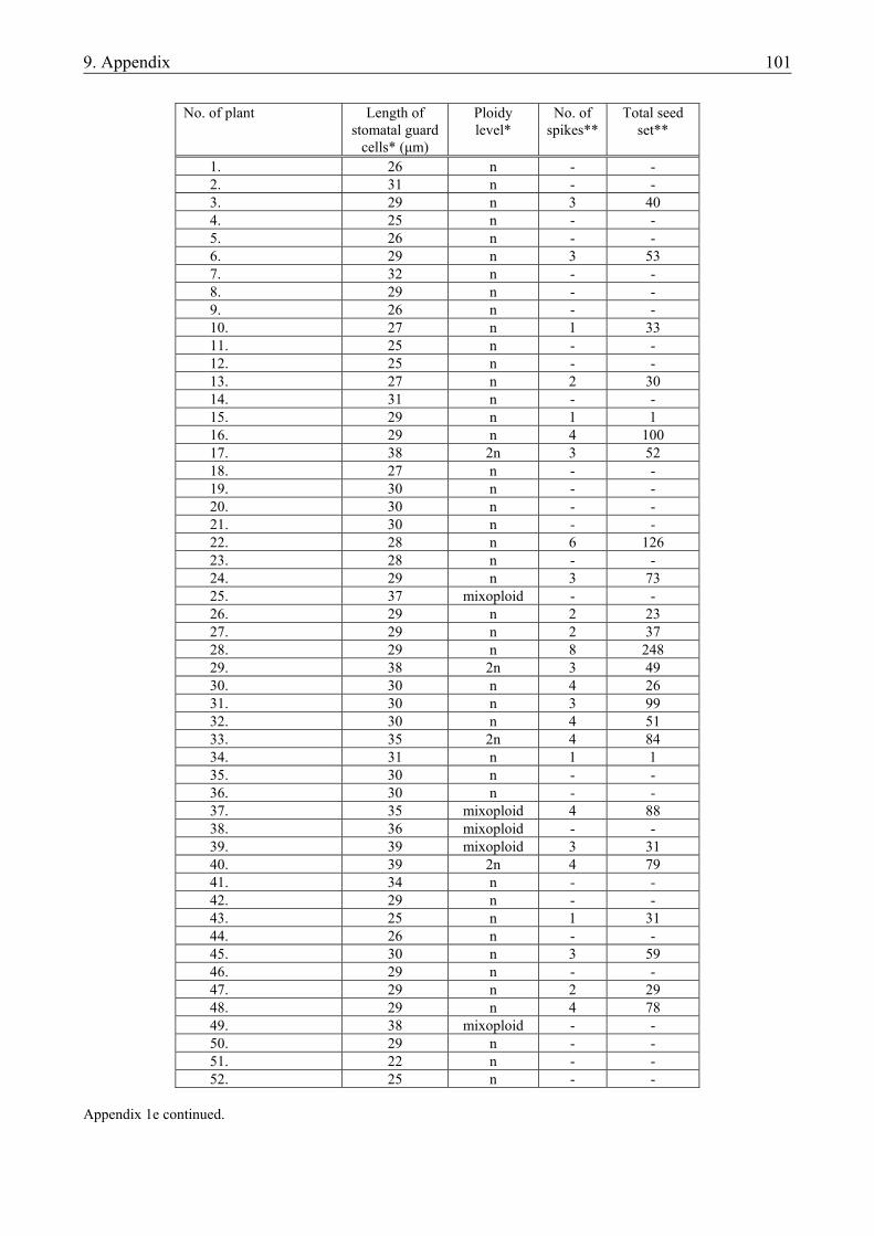
9. Appendix 101
No. of plant Length of
stomatal guard cells* (µm)
Ploidy level*
No. of spikes**
Total seed set**
1. 26 n - - 2. 31 n - - 3. 29 n 3 40 4. 25 n - - 5. 26 n - - 6. 29 n 3 53 7. 32 n - - 8. 29 n - - 9. 26 n - - 10. 27 n 1 33 11. 25 n - - 12. 25 n - - 13. 27 n 2 30 14. 31 n - - 15. 29 n 1 1 16. 29 n 4 100 17. 38 2n 3 52 18. 27 n - - 19. 30 n - - 20. 30 n - - 21. 30 n - - 22. 28 n 6 126 23. 28 n - - 24. 29 n 3 73 25. 37 mixoploid - - 26. 29 n 2 23 27. 29 n 2 37 28. 29 n 8 248 29. 38 2n 3 49 30. 30 n 4 26 31. 30 n 3 99 32. 30 n 4 51 33. 35 2n 4 84 34. 31 n 1 1 35. 30 n - - 36. 30 n - - 37. 35 mixoploid 4 88 38. 36 mixoploid - - 39. 39 mixoploid 3 31 40. 39 2n 4 79 41. 34 n - - 42. 29 n - - 43. 25 n 1 31 44. 26 n - - 45. 30 n 3 59 46. 29 n - - 47. 29 n 2 29 48. 29 n 4 78 49. 38 mixoploid - - 50. 29 n - - 51. 22 n - - 52. 25 n - -
Appendix 1e continued.
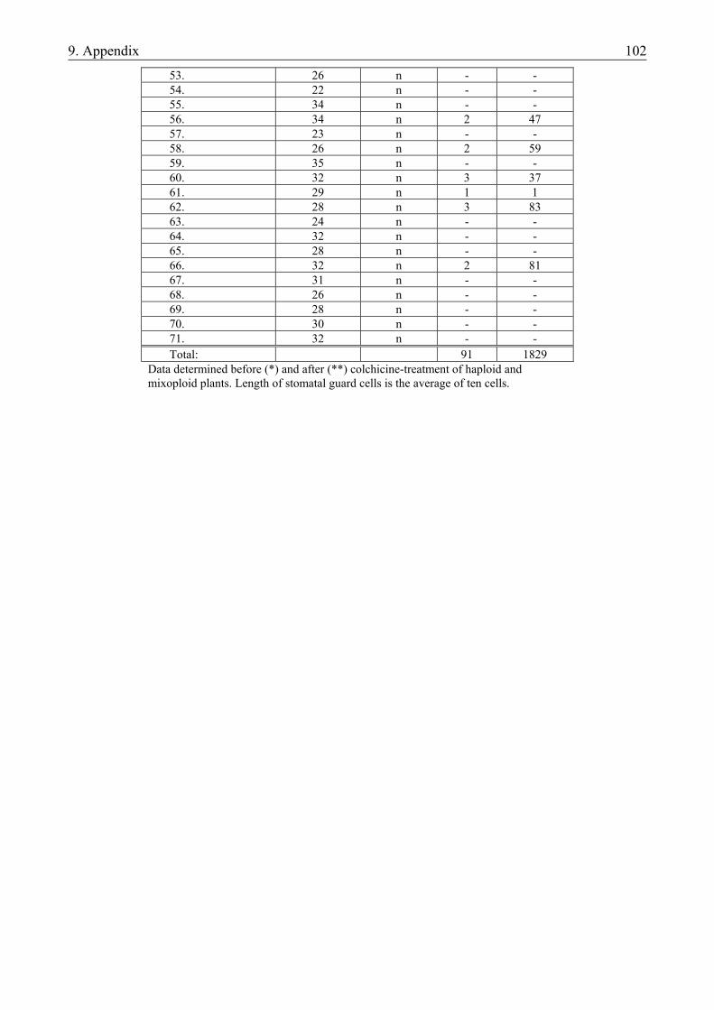
9. Appendix 102
53. 26 n - - 54. 22 n - - 55. 34 n - - 56. 34 n 2 47 57. 23 n - - 58. 26 n 2 59 59. 35 n - - 60. 32 n 3 37 61. 29 n 1 1 62. 28 n 3 83 63. 24 n - - 64. 32 n - - 65. 28 n - - 66. 32 n 2 81 67. 31 n - - 68. 26 n - - 69. 28 n - - 70. 30 n - - 71. 32 n - - Total: 91 1829
Data determined before (*) and after (**) colchicine-treatment of haploid and mixoploid plants. Length of stomatal guard cells is the average of ten cells.
9. Appendix 103
Appendix 2. Composition of basic media used in microspore cultures of barley and triticale
Component N24* 190-2** Macro elements (mgL-1) NH4Cl 144 (NH4)2SO4 200 KNO3 2457 1000 Ca(NO3)2 x 4H2O 100 CaCl2 x 2H2O 450 MgSO4 x 7H2O 350 200 KH2PO4 200 300 KCl 40 Na2EDTA 37 37 FeSO4 x 7H2O 28 28 Micro elements (mgL-1) MnSO4 x H2O 25.00 6.10 ZnSO4 x H2O 7.50 3.00 H3BO3 5.00 3.00 KI 0.75 0.50 Na2MoO4 x 2H2O 0.25 CuSO4 x 5H2O 0.025 CoCl2 x 6H2O 0.025 Vitamins (mgL-1) Myoinositol 100 100 Nicotinic acid 1 0.5 Pyridoxine HCl 1 0.5 Thiamine HCl 10 1 Amino acids (mgL-1) Glycine 2 Glutamine 222
* Mordhorst and Lörz, 1993 ** Zhuang and Jia, 1983
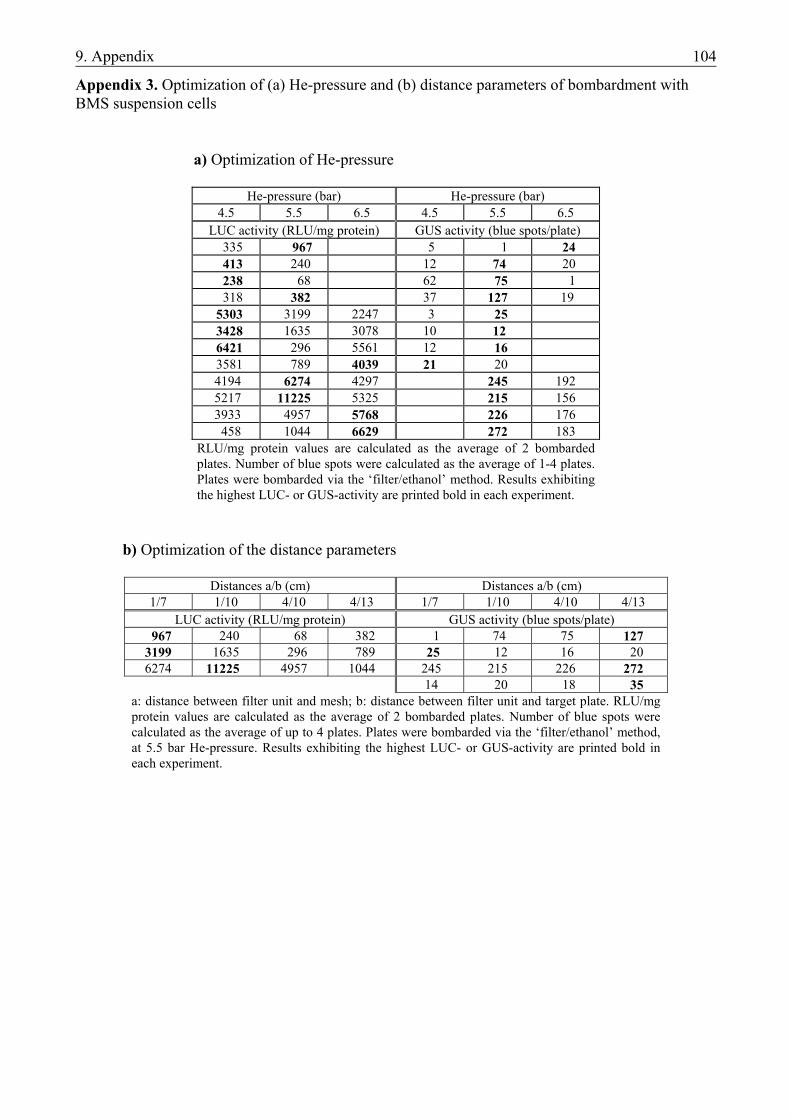
9. Appendix 104
Appendix 3. Optimization of (a) He-pressure and (b) distance parameters of bombardment with BMS suspension cells
a) Optimization of He-pressure
He-pressure (bar) He-pressure (bar)
4.5 5.5 6.5 4.5 5.5 6.5 LUC activity (RLU/mg protein) GUS activity (blue spots/plate) 335 967 5 1 24 413 240 12 74 20 238 68 62 75 1 318 382 37 127 19 5303 3199 2247 3 25 3428 1635 3078 10 12 6421 296 5561 12 16 3581 789 4039 21 20 4194 6274 4297 245 192 5217 11225 5325 215 156 3933 4957 5768 226 176 458 1044 6629 272 183
RLU/mg protein values are calculated as the average of 2 bombarded plates. Number of blue spots were calculated as the average of 1-4 plates. Plates were bombarded via the ‘filter/ethanol’ method. Results exhibiting the highest LUC- or GUS-activity are printed bold in each experiment.
b) Optimization of the distance parameters
Distances a/b (cm) Distances a/b (cm) 1/7 1/10 4/10 4/13 1/7 1/10 4/10 4/13
LUC activity (RLU/mg protein) GUS activity (blue spots/plate) 967 240 68 382 1 74 75 127 3199 1635 296 789 25 12 16 20 6274 11225 4957 1044 245 215 226 272
14 20 18 35 a: distance between filter unit and mesh; b: distance between filter unit and target plate. RLU/mg protein values are calculated as the average of 2 bombarded plates. Number of blue spots were calculated as the average of up to 4 plates. Plates were bombarded via the ‘filter/ethanol’ method, at 5.5 bar He-pressure. Results exhibiting the highest LUC- or GUS-activity are printed bold in each experiment.
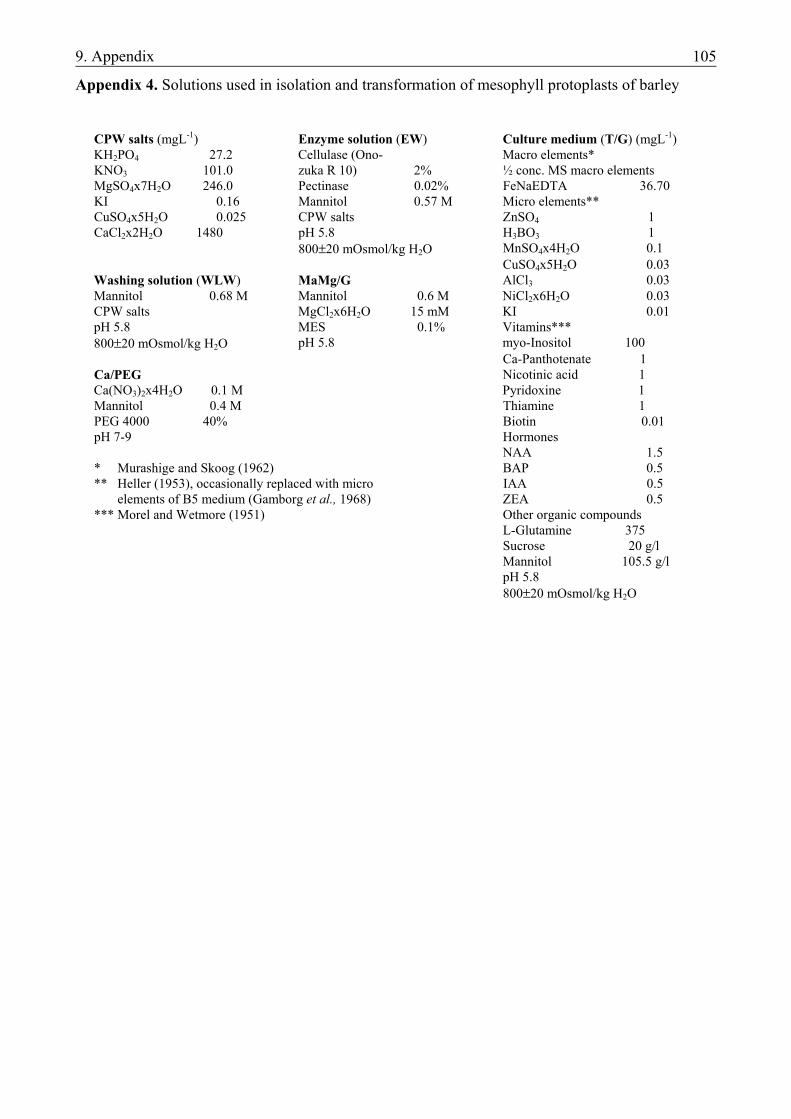
9. Appendix 105
Appendix 4. Solutions used in isolation and transformation of mesophyll protoplasts of barley
CPW salts (mgL-1) Enzyme solution (EW) Culture medium (T/G) (mgL-1) KH2PO4 27.2 Cellulase (Ono- Macro elements* KNO3 101.0 zuka R 10) 2% ½ conc. MS macro elements MgSO4x7H2O 246.0 Pectinase 0.02% FeNaEDTA 36.70 KI 0.16 Mannitol 0.57 M Micro elements** CuSO4x5H2O 0.025 CPW salts ZnSO4 1 CaCl2x2H2O 1480 pH 5.8 H3BO3 1 800±20 mOsmol/kg H2O MnSO4x4H2O 0.1 CuSO4x5H2O 0.03 Washing solution (WLW) MaMg/G AlCl3 0.03 Mannitol 0.68 M Mannitol 0.6 M NiCl2x6H2O 0.03 CPW salts MgCl2x6H2O 15 mM KI 0.01 pH 5.8 MES 0.1% Vitamins*** 800±20 mOsmol/kg H2O pH 5.8 myo-Inositol 100 Ca-Panthotenate 1 Ca/PEG Nicotinic acid 1 Ca(NO3)2x4H2O 0.1 M Pyridoxine 1 Mannitol 0.4 M Thiamine 1 PEG 4000 40% Biotin 0.01 pH 7-9 Hormones NAA 1.5 * Murashige and Skoog (1962) BAP 0.5 ** Heller (1953), occasionally replaced with micro IAA 0.5 elements of B5 medium (Gamborg et al., 1968) ZEA 0.5 *** Morel and Wetmore (1951) Other organic compounds L-Glutamine 375 Sucrose 20 g/l Mannitol 105.5 g/l pH 5.8 800±20 mOsmol/kg H2O
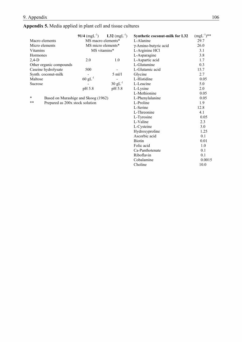
9. Appendix 106
Appendix 5. Media applied in plant cell and tissue cultures
91/4 (mgL-1) L32 (mgL-1) Synthetic coconut-milk for L32 (mgL-1)** Macro elements MS macro elements* L-Alanine 29.7 Micro elements MS micro elements* γ-Amino-butyric acid 26.0 Vitamins MS vitamins* L-Arginine HCl 3.1 Hormones L-Asparagine 3.8 2,4-D 2.0 1.0 L-Aspartic acid 1.7 Other organic compounds L-Glutamine 0.3 Caseine hydrolysate 500 - L-Glutamic acid 15.7 Synth. coconut-milk - 5 ml/l Glycine 2.7 Maltose 60 gL-1 - L-Histidine 0.05 Sucrose - 30 gL-1 L-Leucine 5.0 pH 5.8 pH 5.8 L-Lysine 2.0 L-Methionine 0.05 * Based on Murashige and Skoog (1962) L-Phenylalanine 0.05 ** Prepared as 200x stock solution L-Proline 1.9 L-Serine 12.8 L-Threonine 4.1 L-Tyrosine 0.05 L-Valine 2.3 L-Cysteine 3.0 Hydroxyproline 1.25 Ascorbic acid 0.1 Biotin 0.01 Folic acid 1.0 Ca-Panthotenate 0.1 Riboflavin 0.1 Cobalamine 0.0015 Choline 10.0
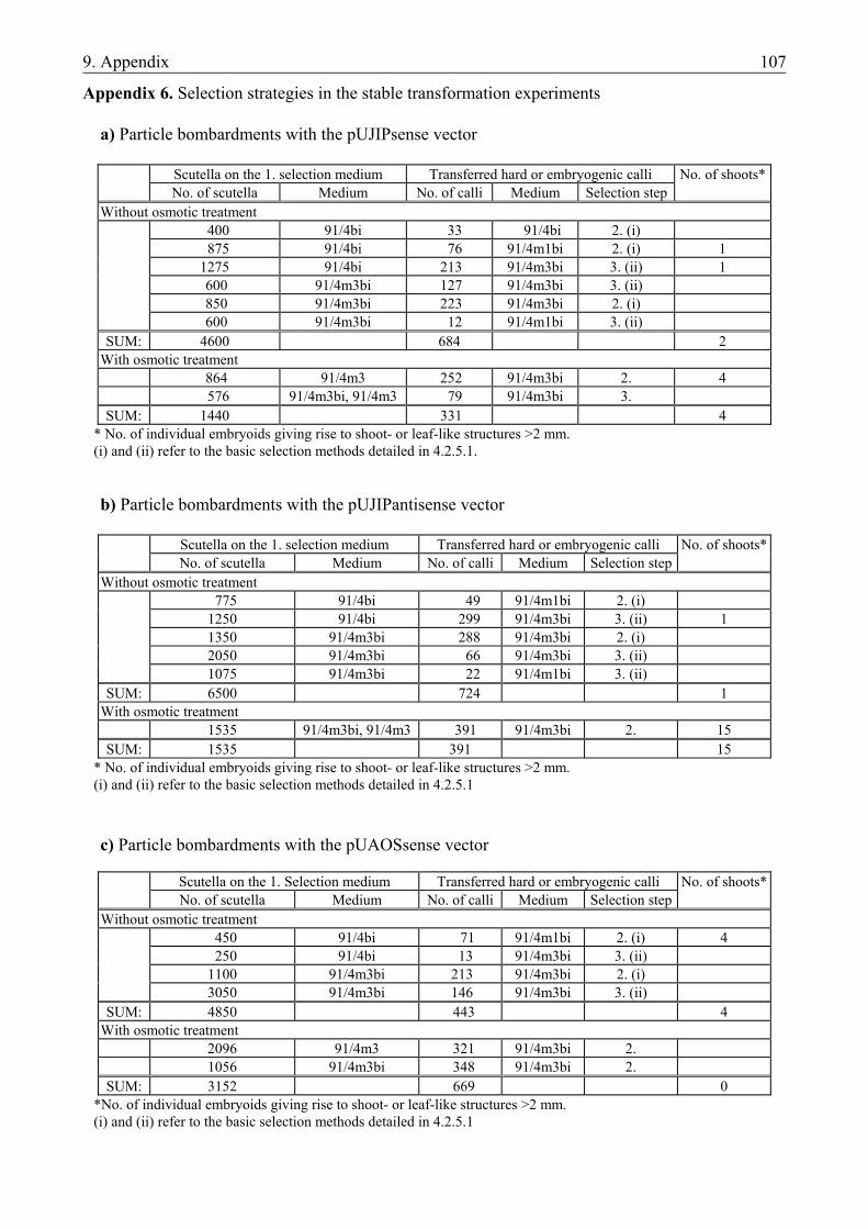
9. Appendix 107
Appendix 6. Selection strategies in the stable transformation experiments
a) Particle bombardments with the pUJIPsense vector
Scutella on the 1. selection medium Transferred hard or embryogenic calli No. of shoots* No. of scutella Medium No. of calli Medium Selection step
Without osmotic treatment 400 91/4bi 33 91/4bi 2. (i) 875 91/4bi 76 91/4m1bi 2. (i) 1 1275 91/4bi 213 91/4m3bi 3. (ii) 1 600 91/4m3bi 127 91/4m3bi 3. (ii) 850 91/4m3bi 223 91/4m3bi 2. (i) 600 91/4m3bi 12 91/4m1bi 3. (ii)
SUM: 4600 684 2 With osmotic treatment
864 91/4m3 252 91/4m3bi 2. 4 576 91/4m3bi, 91/4m3 79 91/4m3bi 3.
SUM: 1440 331 4 * No. of individual embryoids giving rise to shoot- or leaf-like structures >2 mm. (i) and (ii) refer to the basic selection methods detailed in 4.2.5.1. b) Particle bombardments with the pUJIPantisense vector
Scutella on the 1. selection medium Transferred hard or embryogenic calli No. of shoots* No. of scutella Medium No. of calli Medium Selection step
Without osmotic treatment 775 91/4bi 49 91/4m1bi 2. (i) 1250 91/4bi 299 91/4m3bi 3. (ii) 1 1350 91/4m3bi 288 91/4m3bi 2. (i) 2050 91/4m3bi 66 91/4m3bi 3. (ii) 1075 91/4m3bi 22 91/4m1bi 3. (ii)
SUM: 6500 724 1 With osmotic treatment
1535 91/4m3bi, 91/4m3 391 91/4m3bi 2. 15 SUM: 1535 391 15
* No. of individual embryoids giving rise to shoot- or leaf-like structures >2 mm. (i) and (ii) refer to the basic selection methods detailed in 4.2.5.1 c) Particle bombardments with the pUAOSsense vector
Scutella on the 1. Selection medium Transferred hard or embryogenic calli No. of shoots* No. of scutella Medium No. of calli Medium Selection step
Without osmotic treatment 450 91/4bi 71 91/4m1bi 2. (i) 4 250 91/4bi 13 91/4m3bi 3. (ii) 1100 91/4m3bi 213 91/4m3bi 2. (i) 3050 91/4m3bi 146 91/4m3bi 3. (ii)
SUM: 4850 443 4 With osmotic treatment
2096 91/4m3 321 91/4m3bi 2. 1056 91/4m3bi 348 91/4m3bi 2.
SUM: 3152 669 0 *No. of individual embryoids giving rise to shoot- or leaf-like structures >2 mm. (i) and (ii) refer to the basic selection methods detailed in 4.2.5.1
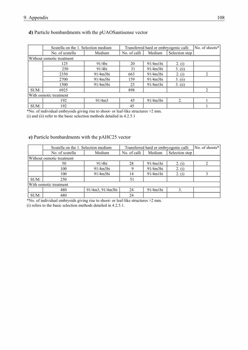
9. Appendix 108
d) Particle bombardments with the pUAOSantisense vector
Scutella on the 1. Selection medium Transferred hard or embryogenic calli No. of shoots* No. of scutella Medium No. of calli Medium Selection step
Without osmotic treatment 125 91/4bi 20 91/4m1bi 2. (i) 250 91/4bi 31 91/4m3bi 3. (ii) 2350 91/4m3bi 663 91/4m3bi 2. (i) 2 2700 91/4m3bi 159 91/4m3bi 3. (ii) 1500 91/4m3bi 25 91/4m1bi 3. (ii)
SUM: 6925 898 2 With osmotic treatment
192 91/4m3 45 91/4m3bi 2. 1 SUM: 192 45 1
*No. of individual embryoids giving rise to shoot- or leaf-like structures >2 mm. (i) and (ii) refer to the basic selection methods detailed in 4.2.5.1 e) Particle bombardments with the pAHC25 vector
Scutella on the 1. Selection medium Transferred hard or embryogenic calli No. of shoots* No. of scutella Medium No. of calli Medium Selection step
Without osmotic treatment 50 91/4bi 28 91/4m1bi 2. (i) 2 100 91/4m3bi 9 91/4m3bi 2. (i) 100 91/4m3bi 14 91/4m1bi 2. (i) 3
SUM: 250 51 With osmotic treatment
480 91/4m3, 91/4m3bi 24 91/4m1bi 3. SUM: 480 24
*No. of individual embryoids giving rise to shoot- or leaf-like structures >2 mm. (i) refers to the basic selection methods detailed in 4.2.5.1.
Acknowledgements 109
ACKNOWLEDGEMENTS
First of all, I owe thanks to my supervisors in the two research topics reported in this thesis. Dr. Bettina Hause (IPB, Halle), whose name I could not write on the second page of this work for administrative reasons, I thank for offering me the challenging research theme of barley transformation, for her help in organizing my life in Halle, for her help in the preparation of Western-blots and for her critical comments on the manuscript. I thank Dr. János Pauk (CRI, Szeged) for the long years of thorough supervision over my experiments with microspores, for the possibility to work in his lab, for offering me publication opportunities and for the regular stimulating discussions. I would also like to thank Dr. Beáta Barnabás, Dr. Barnabás Jenes, Dr. Ferenc Sági and Prof. Dr. Claus Wasternack for their constructive criticism while reviewing my thesis. I am grateful to Ágnes Fejérvári, Orsolya Kiss and Krisztina Talpas (CRI, Szeged) for their indispensable help in the preparation of culture media and taking care of the microspore cultures as well as for the friendly labor atmosphere. I also thank Dr. Klára Tóth-Lökös (Szent István University, Gödöllő) for her enormous help in the statistical analysis of the triticale experiments. My special thanks go to Dr. Helmut Maucher for introducing me to molecular biological methods and always being ready to answer my numerous ‘technical’ questions. Silvia Wegener I thank for helping me to find everything in the lab, in the building of IPB and in Halle. I thank Regina Weiss for DNA sequencing. I would also like to thank Dr. Jutta Schulze, Dr. Vijendra K. Sharma and Dr. Robert Hänsch (TU, Braunschweig) for the continuous discussion about tissue culture and particle bombardment, for the plasmids and BMS suspension stock culture and for preparing the PAT-assays reported in this work. Furthermore, I would like to express my gratitude to those members of CRI and IPB whose useful comments contributed to the success of my work: Dr. János Matuz, Dr. Zoltán Kertész, Dr. Irene Stenzel, Dr. Jon Page, Dr. Stephan Bau, Dr. Magdalena Krzymowska, Dr. Sándor Mórocz and Dr. Béla Szarka. I thank Prof. Dr. László Heszky (Szent István University, Gödöllő) for his useful suggestions and for giving a comprehensive overview of ‘molecular plant breeding’. I also thank the current and former directors of the College of Agriculture in Hódmezővásárhely and the current and former heads of the Dept. of Plant Production and Horticulture, Dr. Gyula Bicsérdy, Dr. Imre Molnár, Prof. Dr. Imre Mucsi, Prof. Dr. József Frank and Prof. Dr. György Sinkovics, for supporting my Ph.D. studies and my research. The research performed at the IPB, Halle was supported by the DFG project “The role of jasmonates in the development of barley” - thank you. I am indebted forever to my parents for supporting my studies and encouraging me in every field of life. And last but not least, I owe my biggest thanks to my wife, Orsi, and to my daughter, Eszter, for their sympathy, mental support, the warm family atmosphere, for putting up with my two-year absence due to working abroad and for everything in general.
Acknowledgements 110
SUMMARY
MICROSPORE CULTURE AND GENETIC TRANSFORMATION STUDIES IN BARLEY
AND TRITICALE
MONOSTORI TAMÁS
Microspore cultures are optimal objects for biotechnological and plant breeding studies of practical
aspect. Besides, the decisive factors of haploid embryogenesis can be studied here in a
synchronized unicellular system. In the first part of the current work, the protocol for the
regeneration of haploid/dihaploid plants from isolated microspores of triticale is described. The
method was established in experiments with five genotypes and it is suitable for the application in
plant breeding. Its further practical advantage is that through the microspore isolation method used
here (microblending of spikes), the laboursome process of anther isolation can be avoided. In
further studies, it was observed that the induction of haploid embryogenesis does not require
exogenous hormone-supplement neither in triticale nor in barley microspore culture. It is the first
description in unicellular systems of these two monocot species, that hormones are not essential
exogenous factors required for the induction of haploid embryogenesis. Thus, these results suggest
that some critical factors of androgenesis (e.g. stress required for the induction of embryogenesis)
are of general validity irrespective of species and genotype, while others (e.g. hormone-requirement
of plant regeneration, optimal composition of media) are specific for species and genotype.
Barley can be regarded as a model system of studies on the role of the jasmonates and jasmonate-
induced proteins (JIPs) in monocots. Our current knowledge on the role of jasmonates and JIPs in
monocots is based on treatment with JAs or on stress-induced JA-formation. In the second part of
the current work, the prerequisites were prepared for further transgenic studies on the role of
jasmonates in the development of barley. Novel plasmid vector constructs have been prepared,
which contain the barley AOS1 or JIP23 cDNAs in sense or antisense orientation, respectively,
under the control of the Ubi-1 promoter. Based on the same construct, vectors have been prepared,
which contain a multiple cloning site (MCS) in sense or antisense orientation, respectively,
downstream of the Ubi-1 promoter. The cloning site includes restriction sites for at least ten
enzymes. Thus, these vectors can be used for the cloning (and expression) of further genes/cDNAs.
The functionality of the novel vector constructs has been revealed by the transient expression of the
pat resistance marker gene and that of the JIP23 cDNS in barley mesophyll protoplasts transformed
with the sense vector constructs. Thus, it is suggested that the AOS and JIP23 sense and antisense

Acknowledgements 111
vector constructs are suitable for the modification of endogenous jasmonate and JIP23 levels in
barley an other cereals via genetic transformation.
Related Documents