Microsoft PowerPoint - core_course_lecture
Aug 20, 2015
Welcome message from author
This document is posted to help you gain knowledge. Please leave a comment to let me know what you think about it! Share it to your friends and learn new things together.
Transcript


Radiations Used in Medical RadiologyRadiations Used in Medical Radiology
Positrons
Electrons
MRIGamma Rays
Ultrasound X Rays
Non-ionizing RadiationsIonizing Radiations
What are the potential risks?
Induced neoplasmInjuryMaldevelopment during pregnancyGenetically heritable effects
(malformation, disease)

From: MacKenzie I. Breast cancer following multiple fluoroscopies. Br J Cancer 1965; 19: 1-8

How bad is the risk from exposure to X rays?
Induced Neoplasm – risk increases as dose increases and not ever known to be zero. Principle for safe practice is ALARA.
Injury – requires a minimum dose to be induced. Risk is zero under this minimum. Over it risk is substantial and severity increases as dose goes up.
Prenatal – Risk depends on gestation age and dose.
Heritable – risk increases as dose increases and not ever known to be zero. Principle for safe practice is ALARA.


What is a DOSE of radiation?Dose is defined as the energy deposited in tissue
per unit mass of tissue. Physically, dose conveys the magnitude of biochemical disruption that occurs from an exposure, which in turn is related to health risk.
Units of dose are milligray (mGy) or millisievert(mSv), where for our purposes milligray and miilisievert are the same.
Note: if dose is given in rad or mrad, divide the rad-type dose by 100 to get the gray-type dose. That is:
1 rad = 0.01 Gy and 1 mrad = 0.01 mGy

How bad is the risk from exposure to X rays?
NonexistentTrivialSmallModestConsiderableSevere

RISK DEPENDS ON THE DOSE!!
Two types of Dose ----
Tissue dose --- used to assess the risk for injury to specific exposed tissues.
Effective dose ---- used to assess risk for neoplastic development and heritable genetic effects. (Converts tissue dose to whole-body dose of effectively the same risk.)

Annual Human Exposure to Ionizing Radiation
Source EffectiveWhole-Body
Dose
Naturally existing radiations……...…….. 3 mSvMedical diagnosis …………………….....~1 mSvConsumer products …….....................0.12 mSvMiscellaneous (e.g., nuclear power) …0.01mSv
Lesson: Medicine is responsible for the greatest exposures of the population to man-made radiation.


British Radiologists vs British Male Medical Practitioners SMR
0.711.121.241.75*Cancer
0.68*1.000.920.97All Causes
1955 -1979
1936 –1954
1921 –1935
1897 –1920
Cause of Death
Significant at the p < 0.001 level

Cancer Deaths from 1950 to 1985 in those exposed at Hiroshima and Nagasaki
0 (0%)1,693 (7.3%)1,702 (7.3%)23,32110 - 100
131 (1.1%)987 (8.4%)856 (7.3%)11,730100 – 500
268 (4.0%)755 (11.3%)487 (7.3%)6,668>500
Excess Deaths
Ca. Deaths in Exposed
Ca. Deaths in Controls
Exposed Population
Dose (mGy)
Shimiziu Y, et al. Life Span Study Report II. Part I. RERF; 1987:8.

3 months0.7 mSvMammography
3 years8 mSvComputed Tomography (CT)-Chest
10 days0.1 mSvRadiography-Chest
8 months2 mSvComputed Tomography (CT)-Head
8 months2 mSvRadiography-Upper GI Tract16 months4 mSvRadiography-Lower GI Tract
6 months1.6 mSvIntravenous Pyelogram(IVP)
3 years10 mSvComputed Tomography (CT)-Body
3 years10 mSvComputed Tomography (CT)-Abdomen
Comparable to natural background radiation for:
Your effective radiation dose is:For this procedure:

Conclusions regarding diagnostic examinations and risk
• For individual diagnostic studies in adults, the effective dose is below the level at which there is data suggesting that X rays induce neoplasms.
• This is not a license for a cavalier attitude!• Practice is to manage radiation exposure to
levels that are necessary for diagnosis and no more (ALARA)


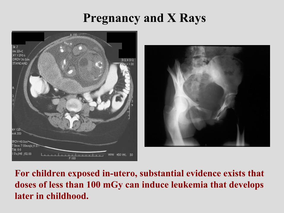
Pregnancy and X Rays
For children exposed in-utero, substantial evidence exists that doses of less than 100 mGy can induce leukemia that develops later in childhood.
Pregnancy and radiation management
For diagnostic doses most likely risk is neoplasm
Dose from studies outside abdomen are due only to scatter, which is trivial.
Only X-rays of the abdomen/pelvis have the potential to produce a substantive dose to the conceptus
Malformation (and induced abortion for early pregnancy) not a concern unless doses exceed 5 – 10 rad
Doses seldom reach 5 – 10 rad.

Cancer after In-utero Irradiation in Humans
0 - 5---<40A-bomb
----0.8 - 2.5<16Twin Pelvimetry
2 - 101.56<15Pelvimetry
Excess in 10,000
exposed to 10 mGy
Relative RiskAges at Risk (yrs)
Study Group
Implication: Expose 10,000 concepti of pregnant women to a dose of 10 mGy and you might induce 0 –10 caners in the offspring.


Heritable EffectsHeritable EffectsHeritable EffectsReproductive risks in humans never definitively
demonstrated
Gonad shields used to protect gene pool, not individual
Shields often not used because obstruct necessary anatomy
Gonad shields difficult to position in girls
Dose to gonads from 3 well performed AP pelvic x-rays in child on order of 1 – 5 mSv, background radiation is more


Diagnostic versus complex interventional uses of x rays
Diagnosis – the intent is to obtain medical information to direct the course of medical care. Radiation risk must be very small.
Simple intervention – the intent is to treat a condition that is not usually life threatening. Radiation risks are typically small.
Complex intervention – the intent is to treat a serious condition with often life-saving results. In this case, much higher radiation risks are acceptable if required by the procedure.

Coronary Angioplasty
TIPS placementRadiofrequency Ablation
NeuroembolizationUterine embolization Renal angioplasty
Types of procedures associated with severe injuries
Courtesy: Shope, FDA
Vañó, Br J Radiol1998; 71, 510 - 516
Nahass et al, Am J Gastroent1998; 93: 1546-9
Dandurand et al, Ann DermVener 1999; 126: 413-417
Courtesy F Mettler MD
35

Face shield
65 cm
55 cm
45 cm2.1 Dose Units - Grid in1.4 Dose Units - Grid out
1.0 Dose UnitsGrid in
35 cm
Leg Shie ld

• Use patient as a shield
• Keep hands out of beam
• Wear a hand dosimeter
Rules to protect hands while performing fluoroscopic procedures

Protection of personnel
1. Maximize distance from source (Scatter at 2 meters is < 0.1% of what patient gets but it is cumulative.)
2. Use a shield3. Use radiation sparingly


IMPORTANT INFO ABOUT BENEFIT AND RISK WITH MEDICAL X RAYS!
The doses to patients from medical x rays range from the trivially small to the extremely large.
Doses from medical uses must always be justified by the benefits.
For simple diagnoses of non-critical health risks the doses must be very small.
For life-saving interventional work the doses might be very large and might cause skin injury that could be severe.
Users of radiation must be trained in the safe applications of these radiations for the protection of both the patient and the users.
Related Documents


](https://static.cupdf.com/doc/110x72/577cc1011a28aba71191ebaa/11040-microsoft-powerpoint-11040-microsoft-powerpoint-securtization-compatibility.jpg)