Microscopy Microscopy Chapter 3 Chapter 3 Biology Biology Mr. Mr. Gilbertson Gilbertson

Microscopy Chapter 3 Biology Mr. Gilbertson. DISCOVERY OF CELL PRECEDED BY THE INVENTION OF THE MICROSCOPE ROBERT HOOKE - NAMED “CELLS” BECAUSE THEY LOOKED.
Dec 26, 2015
Welcome message from author
This document is posted to help you gain knowledge. Please leave a comment to let me know what you think about it! Share it to your friends and learn new things together.
Transcript
MicroscopyMicroscopy
Chapter 3Chapter 3
BiologyBiology
Mr. Gilbertson Mr. Gilbertson
DISCOVERY OF CELLDISCOVERY OF CELL• PRECEDED BY THE INVENTION OF THE
MICROSCOPE
• ROBERT HOOKE - NAMED “CELLS” BECAUSE THEY LOOKED LIKE LITTLE ROOMS FOUND IN A MONASTERY ABOUT 1665– LOOKING AT CORK BARK FROM MEDITERRANEAN
CORK OAK– ALSO OBSERVED STEMS OF ELDER, CARROT, AND FERN– ALL WERE FOUND TO BE SIMILAR– ACTUALLY HE OBSERVED ONLY DEAD CELLS
• ANTOINE VAN LEEWENHOEK - – DUTCH LENS AND MICROSCOPE MAKER– FIRST TO OBSERVE LIVING CELLS ABOUT 1675
Looking at CellsLooking at Cells
• Cells are too small to be viewed with the naked Cells are too small to be viewed with the naked eye. (most smaller than a grain of sand)eye. (most smaller than a grain of sand)
• Requires the use of a microscope to view.Requires the use of a microscope to view.• Measurement is made using the SI system.Measurement is made using the SI system.
– Based on powers of ten – like the money systemBased on powers of ten – like the money system
– Uses common prefixes – makes conversion easyUses common prefixes – makes conversion easy
– Truly international systemTruly international system
• Micrometer is the unit used for cell size (one Micrometer is the unit used for cell size (one millionth of a meter, 1000millionth of a meter, 1000thth of a mm) of a mm)
MicroscopesMicroscopes• Light microscopesLight microscopes – also known as compound – also known as compound
microscopesmicroscopes– Use light and lenses to focus images of very small Use light and lenses to focus images of very small
objects.objects.
– Maximum magnification about 2000XMaximum magnification about 2000X
• Electron MicroscopesElectron Microscopes– Use a beam of electrons focused by magnetic fields to Use a beam of electrons focused by magnetic fields to
form images of objects.form images of objects.
– Two types – Two types – • TEMTEM (transmission electron microscope) mag. up to 200,000X (transmission electron microscope) mag. up to 200,000X
• SEMSEM (scanning electron microscope) max mag 50,000X (scanning electron microscope) max mag 50,000X
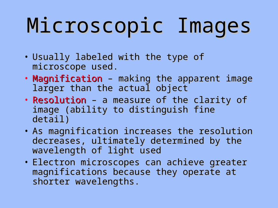
Microscopic ImagesMicroscopic Images• Usually labeled with the type of microscope used.Usually labeled with the type of microscope used.• MagnificationMagnification – making the apparent image larger – making the apparent image larger
than the actual objectthan the actual object• Resolution Resolution – a measure of the clarity of image – a measure of the clarity of image
(ability to distinguish fine detail)(ability to distinguish fine detail)• As magnification increases the resolution As magnification increases the resolution
decreases, ultimately determined by the decreases, ultimately determined by the wavelength of light usedwavelength of light used
• Electron microscopes can achieve greater Electron microscopes can achieve greater magnifications because they operate at shorter magnifications because they operate at shorter wavelengths.wavelengths.
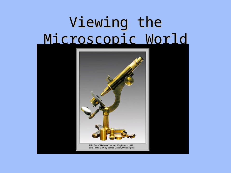
Viewing the Microscopic WorldViewing the Microscopic World
The Simple MicroscopeThe Simple Microscope
• Hand lens or a magnifying glass is a simple Hand lens or a magnifying glass is a simple microscope.microscope.
• Used to magnify, allows us to see fine details of Used to magnify, allows us to see fine details of structurestructure
• Power of Magnification is the number of times the Power of Magnification is the number of times the apparent size of an object is increased. (10X – ten apparent size of an object is increased. (10X – ten times)times)
• Focusing – the process of moving the position of Focusing – the process of moving the position of the lenses to bring the image into clear focusthe lenses to bring the image into clear focus
The Compound The Compound Light MicroscopeLight Microscope
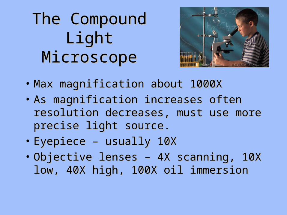
• Max magnification about 1000XMax magnification about 1000X
• As magnification increases often resolution As magnification increases often resolution decreases, must use more precise light decreases, must use more precise light source.source.
• Eyepiece – usually 10XEyepiece – usually 10X
• Objective lenses – 4X scanning, 10X low, Objective lenses – 4X scanning, 10X low, 40X high, 100X oil immersion40X high, 100X oil immersion
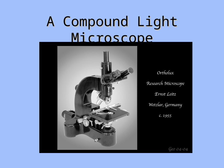
A Compound Light MicroscopeA Compound Light Microscope
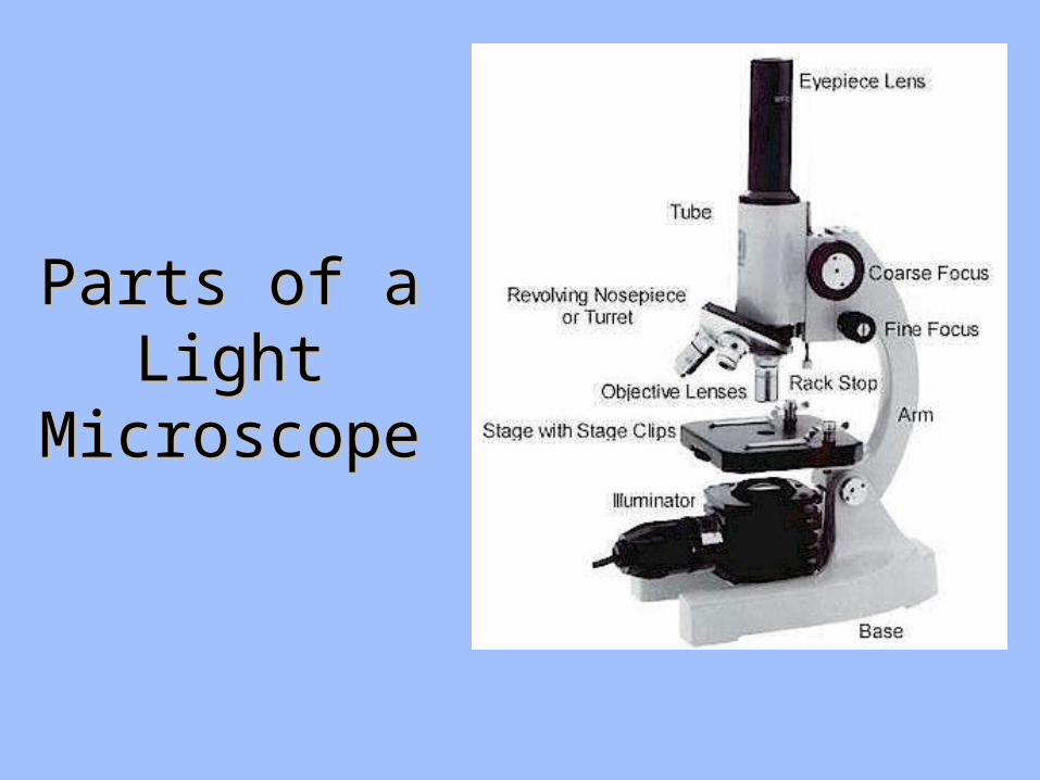
Parts of a Parts of a Light Light
MicroscopeMicroscope
Parts of a Light MicroscopeParts of a Light Microscope
• Light trainLight train is made up of the condenser, is made up of the condenser, diaphragm, slide, specimen, objective, body tube, diaphragm, slide, specimen, objective, body tube, and eyepiece.and eyepiece.
• Coarse adjustmentCoarse adjustment, brings the sample into basic , brings the sample into basic focus.focus.
• Fine adjustmentFine adjustment brings sample into clear focus brings sample into clear focus (hopefully).(hopefully).
• Always start out on low power and work up.Always start out on low power and work up.• To view sample under oil immersion, oil must be To view sample under oil immersion, oil must be
used between objective and cover slip.used between objective and cover slip.
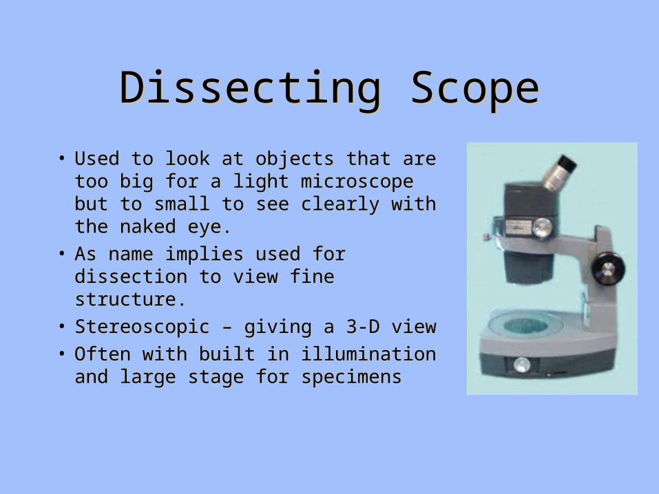
Dissecting ScopeDissecting Scope
• Used to look at objects that are too Used to look at objects that are too big for a light microscope but to big for a light microscope but to small to see clearly with the naked small to see clearly with the naked eye.eye.
• As name implies used for As name implies used for dissection to view fine structure.dissection to view fine structure.
• Stereoscopic – giving a 3-D viewStereoscopic – giving a 3-D view• Often with built in illumination Often with built in illumination
and large stage for specimensand large stage for specimens
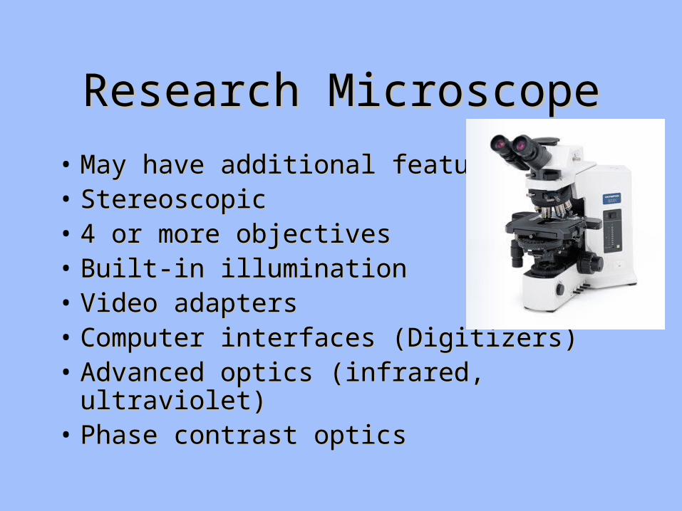
Research MicroscopeResearch Microscope
• May have additional featuresMay have additional features• Stereoscopic Stereoscopic • 4 or more objectives4 or more objectives• Built-in illuminationBuilt-in illumination• Video adaptersVideo adapters• Computer interfaces (Digitizers)Computer interfaces (Digitizers)• Advanced optics (infrared, ultraviolet)Advanced optics (infrared, ultraviolet)• Phase contrast opticsPhase contrast optics

Electron MicroscopesElectron Microscopes• Uses an electron beam and detectors to Uses an electron beam and detectors to
form an image of the specimen.form an image of the specimen.• Must be fixed (immovable) dead and Must be fixed (immovable) dead and
coated with a reflective material (gold) in coated with a reflective material (gold) in a vacuuma vacuum
• Can magnify up to 2 million times.Can magnify up to 2 million times.• TEM – Transmission Electron TEM – Transmission Electron
MicroscopeMicroscope• SEM – Scanning Electron MicroscopeSEM – Scanning Electron Microscope• STM – Scanning Tunneling MicroscopeSTM – Scanning Tunneling Microscope
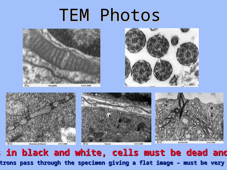
TEM PhotosTEM Photos
Always in black and white, cells must be dead and fixedAlways in black and white, cells must be dead and fixedElectrons pass through the specimen giving a flat image – must be very thin.Electrons pass through the specimen giving a flat image – must be very thin.
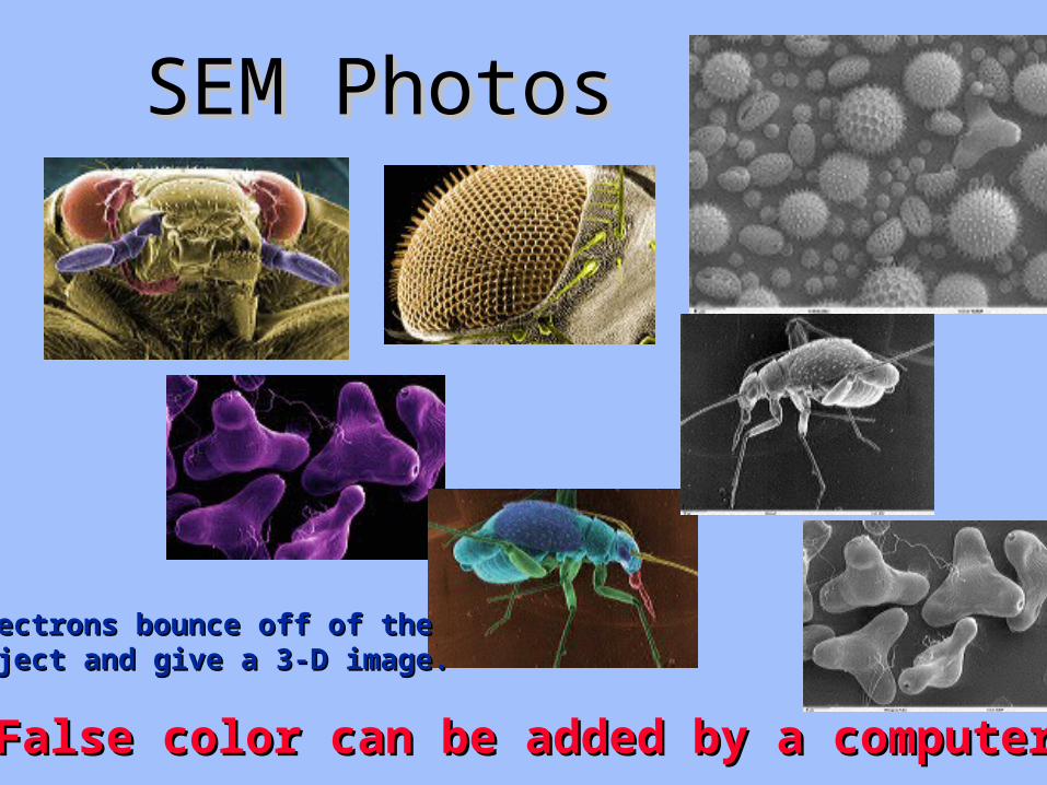
SEM PhotosSEM Photos
False color can be added by a computerFalse color can be added by a computer
Electrons bounce off of the Electrons bounce off of the Object and give a 3-D image.Object and give a 3-D image.
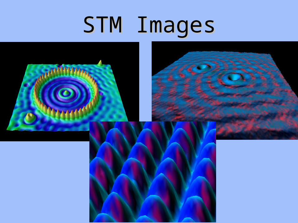
STM ImagesSTM Images
Related Documents