MICROSCOPIC ANATOMY OF BONE FIGURE 5.3 PAGE 116

Welcome message from author
This document is posted to help you gain knowledge. Please leave a comment to let me know what you think about it! Share it to your friends and learn new things together.
Transcript

MICROSCOPIC ANATOMY OF BONE
FIGURE 5.3 PAGE 116
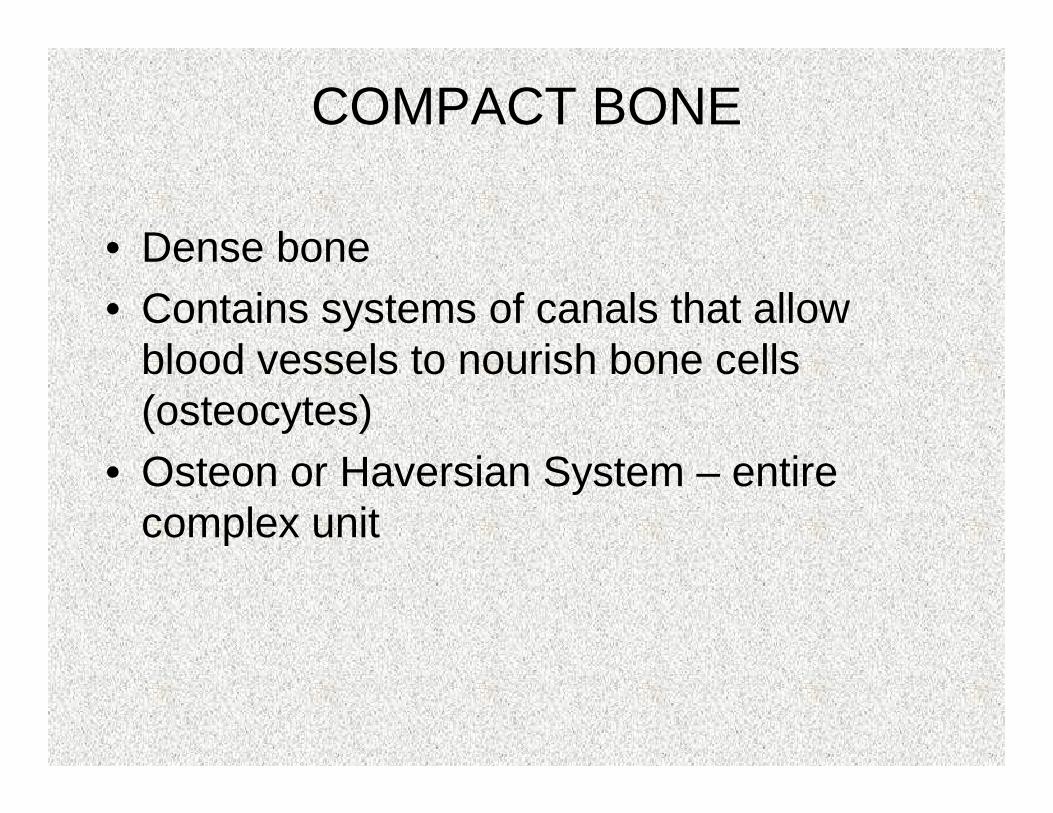
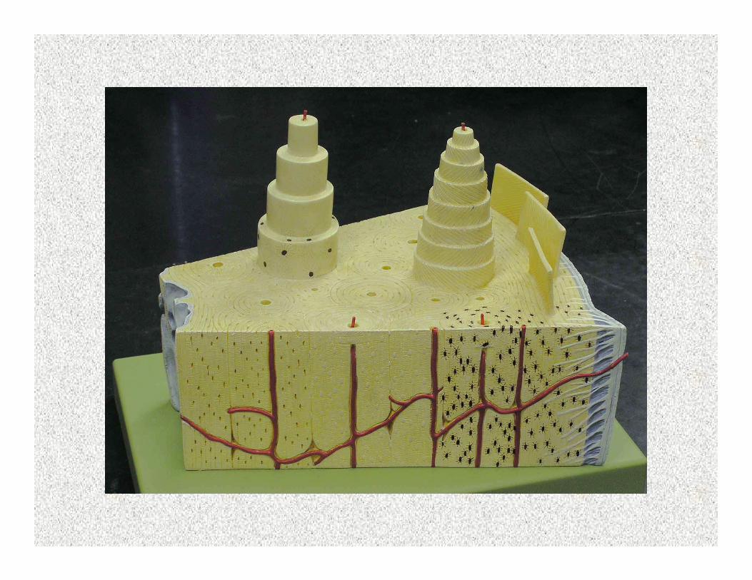
COMPACT BONE
• Dense bone• Contains systems of canals that allow
blood vessels to nourish bone cells (osteocytes)
• Osteon or Haversian System – entire complex unit


STRUCTURE
• Lamellae – “little layers” ; concentric rings formed by lacunae
• Lacunae – “little lakes”; cavity containing osteocyte
• Canaliculi – “little channels”; outward canals that lead to all lacunae ensuring all osteocytes are nourished
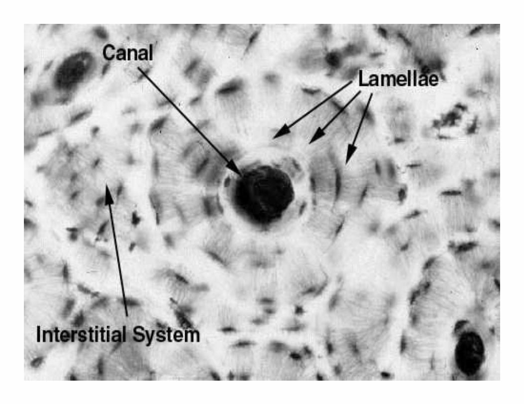
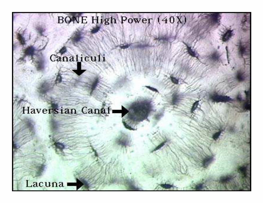

• Haversian Canal – run longitudinally through the center of the osteon
• Volkmann’s Canal – run horizontally connecting each osteon
• Both contain blood vessels which nourish the bone cells
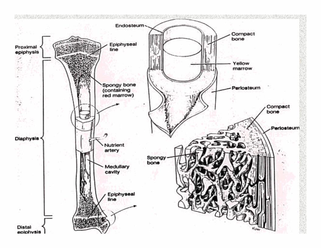
SPONGY BONE(CANCELLOUS BONE)
• Porous• Most abundant in short, flat & irregular
bones• Consists of interconnecting rods called
trabeculae• Support bone & store marrow• Cavities that contain osteoblasts –
immature bone cells

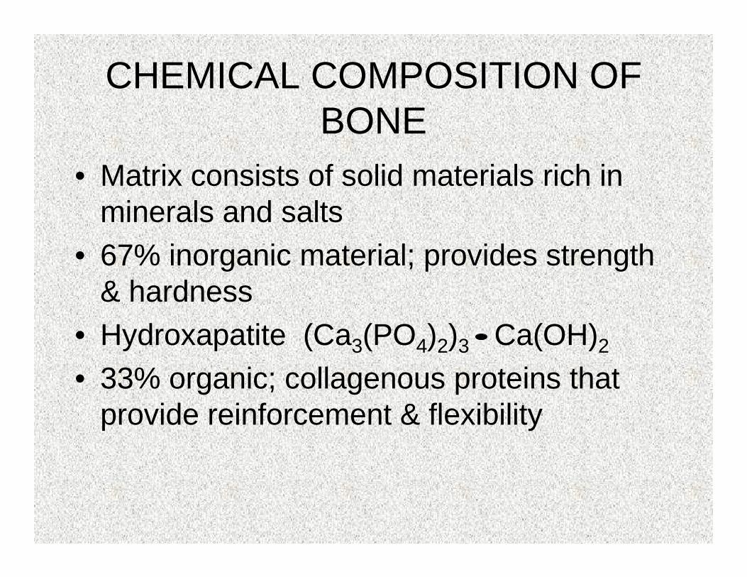
CHEMICAL COMPOSITION OF BONE
• Matrix consists of solid materials rich in minerals and salts
• 67% inorganic material; provides strength & hardness
• Hydroxapatite (Ca3(PO4)2)3 Ca(OH)2
• 33% organic; collagenous proteins that provide reinforcement & flexibility

OSSIFICATION
• Formation of new bone from hyaline cartilage
• Cartilage model is covered with bone matrix by the osetoblast
• Hyaline model is digested away opening up the medullary canal
• Digesting occurs in all areas except on ends of bone & growth plates

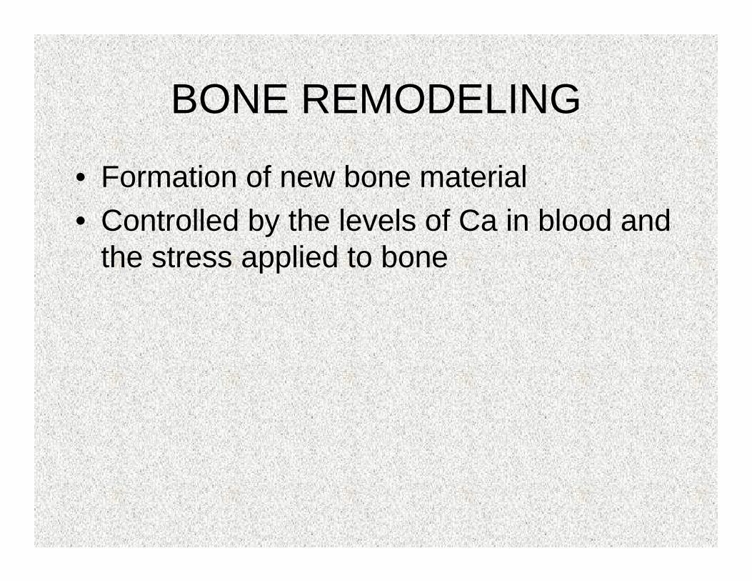
BONE REMODELING
• Formation of new bone material• Controlled by the levels of Ca in blood and
the stress applied to bone
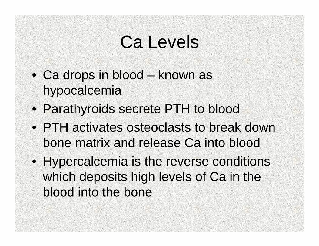
Ca Levels
• Ca drops in blood – known as hypocalcemia
• Parathyroids secrete PTH to blood• PTH activates osteoclasts to break down
bone matrix and release Ca into blood• Hypercalcemia is the reverse conditions
which deposits high levels of Ca in the blood into the bone
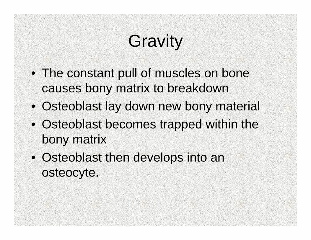
Gravity
• The constant pull of muscles on bone causes bony matrix to breakdown
• Osteoblast lay down new bony material• Osteoblast becomes trapped within the
bony matrix• Osteoblast then develops into an
osteocyte.
Related Documents