RESEARCH ARTICLE Microsaccades during reading Norick R. Bowers 1 , Martina Poletti 2¤ * 1 Vision Science, University of California, Berkeley, CA, United States of America, 2 Department of Psychological and Brain Sciences, Boston University, Boston, MA, United States of America ¤ Current address: Department of Neuroscience, University of Rochester Medical Center, Rochester, NY * [email protected] Abstract Recent research has shown that microsaccades contribute to high acuity vision. However, little is known about whether microsaccades also play a role in daily activities, such as read- ing, that do not involve stimuli at the limit of spatial resolution. While the functions of larger saccades in reading have been extensively examined, microsaccades are commonly regarded as oculomotor noise in this context. We used high-resolution eyetracking and pre- cise gaze localization to investigate fine oculomotor behavior during reading. Our findings show that microsaccade characteristics differ from those measured during sustained fixa- tion: microsaccades are larger in size and primarily leftwards during reading, i.e. they move the line of sight backward on the text. Analysis of how microsaccades shift gaze relative to the text suggests that these movements serve two important functions: (1) a corrective func- tion, by moving the gaze regressively within longer words when the preceding saccade lands too far toward the end of these words, and (2) an exploratory function, by shifting the gaze on adjacent words to gain additional information before the execution of the next saccade. Thus, microsaccades may benefit reading by enhancing the visibility of nearby words. This study highlights the importance of examining fine oculomotor behavior in reading, and calls for further research to investigate the possible roles of microsaccades in reading difficulties. Introduction It is well established that analysis of eye movements during reading provides insights on the syntactic and semantic processing of sentences [1]. Most of the oculomotor research on read- ing has so far focused on the control of saccades larger than half a degree. In contrast, little attention has been paid to the possible functions of very small saccades (microsaccades). This has been a consequence of both the technical difficulty inherent in studying small eye move- ments, and the common view that microsaccades are too small to be beneficial in normal read- ing conditions [2–4]. Furthermore, previous research that looked at microsaccades during reading reported them to be rare [2, 3]. As a result, microsaccades have been often discarded as oculomotor noise in the reading literature [1]. Yet, recent studies have shown that microsaccades are important for high-acuity vision [5], and are finely tuned based on the task performed [5, 6]. Microsaccades are controlled similarly to large saccades [7–9], and their production appears to be mediated by the same neural PLOS ONE | https://doi.org/10.1371/journal.pone.0185180 September 21, 2017 1 / 14 a1111111111 a1111111111 a1111111111 a1111111111 a1111111111 OPEN ACCESS Citation: Bowers NR, Poletti M (2017) Microsaccades during reading. PLoS ONE 12(9): e0185180. https://doi.org/10.1371/journal. pone.0185180 Editor: Nicholas Seow Chiang Price, Monash University, AUSTRALIA Received: March 8, 2017 Accepted: September 7, 2017 Published: September 21, 2017 Copyright: © 2017 Bowers, Poletti. This is an open access article distributed under the terms of the Creative Commons Attribution License, which permits unrestricted use, distribution, and reproduction in any medium, provided the original author and source are credited. Data Availability Statement: All relevant data are within the paper. Funding: This work was supported by National Science Foundation Grants BCS-1534932 (M.P.) and BCS-1420212, and National Institutes of Health Grant R01-EY18363. Competing interests: The authors have declared that no competing interests exist.

Welcome message from author
This document is posted to help you gain knowledge. Please leave a comment to let me know what you think about it! Share it to your friends and learn new things together.
Transcript
RESEARCH ARTICLE
Microsaccades during reading
Norick R. Bowers1, Martina Poletti2¤*
1 Vision Science, University of California, Berkeley, CA, United States of America, 2 Department of
Psychological and Brain Sciences, Boston University, Boston, MA, United States of America
¤ Current address: Department of Neuroscience, University of Rochester Medical Center, Rochester, NY
Abstract
Recent research has shown that microsaccades contribute to high acuity vision. However,
little is known about whether microsaccades also play a role in daily activities, such as read-
ing, that do not involve stimuli at the limit of spatial resolution. While the functions of larger
saccades in reading have been extensively examined, microsaccades are commonly
regarded as oculomotor noise in this context. We used high-resolution eyetracking and pre-
cise gaze localization to investigate fine oculomotor behavior during reading. Our findings
show that microsaccade characteristics differ from those measured during sustained fixa-
tion: microsaccades are larger in size and primarily leftwards during reading, i.e. they move
the line of sight backward on the text. Analysis of how microsaccades shift gaze relative to
the text suggests that these movements serve two important functions: (1) a corrective func-
tion, by moving the gaze regressively within longer words when the preceding saccade lands
too far toward the end of these words, and (2) an exploratory function, by shifting the gaze on
adjacent words to gain additional information before the execution of the next saccade.
Thus, microsaccades may benefit reading by enhancing the visibility of nearby words. This
study highlights the importance of examining fine oculomotor behavior in reading, and calls
for further research to investigate the possible roles of microsaccades in reading difficulties.
Introduction
It is well established that analysis of eye movements during reading provides insights on the
syntactic and semantic processing of sentences [1]. Most of the oculomotor research on read-
ing has so far focused on the control of saccades larger than half a degree. In contrast, little
attention has been paid to the possible functions of very small saccades (microsaccades). This
has been a consequence of both the technical difficulty inherent in studying small eye move-
ments, and the common view that microsaccades are too small to be beneficial in normal read-
ing conditions [2–4]. Furthermore, previous research that looked at microsaccades during
reading reported them to be rare [2, 3]. As a result, microsaccades have been often discarded
as oculomotor noise in the reading literature [1].
Yet, recent studies have shown that microsaccades are important for high-acuity vision [5],
and are finely tuned based on the task performed [5, 6]. Microsaccades are controlled similarly
to large saccades [7–9], and their production appears to be mediated by the same neural
PLOS ONE | https://doi.org/10.1371/journal.pone.0185180 September 21, 2017 1 / 14
a1111111111
a1111111111
a1111111111
a1111111111
a1111111111
OPENACCESS
Citation: Bowers NR, Poletti M (2017)
Microsaccades during reading. PLoS ONE 12(9):
e0185180. https://doi.org/10.1371/journal.
pone.0185180
Editor: Nicholas Seow Chiang Price, Monash
University, AUSTRALIA
Received: March 8, 2017
Accepted: September 7, 2017
Published: September 21, 2017
Copyright: © 2017 Bowers, Poletti. This is an open
access article distributed under the terms of the
Creative Commons Attribution License, which
permits unrestricted use, distribution, and
reproduction in any medium, provided the original
author and source are credited.
Data Availability Statement: All relevant data are
within the paper.
Funding: This work was supported by National
Science Foundation Grants BCS-1534932 (M.P.)
and BCS-1420212, and National Institutes of
Health Grant R01-EY18363.
Competing interests: The authors have declared
that no competing interests exist.
structures [10, 11]. Moreover, these small eye movements have been shown to indicate the
peripheral allocation of attention [12, 13]. These considerations call for a reassessment of
microsaccade characteristics during reading.
When examining microsaccades it is crucial to keep in mind that different studies have
used very different amplitude thresholds for defining these movements, ranging from 0.2˚ [2],
to 2˚ [14] (see [15–17] on this issue). In fact, setting a threshold to define these eye movements
is arbitrary given that saccade amplitude distributions are unimodal. Here we adopt a defini-
tion based on anatomical and physiological considerations, and define microsaccades as the
saccades smaller than the radius of the foveola (<0.5˚), the rod and capillary-free high acuity
region of the fovea [18, 19]. Saccades in this amplitude range maintain the fixated stimulus
within the foveola, yielding more than 50% overlap in the pre and the post-saccadic retinal
input.
It is known that the amplitude of saccades scales with the size of the characters in the text
[20]. These findings suggest that the range of text size for which microsaccades may be used
similarly to saccades is too small to be functional for effective reading as more than two words
should fit in 1˚. However, it is possible that microsaccades play a role complementary to that
of saccades with normal size text, or with text sizes slightly smaller than the most effective
range for reading (i.e., character size <0.2˚) [21]. Even if microsaccades shift the gaze over
only a few characters in the text, they might still contribute to improve the discernibility not
only of nearby, but also of more peripheral characters thanks to the shifts in attention with
which they have been associated [22].
To investigate these possibilities, here we examined how microsaccades shift the line of
sight with respect to the text. This is a challenging problem because of the difficulty in precisely
localizing the center of gaze. Even with high-resolution eyetracking techniques, the error in
gaze localization can be as large as 1 deg2 [23], an error too large to reliably examine where
small eye movements shift the gaze on the text. To achieve high accuracy in gaze localization
and to examine fine oculomotor behavior during reading, we relied on a high-precision Dual
Purkinje Image (DPI) eyetracker [24, 25], and a state-of-the-art gaze-contingent calibration
procedure that we recently developed [17, 26].
Materials and methods
Subjects
Ten emmetropic native English speakers (6 females, 4 males, average age: 23) took part in the
experiment. A written informed consent was obtained from all participants following the pro-
cedures approved by the Boston University Charles River Campus Institutional Review Board.
Participants were recruited using on-campus ads. All the recruited participants completed the
study.
Apparatus
The experiment was conducted in a dimly illuminated room. Stimuli were displayed on a fast
phosphor monitor (Iyamaya HM204DT) at a resolution of 1024×768 pixels and vertical refresh
rate of 85 Hz. Subjects were kept at a fixed distance of 123 cm from the monitor. A dental
imprint bite bar and a head rest prevented movements of the head. The movements of the
right eye were measured by means of a Generation 6 Dual Purkinje Image (DPI) eyetracker
(Fourward Technologies). The internal noise of this system is *20 arcsec [17, 24], enabling a
resolution of eye movements of approximately 10 (10 = 1/60th of a degree) [25]. Vertical and
horizontal eye positions were sampled at 1 kHz and recorded for subsequent analysis. Stimuli
were observed monocularly, with the left eye patched, and were rendered by means of EyeRIS,
Microsaccades during reading
PLOS ONE | https://doi.org/10.1371/journal.pone.0185180 September 21, 2017 2 / 14
a hardware/software system for gaze contingent display control that enables precise synchroni-
zation between eye movement data and the refresh of the image on the monitor [26].
Task and procedure
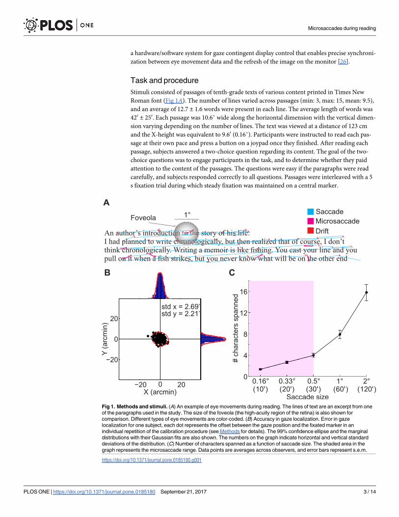
Stimuli consisted of passages of tenth-grade texts of various content printed in Times New
Roman font (Fig 1A). The number of lines varied across passages (min: 3, max: 15, mean: 9.5),
and an average of 12.7 ± 1.6 words were present in each line. The average length of words was
420 ± 250. Each passage was 10.6˚ wide along the horizontal dimension with the vertical dimen-
sion varying depending on the number of lines. The text was viewed at a distance of 123 cm
and the X-height was equivalent to 9.60 (0.16˚). Participants were instructed to read each pas-
sage at their own pace and press a button on a joypad once they finished. After reading each
passage, subjects answered a two-choice question regarding its content. The goal of the two-
choice questions was to engage participants in the task, and to determine whether they paid
attention to the content of the passages. The questions were easy if the paragraphs were read
carefully, and subjects responded correctly to all questions. Passages were interleaved with a 5
s fixation trial during which steady fixation was maintained on a central marker.
Fig 1. Methods and stimuli. (A) An example of eye movements during reading. The lines of text are an excerpt from one
of the paragraphs used in the study. The size of the foveola (the high-acuity region of the retina) is also shown for
comparison. Different types of eye movements are color-coded. (B) Accuracy in gaze localization. Error in gaze
localization for one subject, each dot represents the offset between the gaze position and the fixated marker in an
individual repetition of the calibration procedure (see Methods for details). The 99% confidence ellipse and the marginal
distributions with their Gaussian fits are also shown. The numbers on the graph indicate horizontal and vertical standard
deviations of the distribution. (C) Number of characters spanned as a function of saccade size. The shaded area in the
graph represents the microsaccade range. Data points are averages across observers, and error bars represent s.e.m.
https://doi.org/10.1371/journal.pone.0185180.g001
Microsaccades during reading
PLOS ONE | https://doi.org/10.1371/journal.pone.0185180 September 21, 2017 3 / 14
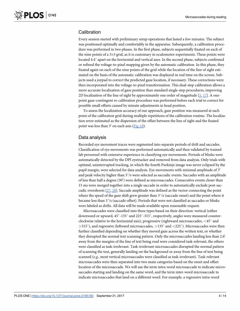
Calibration
Every session started with preliminary setup operations that lasted a few minutes. The subject
was positioned optimally and comfortably in the apparatus. Subsequently, a calibration proce-
dure was performed in two phases. In the first phase, subjects sequentially fixated on each of
the nine points of a 3×3 grid, as it is customary in oculomotor experiments. These points were
located 4.4˚ apart on the horizontal and vertical axes. In the second phase, subjects confirmed
or refined the voltage-to-pixel mapping given by the automatic calibration. In this phase, they
fixated again on each of the nine points of the grid while the location of the line of sight esti-
mated on the basis of the automatic calibration was displayed in real time on the screen. Sub-
jects used a joypad to correct the predicted gaze location, if necessary. These corrections were
then incorporated into the voltage-to-pixel transformation. This dual-step calibration allows a
more accurate localization of gaze position than standard single-step procedures, improving
2D localization of the line of sight by approximately one order of magnitude [5, 17]. A one-
point gaze-contingent re-calibration procedure was performed before each trial to correct for
possible small offsets caused by minute adjustments in head position.
To assess the localization accuracy of our approach, gaze position was measured at each
point of the calibration grid during multiple repetitions of the calibration routine. The localiza-
tion error estimated as the dispersion of the offset between the line of sight and the fixated
point was less than 30 on each axis (Fig 1B).
Data analysis
Recorded eye movement traces were segmented into separate periods of drift and saccades.
Classification of eye movements was performed automatically and then validated by trained
lab personnel with extensive experience in classifying eye movements. Periods of blinks were
automatically detected by the DPI eyetracker and removed from data analysis. Only trials with
optimal, uninterrupted tracking, in which the fourth Purkinje image was never eclipsed by the
pupil margin, were selected for data analysis. Eye movements with minimal amplitude of 30
and peak velocity higher than 3˚/s were selected as saccadic events. Saccades with an amplitude
of less than half a degree (300) were defined as microsaccades. Consecutive events closer than
15 ms were merged together into a single saccade in order to automatically exclude post-sac-
cadic overshoots [27, 28]. Saccade amplitude was defined as the vector connecting the point
where the speed of the gaze shift grew greater than 3˚/s (saccade onset) and the point where it
became less than 3˚/s (saccade offset). Periods that were not classified as saccades or blinks
were labeled as drifts. All data will be made available upon reasonable request.
Microsaccades were classified into three types based on their direction: vertical (either
downward or upward, 45˚-135˚ and 225˚-315˚, respectively, angles were measured counter-
clockwise relative to the horizontal axis), progressive (rightward microsaccades, <45˚ and
>315˚), and regressive (leftward microsaccades, >135˚ and<225˚). Microsaccades were then
further classified depending on whether they moved gaze across the written text, or whether
they disrupted the normal text scanning pattern. Only the microsaccades landing less than 2.60
away from the margins of the line of text being read were considered task-relevant, the others
were classified as task-irrelevant. Task-irrelevant microsaccades disrupted the normal pattern
of scanning the text, generally landing on the background or away from the line of text being
scanned (e.g., most vertical microsaccades were classified as task-irrelevant). Task-relevant
microsaccades were then separated into two main categories based on the onset and offset
location of the microsaccade. We will use the term intra-word microsaccade to indicate micro-
saccades starting and landing on the same word, and the term inter-word microsaccade to
indicate microsaccades that land on a different word. For example, a regressive intra-word
Microsaccades during reading
PLOS ONE | https://doi.org/10.1371/journal.pone.0185180 September 21, 2017 4 / 14
microsaccade moves the gaze leftward on a single word, whereas a regressive inter-words
microsaccade brings the gaze from one word to the preceding one. Spatial locations of the
words on the display were obtained using the Optical Character Recognition toolbox in
Matlab. The spatial borders of a word were defined as a rectangle encompassing all the charac-
ters in the word.
To determine whether the observed pattern of microsaccades was different from chance we
used Monte Carlo simulations (N = 2,000). In one simulation the microsaccades performed
during reading were replaced with microsaccades randomly chosen from those occurring dur-
ing sustained fixation on a marker. While in another simulation microsaccades performed
during sustained fixation were randomly placed over the text. The proportions of task-relevant
and irrelevant microsaccades were calculated per each subject in each simulation and com-
pared with the observed data.
Results
We examined how microsaccades (here referred to as saccades smaller than 300) relocate the
line of sight with respect to the words in the text during reading of 10th grade passages
(Fig 1A). The size of the text was such that * 8 characters fitted within the foveola. As shown
in Fig 1C, microsaccades between 100 to 300 move the gaze over * 1 to * 4 characters, while
larger saccades shift the gaze across up to * 16 characters.
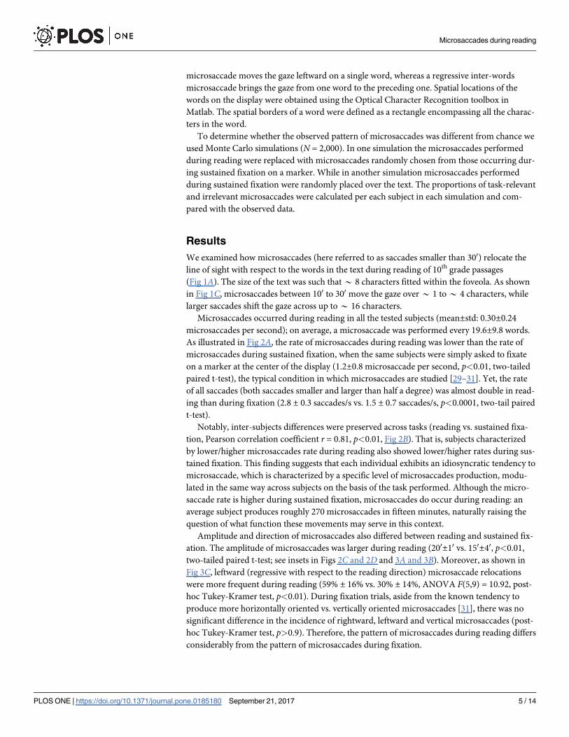
Microsaccades occurred during reading in all the tested subjects (mean±std: 0.30±0.24
microsaccades per second); on average, a microsaccade was performed every 19.6±9.8 words.
As illustrated in Fig 2A, the rate of microsaccades during reading was lower than the rate of
microsaccades during sustained fixation, when the same subjects were simply asked to fixate
on a marker at the center of the display (1.2±0.8 microsaccade per second, p<0.01, two-tailed
paired t-test), the typical condition in which microsaccades are studied [29–31]. Yet, the rate
of all saccades (both saccades smaller and larger than half a degree) was almost double in read-
ing than during fixation (2.8 ± 0.3 saccades/s vs. 1.5 ± 0.7 saccades/s, p<0.0001, two-tail paired
t-test).
Notably, inter-subjects differences were preserved across tasks (reading vs. sustained fixa-
tion, Pearson correlation coefficient r = 0.81, p<0.01, Fig 2B). That is, subjects characterized
by lower/higher microsaccades rate during reading also showed lower/higher rates during sus-
tained fixation. This finding suggests that each individual exhibits an idiosyncratic tendency to
microsaccade, which is characterized by a specific level of microsaccades production, modu-
lated in the same way across subjects on the basis of the task performed. Although the micro-
saccade rate is higher during sustained fixation, microsaccades do occur during reading: an
average subject produces roughly 270 microsaccades in fifteen minutes, naturally raising the
question of what function these movements may serve in this context.
Amplitude and direction of microsaccades also differed between reading and sustained fix-
ation. The amplitude of microsaccades was larger during reading (200±10 vs. 150±40, p<0.01,
two-tailed paired t-test; see insets in Figs 2C and 2D and 3A and 3B). Moreover, as shown in
Fig 3C, leftward (regressive with respect to the reading direction) microsaccade relocations
were more frequent during reading (59% ± 16% vs. 30% ± 14%, ANOVA F(5,9) = 10.92, post-
hoc Tukey-Kramer test, p<0.01). During fixation trials, aside from the known tendency to
produce more horizontally oriented vs. vertically oriented microsaccades [31], there was no
significant difference in the incidence of rightward, leftward and vertical microsaccades (post-
hoc Tukey-Kramer test, p>0.9). Therefore, the pattern of microsaccades during reading differs
considerably from the pattern of microsaccades during fixation.
Microsaccades during reading
PLOS ONE | https://doi.org/10.1371/journal.pone.0185180 September 21, 2017 5 / 14
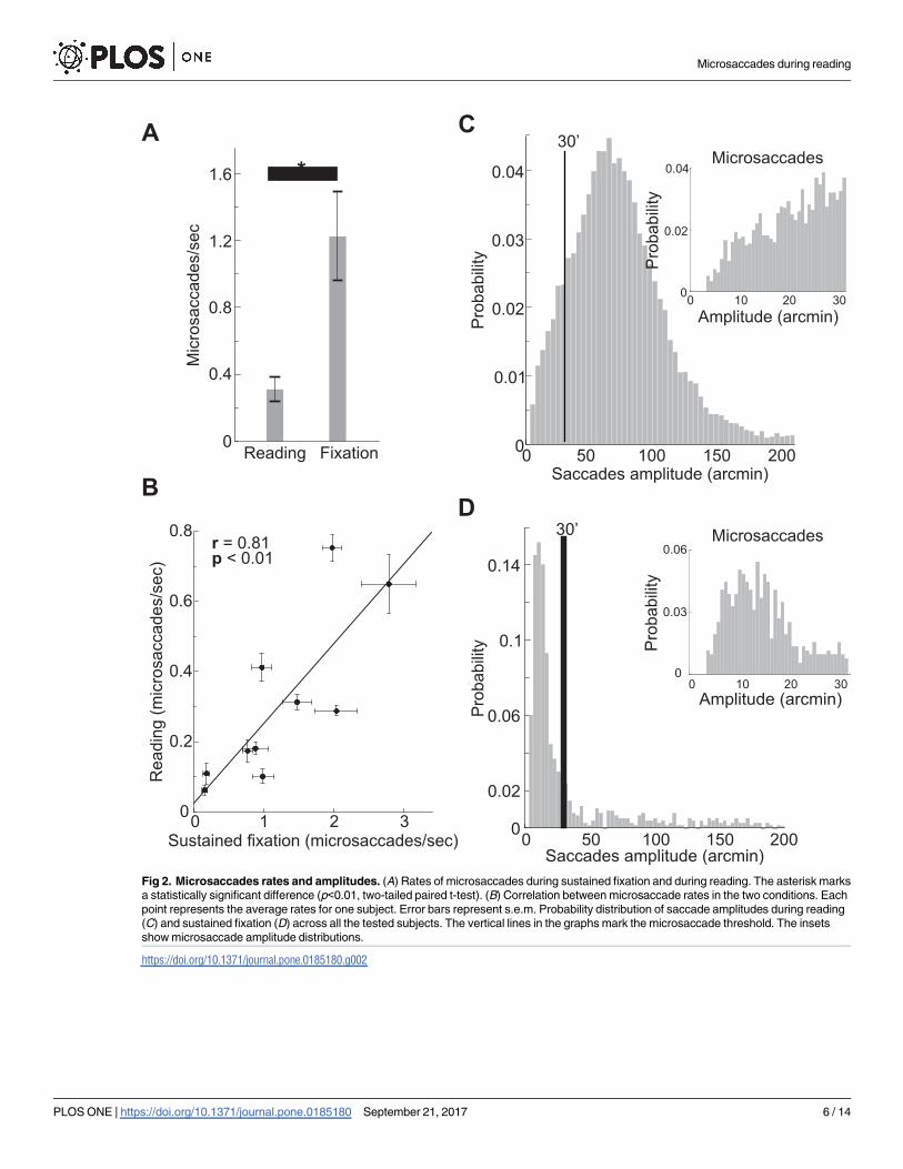
Fig 2. Microsaccades rates and amplitudes. (A) Rates of microsaccades during sustained fixation and during reading. The asterisk marks
a statistically significant difference (p<0.01, two-tailed paired t-test). (B) Correlation between microsaccade rates in the two conditions. Each
point represents the average rates for one subject. Error bars represent s.e.m. Probability distribution of saccade amplitudes during reading
(C) and sustained fixation (D) across all the tested subjects. The vertical lines in the graphs mark the microsaccade threshold. The insets
show microsaccade amplitude distributions.
https://doi.org/10.1371/journal.pone.0185180.g002
Microsaccades during reading
PLOS ONE | https://doi.org/10.1371/journal.pone.0185180 September 21, 2017 6 / 14
Fig 3. Microsaccades and saccades direction. Average 2D polar histograms of microsaccades direction and amplitude during reading
(A) and sustained fixation (B). (C) Average probability of microsaccade direction during reading and during sustained fixation.
Microsaccade direction was classified into three different categories; progressive (rightward), regressive (leftward) and vertical (up and
downward). (D) Average probability of regressive and progressive saccades during reading as a function of saccade amplitude. Error bars
represent s.e.m.
https://doi.org/10.1371/journal.pone.0185180.g003
Microsaccades during reading
PLOS ONE | https://doi.org/10.1371/journal.pone.0185180 September 21, 2017 7 / 14
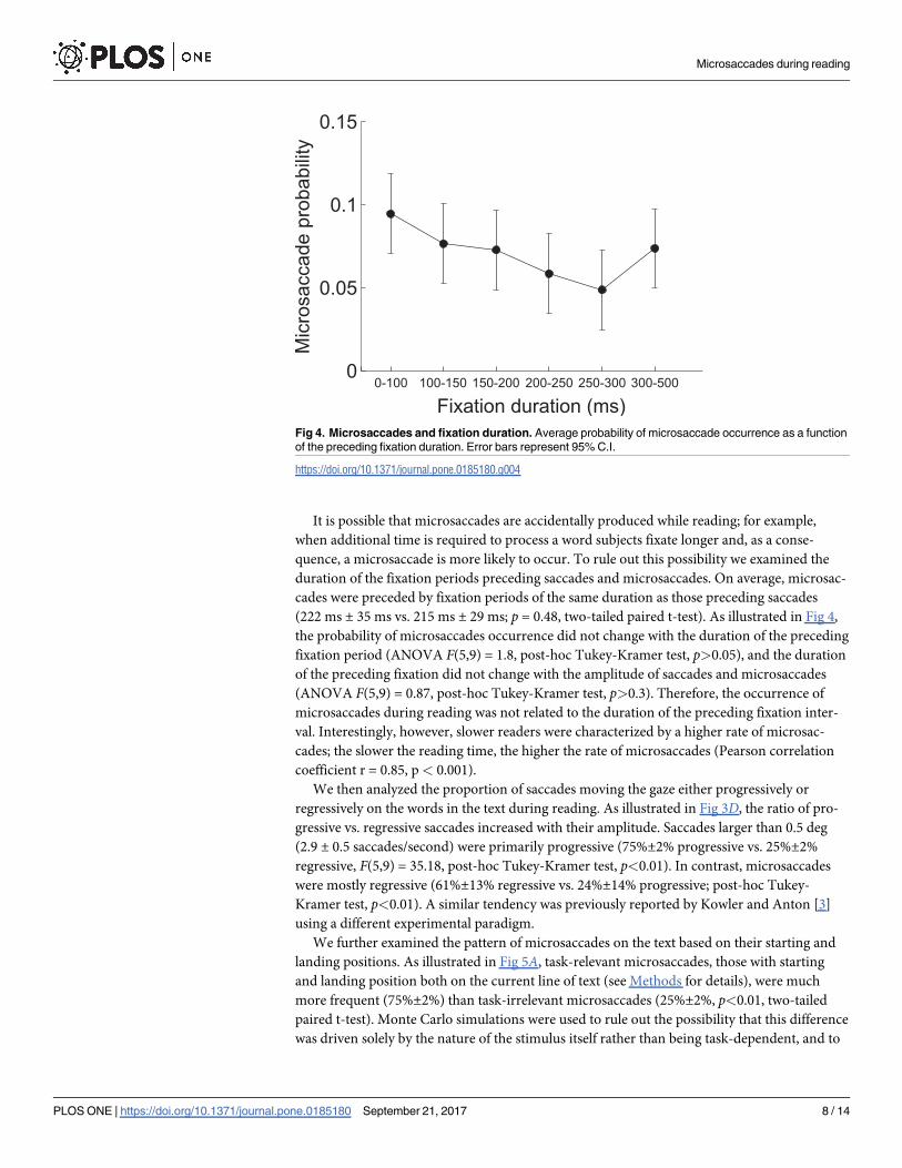
It is possible that microsaccades are accidentally produced while reading; for example,
when additional time is required to process a word subjects fixate longer and, as a conse-
quence, a microsaccade is more likely to occur. To rule out this possibility we examined the
duration of the fixation periods preceding saccades and microsaccades. On average, microsac-
cades were preceded by fixation periods of the same duration as those preceding saccades
(222 ms ± 35 ms vs. 215 ms ± 29 ms; p = 0.48, two-tailed paired t-test). As illustrated in Fig 4,
the probability of microsaccades occurrence did not change with the duration of the preceding
fixation period (ANOVA F(5,9) = 1.8, post-hoc Tukey-Kramer test, p>0.05), and the duration
of the preceding fixation did not change with the amplitude of saccades and microsaccades
(ANOVA F(5,9) = 0.87, post-hoc Tukey-Kramer test, p>0.3). Therefore, the occurrence of
microsaccades during reading was not related to the duration of the preceding fixation inter-
val. Interestingly, however, slower readers were characterized by a higher rate of microsac-
cades; the slower the reading time, the higher the rate of microsaccades (Pearson correlation
coefficient r = 0.85, p< 0.001).
We then analyzed the proportion of saccades moving the gaze either progressively or
regressively on the words in the text during reading. As illustrated in Fig 3D, the ratio of pro-
gressive vs. regressive saccades increased with their amplitude. Saccades larger than 0.5 deg
(2.9 ± 0.5 saccades/second) were primarily progressive (75%±2% progressive vs. 25%±2%
regressive, F(5,9) = 35.18, post-hoc Tukey-Kramer test, p<0.01). In contrast, microsaccades
were mostly regressive (61%±13% regressive vs. 24%±14% progressive; post-hoc Tukey-
Kramer test, p<0.01). A similar tendency was previously reported by Kowler and Anton [3]
using a different experimental paradigm.
We further examined the pattern of microsaccades on the text based on their starting and
landing positions. As illustrated in Fig 5A, task-relevant microsaccades, those with starting
and landing position both on the current line of text (see Methods for details), were much
more frequent (75%±2%) than task-irrelevant microsaccades (25%±2%, p<0.01, two-tailed
paired t-test). Monte Carlo simulations were used to rule out the possibility that this difference
was driven solely by the nature of the stimulus itself rather than being task-dependent, and to
Fig 4. Microsaccades and fixation duration. Average probability of microsaccade occurrence as a function
of the preceding fixation duration. Error bars represent 95% C.I.
https://doi.org/10.1371/journal.pone.0185180.g004
Microsaccades during reading
PLOS ONE | https://doi.org/10.1371/journal.pone.0185180 September 21, 2017 8 / 14
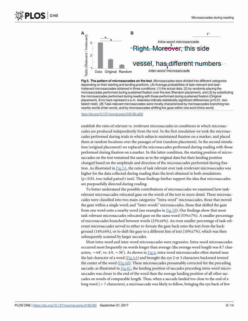
establish the ratio of relevant vs. irrelevant microsaccades in conditions in which microsac-
cades are produced independently from the text. In the first simulation we took the microsac-
cades performed during trials in which subjects maintained fixation on a marker, and placed
them at random locations over the passages of text (random placement). In the second simula-
tion (original placement) we replaced the microsaccades performed during reading with those
performed during fixation on a marker. In this latter condition, the starting position of micro-
saccades on the text remained the same as in the original data but their landing position
changed based on the amplitude and direction of the microsaccades performed during fixa-
tion. As illustrated in Fig 5A, the ratio of task relevant over task-irrelevant microsaccades was
higher for the data collected during reading than the level obtained in both simulations
(p<0.01, two-tailed paired t-test). These findings further support the idea that microsaccades
are purposefully directed during reading.
To better understand the possible contributions of microsaccades we examined how task-
relevant microsaccades relocated gaze on the words of the text in more detail. These microsac-
cades were classified into two main categories: “Intra-word” microsaccades, those that moved
the gaze within a single word; and “Inter-words” microsaccades, those that shifted the gaze
from one word onto a nearby word (see examples in Fig 5B). Our findings show that most
task-relevant microsaccades relocated gaze on the same word (53%±7%). A smaller percentage
of microsaccades branched between words (23%±6%). An even smaller percentage of task-rel-
evant microsaccades served to either re-foveate the gaze back onto the text from the back-
ground (14%±6%), or to shift the gaze to a different line of text (10%±7%), which was then
subsequently scanned by larger saccades.
Most intra-word and inter-word microsaccades were regressive. Intra-word microsaccades
occurred most frequently on words longer than average (the average word length was 8.7 char-
acters, *640, vs. 4.9, *360). As shown in Fig 6, intra-word microsaccades often started near
the last character of a word (Fig 6A) and brought the eye 2 or 3 characters backward toward
the center of the word (Fig 6B). These microsaccades presumably corrected for the preceding
saccade; as illustrated in Fig 6C, the landing position of saccades preceding intra-word micro-
saccades was closer to the end of the word than the average landing position of all other sac-
cades on words of comparable length. Thus, when a saccade landed too close to the end of a
long word (> 7 characters), a microsaccade was likely to follow, bringing the eye back of few
Fig 5. The pattern of microsaccades on the text. Microsaccades were divided into different categories
depending on their starting and landing positions. (A) Average probabilities of task-relevant and task-
irrelevant microsaccades obtained in three conditions: (1) the actual data, (2) by randomly placing the
microsaccades performed during sustained fixation over the text (Random placement), and (3) by substituting
the microsaccades performed during reading with those performed during sustained fixation (Original
placement). Error bars represent s.e.m. Asterisks indicate statistically significant differences (p<0.01, two-
tailed t-test). (B) Task-relevant microsaccades were mostly characterized by microsaccades branching two
nearby words (Inter-word), and by microsaccades shifting the gaze within one word (Intra-word).
https://doi.org/10.1371/journal.pone.0185180.g005
Microsaccades during reading
PLOS ONE | https://doi.org/10.1371/journal.pone.0185180 September 21, 2017 9 / 14
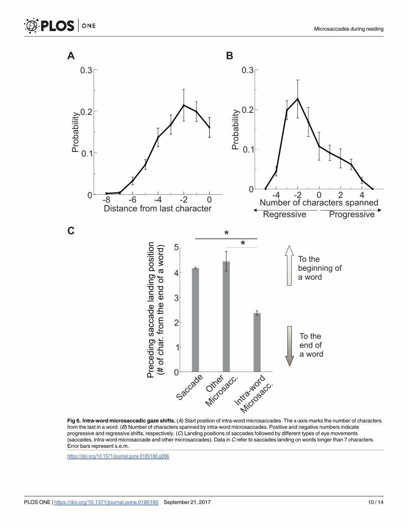
Fig 6. Intra-word microsaccadic gaze shifts. (A) Start position of intra-word microsaccades. The x-axis marks the number of characters
from the last in a word. (B) Number of characters spanned by intra-word microsaccades. Positive and negative numbers indicate
progressive and regressive shifts, respectively. (C) Landing positions of saccades followed by different types of eye movements
(saccades, intra-word microsaccade and other microsaccades). Data in C refer to saccades landing on words longer than 7 characters.
Error bars represent s.e.m.
https://doi.org/10.1371/journal.pone.0185180.g006
Microsaccades during reading
PLOS ONE | https://doi.org/10.1371/journal.pone.0185180 September 21, 2017 10 / 14
characters, an adjustment that appears to ensure optimal placement of the word within the
foveola. These microsaccades, like those shifting gaze from the background to the text, appear
to correct for saccades landing errors. In contrast, inter-words microsaccades and microsac-
cades shifting to a new line of text appear to serve a more exploratory function, in particular
inter-words microsaccades may gather additional information on a word surroundings before
planning the next saccade.
Discussion
Our findings show that microsaccades are relatively frequent during reading, and that they are
not simply the mere outcome of motor errors. The pattern of microsaccades during reading
differs considerably from that occurring under sustained fixation. During reading microsac-
cades are predominantly leftwards and possess larger amplitudes. Whereas during sustained
fixation microsaccades occur in all directions, and they cooperate with ocular drift to maintain
the gaze on the target [32–34], during reading, microsaccades seem to play a role complemen-
tary to that of larger saccades.
Based on these findings we speculate that microsaccades during reading could possibly
enhance visibility in two main ways: by correcting for saccadic landing errors and by moving
the gaze, mostly regressively, over nearby words. Corrective microsaccades occur when sac-
cades land toward the last few characters of a long word (> 7 characters); as high acuity vision
starts to deteriorate already 100 away from the preferred locus of fixation [5], landing at the
end of a long word will cause part of its characters to fall away from the point of highest visual
acuity. An intra-word regressive microsaccade can, therefore, optimally relocate the foveola on
such long words allowing for a better visibility of all its characters. Importantly, microsaccades
not only correct for saccades landing errors, they also enable exploration of nearby words. The
word preceding the current fixation typically falls on a retinal region where vision is subopti-
mal, both because of retinal sampling and the perceptual span; the perceptual span during
reading extends up to 14 letters to the right of the fixated location, but only a few letters to its
left [35, 36]. If extra processing is needed, as for example, to eliminate semantic ambiguities,
executing a microsaccade can be a very efficient strategy to acquire further information. There-
fore, microsaccades may be particularly important for readers characterized by smaller percep-
tual spans, who typically tend to also be slower readers [37]. Consistent with this idea, our data
show that reading times are positively correlated with the rate of microsaccades.
Our work represents a first step toward a better understanding of the role of microsaccades
in reading. A natural question that arises based on our findings regards the actual impact of
microsaccades on words visibility and comprehension. Future research should examine this
issue, which can be addressed, for example, by having readers respond to a number of targeted
questions on specific portions of the text, and evaluate their performance as a function of
microsaccade occurrence on those sections of the text.
Importantly, microsaccades during reading are primarily regressive, moving the gaze back-
ward on the text. Previous research linked the amount of regressive saccades to difficulties in
reading [1, 38], and reported that regressive saccades normally represent only * 10–15% of
the total amount of produced saccades [39]. Here we show that these previous results do not
extend to microsaccades. We report that regressive microsaccades represent the majority of
microsaccades even for expert readers when reading simple text, indicating that they play an
important role in normal reading and are not necessarily linked to reading difficulties.
While the perceptual span during reading is often treated as a static window that moves
with the gaze, recent research [40] showed that it expands in the direction of the upcoming
saccade, similarly to what happens during scene viewing [41, 42]. Therefore, microsaccades
Microsaccades during reading
PLOS ONE | https://doi.org/10.1371/journal.pone.0185180 September 21, 2017 11 / 14
may be beneficial in reading not only by optimally relocating the stimuli within the foveola,
but also by expanding the perceptual span, further enhancing visibility of nearby words. More-
over, the advantage of microsaccades likely extends to more peripheral words. It is known that
microsaccades are associated with shifts of covert attention [12, 13], this is particularly impor-
tant when reading as they may bring the benefits of a covert attentional shift toward peripheral
words while maintaining the gaze on the word being read.
Our results highlight the importance of examining microsaccades during reading, and calls
for further research investigating how microsaccades vary across individuals with different
reading capabilities.
Acknowledgments
This work was supported by National Science Foundation Grants BCS-1534932 (M.P.) and
1420212, and National Institutes of Health Grant R01-EY18363. We thank Dr. Matthew H.
Schneps for providing the passages of text used in this study, and Dr. M. Rucci for the helpful
comments.
Author Contributions
Conceptualization: Martina Poletti.
Data curation: Norick R. Bowers, Martina Poletti.
Formal analysis: Norick R. Bowers.
Funding acquisition: Martina Poletti.
Methodology: Norick R. Bowers, Martina Poletti.
Project administration: Martina Poletti.
Supervision: Martina Poletti.
Validation: Martina Poletti.
Visualization: Norick R. Bowers, Martina Poletti.
Writing – original draft: Norick R. Bowers, Martina Poletti.
Writing – review & editing: Martina Poletti.
References1. Rayner K. Eye movements in reading and information processing: 20 years of research. Psychol Bull.
1998; 124(3):372–422. https://doi.org/10.1037/0033-2909.124.3.372 PMID: 9849112
2. Cunitz RJ, Steinman RM. Comparison of saccadic eye movements during fixation and reading. Vision
Res. 1969; 9:683–693. https://doi.org/10.1016/0042-6989(69)90125-4 PMID: 5822784
3. Kowler E, Anton S. Reading twisted text: Implications for the role of saccades. Vision Res. 1987; 27(1):
45–60. https://doi.org/10.1016/0042-6989(87)90142-8 PMID: 3617546
4. McConkie GW. Eye movements and perception during reading. Illinois Univ., Urbana. Center for the
Study of Reading.; Bolt, Beranek and Newman, Inc., Cambridge, MA.; 1982.
5. Poletti M, Listorti C, Rucci M. Microscopic eye movements compensate for nonhomogeneous vision
within the fovea. Curr Biol. 2013; 23(17):1691–1695. https://doi.org/10.1016/j.cub.2013.07.007 PMID:
23954428
6. Ko HK, Poletti M, Rucci M. Microsaccades precisely relocate gaze in a high visual acuity task. Nat Neu-
rosci. 2010; 13(12):1549–1553. https://doi.org/10.1038/nn.2663 PMID: 21037583
7. Zuber BL, Stark L, Cook G. Microsaccades and the velocity-amplitude relationship for saccadic eye
movements. Science. 1965; 150(3702):1459–1460. https://doi.org/10.1126/science.150.3702.1459
PMID: 5855207
Microsaccades during reading
PLOS ONE | https://doi.org/10.1371/journal.pone.0185180 September 21, 2017 12 / 14
8. Steinman RM, Cunitz RJ, Timberlake GT, Herman M. Voluntary control of microsaccades during main-
tained monocular fixation. Science. 1967; 155(3769):1577–1579. https://doi.org/10.1126/science.155.
3769.1577 PMID: 6020487
9. Havermann K, Cherici C, Rucci M, Lappe M. Fine-scale plasticity of microscopic saccades. J Neurosci.
2014; 34(35):11665–11672. https://doi.org/10.1523/JNEUROSCI.5277-13.2014 PMID: 25164662
10. Hafed ZM, Goffart L, Krauzlis RJ. A neural mechanism for microsaccade generation in the primate
superior colliculus. Science. 2009; 323(5916):940–943. https://doi.org/10.1126/science.1166112
PMID: 19213919
11. Hafed ZM, Krauzlis RJ. Similarity of superior colliculus involvement in microsaccade and saccade gen-
eration. J Neurophysiol. 2012; 107(7):1904–1916. https://doi.org/10.1152/jn.01125.2011 PMID:
22236714
12. Hafed ZM, Clark JJ. Microsaccades as an overt measure of covert attention shifts. Vision Res. 2002;
42(22):2533–2545. https://doi.org/10.1016/S0042-6989(02)00263-8 PMID: 12445847
13. Yuval-Greenberg S, Merriam EP, Heeger DJ. Spontaneous microsaccades reflect shifts in covert atten-
tion. J Neurosci. 2014; 34(41):13693–13700. https://doi.org/10.1523/JNEUROSCI.0582-14.2014
PMID: 25297096
14. Martinez-Conde S, Macknik SL, Troncoso XG, Dyar TA. Microsaccades counteract fading during fixa-
tion. Neuron. 2006; 49(2):297–305. https://doi.org/10.1016/j.neuron.2005.11.033 PMID: 16423702
15. Collewijn H, Kowler E. The significance of microsaccades for vision and oculomotor control. J Vis. 2008;
8(14):1–21. https://doi.org/10.1167/8.14.20 PMID: 19146321
16. Nystrom M, Hansen DW, Andersson R, Hooge I. Why have microsaccades become larger? Investigat-
ing eye deformations and detection algorithms. Vision Res. 2016; 118:17–24. https://doi.org/10.1016/j.
visres.2014.11.007 PMID: 25481631
17. Poletti M, Rucci M. A compact field guide to the study of microsaccades: Challenges and functions.
Vision Res. 2016; 118:83–97. https://doi.org/10.1016/j.visres.2015.01.018 PMID: 25689315
18. Curcio CA, Sloan KR, Kalina RE, Hendrickson AE. Human photoreceptor topography. J Comp Neurol.
1990; 292(4):497–523. https://doi.org/10.1002/cne.902920402 PMID: 2324310
19. Rossi EA, Roorda A. The relationship between visual resolution and cone spacing in the human fovea.
Nat Neurosci. 2010; 13(2):156–157. https://doi.org/10.1038/nn.2465 PMID: 20023654
20. Morrison RE, Rayner K. Saccade size in reading depends upon character spaces and not visual angle.
Percept Psychophys. 1981; 30(4):395–396. https://doi.org/10.3758/BF03206156 PMID: 7322819
21. Legge GE, Bigelow CA. Does print size matter for reading? A review of findings from vision science and
typography. J Vis. 2011; 11(5):1–22. https://doi.org/10.1167/11.5.8
22. Hafed ZM. Alteration of visual perception prior to microsaccades. Neuron. 2013; 77(4):775–786. https://
doi.org/10.1016/j.neuron.2012.12.014 PMID: 23439128
23. Holmqvist K, Nystrom M, Andersson R, Dewhurst R, Jarodzka H, de Weijer JV. Eye tracking: A compre-
hensive guide to methods and measures. Oxford University Press; 2011.
24. Crane HD, Steele CM. Generation V Dual Purkinje-Image eyetracker. Appl Opt. 1985; 24(4):527–537.
https://doi.org/10.1364/AO.24.000527 PMID: 18216982
25. Ko HK, Snodderly DM, Poletti M. Eye movements between saccades: Measuring ocular drift and
tremor. Vision Res. 2016; 122:93–104. https://doi.org/10.1016/j.visres.2016.03.006 PMID: 27068415
26. Santini F, Redner G, Iovin R, Rucci M. EyeRIS: A general-purpose system for eye movement contin-
gent display control. Behav Res Methods. 2007; 39(3):350–364. https://doi.org/10.3758/BF03193003
PMID: 17958145
27. Deubel H, Bridgeman B. Fourth Purkinje image signals reveal eye-lens deviations and retinal image dis-
tortions during saccades. Vision Res. 1995; 35:529–538. https://doi.org/10.1016/0042-6989(94)00146-
D PMID: 7900293
28. Stevenson SB, Roorda A. Correcting for miniature eye movements in high resolution scanning laser
ophthalmoscopy. In: Manns F, Soderberg P, Ho A, editors. Ophthalmic Technologies XV. SPIE; 2005.
p. 145–151.
29. Ratliff F, Riggs LA. Involuntary motions of the eye during monocular fixation. J Exp Psychol. 1950;
40(6):687–701. https://doi.org/10.1037/h0057754 PMID: 14803643
30. Ditchburn RW, Ginsborg BL. Vision with a stabilized retinal image. Nature. 1952; 170(4314):36–37.
https://doi.org/10.1038/170036a0 PMID: 14957011
31. Cherici C, Kuang X, Poletti M, Rucci M. Precision of sustained fixation in trained and untrained observ-
ers. J Vis. 2012; 12(6):1–16. https://doi.org/10.1167/12.6.31
32. Nachmias J. Determiners of the drift of the eye during monocular fixation. J Opt Soc Am. 1961; 51:
761–766. https://doi.org/10.1364/JOSA.51.000761 PMID: 13727312
Microsaccades during reading
PLOS ONE | https://doi.org/10.1371/journal.pone.0185180 September 21, 2017 13 / 14
33. Engbert R, Mergenthaler K. Microsaccades are triggered by low retinal image slip. Proc Natl Acad Sci
USA. 2006; 103(18):7192–7197. https://doi.org/10.1073/pnas.0509557103 PMID: 16632611
34. Poletti M, Rucci M. Fixational eye movements under various conditions of image fading. J Vis. 2010;
10(3)(6):1–18. https://doi.org/10.1167/10.3.6 PMID: 20377283
35. Rayner K, McConkie GW. What guides a reader’s eye movements? Vision Res. 1976; 16(8):829–37.
https://doi.org/10.1016/0042-6989(76)90143-7 PMID: 960610
36. Miellet S, O’Donnell PJ, Sereno SC. Parafoveal magnification: Visual acuity does not modulate the per-
ceptual span in reading. Psychol Sci. 2009; 20(6):721–728. https://doi.org/10.1111/j.1467-9280.2009.
02364.x PMID: 19470124
37. Rayner K, Slattery TJ, Belanger NN. Eye movements, the perceptual span, and reading speed. Psy-
chon Bull Rev. 2010; 17(6):834–839. https://doi.org/10.3758/PBR.17.6.834 PMID: 21169577
38. Hyona J, Olson RK. Eye fixation patterns among dyslexic and normal readers: Effects of word length
and word frequency. Psychol Learn Mem Cogn. 1995; 21(6):1430–1440. https://doi.org/10.1037/0278-
7393.21.6.1430
39. Rayner K. Eye movements and attention in reading, scene perception, and visual search. Q J Exp Psy-
chol. 2009; 62(8):1457–506. https://doi.org/10.1080/17470210902816461
40. Apel JK, Henderson JM, Ferreira F. Targeting regressions: Do readers pay attention to the left? Psy-
chon Bull Rev. 2012; 19(6):1108–1113. https://doi.org/10.3758/s13423-012-0291-1 PMID: 22791218
41. Gersch TM, Kowler E, Schnitzer BS, Dosher BA. Attention during sequences of saccades along marked
and memorized paths. Vision Res. 2009; 49(10):1256–1266. https://doi.org/10.1016/j.visres.2007.10.
030 PMID: 18226827
42. Rolfs M, Jonikaitis D, Deubel H, Cavanagh P. Predictive remapping of attention across eye movements.
Nat Neurosci. 2011; 14(2):252–256. https://doi.org/10.1038/nn.2711 PMID: 21186360
Microsaccades during reading
PLOS ONE | https://doi.org/10.1371/journal.pone.0185180 September 21, 2017 14 / 14
Related Documents


![Active Perception based Formation Control for Multiple ...[11], active perception based formation control is addressed using a decentralized non-linear model predictive controller.](https://static.cupdf.com/doc/110x72/5f085c617e708231d421a0af/active-perception-based-formation-control-for-multiple-11-active-perception.jpg)