MicroRNAs miR-1, miR-133a, miR-133b, miR-208a and miR-208b are dysregulated in Chronic Chagas disease Cardiomyopathy Ludmila Rodrigues Pinto Ferreira a,b,c , Amanda Farage Frade a,b,c , Ronaldo Honorato Barros Santos d , Priscila Camillo Teixeira a,b,c,1 , Monique Andrade Baron a,b,c , Isabela Cunha Navarro a,b,c , Luiz Alberto Benvenuti e , Alfredo Inácio Fiorelli d , Edimar Alcides Bocchi d , Noedir Antonio Stolf d , Christophe Chevillard f , Jorge Kalil b,c , Edecio Cunha-Neto a,b,c, ⁎ a Laboratory of Immunology, Heart Institute (InCor), University of São Paulo, School of Medicine, São Paulo, Brazil b Division of Clinical Immunology and Allergy, University of São Paulo, School of Medicine, São Paulo, Brazil c Institute for Investigation in Immunology (iii), INCT, 05403-001, São Paulo, Brazil. d Division of Surgery, Heart Institute (InCor), University of São Paulo, School of Medicine, São Paulo, Brazil e Division of Pathology, Heart Institute (InCor), University of São Paulo, School of Medicine, São Paulo, Brazil f INSERM, U906, Aix-Marseille University AMU, Faculté de Médecine, Marseille, France abstract article info Article history: Received 2 November 2013 Received in revised form 25 April 2014 Accepted 11 May 2014 Available online 17 May 2014 Keywords: Chagas disease Trypanosoma cruzi miRNA Heart Background/methods: Chagas disease is caused by an intracellular parasite, Trypanosoma cruzi, and it is a leading cause of heart failure in Latin America. The main clinical consequence of the infection is the development of a Chronic Chagas disease Cardiomyopathy (CCC), which is characterized by myocarditis, hypertrophy and fibrosis and affects about 30% of infected patients. CCC has a worse prognosis than other cardiomyopathies, like idiopathic dilated cardiomyopathy (DCM). It is well established that myocardial gene expression patterns are altered in CCC, but the molecular mechanisms underlying these differences are not clear. MicroRNAs are recently discovered regulators of gene expression, and are recognized as important factors in heart development and cardiovascular disorders (CD). We analyzed the expression of nine different miRNAs in myocardial tissue samples of CCC patients in comparison to DCM patients and samples from heart transplant donors. Using the results of a cDNA microarray database on CCC and DCM myocardium, signaling networks were built and nodal molecules were identified. Results: We observed that five miRNAs were significantly altered in CCC and three in DCM; importantly, three miRNAs were significantly reduced in CCC as compared to DCM. We observed that multiple gene targets of the differentially expressed miRNAs showed a concordant inverse expression in CCC. Significantly, most gene targets and involved networks belong to crucial disease-related signaling pathways. Conclusion: These results suggest that miRNAs may play a major role in the regulation of gene expression in CCC pathogenesis, with potential implication as diagnostic and prognostic tools. © 2014 Elsevier Ireland Ltd. All rights reserved. 1. Introduction Chagas disease is caused by an infection with the parasite Trypanosoma cruzi naturally transmitted through hematophagous reduviid bugs or by blood transfusions, organ transplants and congeni- tally. The disease is endemic in Latin America with an estimated 8 to 10 million people infected, but has become a worldwide concern, be- cause it has been detected in non-endemic countries in North America (Canada and the United States), Western Pacific Region (mainly Australia and Japan), and more recently in Europe [1]. The main clin- ical consequence of infection by the parasite is the development of a Chronic Chagas disease Cardiomyopathy (CCC), an inflammatory dilated cardiomyopathy that comprises a wide range of life threatening manifestations, including cardiac hypertrophy, fibrosis, heart failure, arrhythmias, heart blocks, sudden death, thromboembolism, and stroke. About 30% of infected patients develop CCC 5 to 30 years after initial infection, and have a worse prognosis than patients with other cardiomyopathies, such as dilated cardiomyopathy (DCM) [2]. Our group has shown that the myocardial gene expression profiles in CCC patients are profoundly different from those of both heart donors and DCM patients as assessed with the 10,386 element cDNA microarray Cardiochip [3]. Although the gene expression profiling provided evi- dence for strong IFN-γ signaling in the CCC myocardium, the determi- nants of gene expression of the majority of differentially expressed genes remained unknown. MicroRNAs (miRNAs or miRs) are noncod- ing single stranded RNA molecules of 18–26 nt in length capable of silencing the gene expression by inhibiting either protein translation International Journal of Cardiology 175 (2014) 409–417 ⁎ Corresponding author at: Laboratory of Immunology, Heart Institute (Incor), Avenida Dr. Enéas de Carvalho Aguiar, 44, Bloco II, 9°, Andar, São Paulo 05403-900, Brazil. E-mail address: [email protected] (E. Cunha-Neto). 1 Present address: F. Hoffmann-La Roche, Basel, Switzerland. http://dx.doi.org/10.1016/j.ijcard.2014.05.019 0167-5273/© 2014 Elsevier Ireland Ltd. All rights reserved. Contents lists available at ScienceDirect International Journal of Cardiology journal homepage: www.elsevier.com/locate/ijcard

Welcome message from author
This document is posted to help you gain knowledge. Please leave a comment to let me know what you think about it! Share it to your friends and learn new things together.
Transcript
International Journal of Cardiology 175 (2014) 409–417
Contents lists available at ScienceDirect
International Journal of Cardiology
j ourna l homepage: www.e lsev ie r .com/ locate / i j ca rd
MicroRNAs miR-1, miR-133a, miR-133b, miR-208a and miR-208b aredysregulated in Chronic Chagas disease Cardiomyopathy
Ludmila Rodrigues Pinto Ferreira a,b,c, Amanda Farage Frade a,b,c, Ronaldo Honorato Barros Santos d,Priscila Camillo Teixeira a,b,c,1, Monique Andrade Baron a,b,c, Isabela Cunha Navarro a,b,c, Luiz Alberto Benvenuti e,Alfredo Inácio Fiorelli d, Edimar Alcides Bocchi d, Noedir Antonio Stolf d, Christophe Chevillard f,Jorge Kalil b,c, Edecio Cunha-Neto a,b,c,⁎a Laboratory of Immunology, Heart Institute (InCor), University of São Paulo, School of Medicine, São Paulo, Brazilb Division of Clinical Immunology and Allergy, University of São Paulo, School of Medicine, São Paulo, Brazilc Institute for Investigation in Immunology (iii), INCT, 05403-001, São Paulo, Brazil.d Division of Surgery, Heart Institute (InCor), University of São Paulo, School of Medicine, São Paulo, Brazile Division of Pathology, Heart Institute (InCor), University of São Paulo, School of Medicine, São Paulo, Brazilf INSERM, U906, Aix-Marseille University AMU, Faculté de Médecine, Marseille, France
⁎ Corresponding author at: Laboratory of Immunology,Dr. Enéas de Carvalho Aguiar, 44, Bloco II, 9°, Andar, São P
E-mail address: [email protected] (E. Cunha-Neto).1 Present address: F. Hoffmann-La Roche, Basel, Switze
http://dx.doi.org/10.1016/j.ijcard.2014.05.0190167-5273/© 2014 Elsevier Ireland Ltd. All rights reserved
a b s t r a c t
a r t i c l e i n f oArticle history:
Received 2 November 2013Received in revised form 25 April 2014Accepted 11 May 2014Available online 17 May 2014Keywords:Chagas diseaseTrypanosoma cruzimiRNAHeart
Background/methods: Chagas disease is caused by an intracellular parasite, Trypanosoma cruzi, and it is a leadingcause of heart failure in Latin America. The main clinical consequence of the infection is the development of aChronic Chagas disease Cardiomyopathy (CCC), which is characterized by myocarditis, hypertrophy and fibrosisand affects about 30% of infected patients. CCC has aworse prognosis than other cardiomyopathies, like idiopathicdilated cardiomyopathy (DCM). It iswell established thatmyocardial gene expression patterns are altered in CCC,but the molecular mechanisms underlying these differences are not clear. MicroRNAs are recently discoveredregulators of gene expression, and are recognized as important factors in heart development and cardiovasculardisorders (CD).Weanalyzed the expression of nine differentmiRNAs inmyocardial tissue samples of CCC patientsin comparison to DCMpatients and samples from heart transplant donors. Using the results of a cDNAmicroarraydatabase on CCC and DCM myocardium, signaling networks were built and nodal molecules were identified.
Results: We observed that five miRNAs were significantly altered in CCC and three in DCM; importantly, threemiRNAs were significantly reduced in CCC as compared to DCM. We observed that multiple gene targets of thedifferentially expressedmiRNAs showed a concordant inverse expression in CCC. Significantly, most gene targetsand involved networks belong to crucial disease-related signaling pathways.Conclusion: These results suggest that miRNAs may play a major role in the regulation of gene expression in CCCpathogenesis, with potential implication as diagnostic and prognostic tools.© 2014 Elsevier Ireland Ltd. All rights reserved.
1. Introduction
Chagas disease is caused by an infection with the parasiteTrypanosoma cruzi naturally transmitted through hematophagousreduviid bugs or by blood transfusions, organ transplants and congeni-tally. The disease is endemic in Latin America with an estimated 8 to10 million people infected, but has become a worldwide concern, be-cause it has been detected in non-endemic countries in North America(Canada and the United States), Western Pacific Region (mainlyAustralia and Japan), and more recently in Europe [1]. The main clin-ical consequence of infection by the parasite is the development of a
Heart Institute (Incor), Avenidaaulo 05403-900, Brazil.
rland.
.
Chronic Chagas disease Cardiomyopathy (CCC), an inflammatorydilated cardiomyopathy that comprises a wide range of life threateningmanifestations, including cardiac hypertrophy, fibrosis, heart failure,arrhythmias, heart blocks, sudden death, thromboembolism, andstroke. About 30% of infected patients develop CCC 5 to 30 years afterinitial infection, and have a worse prognosis than patients with othercardiomyopathies, such as dilated cardiomyopathy (DCM) [2]. Ourgroup has shown that the myocardial gene expression profiles in CCCpatients are profoundly different from those of both heart donors andDCM patients as assessed with the 10,386 element cDNA microarrayCardiochip [3]. Although the gene expression profiling provided evi-dence for strong IFN-γ signaling in the CCC myocardium, the determi-nants of gene expression of the majority of differentially expressedgenes remained unknown. MicroRNAs (miRNAs or miRs) are noncod-ing single stranded RNA molecules of 18–26 nt in length capable ofsilencing the gene expression by inhibiting either protein translation
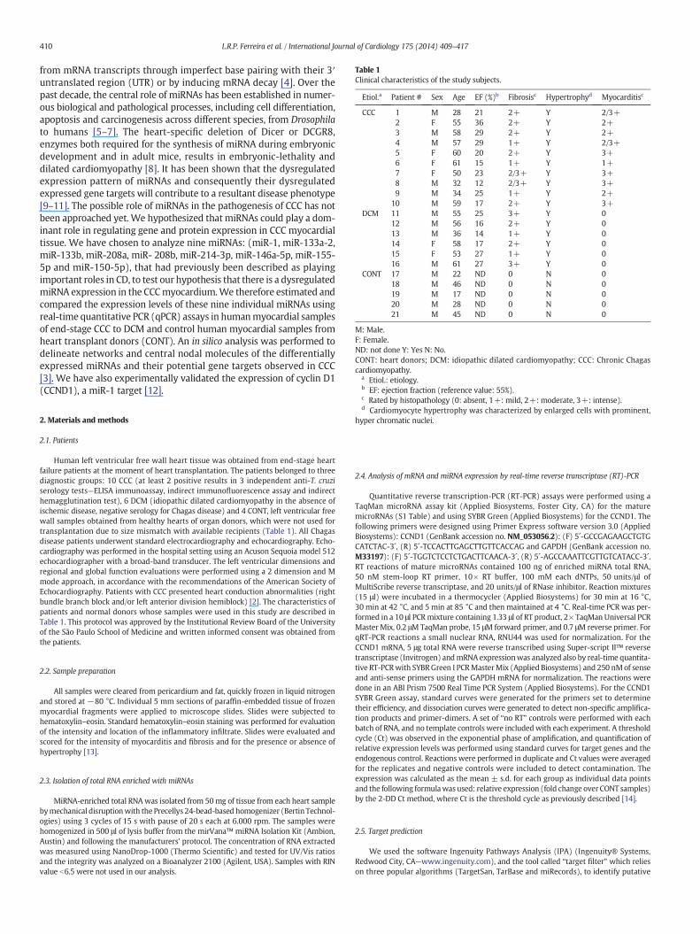
Table 1Clinical characteristics of the study subjects.
Etiol.a Patient # Sex Age EF (%)b Fibrosisc Hypertrophyd Myocarditisc
CCC 1 M 28 21 2+ Y 2/3+2 F 55 36 2+ Y 2+3 M 58 29 2+ Y 2+4 M 57 29 1+ Y 2/3+5 F 60 20 2+ Y 3+6 F 61 15 1+ Y 1+7 F 50 23 2/3+ Y 3+8 M 32 12 2/3+ Y 3+9 M 34 25 1+ Y 2+
10 M 59 17 2+ Y 3+DCM 11 M 55 25 3+ Y 0
12 M 56 16 2+ Y 013 M 36 14 1+ Y 014 F 58 17 2+ Y 015 F 53 27 1+ Y 016 M 61 27 3+ Y 0
CONT 17 M 22 ND 0 N 018 M 46 ND 0 N 019 M 17 ND 0 N 020 M 28 ND 0 N 021 M 45 ND 0 N 0
M: Male.F: Female.ND: not done Y: Yes N: No.CONT: heart donors; DCM: idiopathic dilated cardiomyopathy; CCC: Chronic Chagascardiomyopathy.
a Etiol.: etiology.b EF: ejection fraction (reference value: 55%).c Rated by histopathology (0: absent, 1+: mild, 2+: moderate, 3+: intense).d Cardiomyocyte hypertrophy was characterized by enlarged cells with prominent,
hyper chromatic nuclei.
410 L.R.P. Ferreira et al. / International Journal of Cardiology 175 (2014) 409–417
from mRNA transcripts through imperfect base pairing with their 3′untranslated region (UTR) or by inducing mRNA decay [4]. Over thepast decade, the central role of miRNAs has been established in numer-ous biological and pathological processes, including cell differentiation,apoptosis and carcinogenesis across different species, from Drosophilato humans [5–7]. The heart-specific deletion of Dicer or DCGR8,enzymes both required for the synthesis of miRNA during embryonicdevelopment and in adult mice, results in embryonic-lethality anddilated cardiomyopathy [8]. It has been shown that the dysregulatedexpression pattern of miRNAs and consequently their dysregulatedexpressed gene targets will contribute to a resultant disease phenotype[9–11]. The possible role of miRNAs in the pathogenesis of CCC has notbeen approached yet. We hypothesized that miRNAs could play a dom-inant role in regulating gene and protein expression in CCC myocardialtissue. We have chosen to analyze nine miRNAs: (miR-1, miR-133a-2,miR-133b, miR-208a, miR- 208b, miR-214-3p, miR-146a-5p, miR-155-5p and miR-150-5p), that had previously been described as playingimportant roles in CD, to test our hypothesis that there is a dysregulatedmiRNA expression in the CCCmyocardium.We therefore estimated andcompared the expression levels of these nine individual miRNAs usingreal-time quantitative PCR (qPCR) assays in humanmyocardial samplesof end-stage CCC to DCM and control human myocardial samples fromheart transplant donors (CONT). An in silico analysis was performed todelineate networks and central nodal molecules of the differentiallyexpressed miRNAs and their potential gene targets observed in CCC[3]. We have also experimentally validated the expression of cyclin D1(CCND1), a miR-1 target [12].
2. Materials and methods
2.1. Patients
Human left ventricular free wall heart tissue was obtained from end-stage heartfailure patients at the moment of heart transplantation. The patients belonged to threediagnostic groups: 10 CCC (at least 2 positive results in 3 independent anti-T. cruziserology tests—ELISA immunoassay, indirect immunofluorescence assay and indirecthemagglutination test), 6 DCM (idiopathic dilated cardiomyopathy in the absence ofischemic disease, negative serology for Chagas disease) and 4 CONT, left ventricular freewall samples obtained from healthy hearts of organ donors, which were not used fortransplantation due to size mismatch with available recipients (Table 1). All Chagasdisease patients underwent standard electrocardiography and echocardiography. Echo-cardiography was performed in the hospital setting using an Acuson Sequoia model 512echocardiographer with a broad-band transducer. The left ventricular dimensions andregional and global function evaluations were performed using a 2 dimension and Mmode approach, in accordance with the recommendations of the American Society ofEchocardiography. Patients with CCC presented heart conduction abnormalities (rightbundle branch block and/or left anterior division hemiblock) [2]. The characteristics ofpatients and normal donors whose samples were used in this study are described inTable 1. This protocol was approved by the Institutional Review Board of the Universityof the São Paulo School of Medicine and written informed consent was obtained fromthe patients.
2.2. Sample preparation
All samples were cleared from pericardium and fat, quickly frozen in liquid nitrogenand stored at −80 °C. Individual 5 mm sections of paraffin-embedded tissue of frozenmyocardial fragments were applied to microscope slides. Slides were subjected tohematoxylin–eosin. Standard hematoxylin–eosin staining was performed for evaluationof the intensity and location of the inflammatory infiltrate. Slides were evaluated andscored for the intensity of myocarditis and fibrosis and for the presence or absence ofhypertrophy [13].
2.3. Isolation of total RNA enriched with miRNAs
MiRNA-enriched total RNA was isolated from 50mg of tissue from each heart samplebymechanical disruptionwith the Precellys 24-bead-based homogenizer (Bertin Technol-ogies) using 3 cycles of 15 s with pause of 20 s each at 6.000 rpm. The samples werehomogenized in 500 μl of lysis buffer from the mirVana™ miRNA Isolation Kit (Ambion,Austin) and following the manufacturers' protocol. The concentration of RNA extractedwas measured using NanoDrop-1000 (Thermo Scientific) and tested for UV/Vis ratiosand the integrity was analyzed on a Bioanalyzer 2100 (Agilent, USA). Samples with RINvalue b6.5 were not used in our analysis.
2.4. Analysis of mRNA and miRNA expression by real-time reverse transcriptase (RT)-PCR
Quantitative reverse transcription-PCR (RT-PCR) assays were performed using aTaqMan microRNA assay kit (Applied Biosystems, Foster City, CA) for the maturemicroRNAs (S1 Table) and using SYBR Green (Applied Biosystems) for the CCND1. Thefollowing primers were designed using Primer Express software version 3.0 (AppliedBiosystems): CCND1 (GenBank accession no. NM_053056.2): (F) 5´-GCCGAGAAGCTGTGCATCTAC-3´, (R) 5´-TCCACTTGAGCTTGTTCACCAG and GAPDH (GenBank accession no.M33197): (F) 5´-TGGTCTCCTCTGACTTCAACA-3´, (R) 5´-AGCCAAATTCGTTGTCATACC-3´.RT reactions of mature microRNAs contained 100 ng of enriched miRNA total RNA,50 nM stem-loop RT primer, 10× RT buffer, 100 mM each dNTPs, 50 units/μl ofMultiScribe reverse transcriptase, and 20 units/μl of RNase inhibitor. Reaction mixtures(15 μl) were incubated in a thermocycler (Applied Biosystems) for 30 min at 16 °C,30 min at 42 °C, and 5 min at 85 °C and then maintained at 4 °C. Real-time PCR was per-formed in a 10 μl PCR mixture containing 1.33 μl of RT product, 2× TaqMan Universal PCRMasterMix, 0.2 μMTaqMan probe, 15 μM forward primer, and 0.7 μM reverse primer. ForqRT-PCR reactions a small nuclear RNA, RNU44 was used for normalization. For theCCND1 mRNA, 5 μg total RNA were reverse transcribed using Super-script II™ reversetranscriptase (Invitrogen) andmRNAexpressionwas analyzed also by real-time quantita-tive RT-PCRwith SYBRGreen I PCRMasterMix (Applied Biosystems) and 250 nMof senseand anti-sense primers using the GAPDH mRNA for normalization. The reactions weredone in an ABI Prism 7500 Real Time PCR System (Applied Biosystems). For the CCND1SYBR Green assay, standard curves were generated for the primers set to determinetheir efficiency, and dissociation curves were generated to detect non-specific amplifica-tion products and primer-dimers. A set of “no RT” controls were performed with eachbatch of RNA, and no template controls were includedwith each experiment. A thresholdcycle (Ct) was observed in the exponential phase of amplification, and quantification ofrelative expression levels was performed using standard curves for target genes and theendogenous control. Reactions were performed in duplicate and Ct values were averagedfor the replicates and negative controls were included to detect contamination. Theexpression was calculated as the mean ± s.d. for each group as individual data pointsand the following formulawas used: relative expression (fold change over CONT samples)by the 2-DD Ct method, where Ct is the threshold cycle as previously described [14].
2.5. Target prediction
We used the software Ingenuity Pathways Analysis (IPA) (Ingenuity® Systems,Redwood City, CA—www.ingenuity.com), and the tool called “target filter” which relieson three popular algorithms (TargetSan, TarBase and miRecords), to identify putative
411L.R.P. Ferreira et al. / International Journal of Cardiology 175 (2014) 409–417
targets and targets within lists of differentially expressed genes from ourmicroarray anal-ysis of the five differentially expressed miRNAs for CCC and three for DCM samples.
2.6. Network pathway analysis
IPANetworkmaintains a graphical database of networks of interacting genes (IngenuityKnowledge Base, IKB).Two lists of miRNAs and their target mRNAs, differentially expressedin CCC and DCM compared to CONT, were uploaded in the IPA and analyzed based on thecontent of date 2013–10. The significance of the association between each list and thepathwaywasmeasured by Fisher's exact test. As a result, a P-valuewas obtained, determin-ing the probability that the association between the genes in our data set and the networkgenerated can be explained by chance alone. Molecules are represented as nodes, and thebiological relationship between two nodes is represented as an edge (line). All edges aresupported by at least one reference from the literature, from a textbook, or from canonicalinformation stored in the IKB.
2.7. Statistical analysis
Groups were compared by a non-parametrical test (Mann–Whitney Rank Sum Test)with GraphPad Prism ® software (version 5.0; GraphPad). Results were expressed asmedians and interquartile ranges. P-values were considered significant if b0.05.
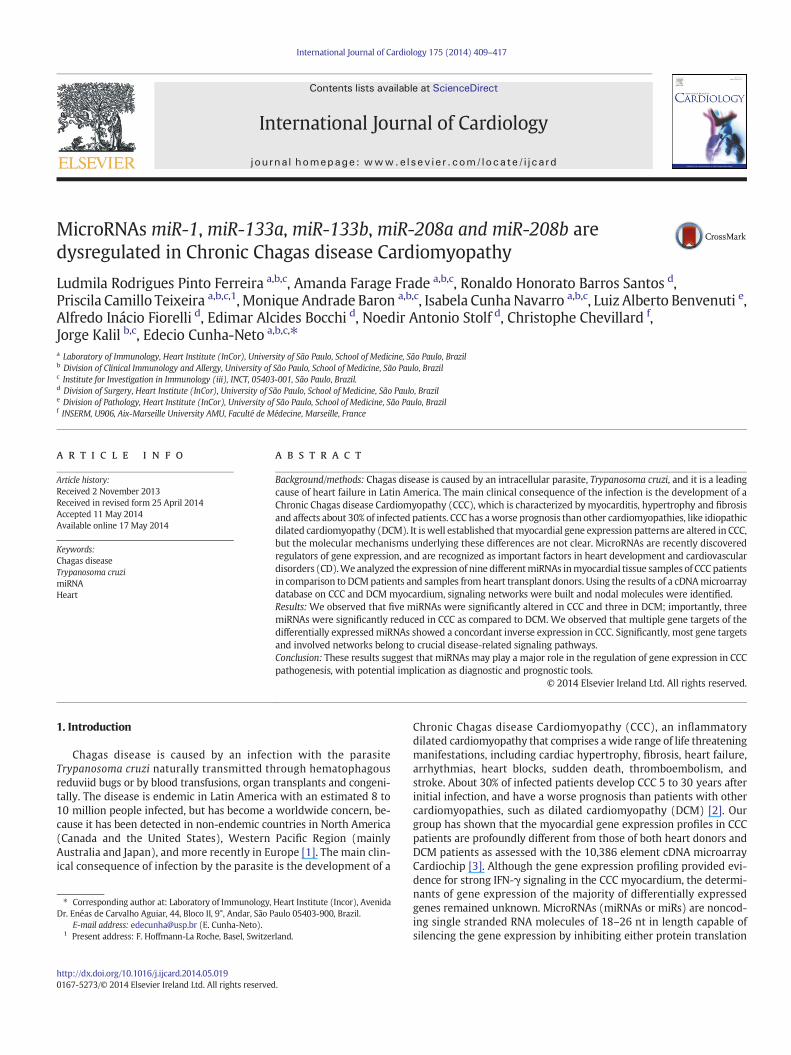
Fig. 1. Individual maturemiRNAs: A)miR-133a, B)miR-133b, C)miR-1, D)miR-208a and E)miR-5), or end-stage CCC (n=10) andDCM (n=6).Weused TaqManMicroRNA single assays (Applieas the mean ± s.d. for each group as individual data points using the relative expression (foldcompared by a non-parametrical test (Mann–Whitney Rank SumTest)withGraphPad Prism® soP-values were considered significant if b0.05.
3. Results
3.1. Muscle-specific miRNAs are downregulated in CCC
We have assessed the expression levels of nine miRNAs that hadpreviously been described as playing important roles in CD: miR-1,miR-133a-2, miR-133b, miR-208a, miR-208b, miR-214-3p, miR-146a-5p, miR-155-5p and miR-150-5p. From those, five miRNAs: miR-1,miR-133a-2, miR-133b, miR-208a, and 208b were significantly down-regulated in CCC samples as compared to CONT (p = 0.0007). Threeof them (miR-133b, miR-208a and miR-208b) were significantlyreduced in DCM samples as compared to controls. Importantly, miR-1,miR-133a-2 and miR-208b were significantly less expressed amongCCC than DCM samples (Fig. 1A to E). Levels of the other four miRNAsanalyzed: miR-214-3p, miR-146-5p, miR-155-5p and miR-150-5pwere not significantly different between groups (Fig. 2: A to D).However, the difference in expression of these miRNAs did not reachstatistical significance; miR-155 showed a trend toward higher expres-sion in CCC compared with CONT samples (Fig. 2C).
208bweremeasured in humanmyocardial samples from heart transplant donors (CONT=dBiosystems®) and the sampleswere normalized to RNU44. The expressionwas calculatedchange over CONT) by the 2-DD Ct method, where Ct is the threshold cycle. Groups wereftware (version 5.0; GraphPad). Resultswere expressed asmedians and interquartile ranges.
Fig. 2. IndividualmaturemiRNAs: A)miR-150-5p, B)miR-214-3p, C)miR-155-5p andD)miR-146a-5pweremeasured inhumanmyocardial samples fromheart transplant donors (CONT=5),or end-stage CCC (n=10) andDCM(n=6).Weused TaqManMicroRNA single assays (Applied Biosystems®) and the sampleswere normalized to RNU44. The expressionwas calculatedas the mean ± s.d. for each group as individual data points using the relative expression (fold change over CONT) by the 2-DD Ct method, where Ct is the threshold cycle. Groups werecompared by a non-parametrical test (Mann–Whitney Rank Sum Test) with GraphPad Prism ® software (version 5.0; GraphPad). Results were expressed as medians and interquartileranges. P-values were considered significant if b0.05.
412 L.R.P. Ferreira et al. / International Journal of Cardiology 175 (2014) 409–417
3.2. miRNA putative targets prediction
A data set containing each statistically significant miRNAs and thecorresponding values of fold change over CONT for each condition(CCC or DCM) was uploaded into IPA application for miRNA target pre-diction. Considering that miRNAs with the same seed sequence usuallytarget the same RNAs, the IPA software clusters together the mature
Table 2In silico analysis done using the IPA software © Ingenuity Systems, Inc. MiRNA/mRNA target (ecDNA microarray [3].
Targets upregulated in CCC
Symbol Fold change Confidence Gene ID Symbol
miR-1 −6.39 High (predicted) BC000076.2 CCND1miR-1 −6.39 High (predicted) M63485 MATR3miR-1 −6.39 Moderate (predicted) M27166 PDHA1miR-1 −6.39 High (predicted) AF006636 SDCBPmiR-1/miR-133a-3p 6.39/-5.638 High (predicted) L02897 SPTBN1miR-1 −6.39 High (predicted) 6908 TBPmiR-1 −6.39 Experimentally Observed M17733 TMSB10miR-1/miR-133a-3p 6.39/-5.638 Experimentally Observed X04588 TPM3miR-133a-3p −5.638 High (predicted) D28475 CLCN6miR-133a-3p −5.638 High (predicted) Z74615 COL1A1miR-133a-3p −5.638 High (predicted) J04617 EEF1A1miR-133a-3p −5.638 High (predicted) M14660 IFIT2miR-133a-3p −5.638 Moderate (predicted) M15856 LPLmiR-133a-3p −5.638 High (predicted) AF043324 NMT1miR-133a-3p −11.6 High (predicted) J03620 DLDTargets upregulated in DCMmiR-133a-3p −1.9 High (predicted) D28475 CLCN6miR-133a-3p −1.9 High (predicted) M69066 MSNmiR-133a-3p −1.9 Experimentally Observed M26252 PKMmiR-208a-3p −7.8 High (predicted) S54705 PRKAR1Targets upregulated in both CCC and DCMmiR-133a-3p −1.9 High (predicted) D28475 CLCN6miR-1 −6.39 High (predicted) NM_053056.2 CCND1
miRNAs that share the same 7-nucleotide seed sequence (nucleotides2–8 from the 5′ end of the mature sequence) into one entity or “node”in order to increase the specificity of targeting information. Two setsout of the five differentially expressed miRNAs analyzed share thesame seed sequence (S1 Table), miR-208a with miR-208b and miR-133a-2 with miR-133b. For CCC samples, the software identified a totalof 2227 putative target transcripts of the five differentially expressed
xpression pairing) in CCC and DCMmyocardial tissue as compared with CONT; Cardiochip
Fold change Entrez gene name
1.8 Cyclin D11.5 Matrin 32.06 Pyruvate dehydrogenase (lipoamide) alpha 11.52 Syndecan binding protein (syntenin)1.6 Spectrin, beta, non-erythrocytic 11.5 TATA box binding protein
/TMSB4X 1.5 Thymosin beta 102 Tropomyosin 31.99 Chloride channel, voltage-sensitive 61.5 Collagen, type I, alpha 12.1 Eukaryotic translation elongation factor 1 alpha 12.7 Interferon-induced protein with tetratricopeptide repeats 21.5 Lipoprotein lipase1.7 N-myristoyltransferase 11.8 Dihydrolipoamide dehydrogenase
1.54 Chloride channel, voltage-sensitive 61.6 Moesin1.8 Pyruvate kinase, muscle
A 1.9 Protein kinase, cAMP-dependent, regulatory, type I, alpha
1.54 Chloride channel, voltage-sensitive 61.8 Cyclin D1
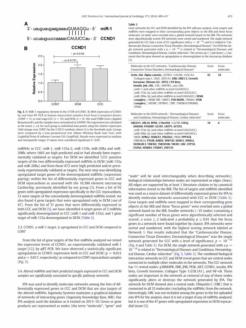
Fig. 3. A) MiR-1 regulatory element in the 3´UTR of CCND1. B) RNA expression of CCND1by real time RT-PCR in human myocardial samples from heart transplant donors(CONT= 5), or end-stage CCC (n=10) and DCM (n=6).We used SYBR Green (AppliedBiosystems®) and the sampleswerenormalized to GAPDH. The expressionwas calculatedas the mean ± s.d. for each group as individual data points using the relative expression(fold change over CONT) by the 2-DD Ct method, where Ct is the threshold cycle. Groupswere compared by a non-parametrical test (Mann–Whitney Rank Sum Test) withGraphPad Prism ® software (version 5.0; GraphPad). Results were expressed as mediansand interquartile ranges. P-values were considered significant if b0.05.
Table 3Gene networks for CCC and DCM identified by the IPA software analysis. Gene targets andmiRNAs were mapped to their corresponding gene objects in the IKB and these focusmolecules (in bold) were overlaid onto a global network based on the IKB. The networkswere algorithmically scored. IPA networks were sorted and numbered. The single networkgenerated for CCC had a score of 55 (significance with p = 10−55) and it is related to “Car-diovascular Disease, Connective TissueDisorders, Dermatological Diseases”. For DCM the sin-gle network generated with a p = 10−18 is related to “Dermatological Diseases andConditions, Hematological Disease, Cardiac infarction”. The arrows up (↑) and down (↓) rep-resent that the gene showed an upregulation or downregulation in the microarray database[3].
Molecules in the CCC network—“Cardiovascular Disease,Connective Tissue Disorders, Dermatological Diseases”
Score Focusmolecules
Actin, Akt, Alpha catenin, ↓CCND1, ↑CLCN6, ↑COL1A1,↑Collagen type I, ↑DLD, ↑EEF1A1, ERK, ERK1/2, Growthhormone, Histone h3, ↑IFIT2, I FN Beta,Insulin, Jnk, LDL, ↑LPL, ↑MATR3, ↓mir-208,↓miR-1 (and other miRNAs w/seed GGAAUGU),↓miR-133a-3p (and other miRNAs w/seed UUGGUCC),↓miR-208a-3p (andothermiRNAsw/seedUAAGACG),NFkB(complex), ↑NFkB-TBP, ↑NMT1, P38 MAPK, ↑PDHA1, PI3K(complex), ↑SDCBP, ↑SPTBN1, ↑TBP, ↑TMSB10/TMSB4X,↑TPM3
55 18
Molecules in the DCM network—“Dermatological Diseasesand Conditions, Hematological Disease, Cardiac infarction”
Score Focusmolecules
ARGLU1, ARL16, BPHL, C19orf44, ↑CLCN6, COLQ,DHODH, FN3KRP, GLYR1, GRTP1, KCTD3,↓miR-133a-3p (and other miRNAs w/seed UUGGUCC),↓miR-208a-3p (and other miRNAs w/seed UAAGACG),↑MSN, NPRL2, NSMCE1, PHLDA2, ↑PKM, PPP3CB,↑PRKAR1A, PTGIR, RAB5C, RNF145, SEMG1, SEMG2,SH3BGRL3, TMEM2, TMEM100, TRUB1, UBC, UTP18,UVSSA, WDR83, YTHDF1, ZNF135
18 6
413L.R.P. Ferreira et al. / International Journal of Cardiology 175 (2014) 409–417
miRNAs in CCC: miR-1, miR-133a-2, miR-133b, miR-208a and miR-208b, where 1665 are high predicted and/or had already been experi-mentally validated as targets. For DCM we identified 1231 putativetargets of the two differentially expressed miRNAs in DCM (miR-133aand miR-208a) and from those 872 were high predicted and/or previ-ously experimentally validated as targets. The next step was identifyingupregulated target genes of the downregulated miRNAs (expressionpairing) within the list of differentially expressed genes in CCC andDCM myocardium as assessed with the 10,386 element microarrayCardiochip, previously identified by our group [3]. From a list of 92genes with upregulated expression specifically in the CCC myocardium,12 were targets of the concordantly downregulated miRNAs tested; wealso found 4 gene targets that were upregulated only in DCM (out of47). From the list of 31 genes that were differentially expressed inboth CCC and DCM [3], we have identified 4 target genes of 2 miRNAssignificantly downregulated in CCC (miR-1 and miR-133a) and 1 genetarget of miR-133a downregulated in DCM (Table 2).
3.3. CCND1, a miR-1 target, is upregulated in CCC and DCM compared toCONT
From the list of gene targets of the five miRNAs analyzed we testedthe expression levels of CCND1, an experimentally validated miR-1target [12], by qRT-PCR. We have observed a statistically significantupregulation in CCND1 expression both in CCC and DCM (p = 0.012and p= 0.017, respectively) as compared to CONTmyocardium samples(Fig. 3).
3.4. Altered miRNAs and their predicted targets expressed in CCC and DCMsamples are significantly associated to specific pathway networks
IPA was used to identify molecular networks among the lists of dif-ferentially expressed genes in CCC and DCM that are also targets ofthe altered miRNAs. Ingenuity Systems maintains a graphical databaseof networks of interacting genes (Ingenuity Knowledge Base, IKB). OurIPA analysis used the database as it existed on 2013–10. Genes or geneproducts are represented as nodes (the term “molecule”, “gene” and
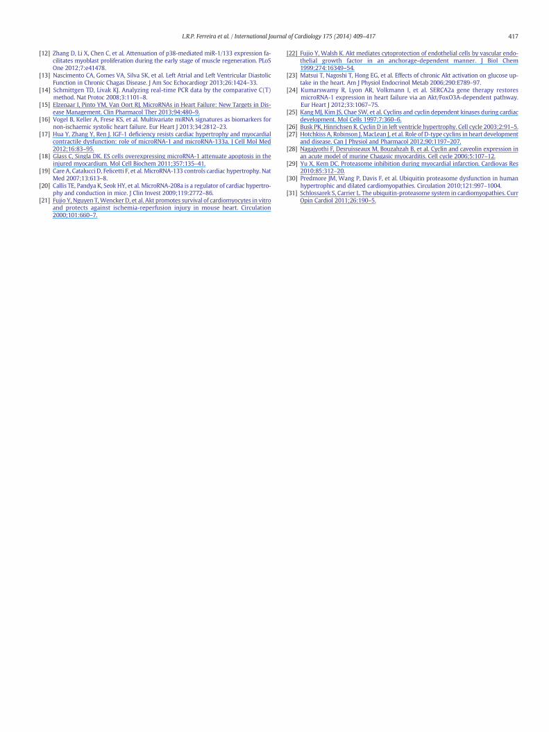
“node” will be used interchangeably when describing networks);biological relationships between nodes are represented as edges (lines).All edges are supported by at least 1 literature citation or by canonicalinformation stored in the IKB. The list of targets and miRNAs identifiedwas used as a source dataset of differentially expressed genes for IPA toidentify molecular networks associated with CCC or DCM (Table 3).Gene targets and miRNAs were mapped to their corresponding geneobjects in the IKB and these “focus genes” were overlaid onto a globalnetwork based on the IKB. Smaller networks (b35 nodes) containing asignificant number of focus genes were algorithmically selected andscored; a score ≥ 2 indicated a probability p ≤ 0.01 that the focusgenes in a network were found together by chance. IPA networks weresorted and numbered, with the highest scoring network labeled asNetwork 1. Our results indicated that the “Cardiovascular Disease,Connective Tissue Disorders, Dermatological Diseases” was the singlenetwork generated for CCC with a level of significance, p = 10−55
(Fig. 4 and Table 3). For DCM, the single network generated with a p =10−18 is related to “Dermatological Diseases and Conditions, Hematolog-ical Disease, Cardiac infarction” (Fig. 5, Table 3). The combined biologicalinteraction networks in CCC and DCM reveal genes that are central nodesconnected tomultiple othermolecules in the networks. The CCC networkhas 11 central nodes: p38MAPK, ERK, JNK, PI3K, AKT, CCND1, insulin, IFNbeta, Growth hormone, Collagen Type 1(COL1A1), and NF-κB. Thesenodes are important in the network as removal of any of these nodessignificantly alters or destroys the network generated by IPA. Thenetwork for DCM showed also a central node, Ubiquitin C (UBC) that isconnected to all 33molecules (excluding themiRNAs) from the network.Interestingly, UBCwas not included among the genes in the list uploadedinto IPA for the analysis, since it is not a target of any ofmiRNAs analyzed,but it is one of the 47 geneswith upregulated expression in DCMmyocar-dial tissue [3].

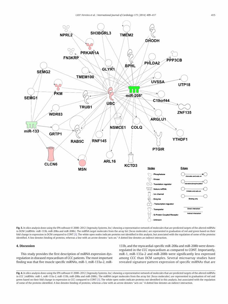
Fig. 5. In silico analysis done using the IPA software© 2000–2012 (Ingenuity Systems, Inc) showing a representative network of molecules that are predicted targets of the alteredmiRNAsin DCM (miRNAs: miR-133b, miR-208a and miR-208b). The miRNA target molecules from the array list (focus molecules) are represented in graduation of red and green based on theirfold change in expression in DCM compared to CONT [3]. The white open nodes indicate proteins not identified in this analysis, but associatedwith the regulation of some of the proteinsidentified. A line denotes binding of proteins, whereas a line with an arrow denotes “acts on.” A dotted line denotes an indirect interaction.
415L.R.P. Ferreira et al. / International Journal of Cardiology 175 (2014) 409–417
4. Discussion
This study provides the first description of miRNA expression dys-regulation in diseasedmyocardium of CCC patients. Themost importantfinding was that five muscle specific miRNAs, miR-1, miR-133a-2, miR-
Fig. 4. In silico analysis done using the IPA software© 2000–2012 (Ingenuity Systems, Inc) showin CCC (miRNAs: miR-1, miR-133a-2, miR-133b, miR-208a and miR-208b). The miRNA targetgreen based on their fold change in expression in CCC compared to CONT [3]. The white openof some of the proteins identified. A line denotes binding of proteins, whereas a line with an a
133b, and themyocardial-specificmiR-208a andmiR-208bwere down-regulated in the CCC myocardium as compared to CONT. Importantly,miR-1, miR-133a-2 and miR-208b were significantly less expressedamong CCC than DCM samples. Several microarray studies haverevealed signature pattern expression of specific miRNAs that are
ing a representative network of molecules that are predicted targets of the alteredmiRNAsmolecules from the array list (focus molecules) are represented in graduation of red andnodes indicate proteins not identified in this analysis, but associated with the regulationrrow denotes “acts on.” A dotted line denotes an indirect interaction.

416 L.R.P. Ferreira et al. / International Journal of Cardiology 175 (2014) 409–417
consistently aberrantly expressed in patients with different types of CD:i.e., miR-1, miR-29, miR-30, miR-133 and miR-150 are found to bedownregulated in heart failure patients [10,15], whereas miRNAs:miR-21, miR-23a, miR-125, miR-146, miR-195, miR-199 and miR-214are upregulated [16]. MiR-1 and miR-133 have an impaired expressionin patients with hypertrophic cardiomyopathy and atrial dilation aswell as in different murine models of cardiac hypertrophy (i.e., trans-verse aortic constriction and overexpression of active cardiospecificAkt kinase) [17,18]. Overexpression of miR-133 resulted in inhibitionof hypertrophic growth induced by endothelin-1 and phenylephrineand expression inhibition of endogenous miR-133 using a targeted 3′UTR decoy resulted in cell hypertrophy, increased fetal gene expression,protein synthesis, and expression of atrial natriuretic factor (ANF) [19].Zhang et al. showed that both miR-1 and miR-133 are importantinducers of myoblast growth arrest to G1 phase by direct targetingCCND1 and Sp1, respectively, which in turn mediate the effect of miR-133 and miR-1 on cell cycle progression [12]. Callis et al. have shownthat transgenic overexpression of miR-208a (encoded within an intronof cardiacmusclemyosin heavy chain gene,Myh6) in the heart was suf-ficient to induce hypertrophic growth in mice, resulting in pronouncedrepression of the miR-208 regulatory targets thyroid hormone-associated protein 1 and myostatin 2, negative regulators of musclegrowth and hypertrophy [20]. Furthermore, analysis of mice lackingmiR-208a indicated that this miRNA is important for proper cardiacconduction and expression of the cardiac transcription factorshomeodomain-only protein and GATA4 and the gap junction proteinconnexin 40 [20]. We observed that miRNA expression profiles of CCCand DCM had some predictable similarities, like the three miRNAs:miR-133b, miR-208a and miR-208b that were also downregulated inDCM samples compared to CONT. To move beyond the singlemiRNA or single gene target approach, we analyzed our target scansignatures using IPA, which allows for identification of enriched net-works. The IPA ingenuity analysis revealed different gene networksrelated to each disease condition with different node molecules.These nodes relate to one transcription factor, i.e., the inflammatorytranscription factor and a known mediator in cardiac dysfunction,NF-κB and protein kinases, i.e., mitogen-activated protein kinases(MAPK) including p38MAPK, ERK1/2, c-Jun N-terminal kinases(JNK), phosphatidylinositide 3-kinases (PI3K), and the proteinkinase B (AKT), enzymes that play important roles in signaling path-ways leading to cardiac hypertrophy. Although studies have shownthat acute activation of Akt is cardio-protective in vitro and in vivodue to its ability to inhibit apoptosis [21,22], chronic Akt activationin the heart has been shown to be prejudicial [23]. Kumarswamyet al. have shown that Akt activation negatively regulates cardio-myocyte miR-1 expression in vitro and in failing hearts in vivo, bythe phosphorylation of the transcription factor, Forkhead box O3(FoxO3a) which binds to the miR-1 promoter and is responsible fora decreased miR-1 expression observed in chronic failing hearts.It has also been shown that this decreased miR-1 expression isreversed in sarcoplasmic reticulum calcium ATPase 2a (SERCA2a)treated hearts [24]. This pathway might be also important andshould be further investigated in CCC, as the present study resultsare showing amiR-1 downregulation; significantly, the gene expressioncorroborates that AKT is upregulated, while SERCA2a is downregulatedin the myocardium of CCC patients [3]. CDND1, an important nodemolecule in the CCC network, is connected to all the other eightnodes. This protein, along with other D-type cyclins (D2 and D3) is apositive cell cycle regulator that plays an important role in controllingproliferation of cardiomyocytes during normal heart development[25]. The central position of CDND1 is in linewith earlier studies reveal-ing that this protein is a key element for cardiac hypertrophic growth[26]. Importantly, the expression of D-type cyclins is generally low inthe adult heart and is increased in the diseased heart, where their up-regulation may promote cardiac hypertrophy instead of cell prolifera-tion [27]. Accordingly, a previous study has shown that CCND1
expression is upregulated during T. cruzi acute infection in mice andthat the expression of CDND1 and other types of cyclins like A1, B1and E1 is increased in heart lysates of mice acutely infected withT. cruzi compared with uninfected controls [28]. Here we show thatmiR-1 controlled CDND1 might also be a key element in CCC. The net-work generated for DCM showed a central node, the UBC, apolyubiquitin precursor. Ubiquitin is a small, ubiquitously expressedprotein that is covalently attached to proteins in linear chains via a spe-cific enzymatic reaction called ubiquitination which is associated withprotein degradation, DNA repair, cell cycle regulation, kinase modifica-tion, endocytosis, and regulation of other cell signaling pathways. Im-portantly, inhibition of the cardiac proteasome has been shown to becardioprotective under some circumstances [29–31]. The concordantexpression of miRs and their targets in CCC and DCM heart tissue, aswell as the results of the iterative analysis presented here indicatesthat miRNAs may play an important role in the regulation of gene ex-pression, modulating key pathways resulting in the manifestation ofCCC and DCM phenotype. The results presented here will guide furtherstudies on the contribution ofmiRNAs and their target genes to CCC andDCM pathogenesis and treatment.
Supplementary data to this article can be found online at http://dx.doi.org/10.1016/j.ijcard.2014.05.019.
Competing interests
The authors have declared that no competing interests exist.
Acknowledgments
This research was supported by Brazilian Council for Scientificand Technological Development—CNPq (57.3879/2008-7) and theSão Paulo State Research Funding Agency—FAPESP (grant numbers2008/57881-0 ;2012/08107-6; 2013/50302-3). Authors also re-ceived financial assistance from the Institut National de la Santé etde la Recherche Médicale (INSERM), the Aix-Marseille University(Direction des Relations Internationales), USP-COFECUB program,and the ARCUS II PACA Brésil program. LRPF is recipient of a CNPq fel-lowship. ECN and JK are recipients of the Brazilian Council for Scien-tific and Technological Development—CNPq productivity awards,AFF, MAB, ICN are recipients of a FAPESP fellowship. ECN and CCare recipients of an international program funded by both, theFrench ANR (Br-Fr-CHAGAS) and the Brazilian FAPESP agencies. CCwas a recipient of a temporary professor position supported by theFrench consulate in Brazil and the University of São Paulo (USP).The funders had no role in study design, data collection and analysis,decision to publish, or preparation of the manuscript.
References
[1] Cortez J, Providencia R, Ramos E, et al. Emerging and under-recognized Chagas car-diomyopathy in non-endemic countries. World J Cardiol 2012;4:234–9.
[2] Bocchi EA, Guimaraes G, Tarasoutshi F, et al. Cardiomyopathy, adult valve diseaseand heart failure in South America. Heart 2009;95:181–9.
[3] Cunha-Neto E, Dzau VJ, Allen PD, et al. Cardiac gene expression profiling providesevidence for cytokinopathy as a molecular mechanism in Chagas' disease cardiomy-opathy. Am J Pathol 2005;167:305–13.
[4] Ambros V. MicroRNAs and developmental timing. Curr Opin Genet Dev2011;21:511–7.
[5] Ambros V. The functions of animal microRNAs. Nature 2004;431:350–5.[6] Carrington JC, Ambros V. Role of microRNAs in plant and animal development. Sci-
ence 2003;301:336–8.[7] Karp X, Ambros V. Developmental biology. Encountering microRNAs in cell fate sig-
naling Science 2005;310:1288–9.[8] Yang WJ, Yang DD, Na S, et al. Dicer is required for embryonic angiogenesis during
mouse development. J Biol Chem 2005;280:9330–5.[9] Bhaskaran M, Mohan M. MicroRNAs: History, Biogenesis, and Their Evolving Role in
Animal Development and Disease. Vet Pathol 2013 Sep 17;51(4):759–74.[10] Hagiwara S, Kantharidis P. Cooper. M E microRNA as Biomarkers and Regulator of
Cardiovascular Development and Disease Curr Pharm Des 2014;20:2347–70.[11] Lau P, Bossers K, Janky R, et al. Alteration of the microRNA network during the pro-
gression of Alzheimer's disease. EMBO Mol Med 2013;5:1613–34.
417L.R.P. Ferreira et al. / International Journal of Cardiology 175 (2014) 409–417
[12] Zhang D, Li X, Chen C, et al. Attenuation of p38-mediated miR-1/133 expression fa-cilitates myoblast proliferation during the early stage of muscle regeneration. PLoSOne 2012;7:e41478.
[13] Nascimento CA, Gomes VA, Silva SK, et al. Left Atrial and Left Ventricular DiastolicFunction in Chronic Chagas Disease. J Am Soc Echocardiogr 2013;26:1424–33.
[14] Schmittgen TD, Livak KJ. Analyzing real-time PCR data by the comparative C(T)method. Nat Protoc 2008;3:1101–8.
[15] Elzenaar I, Pinto YM, Van Oort RJ. MicroRNAs in Heart Failure: New Targets in Dis-ease Management. Clin Pharmacol Ther 2013;94:480–9.
[16] Vogel B, Keller A, Frese KS, et al. Multivariate miRNA signatures as biomarkers fornon-ischaemic systolic heart failure. Eur Heart J 2013;34:2812–23.
[17] Hua Y, Zhang Y, Ren J. IGF-1 deficiency resists cardiac hypertrophy and myocardialcontractile dysfunction: role of microRNA-1 and microRNA-133a. J Cell Mol Med2012;16:83–95.
[18] Glass C, Singla DK. ES cells overexpressing microRNA-1 attenuate apoptosis in theinjured myocardium. Mol Cell Biochem 2011;357:135–41.
[19] Care A, Catalucci D, Felicetti F, et al. MicroRNA-133 controls cardiac hypertrophy. NatMed 2007;13:613–8.
[20] Callis TE, Pandya K, Seok HY, et al. MicroRNA-208a is a regulator of cardiac hypertro-phy and conduction in mice. J Clin Invest 2009;119:2772–86.
[21] Fujio Y, Nguyen T,Wencker D, et al. Akt promotes survival of cardiomyocytes in vitroand protects against ischemia-reperfusion injury in mouse heart. Circulation2000;101:660–7.
[22] Fujio Y, Walsh K. Akt mediates cytoprotection of endothelial cells by vascular endo-thelial growth factor in an anchorage-dependent manner. J Biol Chem1999;274:16349–54.
[23] Matsui T, Nagoshi T, Hong EG, et al. Effects of chronic Akt activation on glucose up-take in the heart. Am J Physiol Endocrinol Metab 2006;290:E789–97.
[24] Kumarswamy R, Lyon AR, Volkmann I, et al. SERCA2a gene therapy restoresmicroRNA-1 expression in heart failure via an Akt/FoxO3A-dependent pathway.Eur Heart J 2012;33:1067–75.
[25] KangMJ, Kim JS, Chae SW, et al. Cyclins and cyclin dependent kinases during cardiacdevelopment. Mol Cells 1997;7:360–6.
[26] Busk PK, Hinrichsen R. Cyclin D in left ventricle hypertrophy. Cell cycle 2003;2:91–5.[27] Hotchkiss A, Robinson J, MacLean J, et al. Role of D-type cyclins in heart development
and disease. Can J Physiol and Pharmacol 2012;90:1197–207.[28] Nagajyothi F, Desruisseaux M, Bouzahzah B, et al. Cyclin and caveolin expression in
an acute model of murine Chagasic myocarditis. Cell cycle 2006;5:107–12.[29] Yu X, Kem DC. Proteasome inhibition during myocardial infarction. Cardiovas Res
2010;85:312–20.[30] Predmore JM, Wang P, Davis F, et al. Ubiquitin proteasome dysfunction in human
hypertrophic and dilated cardiomyopathies. Circulation 2010;121:997–1004.[31] Schlossarek S, Carrier L. The ubiquitin-proteasome system in cardiomyopathies. Curr
Opin Cardiol 2011;26:190–5.
Related Documents


