SUPPLEMENTAL MATERIAL MicroRNA-328 Contributes to Adverse Electrical Remodeling in Atrial Fibrillation Yanjie Lu, MD, PhD*; Ying Zhang, MD, PhD*; Ning Wang, MD, PhD; Zhenwei Pan, MD, PhD; Xu Gao, MD, PhD; Fengmin Zhang, MD, PhD; Yong Zhang, MD, PhD; Hongli Shan, MD, PhD; Xiaobin Luo, MSc; Yunlong Bai, MD, PhD; Lihua Sun, MD, PhD; Wuqi Song, MD, PhD; Chaoqian Xu, MD, PhD; Zhiguo Wang, PhD; & Baofeng Yang, MD, PhD From Department of Pharmacology (the State-Province Key Laboratories of Biomedicine- Pharmaceutics of China) (Y.L., Y.Z., N.W., Z.P., Y.Z., H.S., Y.B., L.S., C.X., X.L., Z.W., B.Y.), and Department of Biochemistry (X.G.), Department of Microbiology (F.Z., W.S.), Harbin Medical University, Harbin, Heilongjiang 150081, P. R. China Running Title: Control of Atrial Fibrillation by miR-328 * The first 2 authors contributed equally to this work. Corresponding to Baofeng Yang, MD, PhD; Department of Pharmacology (the State-Province Key Laboratories of Biomedicine-Pharmaceutics of China), Harbin Medical University, Harbin, Heilongjiang 150081 P.R. China; Tel. +86 451 8666-9473; E-mail: [email protected] . Corresponding to Zhiguo Wang, PhD, FAHA, FESC; Research Center, Montreal Heart Institute, 5000 Belanger East, Montreal, PQ H1T 1C8, Canada; Tel.: (514) 376-3330-3517. Fax: (514) 376-1355; E-mail: [email protected] or [email protected] 1

Welcome message from author
This document is posted to help you gain knowledge. Please leave a comment to let me know what you think about it! Share it to your friends and learn new things together.
Transcript
SUPPLEMENTAL MATERIAL
MicroRNA-328 Contributes to Adverse Electrical Remodeling in
Atrial Fibrillation
Yanjie Lu, MD, PhD*; Ying Zhang, MD, PhD*; Ning Wang, MD, PhD; Zhenwei Pan, MD, PhD;
Xu Gao, MD, PhD; Fengmin Zhang, MD, PhD; Yong Zhang, MD, PhD; Hongli Shan, MD, PhD;
Xiaobin Luo, MSc; Yunlong Bai, MD, PhD; Lihua Sun, MD, PhD; Wuqi Song, MD, PhD;
Chaoqian Xu, MD, PhD; Zhiguo Wang, PhD; & Baofeng Yang, MD, PhD
From Department of Pharmacology (the State-Province Key Laboratories of Biomedicine-
Pharmaceutics of China) (Y.L., Y.Z., N.W., Z.P., Y.Z., H.S., Y.B., L.S., C.X., X.L., Z.W., B.Y.),
and Department of Biochemistry (X.G.), Department of Microbiology (F.Z., W.S.), Harbin
Medical University, Harbin, Heilongjiang 150081, P. R. China
Running Title: Control of Atrial Fibrillation by miR-328
* The first 2 authors contributed equally to this work.
Corresponding to Baofeng Yang, MD, PhD; Department of Pharmacology (the State-Province
Key Laboratories of Biomedicine-Pharmaceutics of China), Harbin Medical University,
Harbin, Heilongjiang 150081 P.R. China; Tel. +86 451 8666-9473; E-mail:
Corresponding to Zhiguo Wang, PhD, FAHA, FESC; Research Center, Montreal Heart Institute,
5000 Belanger East, Montreal, PQ H1T 1C8, Canada; Tel.: (514) 376-3330-3517. Fax:
(514) 376-1355; E-mail: [email protected] or [email protected]
1

This PDF file includes:Supplementary MethodsSupplementary ResultsFigs. S1 to S10Tables S1
2
Supplementary Methods
Computational Prediction of miRNA Target
We used five established miRNA target prediction algorithms to identify the candidate miRNAs
that have the potential to target the ion channel genes; these algorithms include DIANA-
microT3.1, miRanda, PITA, RNAhybrid, and TargetScan5.1. Only the gene predicted by at least
three of the five algorithms to be a target for a given miRNA (miR-328, miR-223, or miR-664)
was considered as a candidate for further analysis.
Canine Model of Atrial Fibrillation (AF)
Mongrel dogs (20 to 30 kg) of either sex were randomly divided into two groups: sham control
(Ctl, n=10) and atrial tachypacing (A-TP, n=17) groups. For animals in the A-TP group, dogs
were sedated and anesthetized with morphine (2 mg/kg SC) and α-chloralose (120 mg/kg IV
load, 29.25 mg/kg/h infusion), for electrode implantation via the jugular veins. A programmable
pacemaker was inserted in a subcutaneous pocket with sterile techniques, and a tined atrial pacing
lead was positioned in the right atrial appendage under fluoroscopic guidance.1 The dogs were
subjected to continuous right atrial pacing at 400 bpm for 56 days (8 weeks) before experimental
studies. The control dogs were sham-operated in the same way as A-TP dogs but without
tachypacing. On study days, dogs were anaesthetized with morphine and α-chloralose and
ventilated to maintain physiological arterial blood gases. Body temperature was maintained at
37°C. A median sternotomy was performed, and bipolar, Teflon-coated, stainless steel electrodes
were hooked into the right and left atrial appendages for recording and stimulation. A
programmable stimulator was used to deliver 2-ms pulses at twice-threshold current. The surface
ECG and direct atrial activation electrograms were recorded. All animal procedures were
3
previously approved by the Animal Care and Use Committee at the Harbin Medical University
(same below).
AF vulnerability was tested at a basic cycle length (S1−S1 interval) of 300 ms, with single
premature S2 extrastimuli delivered at each site by setting the coupling interval initially to 200
ms and decreasing by 10 ms decrements until AF was induced or failure to capture occurs. For
the purpose of measuring AF duration, AF was induced by burst atrial pacing with 4x threshold
4-ms pulses at 20 Hz at a basic cycle length (BCL) of 300 ms. AF was considered sustained if it
required electrical cardioversion for termination (cardioversion was performed after 30 min AF).
To estimate the mean duration of AF, AF was induced 10 times if AF duration was <5 min, 5
times for AF between 5 and 20 min and 3 times for AF >20 min.1 Measurements were made
before drug treatment and repeated 12 h after treatment (adenovirus infection or lipofectamine
transfection of miR-328 and other constructs).
Atrial Samples from Patients
Human tissues (right atrium appendage) were provided by the Second Affiliated Hospital of the
Harbin Medical University under the procedures approved by the Ethnic Committee for Use of
Human Samples of Harbin Medical University. The tissues were obtained from 22 individuals
undergoing heart surgery, ten of them with no atrial fibrillation (AF) and twelve with AF, who
undergoing surgical procedures (see Supplementary Table 1 online). These preparations were
used to isolate total RNA for real-time RT-PCR quantification of miRNAs.
Microarray Analysis
The hearts were removed from dogs and RNA samples were extracted for miRNA expression
analysis. The RNA samples from 7 AF dogs and from 7 control dogs were pooled into three
pairs, respectively for miRNA profiling. miRNA expression profile was analyzed using the
4
miRNA microarray technology miRCURY™ LNA Array (Exiqon Company, Denmark).
miRCURY™ LNA Array, representing 540 mature human miRNAs plus 576 mature rodent
miRNAs, incorporates Locked Nucleic Acid into an oligonucleotide probe, which greatly
increases the affinity and specificity of that oligonucleotide for its complementary DNA or RNA
target. Slides were scanned by the Genepix 4000B at 635 nm and the expression level was
analyzed by Genepix Pro 6.0. The array output was received in Excel spreadsheets as lists of raw
data and also as “simple detectable” data, which were the average of 4 signal values for each
miRNA on the array. Differentially regulated miRNAs were defined as those with >2-fold
increase and >50% decrease of miRNA levels in AF dogs compared with the baseline expression
levels from sham-operated dogs.
Quantitative Real-Time RT-PCR Analysis
The mirVana™ qRT-PCR miRNA Detection Kit (Ambion) was used in conjunction with real-
time PCR with TaqMan for quantification of miRNAs in our study, as previously described in
detail.2-4 The total RNA samples were isolated with Ambion’s mirVana miRNA Isolation Kit,
from canine left atrial preparations, from cultured neonatal rat atrial myocytes, and from mouse
left atrium. Reactions contained mirVana qRT-PCR Primer sets specific for human, canine, rat
and mouse miR-328, and a scrambled miRNA as a negative control. qRT-PCR was performed on
a thermocycler ABI Prism® 7500 fast (Applied Biosystems) for 40 cycles. Fold variations in
expression of an mRNA between RNA samples were calculated. The threshold cycle (CT) is
defined as the fractional cycle number at which the fluorescence passes the fixed threshold. To
estimate copy numbers of transcript in a cardiac cells, a standard curve was generated by using a
series of concentrations of synthetic miR-328 and converting TaqMan CT values into absolute
copy numbers using the standard curve assuming 30 pg of total RNA in each cell.
5
Northern Blot Analysis
Total miRNAs were extracted from dog tissue with mirVana mRNA isolation kit (Ambion,
Cat.No. AM1560). RNA samples were run on 12.5% acrylamide denaturing (urea) gels and then
transferred to Nylon Positively Charged Membranes (Roche, Cat. No. 1 209 272) by semi-dry
electrophoresis (OWL SEPARATION SYSTEMS, HEP-1Semidry Electroblotter). After transfer,
they were crosslinked with 120 mjoules of UV and baked at 80°C for 1 hour. Oligonucleotide
probes were labeled using DIG oligonucleotide tailing Kit, 2nd generation (Roche, Cat. No.
3353583) and hybridized to the membranes at 50°C overnight. Membranes were washed twice
with 2XSSC, 0.1% SDS and twice with 0.5XSSC, 0.1% SDS. The blots were exposed on X-ray
film (Clonex corporation, Bioflex MSI Film for maximum sensitivity imaging, Cat. CLMS810).
The oligonucleotide probes used were all LNA-modified (synthesized by IDT) including miR-
328 probe: ACGGAAGGGCAGAGAGGGCCAG; miR-1 probe:
TACATACTTCTTTACATTCCA; and U6 snRNA probe:
TAAAAATATGGAACGCTTCACGAATTTGCGTGTCATCCTTGCGCAGGGGCCATGCTA
AT.
Western Blot Analysis
The protein samples (membrane and cytosolic samples separately) were extracted from the left
atrium of the dogs, cultured rat atrial myocytes, and atrial tissues of transgenic mice for
immunoblotting analysis, with the procedures essentially the same as described in detail
elsewhere.2-5 The protein content was determined by BCA Protein Assay Kit using bovine serum
albumin as the standard. Protein sample (~50 µg) was fractionated by SDS-PAGE (12%
polyacrylamide gels) and transferred to PVDF membrane (Millipore, Bedford, MA). The sample
was incubated overnight at 4°C with the primary antibodies in 1:200. Affinity purified goat
6

polyclonal anti-CACNB1 (Santa Cruz Biotechnology Inc.) and goat polyclonal anti-Cav1.2
(Alomone Labs), and goat polyclonal antibodies to Kir2.1, Kv4.2, PLN and Cx43 (Santa Cruz
Biotechnology Inc.) were used as the primary antibodies. Inhibitory peptide for each antibody
was used to test the antibody specificity. Next day, the membrane was incubated with secondary
antibodies (Molecular Probes) diluted in PBS for 2 h at room temperature. Finally, the membrane
was rinsed with PBS before scanning using the Infrared Imaging System (LI-COR Biosciences).
GAPDH was used as an internal control for equal input of protein samples, using anti-GAPDH
antibody. Western blot bands were quantified using QuantityOne software by measuring the band
intensity (Area x OD) for each group and normalizing to GAPDH. The final results are expressed
as fold changes by normalizing the data to the control values.
Synthesis of miRNAs and anti-miRNA Antisense Inhibitors
miR-328 (5’-CUGGCCCUCUCUGCCCUUCCGU-3’) and its antisense oligonucleotides
(AMOs: 5’- ACGGAAGGGCAGAGAGGGCCAG-3’) were synthesized by Integrated DNA
Technologies Inc (IDT), as described previously. Five nucleotides or deoxynucleotides at both
ends of the antisense molecules were locked (the ribose ring is constrained by a methylene bridge
between the 2’-O- and the 4’-C atoms). Additionally, a scrambled RNA was used as negative
control; sense: 5'-UUCUCCGAACGUGUCACGUAA-3' and antisense: 5'-
ACGUGACACGUUCGGAGAAUU-3'.
Construction of Luciferase-miRNA-Target Site Fusion Plasmids
To construct reporter vectors bearing miRNA-target sites, we synthesized fragments containing
the exact target sites for miR-328 through Invitrogen, and the 3’UTR of miR-328 target genes
(CACNA1C and CACNB1) by PCR amplification. The sense and antisense strands of the
oligonucleotides were annealed by adding 2 µg of each oligonucleotides to 46 µl of annealing
7
solution (100 mM K-acetate, 30 mM HEPES-KOH, pH 7.4 and 2 mM Mg-acetate) and incubated
at 90oC for 5 min and then at 37oC for 1 h. The annealed oligonucleotides were digested with
HindIII and SpeI. These inserts were ligated into HindIII and SpeI sites in the pMIR-REPORTTM
luciferase miRNA expression reporter vector (Ambion).2-5
Construction of Adenovirus and Infection
The procedures were similar to the study reported by van Rooij et al6 Rno-miR-328 precursor
DNA (5'-GGATCCgACCCCGTCCCCCCGTCCTCC
CCGAGTCCCTCTTTCGTAGATGTCGGGGACCGGGAGAGACGGGAAGGCAGGGGACA
GGGGTTTAttttttAAGCTT-3') was synthesized by GenScript (Nanjing, P.R. China). The
fragment was first inserted into adenovirus shuttle plasmid pDC316-EGFP-U6 (Microbix
Biosystems Inc, Canada). pDC316-EGFP-U6 was then cotransfected with the infectious
adenovirus genomic plasmid pBHGlox∆E1,3Cre into 293 cells by liposome reagent. Following
co-transfection of these two DNAs, homologous recombination occurred to generate a
recombinant adenovirus in which the transgene (pre-miR-328) is incorporated into the viral
genome, replacing the ∆E1 region7 (Supplementary Figure 1). Mismatched miR-328 was
generated by substituting 6 nts within the seed motif as indicated by underlined and italic letters
(5'-
GGATCCgACCCCGTCCCCCCGTCCTCCCCGAGTCCCTCTTTCGTAGATGTCGGGGGTT
GAACGAGACGGGAAGGCAGGGGACAGGGGTTTAttttttAAGCTT-3').
In vivo Gene Transfer
Mongrel dogs (20 to 30 kg) of either sex were randomly divided into 4 groups: sham-operated
control (control, n=7), adenovirus empty vector control (Adv-pDC316, n=7), adenovirus pre-
miR-328 (n=7), and Adv pre-miR-328+AMO-328 (n=7). The dogs were initially anesthetized
8
using 30 mg/kg of sodium pentobarbital delivered intravenously. Additional anesthesia was
administered as needed throughout the experimental study. A right-sided thoracotomy was
performed at the first intercostal space. A pericardial cradle was created, and adenovirus (1x10⁹
pfu/ml, 300 µl) was injected through a 26-gauge needle into multiple sites (~10 sites within an
area of 1 cm2) of the right atrium. After that, a stimulus electrode with five pairs of electrodes
was hooked into the injected sites of the right atrium. Twelve hours after drug injection, atrial
activation electrograms were recorded.
Generation of miR-328 Transgenic Mice
A fragment (320 bp) containing precursor miR-328 (pre-miR-328) sequence was PCR amplified
from the mouse genomic DNA (accession no.: NT_078575). The fragment was then subcloned
into the Sal I and Hind III sites of Bluescript vector (Promega) carrying the cardiac-specific α
myosin heavy chain (αMHC) promoter and human growth hormone poly(A)+ signal
(Supplementary Figure 3).8 The plasmid was digested at the Spe I site to release the pre-miR-
328 sequence flanked by 5’end αMHC promoter and 3’end poly(A). The fragment was separated
on an agarose gel and purified by QIAEX II gel extraction kit (Qiagen # C 04539). The DNA
sample was prepared at a concentration of 3 ng/µl ready for injection. A Tg mouse line carrying a
mismached pre-miR-328 sequence as indicated above was also generated for negative control
experiments.
Sexually immature female mice (4-5 weeks of age) were superovulated by consecutive
PMS and HCG hormone injections to obtain sufficient quantity of (>250) eggs for injection.
These female mice were mated with stud males immediately following the HCG injection. Eggs
were harvested the next day from the ampulla of the oviduct of the mated females, and treated
with hyaluronidase to remove nurse cells. Fertilized eggs were then stored in M16 media (37oC,
9

5% CO2) until injection. Each egg was individually micro-injected with the DNA fragment and
the eggs which did not survive injection were removed. Pseudo-pregnant female mice were
prepared by mating with the vasectomized males. On the day of micro-injection, the pseudo-
pregnant females were anesthetized with 0.5% pentobarbital (20 ml/kg). The injected eggs were
then implanted in a group of 10-15 bilaterally into the oviduct of these animals. The animals were
allowed to recover from anesthesia on a warming plate, and then returned to the animal room.
They were kept under sterile conditions throughout their pregnancy.
The genomic DNA was prepared from tail tissue of the transgenic mice and subjected to
PCR verification for the presence of miR-328 transgene. The forward primer was designed to
recognize αMHC (position: 5250-5268): 5'-CCTTACCCCACATAGACCT-3', and the reverse
primer was for miR-328 (position: 57-39): 5'-CTGTAGATACTTTCTCCCT-3'. The PCR
profiling was composed of an initial denaturing step at 94oC for 2 min and 35 cycles of 94oC (20
s), 60oC (7 s) and 72oC (20 s), followed by a final extension step at 72oC for 5 min.
Generation of miR-328 Sponge Transgenic Mice
A fragment containing six anti-miR-328 antisense units 5’-gtcgacacggaagggcctc-
agggccagaattacggaagggcctc-agggccagaattacggaagggcctc-agggccagaattacggaagggcctc-
agggccagaattacggaagggcctc-agggccagaattacggaagggcctc-agggccagaagctt-3’ was synthesized by
Shanghai Biological Engineering Inc. The fragment was then subcloned into the Sal I and Hind
III sites of Bluescript vector (Promega) carrying the cardiac-specific α myosin heavy chain
(αMHC) promoter and human growth hormone poly(A)+ signal. The same procedures as
described above for miR-328 were followed.8 Knockdown of endogenous miR-328 in F0 was
verified and the transgenic mice of 2 months old were used experimental studies.
Myocyte Isolation and Primary Cell Culture
10
The enzymatic dispersion techniques used to isolate single atrial myocytes from dog, neonatal rat,
and mouse have been previously described in detail.1,2 Canine myocardial specimens from left
atria were cut into chunks and washed three times in oxygenated Ca2+-free Tyrode solution at
37°C. The tissues were then incubated in 10 ml Ca2+-free Tyrode’s solution containing
collagenase (0.25 mg/ml, Type α, Sigma) and BSA (0.2 mg/ml) for 40 min at 37°C, with the
solution constantly gassed with 100% oxygen. Afterwards, tissue were transferred to fresh Ca2+-
free Tyrode’s solution containing collagenase (0.13 mg/ml, Type α) until atrial myocytes were
dispersed. Isolated myocytes were stored in KB solution (in mM: glutamic acid 70, taurine 15,
KCl 30, KH2PO4 10, HEPES 10, MgCl2·6H2O 0.5, glucose 10, and EGTA 0.5; pH 7.4 with KOH)
at 4°C until use.
Neonatal rat atrial cardiomyocytes were isolated and cultured with the procedures similar
to previously described.2 Briefly, 1-3 days old rats were decapitated and their hearts were
aseptically removed. The atria were dissected, minced and trypsinized at 37°C for 10 min.
Dissociated cells were plated in 24-well plates in Dulbecco’s Modified Eagle Medium (DMEM,
Ivitrogen) containing 10% FBS and 0.1 mM bromodeoxyuridine (Sigma) and the non-adherent
cardiomyocytes were removed. The cells (1x10⁵/well) were seeded in a 24-well plate for further
experiments. This procedure yielded cultures with 90±5% myocytes, as assessed by microscopic
observation of cell beating. The cardiomyocytes were also verified by positive staining with an
anti-α-actin monoclonal antibody through immunocytochemistry.
For mice, wild-type and transgenic animals of 2–3 months of age were heparinized,
anaesthetized with 1% pentobarbital (16 ml/kg). The hearts were rapidly removed, and
retrogradely perfused through the aorta using a modified Langendorff apparatus. The preparation
was perfused with standard Tyrode’s solution (in mM: NaCl 126, KCl 5.4, HEPES 10,
11
NaH2PO4·2H2O 0.33, MgCl2·6H2O 1.0, CaCl2 1.8, and glucose 10; pH adjusted to 7.4 with
NaOH) for 5 min, then switched to Ca2+-free Tyrode’s solution until it stopped beating, followed
by perfusion with the same solution containing collagenase II (7 mg/50 ml) and BSA. The freshly
isolated myocytes from the atrial free wall were gently centrifuged and resuspended in the KB
medium. All solutions were gassed with 100% oxygen and warmed to (37 ± 0.5oC). Only single
rod-shaped, Ca2+-tolerant, and quiescent cells with clear cross-striations were selected for
electrophysiological recording.
Cell Culture
HEK293 (human embryonic kidney cell line) used in this study was purchased from American
Type Culture Collection (ATCC, Manassas, VA) and cultured in Dulbecco’s Modified Eagle
Medium (DMEM).
Transfection Procedures
Neonatal rat atrial myocytes were transfected with 1 µg miRNA and/or AMOs, and negative
control AMOs with lipofectamine 2000 (Invitrogen), according to the manufacturer’s
instructions. Forty-eight hours after transfection, cells were used for luciferase assay or were
collected for total RNA or protein purification.
Synthesis and Administration of miR-328 AntagomiR
miR-328 antagomiR was synthesized by Ribobio Co. (Guangzhou, China). The antagomiR is a
single-stranded RNA analogue complementary to the mature miR-328 (5’-
GACCGGGAGAGACGGGAAGGCA-3’), which was chemically modified and cholesterol-
conjugated from a hydoxyprolinol-linked cholesterol solid support and 2’-OMe
phosphoramidites. For negative control experiments, a mismatched miR-328 antagomiR (5’-
GGCAAGACGAAACGAGACGACA-3’) was also synthesized. miR-328 antagomiR was injected
12
into WT or TG mice through the tail-vein at a dosage of 80 mg/kg/d in 0.2 ml saline once a day
for three consecutive days. The surface ECG (lead II) was recorded in anesthetized mice once a
day (1 h) for 2 weeks.
Luciferase Activity Assay
For luciferase assay involving miRNA function, HEK293 cells were transfected with the pMIR-
REPORTTM luciferase miRNA expression reporter vector carrying the 3’UTR of miR-328 target
genes.2-4
For luciferase assay involving analysis of miR-328 promoter activities, neonatal rat atrial
myocytes were similarly transfected with 1 µg PGL3–target DNA (firefly luciferase vector) and
0.1 µg PRL-TK (TK-driven Renilla luciferase expression vector) with lipofectamine 2000.
Following transfection (48 h), luciferase activities were measured with a dual luciferase reporter
assay kit (Promega) on a luminometer (Lumat LB9507). For all experiments, transfection took
place 24 h after starvation of cells in serum-free medium.
Whole-Cell Patch-Clamp Recording
Patch-clamp techniques were applied to isolated atrial myocytes from A-TP dogs and transgenic
mice. The procedures have been described in detail elsewhere.1-3,5 Briefly, the pipette of patch
electrodes had the tip resistance of 2-3 MΩ when filled with pipette solution. The isolated single
cells were placed in a 1-ml chamber mounted on an inverted microscope (IX-70, Olympus) and
perfused with Tyrode solution. Whole-cell recording were performed using an amplifier
(Axopatch 200B, Axon instrument, USA). Signals were filtered at 1 kHz and data were acquired
by A/D conversion (Digidata 1320, Axon Instrument). Ion currents were recorded in the whole-
cell voltage-clamp mode. For the recording of L-type Ca2+ current (ICaL), the pipette solution
contained (in mM) 20 CsCl, 110 Cs-aspartate, 1 MgCl2, 5 MgATP, 0.1 GTP, 5 Na2
13
phosphocreatine, 10 EGTA, and 10 HEPES (pH 7.2 with CsOH). The external Tyrode solution
contained (in mM) 136 tetraethylammonium chloride, 5.4 CsCl, 2 CaCl2, 0.8 MgCl2, 10 HEPES,
and 10 dextrose (pH 7.4 with CsOH). Niflumic acid (50 µM) was added to inhibit Ca2+-dependent
Cl- current. For recording inward rectifier K+ current (IK1), transient outward K+ current (Ito), and
ultrarapid delayed rectifier K+ current (IKur), The pipette solution contained (in mM) 20 KCl, 110
K-aspartate, 1 MgCl2, 5 MgATP, 0.1 GTP, 5 Na2 phosphocreatine, 10 EGTA, and 10 HEPES (pH
7.2 with KOH); the external Tyrode solution contained (in mM) 136 NaCl, 5.4 KCl, 2 CaCl2, 0.8
MgCl2, 10 HEPES, and 10 dextrose (pH 7.4 with NaOH). And BaCl2 (2 mM) was included to
inhibit ICaL. Experiments were conducted at 36 ± 1oC. Junction potentials were zeroed before
formation of the membrane-pipette seal and they were not corrected for our data analyses. Series
resistance and capacitance were compensated and leak currents were subtracted. Cells with
significant leak currents were rejected. For analysis, the data were collected to an IBM-
compatible computer and analyzed with the use of pCLAMP software system 9.2.
ICa,L was elicited by 300-ms depolarizing pulses delivered from a holding potential of -50
mV at a frequency of 0.1 Hz. IK1 was recorded by 300-ms square pulses ranging from -120 mV to
+10 mV at a holding potential of -20 mV at a frequency of 0.1 Hz. Ito and IKur were evoked by
1000-ms depolarizing pulses ranging from -40 mV to +50 mV from a holding potential of -50
mV at a frequency of 0.1 Hz. Ito was measured as the difference between the peak current
amplitude and the sustained current level, and IKur was defined as the current amplitude at the end
of the 1000-ms pulse. For all recordings, sodium current was inactivated by the holding potentials
at or more positive than -50 mV. Since our study was designed for group comparisons of the
experimental results, the currents were all recorded immediately after membrane rupture and
series resistance compensation in order to minimize the possible time-dependent rundown of
14
currents. Individual currents were normalized to the membrane capacity to control for differences
in cell size, being expressed as current density pA/pF.
Single cell action potentials were recorded under the current-clamp mode and a
stimulatory current which is sufficient to induce action potential was used in this experiment. The
action potential duration for both 50% and 90% repolarization (APD50 and APD90) was analyzed.
To verify rate-dependent APD changes, different stimulatory frequency (0.1 Hz, 1 Hz, and 3 Hz)
was applied to record action potential.
Masson Trichrome Staining of Atrial Tissue
Atrial tissues collected from different ages of transgenic mice (28 days and 2 month) and the age-
matched wild-type littermates were fixed in 4% paraformaldehyde solution and embedded in
paraffin and sectioned into 4-μm slices. Slides were hydrated through a series of down-graded
alcohols (100%, 95%, and 75%) for 15 min each. The slides were then stained with Masson
trichrome for the presence of interstitial collagen fiber accumulation indicative of cardiac
fibrosis. After gently rinsing with water, slides were dehydrated through up-graded alcohols for
15 min each, and finally cleared in xylene and coverslipped. Fibrotic areas were stained blue.
Photomicrographs were obtained using Olympus microscopes (100×). The ratio of interstitial
fibrosis to the total atrial area was calculated from 10 randomly selected microscopic fields (n=5
mice per condition).
Supplementary Results
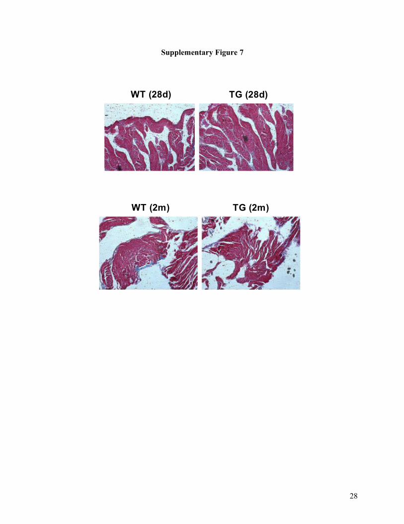
To exclude the possible involvement of structural components such as cardiac fibrosis (in
addition to the electrical alterations) in the observed AF in our miR-328/Tg model, we assessed
15
the anatomical properties of the transgenic heart. In our study, miR-328/Tg mice of 2-month age
were used for data collection. We did not see significant differences in the morphology and size
of the hearts and the thickness of ventricular walls between miR-328/Tg mice of 2-month age.
We observed slightly higher cardiac fibrosis in miR-328/Tg mice than in WT littermates, which
might contribute to the sustained AF in addition to the electrical remodeling process in our miR-
328/Tg mouse model (Supplementary Figure 7 online). Additionally, we consistently observed
sustained AF in younger miR-328/Tg mice (28 days or 4 weeks) after birth that had no cardiac
fibrosis.
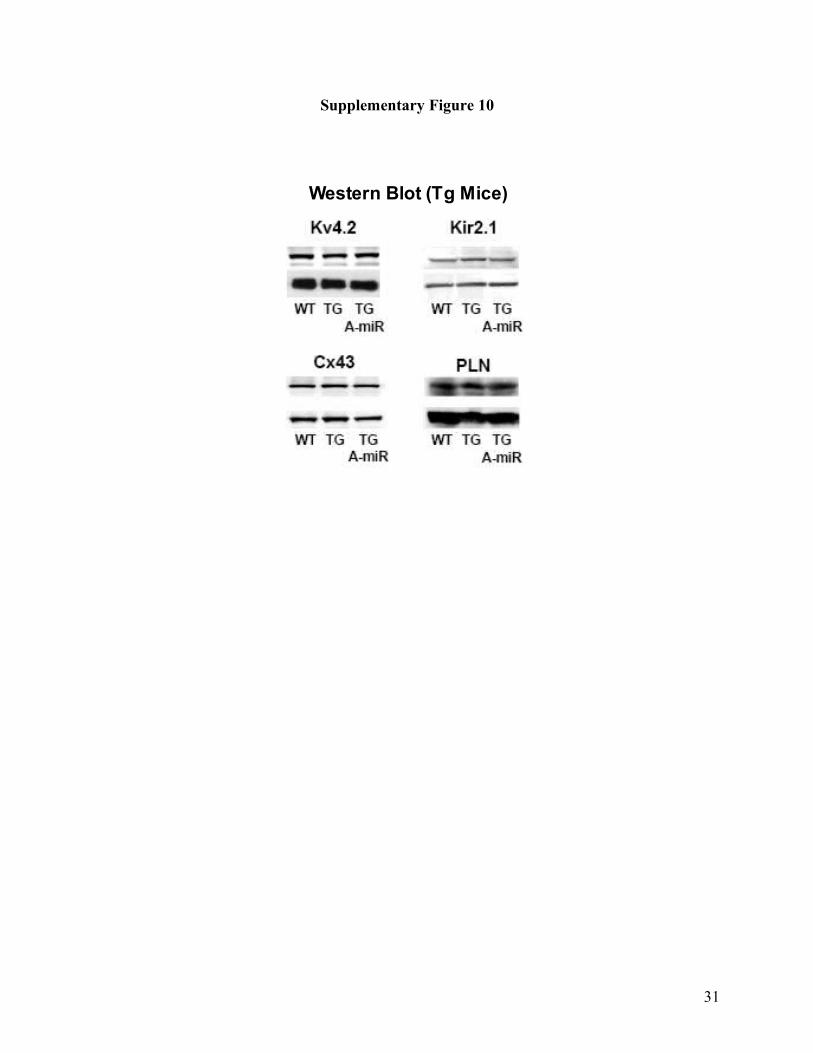
To exclude the possibility that miR-328 induces AF by targeting ion channel subunits
other than CACNA1C and CACNB1, we assessed the effects of miR-328 and antagomiR-328 on
the protein levels of several ion channel genes including KCNJ2 (encoding Kir2.1 for IK1),
KCND2 (encoding Kv4.2 for Ito in mice), GJA1 (encoding gap junction channel protein
connexin43 or Cx43), and phospholamban (a regulator of the Ca2+ pump). As expected, the
protein levels of these genes in miR-328/Tg mice were not different from those in WT littermates
and administration of antagomiR-328 did not affect the expression levels either (Supplementary
Figure 10 online).
Supplementary References
1. Yue L, Feng J, Gaspo R, Li GR, Wang Z, Nattel S. Ionic remodeling underlying action
potential changes in a canine model of atrial fibrillation. Circ Res.1997;81:512–520.
2. Yang B, Lin H, Xiao J, Lu Y, Luo X, Li B, Zhang Y, Xu C, Bai Y, Wang H, Chen G,
Wang Z. The muscle-specific microRNA miR-1 regulates cardiac arrhythmogenic potential by
targeting GJA1 and KCNJ2. Nat Med. 2007;13:486–491.
16
3. Luo X, Lin H, Lu Y, Li B, Xiao J, Yang B, Wang Z. Transcriptional activation by
stimulating protein 1 and post-transcriptional repression by muscle-specific microRNAs of IKs-
encoding genes and potential implications in regional heterogeneity of their expressions. J Cell
Physiol. 2007;212:358–367.
4. Luo X, Lin H, Xiao J, Zhang Y, Lu Y, Yang B, Wang Z. Downregulation of miRNA-
1/miRNA-133 contributes to re-expression of pacemaker channel genes HCN2 and HCN4 in
hypertrophic heart. J Biol Chem. 2008;283:20045–20052.
5. Xiao J, Luo X, Lin H, Zhang Y, Lu Y, Wang N, Zhang YQ, Yang B, Wang Z. MicroRNA
miR-133 represses HERG K+ channel expression contributing to QT prolongation in diabetic
hearts. J Biol Chem. 2007;282:12363–12367.
6. van Rooij E, Sutherland LB, Liu N, Williams AH, McAnally J, Gerard RD, Richardson
JA, Olson EN. A signature pattern of stress-responsive microRNAs that can evoke cardiac
hypertrophy and heart failure. Proc Natl Acad Sci USA. 2006;103:18255–18260.
7. Wang Y, Huang S. Adenovirus technology for gene manipulation and functional studies.
Drug Discovery Today. 2000;5:10–16.
8. Nakajima O, Okano S, Harada H, Kusaka T, Gao X, Hosoya T, Suzuki N, Takahashi S,
Yamamoto M. Transgenic rescue of erythroid 5-aminolevulinate synthase-deficient mice results
in the formation of ring sideroblasts and siderocytes. Genes Cells. 2006;11:685–700.
17
Supplemental Figure Legends
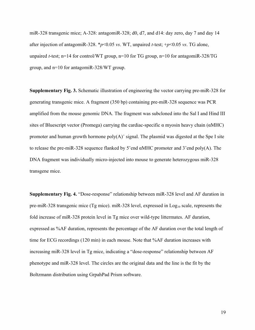
Supplementary Fig. 1. Schematic illustration of construction of adenovirus vector carrying pre-
miR-328. Rat miR-328 precursor DNA (5'-GGATCCgACCCCGTCCCCCCGTCCTCC
CCGAGTCCCTCTTTCGTAGATGTCGGGGACCGGGAGAGACGGGAAGGCAGGGGACA
GGGGTTTAttttttAAGCTT -3') was inserted into adenovirus shuttle plasmid pDC316-EGFP-U6.
pDC316-EGFP-U6 was then cotransfected with the infectious adenovirus genomic plasmid
pBHGlox∆E1,3Cre into 293 cells by lipofectamine. Following co-transfection of these two
DNAs, homologous recombination occurred to generate a recombinant adenovirus in which pre-
miR-328 is incorporated into the viral genome, replacing the ∆E1 region.
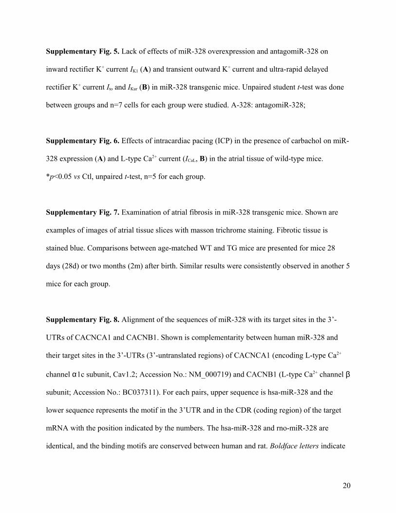
Supplementary Fig. 2. Verification of knockdown of miR-328 by its antisense oligo and
antagomiR. Upper panels: data generated by real-time RT-PCR expressed as mean±SEM; lower
panels: examples of Northern blot bands. (A) Co-application of AMO-328 with Adv-miR-328
effectively prevents the increase in miR-328 level in tissue mass from canine right atrial free wall
subjected to intramuscular injection. *p<0.05 vs pDC316, +p<0.05 vs Adv-miR-328 alone;
unpaired student t-test; n=5 tissue samples or cell batches for each group. pDC316: miR-328-free
adenovirus vector; Adv-miR-328: miR-328-carrying adenovirus vector; +AMO-328: co-infection
with the anti-miR-328 antisense oligo. (B) Co-transfection of AMO-328 with miR-328 prevents
the increase in miR-328 level in neonatal rat atrial cells. *p<0.05 vs Lipo, +p<0.05 vs miR-328
alone; unpaired student t-test; n=5 tissue samples or cell batches for each group. Lipo:
lipofectamine 2000; +AMO-328: co-transfection with the anti-miR-328 antisense oligo. (C) Tail
vein injection of antagomiR-328 rescues overexpression of miR-328 in TG mice (Left panels)
and knockdown endogenous miR-328 in WT mice (right panels). WT: wild-type mice; TG: pre-
18
miR-328 transgenic mice; A-328: antagomiR-328; d0, d7, and d14: day zero, day 7 and day 14
after injection of antagomiR-328. *p<0.05 vs. WT, unpaired t-test; +p<0.05 vs. TG alone,
unpaired t-test; n=14 for control/WT group, n=10 for TG group, n=10 for antagomiR-328/TG
group, and n=10 for antagomiR-328/WT group.
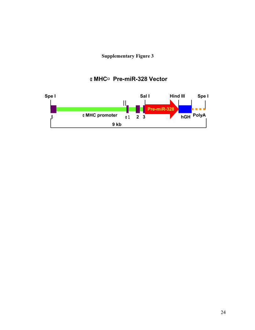
Supplementary Fig. 3. Schematic illustration of engineering the vector carrying pre-miR-328 for
generating transgenic mice. A fragment (350 bp) containing pre-miR-328 sequence was PCR
amplified from the mouse genomic DNA. The fragment was subcloned into the Sal I and Hind III
sites of Bluescript vector (Promega) carrying the cardiac-specific α myosin heavy chain (αMHC)
promoter and human growth hormone poly(A)+ signal. The plasmid was digested at the Spe I site
to release the pre-miR-328 sequence flanked by 5’end αMHC promoter and 3’end poly(A). The
DNA fragment was individually micro-injected into mouse to generate heterozygous miR-328
transgene mice.
Supplementary Fig. 4. “Dose-response” relationship between miR-328 level and AF duration in
pre-miR-328 transgenic mice (Tg mice). miR-328 level, expressed in Log10 scale, represents the
fold increase of miR-328 protein level in Tg mice over wild-type littermates. AF duration,
expressed as %AF duration, represents the percentage of the AF duration over the total length of
time for ECG recordings (120 min) in each mouse. Note that %AF duration increases with
increasing miR-328 level in Tg mice, indicating a “dose-response” relationship between AF
phenotype and miR-328 level. The circles are the original data and the line is the fit by the
Boltzmann distribution using GrpahPad Prism software.
19
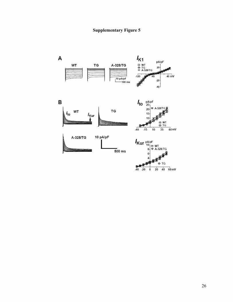
Supplementary Fig. 5. Lack of effects of miR-328 overexpression and antagomiR-328 on
inward rectifier K+ current IK1 (A) and transient outward K+ current and ultra-rapid delayed
rectifier K+ current Ito and IKur (B) in miR-328 transgenic mice. Unpaired student t-test was done
between groups and n=7 cells for each group were studied. A-328: antagomiR-328;
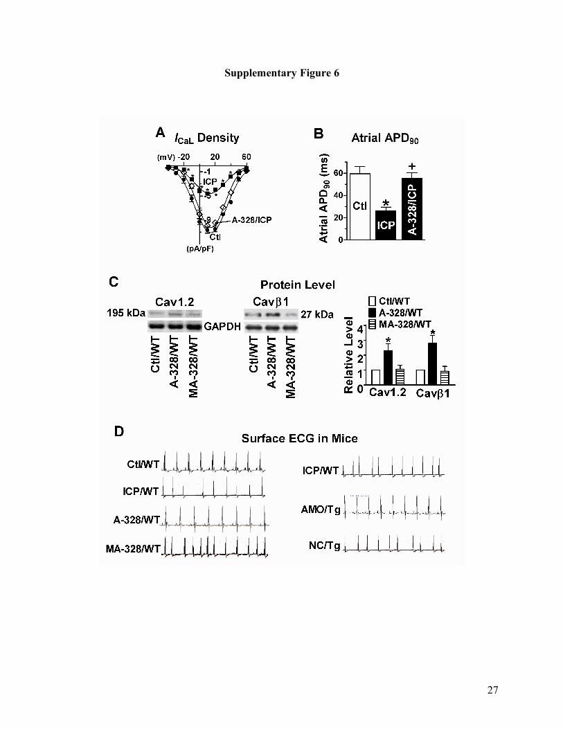
Supplementary Fig. 6. Effects of intracardiac pacing (ICP) in the presence of carbachol on miR-
328 expression (A) and L-type Ca2+ current (ICaL, B) in the atrial tissue of wild-type mice.
*p<0.05 vs Ctl, unpaired t-test, n=5 for each group.
Supplementary Fig. 7. Examination of atrial fibrosis in miR-328 transgenic mice. Shown are
examples of images of atrial tissue slices with masson trichrome staining. Fibrotic tissue is
stained blue. Comparisons between age-matched WT and TG mice are presented for mice 28
days (28d) or two months (2m) after birth. Similar results were consistently observed in another 5
mice for each group.
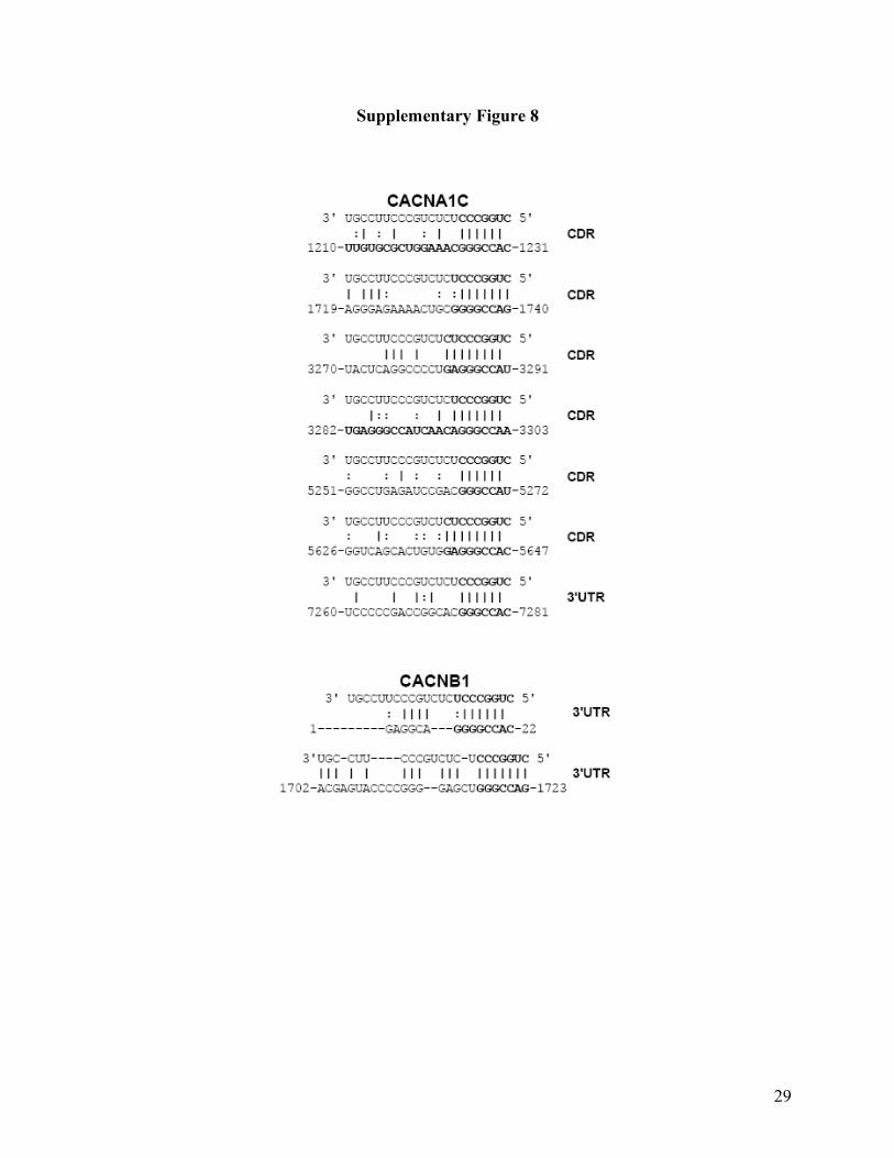
Supplementary Fig. 8. Alignment of the sequences of miR-328 with its target sites in the 3’-
UTRs of CACNCA1 and CACNB1. Shown is complementarity between human miR-328 and
their target sites in the 3’-UTRs (3’-untranslated regions) of CACNCA1 (encoding L-type Ca2+
channel α1c subunit, Cav1.2; Accession No.: NM_000719) and CACNB1 (L-type Ca2+ channel β
subunit; Accession No.: BC037311). For each pairs, upper sequence is hsa-miR-328 and the
lower sequence represents the motif in the 3’UTR and in the CDR (coding region) of the target
mRNA with the position indicated by the numbers. The hsa-miR-328 and rno-miR-328 are
identical, and the binding motifs are conserved between human and rat. Boldface letters indicate
20

that the seed site is critical for miRNA-mRNA binding and interaction and the miRNA::mRNA
base pairings.
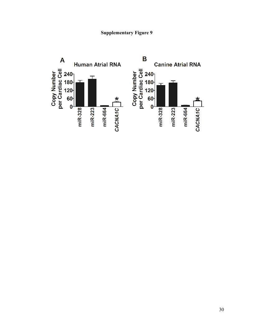
Supplementary Fig. 9. Relative abundance of miR-328, miR-664 and CACNA1C transcripts in
the RNA samples from healthy canine atrial tissues (A) and human atrial tissue (B), determined
by real-time RT-PCR. *p>0.05, unpaired t-test, n=5 batches of cells.
Supplementary Fig. 10. Effects of miR-328 overexpression and antagomiR-328 on the protein
levels of Kv4.2, Kir2.1, connexin43 (Cx43) and phospholamban (PLN), as a comparison with the
effects on Cav1.2 and Cavβ1. A-miR: antagomiR-328.
21
Supplemental Figures
Supplementary Figure 1
22
Supplementary Figure 2
23
Supplementary Figure 3
24
α MHC promoter hGH PolyAPre-miR-328Pre-miR-328
Sal I Hind III Spe ISpe I
β α 1 2 39 kb
α MHC- Pre-miR-328 Vector

Supplementary Figure 4
25
Supplementary Figure 5
26
Supplementary Figure 6
27
Supplementary Figure 7
WT (2m)
WT (28d)
TG (2m)
TG (28d)
28
Supplementary Figure 8
29
Supplementary Figure 9
30
Supplementary Figure 10
Western Blot (Tg Mice)
31
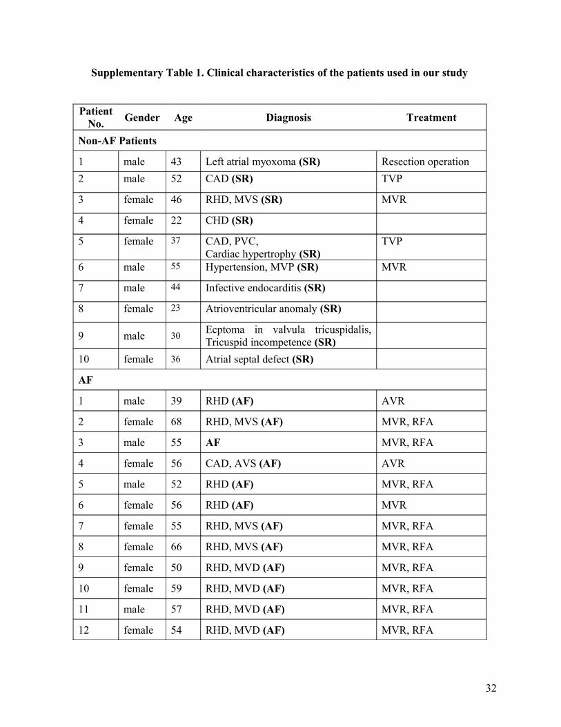
Supplementary Table 1. Clinical characteristics of the patients used in our study
PatientNo. Gender Age Diagnosis Treatment
Non-AF Patients
1 male 43 Left atrial myoxoma (SR) Resection operation2 male 52 CAD (SR) TVP
3 female 46 RHD, MVS (SR) MVR
4 female 22 CHD (SR)
5 female 37 CAD, PVC,Cardiac hypertrophy (SR)
TVP
6 male 55 Hypertension, MVP (SR) MVR
7 male 44 Infective endocarditis (SR)
8 female 23 Atrioventricular anomaly (SR)
9 male 30 Ecptoma in valvula tricuspidalis, Tricuspid incompetence (SR)
10 female 36 Atrial septal defect (SR)
AF
1 male 39 RHD (AF) AVR
2 female 68 RHD, MVS (AF) MVR, RFA
3 male 55 AF MVR, RFA
4 female 56 CAD, AVS (AF) AVR
5 male 52 RHD (AF) MVR, RFA
6 female 56 RHD (AF) MVR
7 female 55 RHD, MVS (AF) MVR, RFA
8 female 66 RHD, MVS (AF) MVR, RFA
9 female 50 RHD, MVD (AF) MVR, RFA
10 female 59 RHD, MVD (AF) MVR, RFA
11 male 57 RHD, MVD (AF) MVR, RFA
12 female 54 RHD, MVD (AF) MVR, RFA
32

AF, atrial fibrillation; AVR, aortic valve replacement; AVS, aortic valve stenosis; CAD, coronary
artery disease; CHD, congenital heart disease; MVP, mitral valve prolapse; MVR, mitral valve
replacement; MVD, mitral valve disease; MVS, mitral valve stenosis; RFA, radiofrequency
ablation; RHD, rheumatic heart disease; SR, sinus rhythm; TVP, tricuspid valvuloplasty.
33
Related Documents