BIOLOGIA PLANTARUM 51 (1): 7-14, 2007 7 Micropropagation of Juniperus phoenicea from adult plant explants and analysis of ploidy stability using flow cytometry J. LOUREIRO, A. CAPELO, G. BRITO, E. RODRIGUEZ, S. SILVA, G. PINTO and C. SANTOS 1 Laboratory of Biotechnology and Cytomics, Department of Biology, University of Aveiro, Campus Universitário de Santiago, 3810-193 Aveiro, Portugal Abstract We report here the successful micropropagation of adult Juniperus phoenicea L. with respective ploidy stability studies. Microcuttings with axillary buds were grown on five media supplemented with different growth regulator combinations. Best elongation rates were achieved on Driver and Kuniyuki (DKW) medium supplemented with kinetin alone or with naphthaleneacetic acid (NAA), while Rugini olive (OM) medium stimulated the development of new branches. Shoots growing on Murashige and Skoog (MS) medium browned and showed necrotic zones. Shoots of second to fourth subcultures usually had higher elongation rates than those of the first culture. For rooting assays, half strength DKW and OM media, different concentrations of growth regulators, auxin continuous exposure vs. dipping and the type of solid matrix were assessed. During rooting assays, two morphotypes were observed with one type having well developed internodes and the other showing hyperhydratation and no internode development. High rooting rates (40 %) were only obtained in the first morphotype shoots exposed for 5 min to 2.4 μM IBA and then transferred to OM medium without growth regulators. Plants were acclimatized in pots containing a mixture of peat and Perlite (3:2) in greenhouse with progressive reduction of relative humidity. A flow cytometric screening for major ploidy changes revealed no differences among the morphotypes and between them and the mother plant. Also the nuclear DNA content of this species was estimated for the first time using flow cytometry (2C = 24.71 pg). Additional key words: in vitro culture, nuclear DNA content, plant acclimatization, rooting studies. Introduction The genus Juniperus has about 75 to 80 species and is the second largest genus of conifers. Juniperus species are widely distributed in semiarid regions where they are used extensively for landscaping, wood and medicinal purposes. Juniperus phoenicea L. is native to some regions of the Mediterranean basin, Canary Islands and North Africa. Recently the taxonomic position of this species was questioned (for details see Rivas-Martínez et al. 1993) and this misleading taxonomy data requires urgent clarification. The lack of pollination, low pollen viability and/or embryo degeneration are important causes for reduced seed production in Juniperus (Ortiz et al. 1998). Also, species that grow upright are more recalcitrant than prostrate ones (Hartman et al. 1990). Dirr and Heuser (1987) reported that cuttings of some Juniperus species could be rooted using up to 4.5 % (m/v) indole-3-butyric acid (IBA). Also, Hartman et al. (1999) reported propagation of J. virginiana and J. procumbens by cuttings and of J. virginiana and J. chinensis by seed germination, but highlighted that the success of rooting may be lower than 10 % when adult trees are ⎯⎯⎯⎯ Received 23 March 2005, accepted 20 February 2006. Abbreviations: 2,4-D - 2,4-dichlorophenoxyacetic acid; BAP - benzylaminopurine; DKW - Driver and Kuniyuki medium; FCM - flow cytometry; IAA - indole-3-acetic acid; IBA - indole-3-butyric acid; KIN - kinetin; MS - Murashige and Skoog medium; NAA - naphthaleneacetic acid; OM - Rugini olive medium; PI - propidium iodide; SH - Schenk and Hildebrandt medium; TRIS - 3-(hydroxymethyl)-aminomethane; WPM - McCown Woody plant medium. Acknowledgements: Conceição Santos is a collaborator of CESAM/UA. This work was supported by FCT project PNAT/1999/AGR/15011/C. The acquisition of the flow cytometer was supported by the FCT/MCT project POCTI/AGR/C/11142/98. The work of João Loureiro in flow cytometry was financed by the fellowship FCT/SFRH/BD/9003/2002 and the work of Glória Pinto was financed by the fellowship FCT/SFRH/BD/8693/2002. Thanks are due to Prof. Jaroslav Doležel for providing the plant DNA standard seeds. Thanks are also due to Eng. Armando Costa for technical assistance in the laboratory and to Dr. Roberto Jardim (Director of the Botanical Garden of Madeira) and Quinta das Palmeiras for providing some of the material used in this study. 1 Corresponding author; fax: (+351) 234426408, e-mail: [email protected]

Welcome message from author
This document is posted to help you gain knowledge. Please leave a comment to let me know what you think about it! Share it to your friends and learn new things together.
Transcript
BIOLOGIA PLANTARUM 51 (1): 7-14, 2007
7
Micropropagation of Juniperus phoenicea from adult plant explants and analysis of ploidy stability using flow cytometry J. LOUREIRO, A. CAPELO, G. BRITO, E. RODRIGUEZ, S. SILVA, G. PINTO and C. SANTOS1 Laboratory of Biotechnology and Cytomics, Department of Biology, University of Aveiro, Campus Universitário de Santiago, 3810-193 Aveiro, Portugal Abstract We report here the successful micropropagation of adult Juniperus phoenicea L. with respective ploidy stability studies. Microcuttings with axillary buds were grown on five media supplemented with different growth regulator combinations. Best elongation rates were achieved on Driver and Kuniyuki (DKW) medium supplemented with kinetin alone or with naphthaleneacetic acid (NAA), while Rugini olive (OM) medium stimulated the development of new branches. Shoots growing on Murashige and Skoog (MS) medium browned and showed necrotic zones. Shoots of second to fourth subcultures usually had higher elongation rates than those of the first culture. For rooting assays, half strength DKW and OM media, different concentrations of growth regulators, auxin continuous exposure vs. dipping and the type of solid matrix were assessed. During rooting assays, two morphotypes were observed with one type having well developed internodes and the other showing hyperhydratation and no internode development. High rooting rates (40 %) were only obtained in the first morphotype shoots exposed for 5 min to 2.4 µM IBA and then transferred to OM medium without growth regulators. Plants were acclimatized in pots containing a mixture of peat and Perlite (3:2) in greenhouse with progressive reduction of relative humidity. A flow cytometric screening for major ploidy changes revealed no differences among the morphotypes and between them and the mother plant. Also the nuclear DNA content of this species was estimated for the first time using flow cytometry (2C = 24.71 pg). Additional key words: in vitro culture, nuclear DNA content, plant acclimatization, rooting studies. Introduction The genus Juniperus has about 75 to 80 species and is the second largest genus of conifers. Juniperus species are widely distributed in semiarid regions where they are used extensively for landscaping, wood and medicinal purposes. Juniperus phoenicea L. is native to some regions of the Mediterranean basin, Canary Islands and North Africa. Recently the taxonomic position of this species was questioned (for details see Rivas-Martínez et al. 1993) and this misleading taxonomy data requires urgent clarification.
The lack of pollination, low pollen viability and/or
embryo degeneration are important causes for reduced seed production in Juniperus (Ortiz et al. 1998). Also, species that grow upright are more recalcitrant than prostrate ones (Hartman et al. 1990). Dirr and Heuser (1987) reported that cuttings of some Juniperus species could be rooted using up to 4.5 % (m/v) indole-3-butyric acid (IBA). Also, Hartman et al. (1999) reported propagation of J. virginiana and J. procumbens by cuttings and of J. virginiana and J. chinensis by seed germination, but highlighted that the success of rooting may be lower than 10 % when adult trees are
⎯⎯⎯⎯ Received 23 March 2005, accepted 20 February 2006. Abbreviations: 2,4-D - 2,4-dichlorophenoxyacetic acid; BAP - benzylaminopurine; DKW - Driver and Kuniyuki medium; FCM - flow cytometry; IAA - indole-3-acetic acid; IBA - indole-3-butyric acid; KIN - kinetin; MS - Murashige and Skoog medium; NAA - naphthaleneacetic acid; OM - Rugini olive medium; PI - propidium iodide; SH - Schenk and Hildebrandt medium; TRIS - 3-(hydroxymethyl)-aminomethane; WPM - McCown Woody plant medium. Acknowledgements: Conceição Santos is a collaborator of CESAM/UA. This work was supported by FCT project PNAT/1999/AGR/15011/C. The acquisition of the flow cytometer was supported by the FCT/MCT project POCTI/AGR/C/11142/98. The work of João Loureiro in flow cytometry was financed by the fellowship FCT/SFRH/BD/9003/2002 and the work of Glória Pinto was financed by the fellowship FCT/SFRH/BD/8693/2002. Thanks are due to Prof. Jaroslav Doležel for providing the plant DNA standard seeds. Thanks are also due to Eng. Armando Costa for technical assistance in the laboratory and to Dr. Roberto Jardim (Director of the Botanical Garden of Madeira) and Quinta das Palmeiras for providing some of the material used in this study. 1 Corresponding author; fax: (+351) 234426408, e-mail: [email protected]
J. LOUREIRO et al.
8
used as mother plants (Edson et al. 1996). Unfortunately, J. phoenicea is not efficiently propagated by traditional methods and results are extremely inconsistent and not reproducible (Brito 2000). For example, and similarly to other Juniperus species, J. phoenicea does not have a high rate of plant production through seed germination (Ortiz et al. 1998).
Therefore, micropropagation should be performed in order to improve the propagation of Juniperus species. Contrarily to Pinus (e.g. Gomez and Segura 1995, 1996, Andersone and Ievinsh 2005) few information is available in Juniperus species. Gomez and Segura (1995) reported the proliferation of J. oxycedrus by axillary shoot proliferation and later found some morphogenic capacity in calluses derived from single cell culture of the same species (Gomez and Segura 1996). More recently, Shanjani (2003) highlighted the importance of nitrogen on callus induction and plant regeneration of J. excelsa, and recently new media formulation based on the explant mineral composition are being proposed as a new strategy for highly recalcitrant species (e.g. Gonçalves 2004). Alternatively, a new method involving germination of in vitro cultured embryos on MS medium was proposed to improve germination and propagation of J. oxycedrus ssp. oxycedrus and ssp. macrocarpa (Cantos et al. 1998).
Due to the possible occurrence of somaclonal variation the analysis of the ploidy stability of micropropagated plants is of particular importance. These types of studies in conifers are rare and the ones using flow cytometry (FCM) are even rarer. Gajdošová et al. (1995) used this technique to analyse the genetic stability of embryogenic calli of silver fir and its hybrids and Libiaková et al. (1995) analysed the genetic stability of Abies concolor × Abies grandis calli and in vitro regenerated shoots. FCM has the advantages that nuclei are analysed individually and at high speed, large populations can be measured in a short time and the presence of subpopulations can be detected (Shapiro 2003). Also, since there is no need to employ tissues with dividing cells and it is easy and rapid to prepare samples, FCM became the preferred method to perform this type of analyses (Doležel and Bartoš 2005).
This investigation describes for the first time a reliable protocol for in vitro regeneration and acclimatization of J. phoenicea plants derived from an adult field tree. The ploidy stability of plantlets derived from the micropropagation protocol and from rooting assays was assessed using FCM and the nuclear DNA content of this species was determined for the first time using leaves of the mother field tree.
Materials and methods In vitro culture establishment: Cuttings (15 - 20 cm long) from terminal branches of adult trees (20-year-old) from Porto Santo Island, collected in the spring, were used as source of microcuttings for in vitro studies. Half of the cuttings were treated immediately and the other half was preconditioned in a greenhouse (22 ± 1 ºC, 16-h photoperiod with irradiance of 400 µmol m-2 s-1) for at least one week with periodic sprays of a fungicide solution: 0.75 g dm-3 Derosal (Hoescht and Schering AgrEvo, Berlin, Germany) and 1.5 cm3 dm-3 PrevicurN (Hoescht and Schering AgrEvo). Cuttings were washed in tap water for 10 min, decontaminated in ethanol 70 % (v/v) for 1 min and then immersed two times (10 min each) in 200 cm3 of a commercial bleach solution (2.5 - 3.0 % available chloride) containing 5 drops of Teepol (Cruz Verde, Lisboa, Portugal). Cuttings were then rinsed in a sterilised fungicide solution of 1g dm-3 Benlate (Rhône-Poulenc, Lisboa, Portugal) for 10 min and washed in sterile water three times (5 min each).
To test the influence of medium composition and growth regulators on shoot propagation, decontaminated initial explants (1 cm tall cuttings with one axillary bud) were placed on different agar media: DKW (Driver and Kuniyuki 1984), MS (Murashige and Skoog 1962), OM (Rugini 1984), SH (Schenk and Hildebrandt 1972) or WPM (McCown and Lloyd 1981), supplemented with different growth regulators (Table 1). Each treatment consisted of ten 400 cm3 flasks (with 50 cm3 medium) containing five explants each giving a total of 50 explants.
Cultures were incubated in a growth chamber at 22 ± 1 ºC, with a 16-h photoperiod and irradiance of 98 µmol m-2 s-1 supplemented by OSRAM (Munich, Germany) L36W/21 lamps.
The influence of culture cycle on shoot multiplication was evaluated by comparing shoots response in different culture periods: 1st culture (shoots derived from the field mother plant), 2nd culture (shoots derived from 2-month old shoots of the 1st culture) and so on.
Shoot survival and morphological characters as shoot length, number of shoots per explant and number of branches per shoot were evaluated in each culture period. Callus production was also evaluated. Treatments were repeated two times in independent experiments. Rooting studies and plant acclimatization: Shoots (2.0 - 3.0 cm long) were transferred to different rooting conditions: various basal culture medium, type and concentration of growth regulator, continues exposure to auxin vs. auxin dipping for 1 min, 5 min or 1 h, and composition of the solid matrix (Table 2). Cultures were incubated under the conditions described previously for shoot culture. Each treatment consisted of eight 400 cm3 flasks containing 5 explants each giving a total of 40 explants. When roots were at least 2 cm long, in vitro plantlets were transferred to pots with sterilised mixture of peat : Perlite (3:2) and treated with a fungicide solution: 0.75 g dm-3 Derosal and 1.5 cm3 dm-3 PrevicurN. After that, they were transferred to a greenhouse and
MICROPROPAGATION OF JUNIPERUS PHOENICEA
9
grown at 22 ± 1 ºC, with a 16-h photoperiod and an irradiance of 400 µmol m-2 s-1, where they were acclimatized to progressive decreasing of relative humidity. Ploidy stability analysis using flow cytometry: Nuclear suspensions from micropropagated and mother plant leaves were prepared according to Galbraith et al. (1983). In short, nuclei were released from cells by chopping the tissue with a razor blade in a Petri dish containing 1 cm3
of Tris-MgCl2 buffer (Pfosser et al. 1995) (200 mM TRIS, 4 mM MgCl2 . 6 H2O, 0.5 % (v/v) Triton X-100, pH 7.5). The buffer was supplemented with 1 % (m/v) PVP-10 to reduce the possible influence of secondary metabolites on propidium iodide (PI) staining. The suspension of nuclei was then filtered through a 50 μm nylon filter and 50 μg dm-3 of PI (Fluka, Buchs, Switzerland) and 50 μg dm-3 of RNAse (Sigma, St. Louis, USA) were added to the samples to stain the DNA. Samples were analysed within a 10 min period in a Coulter EPICS XL (Coulter Electronics, Hialeah, FL, USA) flow cytometer equipped with an air-cooled argon-ion laser tuned at 15 mW and operating at 488 nm. Integral fluorescence and fluorescence pulse height and width emitted from nuclei were collected through a 645-dichroic long-pass filter and a 620-band-pass filter and converted on 1 024 ADC channels. Prior to analysis, the instrument was checked for linearity with Flow-Check fluorespheres (Beckman Coulter, Hialeah, FL, USA). Doublets, partial nuclei, nuclei with associated cytoplasm and other debris were discriminated using a specific gating region defined in a linear-fluorescence light scatter (FL) pulse integral vs. FL pulse height cytogram. Leaves of Secale cereale cv. Dankovske (2C = 15.95 pg, Doležel et al. 1998) were used as an internal reference standard. At least
5 000 nuclei were analysed per sample. To estimate the ploidy level, the position of the G0/G1
peak of the sample on a histogram was compared with the internal reference plant with known ploidy. The size of the nuclear genome of J. phoenicea was estimated according to the following formula: J. phoenicea 2C nuclear DNA content [pg] = (J. phoenicea G0/G1 peak mean / S. cereale G0/G1 peak mean) × 15.95. Conversion into base-pair numbers was performed using the following factor: 1 pg = 978 Mbp (Doležel et al. 2003).
The relative nuclear DNA content of 4 micropropa-gated plantlets obtained in OM medium was estimated and compared with the values obtained from 5 replicates of the adult mother field tree. To assay the possible influence of different rooting conditions on explants ploidy stability, the relative nuclear DNA content of at least one explant in OM medium from each rooting condition was estimated. Statistical analysis: Data from in vitro culture and rooting studies were averaged from two independent analyses and were analysed using the one-way analysis of variance (ANOVA). A multiple comparison Tukey-Kramer test was applied when necessary to determine exactly which groups were different (P < 0.05). Statistical analyses of data from ploidy stability assays were performed using an unpaired t-test (for comparison between micropropagated plantlets and the adult mother plant; P < 0.05) and a two-way ANOVA (for comparison between different rooting conditions – auxinic dipping and composition of solid matrix – and detection of possible interactions between them; P < 0.05). All statistical studies were performed using SigmaStat (Windows Version 3.1, SPSS Inc., Richmond, USA).
Results and discussion In vitro culture establishment: Higher surviving rates (58 % from a total of 30 explants) were obtained with explants derived from preconditioned cuttings (that were periodically treated with a fungicide solution) than with explants derived directly from the field (22 % from a total of 30 explants). The strategy of greenhouse preconditioning of field cuttings to prevent in vitro infections was already applied in micropropagation studies of other field trees with similar success (Pinto et al. 2002). Simultaneously, the method used for explants disinfection was successful.
Explants growing on DKW, OM and SH media presented, in the first culture, similar surviving rates (P ≤ 0.05) that were slightly higher than those of explants growing on WPM (P ≤ 0.1; data not shown) and higher than the surviving rates of explants on MS medium, which was more evident during the 2nd to 4th subcultures. In fact, with the exception of MS and SH media, survival rates increased considerably in subsequent subcultures
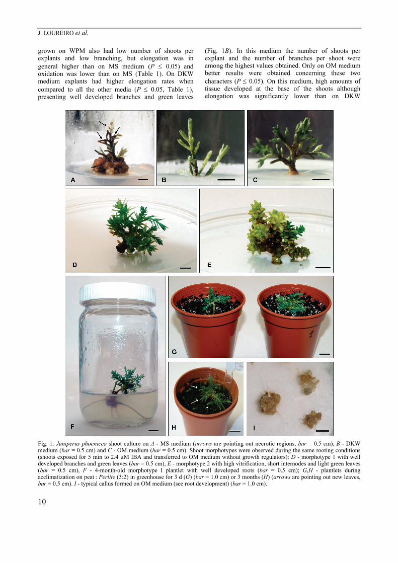
(76 - 100 %). On MS medium, shoots browned and showed apical necrosis that eventually spread during subcultures to the whole shoot leading to its death. Therefore, this medium was not suitable to establish in vitro shoot cultures in this species (Fig. 1A). On SH medium some of shoots dying, while the surviving shoots showed a light green colour and new growing zones.
No significant differences were obtained among different growth regulator combinations in regards of shoot survival rates; 100 % survival rates were obtained on DKW medium with 0.93 µM kinetin (KIN) or with 0.45 µM BAP and on OM medium with 2.74 µM NAA and 1.86 µM KIN.
Independently of the subculture, some morphologic differences (shoot length, number of shoots per explant and number of branches per shoot) were observed. Shoots grown on MS medium showed the lowest survival, lowest number of shoots per explant and a reduced number of branches per shoot (P ≤ 0.05; Table 1). Shoots
J. LOUREIRO et al.
10
grown on WPM also had low number of shoots per explants and low branching, but elongation was in general higher than on MS medium (P ≤ 0.05) and oxidation was lower than on MS (Table 1). On DKW medium explants had higher elongation rates when compared to all the other media (P ≤ 0.05, Table 1), presenting well developed branches and green leaves
(Fig. 1B). In this medium the number of shoots per explant and the number of branches per shoot were among the highest values obtained. Only on OM medium better results were obtained concerning these two characters (P ≤ 0.05). On this medium, high amounts of tissue developed at the base of the shoots although elongation was significantly lower than on DKW
Fig. 1. Juniperus phoenicea shoot culture on A - MS medium (arrows are pointing out necrotic regions, bar = 0.5 cm), B - DKW medium (bar = 0.5 cm) and C - OM medium (bar = 0.5 cm). Shoot morphotypes were observed during the same rooting conditions (shoots exposed for 5 min to 2.4 µM IBA and transferred to OM medium without growth regulators): D - morphotype 1 with well developed branches and green leaves (bar = 0.5 cm), E - morphotype 2 with high vitrification, short internodes and light green leaves (bar = 0.5 cm), F - 4-month-old morphotype I plantlet with well developed roots (bar = 0.5 cm); G,H - plantlets during acclimatization on peat : Perlite (3:2) in greenhouse for 3 d (G) (bar = 1.0 cm) or 3 months (H) (arrows are pointing out new leaves, bar = 0.5 cm). I - typical callus formed on OM medium (see root development) (bar = 1.0 cm).
MICROPROPAGATION OF JUNIPERUS PHOENICEA
11
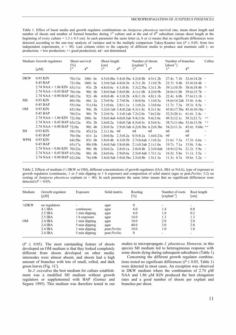
Table 1. Effect of basal media and growth regulator combinations on Juniperus phoenicea survival rate, mean shoot length and number of shoots and number of formed branches during 1st culture and at the end of 4th subculture (mean shoot length at the beginning of every culture = 1.3 ± 0.3 cm). In each parameter the same letter (a, b or c) means that no significant differences were detected according to the one-way analysis of variance and to the multiple comparison Tukey-Kramer test (P ≤ 0.05; from two independent experiments, n = 30). Last column refers to the capacity of different media to produce and maintain calli (- no production; + low production; ++ good production). nd - not determined.
Medium Growth regulators Shoot survival [%]
Shoot length [cm]
Number of shoots [explant-1]
Number of branches [shoot-1]
Callus
[µM] 1st 4th 1st 4th 1st 4th 1st 4th
DKW 0.93 KIN 70±13a 100± 0c 4.5±0.8bc 5.4±0.5bc 4.2±0.8b 4.5±1.2b 27.4± 7.1b 32.6±14.2b - 0.45 BAP 72±10a 100± 0c 3.5±0.5ab 4.8±0.3b 4.7±1.3b 3.1±0.7b 23.7± 9.4b 35.4±18.4b - 2.74 NAA + 1.86 KIN 63±11a 93± 2b 4.8±0.6c 6.1±0.8c 5.3±2.5bc 3.3±1.3b 39.1±10.5b 38.4±18.4b + 2.74 NAA + 0.45 BAP 70±14a 90± 6b 3.8±0.8ab 3.8±0.8b 4.1±1.0b 4.2±0.9b 24.0±11.8b 39.6±15.7b - 2.74 NAA + 0.90 BAP 68±15a 92± 4b 4.0±0.4b 4.1±0.2b 4.8±1.1b 4.8±1.1b 22.4± 6.0b 37.6±11.1b - MS 0.93 KIN 60±18a 66± 2a 2.5±0.9a 2.7±0.9a 1.0±0.0a 1.1±0.3a 18.6±14.2ab 15.4± 6.4a - 0.45 BAP 55±16a 51±14a 2.1±0.6a 2.8±1.1a 1.1±0.2a 1.3±0.6a 11.7± 7.3a 19.3± 8.5a - OM 0.93 KIN 65±16a 94± 2b 3.2±0.1ab 3.4±0.2ab 8.3±1.8c 8.3±1.8c 43.0±17.5bc 45.4±10.1bc - 0.45 BAP 70±21a 90± 7b 2.2±0.5a 3.1±0.1ab 7.2±2.0c 7.6±2.0c 52.2±20.1c 61.6± 2.4c - 2.74 NAA + 1.86 KIN 72±10a 100± 0c 3.0±0.4ab 4.0±0.5ab 9.4±3.0c 9.4±3.0c 49.5±12.1c 59.3±21.7c ++ 2.74 NAA + 0.45 BAP 65±12a 95± 2b 2.6±0.2a 3.0±0.7ab 8.5±0.5c 8.5±0.5c 38.7±11.6bc 33.4±13.5b ++ 2.74 NAA + 0.90 BAP 72±8a 90± 4b 2.8±0.5a 2.9±0.3ab 6.2±0.3bc 6.2±0.3bc 54.2±11.3c 45.6± 9.6bc ++ SH 0.93 KIN 58±15a 45±12a 2.1±1.0a nd nd nd nd nd - 0.45 BAP 70±10a 61± 2a 1.0±0.8a 2.3±0.2a 0.5±0.2a 1.4±0.23a nd nd - WPM 0.93 KIN 64±20a 85± 6b 3.8±0.4b 4.3±0.5b 2.7±0.6ab 1.1±0.2a 21.6± 7.3a 17.3± 6.6a - 0.45 BAP 65±17a 90±10b 3.4±0.5ab 3.8±0.6b 2.1±0.3ab 2.1±1.0a 19.7± 7.1a 15.8± 3.4a - 2.74 NAA + 0.86 KIN 70±22a 90± 0b 2.0±0.2a 2.8±0.1a 2.8±0.4b 2.3±0.8ab 14.9±12.9a 21.2± 5.9a - 2.74 NAA + 0.45 BAP 67±10a 88± 6b 2.6±0.6a 2.9±0.6a 2.5±0.4ab 1.7±1.1a 16.5± 5.8a 11.1± 3.5a - 2.74 NAA + 0.90 BAP 62±24a 76±10b 3.4±0.5ab 3.9±0.5bc 2.5±0.8b 1.5±1.3a 11.3± 4.7a 19.6± 5.2a -
Table 2. Effects of medium (½ DKW or OM), different concentrations of growth regulators (IAA, IBA or NAA), type of exposure to growth regulators (continuous, 1 or 5 min dipping or 1 h exposure) and composition of solid matrix (agar or peat:Perlite; 3:2) on rooting of Juniperus phoenicea explants (n = 40). In each parameter the same letter means that no significant differences were detected (P < 0.05).
Medium Growth regulator [µM]
Exposure Solid matrix Rooting [%]
Number of roots [explant-1]
Root length [cm]
½DKW no regulators - agar 0 - - 4.1 IBA continuous agar 6.0 1.4 0.8 2.5 IBA 1 min dipping agar 6.0 1.0 0.2 2.5 IBA 1 h exposure agar 14.0 1.3 1.3 OM 2.4 IBA 1 min dipping agar 10.0 2.0 2.0 2.4 IBA 5 min dipping agar 40.0 3.0 2.0 2.4 IBA 1 min dipping peat:Perlite 10.0 1.0 1.0 2.4 IBA 5 min dipping peat:Perlite 0 - -
(P ≤ 0.05). The most outstanding feature of shoots developed on OM medium is that they looked completely different from shoots developed on other media: internodes were almost absent, and shoots had a high amount of branches with lots of small, rolled, and dark green leaves (Fig. 1C).
In J. oxicedrus the best medium for culture establish-ment was a modified SH medium without growth regulators or supplemented with BAP (Gomez and Segura 1995). This medium was therefore tested in our
studies to micropropagate J. phoenicea. However, in this species SH medium led to heterogeneous response with some shoots dying during subsequent subcultures (Table 1).
Concerning the different growth regulator combina-tions tested no significant differences (P ≤ 0.05, Table 1) were detected in most cases. An exception was observed in DKW medium where the combination of 2.74 µM NAA and 1.86 µM KIN produced the best elongation rates and a good number of shoots per explant and branches per shoot.
J. LOUREIRO et al.
12
In general higher shoot lengths and number of branches per shoot were observed during subsequent subcultures. This may be explained by the fact that, during 1st culture, growth could be limited by stress imposed by the decontamination process and that shoots need to adjust to the in vitro conditions as they derived from stems of field plants. By other way, shoots in the 2nd to 4th subcultures were already adapted and able to maximise growth by using the conditions supplied by the in vitro culture, and frequently, new formed regions (evident by their light green colour) could be observed at the top of the branches. Rooting studies and plant acclimatization: For rooting experiments two strategies were adopted concerning shoots exposure to auxins (IAA, IBA and NAA): continuous exposure or dipping for 1 min, 5 min or 1 h (Table 2). These studies were performed on half strength DKW (½ DKW) and OM medium as these were the media where the best results were obtained previously. On ½ DKW medium, rooting was only observed when IBA was chosen. For this reason IBA was the growth regulator in the OM medium rooting assays. Concerning the continuous auxin exposure, rooting was only observed on ½ DKW medium with 4.1 µM IBA (Table 2). On this medium root primordia were observed in 6 % of shoots and 4 weeks after transfer to rooting medium (Table 2). When shoots were exposed for 1 h to 2.5 mM IBA, roots appeared in 14 % of them and a 1 min dipping induced roots in 6 % of the shoots.
On OM medium, dipping in 2.4 µM IBA induced higher percentages of rooting (10 % for 1 min exposure and 40 % for 5 min exposure). These values were higher in agar medium when compared to the same conditions on a peat and Perlite mixture (n = 20).
Comparing with previous results in the same genus, this species seems to have different rooting abilities. For example, rooting of J. scoppulorum reached 82 % (Dirr and Heuser 1987). However, this macropropagation depends on the species/ecotype, and is reduced when adult trees are used. Wagner et al. (1994) obtained less than 10 % rooting in cuttings from 12-year-old mother plants. Gomez and Segura (1995) reported low rooting capacity of in vitro shoots of J. oxycedrus, and the genotypes of J. phoenicea from Porto Santo Island were for a long time extremely recalcitrant to both ex vitro and in vitro rooting (Brito 2000). Some of the reasons for this difficulty may be the peculiar environmental conditions of the Island together with the genotype/ecotypes used.
Finally, two morphotypes were observed: one group (app. 70 %) defined as “Morphotype I” had apparently normal shoots with green leaves, well developed internodes and a large number of branches (Fig. 1D); the other group (app. 30 %), defined as “Morphotype II”, was characterized by frequent hyperhydratation and short/almost absent internodes (Fig. 1E). Rooting was only achieved in shoots of the “Morphotype I“ (Fig. 1F).
Plant acclimatization in greenhouse is still in course and restricted to few plants but, in “Morphotype I“, the
acclimatisation is up to the moment 70 % successful (Fig. 1G,H).
When developing a plant regeneration protocol, callus formation should be avoided. However, among the 19 media combinations tested some led to the production of callus tissue. This is the first report on callus production in J. phoenicea (Fig. 1I). Callus production may be important for studies of indirect morphogenesis (e.g. somatic embryogenesis) – that are presently occurring in our laboratory – or for studies of production of secondary metabolites. An interesting example is the production of high contents of podophyllotoxin (a strong anti-tumor agent) in callus tissue from leaves of J. chinensis on SH medium supplemented with 16.2 µM NAA and 0.93 µM KIN (Muranaka et al. 1998). Other growth regulator combinations as 4.5 µM 2,4-dichloro-phenoxyacetic acid (2,4-D) and 0.93 µM KIN or 5.4 µM NAA and 0.93 µM KIN also originated good calli production although not so efficiently (Muranaka et al. 1998). Also, Gomez and Segura (1995, 1996) achieved callus formation from adult J. oxycedrus plants in SH medium with 2,4-D. Our results with J. phoenicea confirm that, similarly to J. chinensis and among the combinations tested, NAA plays an important role on callus production as the media that induced callus had this growth regulator (Table 1). On the other hand, stems showed to be, in this species, a good explant source for callus production. Ploidy stability: FCM analyses were applied to study the ploidy stability of J. phoenicea plantlets derived from the micropropagation protocol and from rooting assays. In J. phoenicea to assure that a sufficient number of nuclei were obtained the amount of material per sample had to be increased.
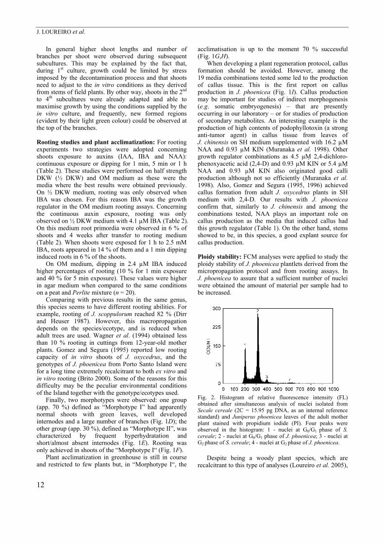
Fig. 2. Histogram of relative fluorescence intensity (FL) obtained after simultaneous analysis of nuclei isolated from Secale cereale (2C = 15.95 pg DNA, as an internal reference standard) and Juniperus phoenicea leaves of the adult mother plant stained with propidium iodide (PI). Four peaks were observed in the histogram: 1 - nuclei at G0/G1 phase of S. cereale; 2 - nuclei at G0/G1 phase of J. phoenicea; 3 - nuclei at G2 phase of S. cereale; 4 - nuclei at G2 phase of J. phoenicea.
Despite being a woody plant species, which are
recalcitrant to this type of analyses (Loureiro et al. 2005),
MICROPROPAGATION OF JUNIPERUS PHOENICEA
13
Table 3. Nuclear DNA content of micropropagated plantlets and adult mother field tree from which they were obtained. Means ± SD, CV - coefficient of variation. The differences were not significant at P ≤ 0.05; n = 4 for micropropagated plantlets and 5 for trees. For detail see Materials and methods.
Nuclear DNA content [pg/2C]
1C genome size [Mbp]
CV [%]
Plantlets 24.46±0.645 11961 3.46 Adult mother tree 24.71±0.178 12083 2.44
Table 4. Nuclear DNA content of Juniperus phoenicea morphotypes cultivated on OM medium and dipped in 5.0 µM IBA for 1 or 5 min before rooting on agar or peat:Perlite (3:2). Means ± SD, CV - coefficient of variation. The differences were not significant at P ≤ 0.05; n = 1 - 3. For detail see Materials and methods.
Morphotype Dipping [min]
Solid matrix
Nuclear DNA content [pg/2C]
CV [%]
n
1 24.94±0.257 3.62 3 5
agar 25.03±0.231 3.38 3
1 25.09 4.45 1
1
5 peat:Perlite
24.48 4.24 1 1 24.61 2.29 1 5
agar 24.86±0.420 2.58 2
1 24.25 3.02 1
2
5 peat:Perlite
25.03±0.101 2.87 2
the mean coefficient of variation (CV) values obtained (Table 3 and 4) were below 5 that is considered the acceptance criterion by Galbraith et al. (2002). As expected, the internal standard S. cereale had a lower 2C distribution (mean CV = 3.0 %; Fig. 2). The development of a protocol that gives low CV values is of crucial importance (this issue is fully addressed by Pinto et al. 2004) as it is a measure of the precision of the analysis. Unfortunately, in many reports this information is not provided and this difficult results interpretation. There is usually a correlation between higher CV values and higher background noise, especially in lower channel
numbers (Emshwiller 2002). This applies to this case where some background noise was observed. Despite that, the FCM protocol established here for J. phoenicea provided good results demonstrating its reliability and potential use in other Juniperus species.
Considering the in vitro culture protocol used to establish J. phoenicea, the mean nuclear DNA fluorescence index (DI = 2CJ. phoenicea/2CS. cereale) obtained for the micropropagated plantlets (DI = 1.533) was very similar (P ≤ 0.05) to the one obtained for the mother field plant (DI = 1.549). These results strongly indicate that the micropropagation protocol described here apparently does not induce major ploidy changes. Nevertheless, the CV values obtained could mask the possible occurrence of small differences in nuclear DNA content (aneuploidy and DNA polymorphism) and therefore complementary studies, such as chromosome counting and micro-satellites, to evaluate this situation, are presently being conducted in our laboratory.
To assay the possible influence of different rooting conditions on explants ploidy stability, the relative nuclear DNA content of plantlets from each morphotype was assessed (Table 4). The DI values obtained for each set of conditions were very similar (P ≤ 0.05). Also, no statistically significant differences were detected between the two morphotypes, revealing that the morphologic differences did not reflect major genetic changes. Up to the moment, all micropropagated plants that were acclimatized showed a normal development. Never-theless, more refine techniques (as microsatellites to assess DNA mutations and DNA methylation to evaluate the influence of epigenetic changes) are currently being performed in our laboratory.
The determination of nuclear DNA content of J. phoenicea L. in absolute units was performed using the adult field tree and the obtained value was 2C = 24.71 ± 0.178 pg (1C genome size = 12 083 Mbp). This is the first report for this species and the obtained value is similar to the values obtained by Dhillon (1987) using Feulgen staining and microspectrophotometry and Hizume et al. (2001) using FCM for other Juniperus species (J. virginiana, J. conferta and J. rigida), and as reported above FCM may also be used as an important tool for taxonomic studies in this genus.
References Andersone, U., Ienvish, G.: In vitro regeneration of mature
Pinus sylvestris buds stored at freezing temperatures. - Biol. Plant. 49: 281-284, 2005.
Brito G.: Micropropagação de duas espécies autóctones da Ilha de Porto Santo (Olea europaea L. ssp. maderensis Lowe e Juniperus phoenicea L.) e estudo da resposta de rebentos in vitro a stress osmótico. - Master Thesis, University of Aveiro, Aveiro 2000.
Cantos, M., Cuerva, J., Zarate, R., Troncoso, A.: Embryo rescue and development of Juniperus oxycedrus subsp. oxycedrus and macrocarpam seed. - Sci. Technol. 26: 193-198, 1998.
Dhillon, S.S.: DNA in tree species. - In: Bouga, J.M., Deuzan,
D.J. (ed.): Cell and Tissue Culture in Forestry. Pp. 298-313. Martinus Nijhoff, Dordrecht 1987.
Dirr, M.A., Heuser, C.W. (ed.): The Reference Manual of Woody Plant Propagation: From Seed to Tissue Culture. - Varsity Press, Athens 1987.
Doležel, J., Bartoš, J.: Plant DNA flow cytometry and estimation of nuclear genome size. - Ann. Bot. 95: 99-110, 2005.
Doležel, J., Bartoš, J., Voglmayr, H., Greilhuber, J.: Nuclear DNA content and genome size of trout and human. - Cytometry Part A 51: 127-128, 2003.
Doležel, J., Greilhuber, J., Lucretti, S., Meister, A., Lysák, M.,
J. LOUREIRO et al.
14
Nardi, L., Obermayer, R.: Plant genome size estimation by flow cytometry: inter-laboratory comparison. - Ann. Bot. 82: 17-26, 1998.
Driver, J.A., Kuniyuki, A.H.: In vitro propagation of paradox walnut rootstock. - HortScience 19: 507-509, 1984.
Edson, J.L., Wenny, D.L., Dumroese, R.K., Leege-Brusven, A.: Mass propagation of rocky mountain juniper from stem cuttings. - Tree Planters’ Notes 47: 94-99, 1996.
Emshwiller, E.: Ploidy levels among species in the Oxalis tuberosa 'Alliance' as inferred by flow cytometry. - Ann. Bot. 89: 741-753, 2002.
Gajdošová, A., Vooková, B., Kormuťák, A., Libiaková, G., Doležel, J.: Induction, protein-composition and DNA-ploidy level of embryogenic calli of silver fir and its hybrids. - Biol. Plant. 37: 169-176, 1995.
Galbraith, D.W., Harkins, K.R., Maddox, J.M., Ayres, N.M., Sharma, D.P., Firoozabady, E.: Rapid flow cytometric analysis of the cell-cycle in intact plant-tissues. - Science 220: 1049-1051, 1983.
Galbraith, D.W., Lambert, G., Macas, J., Doležel, J.: Analysis of nuclear DNA content and ploidy in higher plants. - In: Robinson, J., Azmi, A., Tutois, S. (ed.): Current Protocols in Cytometry. Pp. 7.6.1-7.6.15. John Wiley & Sons, New York 2002.
Gomez, M.P., Segura, J.: Axillary shoot proliferation in cultures of explants from mature Juniperus oxycedrus trees. - Tree Physiol. 15: 625-628, 1995.
Gomez, M.P., Segura, J.: Morphogenesis in leaf and single-cell cultures of mature Juniperus oxycedrus. - Tree Physiol. 16: 681-686, 1996.
Gonçalves, S., Correia, P.J., Martins-Loução, M.A., Romano, A.: A new medium for in vitro rooting of carob tree based on leaf macronutrients concentrations. - Biol. Plant. 49: 277-280, 2004.
Hartman, H.T., Kester, D.E., Davies, F.T. (ed.): Plant Propagation: Principles and Practices. - Prentice Hall International, 1990.
Hartman, H.T., Kester, D.E., Davies, F.T., Geneve, R. (ed.): Plant Propagation: Principles and Practices. - Prentice Hall International, Englewood Cliffs 1999.
Hizume, M., Kondo, T., Shibata, F., Ishizuka, R.: Flow cytometric determination of genome size in the Taxodiaceae, Cupressaceae sensu stricto and Sciadopityaceae. - Cytologia 66: 307-311, 2001.
Libiaková, G., Gajdošová, A., Vooková, B., Kubincová, M., Doležel, J.: Karyological study of Abies concolor × Abies grandis calli and shoots regenerated in vitro. - Biologia 50: 61-64, 1995.
Loureiro, J., Pinto, G., Lopes, T., Doležel, J., Santos, C.:
Assessment of genetic stability of Quercus suber L. somatic embryogenesis process using flow cytometry. - Planta 221: 815-822, 2005.
McCown, B.H., Lloyd, G.: Woody plant medium (WPM) - a mineral nutrient formulation for microculture of woody plant-species. - HortScience 16: 453-453, 1981.
Muranaka, T., Miyata, M., Ito, K., Tachibana, S.: Production of podophyllotoxin in Juniperus chinensis callus cultures treated with oligosaccharides and a biogenetic precursor. - Phytochemistry 49: 491-496, 1998.
Murashige, T., Skoog, F.: A revised medium for rapid growth and bioassays with tobacco tissue cultures. - Physiol. Plant. 15: 473-497, 1962.
Ortiz, P.L., Arista, M., Talavera, S.: Low reproductive success in two subspecies of Juniperus oxycedrus L. - Int. J. Plant Sci. 159: 843-847, 1998.
Pfosser, M., Amon, A., Lelley, T., Heberle-Bors, E.: Evaluation of sensitivity of flow cytometry in detecting aneuploidy in wheat using disomic and ditelosomic wheat-rye addition lines. - Cytometry 21: 387-393, 1995.
Pinto, G., Loureiro, J., Lopes, T., Santos, C.: Analysis of the genetic stability of Eucalyptus globulus Labill. somatic embryos by flow cytometry. - Theor. appl. Genet. 109: 580-587, 2004.
Pinto, G., Valentim, H., Costa, A., Castro, S., Santos, C.: Somatic embryogenesis in leaf callus from a mature Quercus suber L. tree. - In vitro cell. dev. Biol. Plant. 38: 569-572, 2002.
Rivas-Martínez, S., Wildpret, W., Paz, P.L.P.: Datos sobre Juniperus phoenicea aggr. (Cupressaceae). - Itinera Geobot. 7: 509-512, 1993.
Rugini, E.: In vitro propagation of some olive (Olea europaea L. var. sativa) cultivars with different root-ability, and medium development using analytical data from developing shoots and embryos. - Sci. Hort. 24: 123-134, 1984.
Schenk, R.U., Hildebrandt, A.C.: Medium and techniques for induction and growth of monocotyledonous and dicotyledonous plant-cell cultures. - Can. J. Bot. 50: 199, 1972.
Shanjani, P.S.: Nitrogen effect on callus induction and plant regeneration of Juniperus excelsa. - Int. J. agr. Biol. 5: 419-422, 2003.
Shapiro, H.M. (ed.): Practical Flow Cytometry. - Wiley-Liss, New York 2003.
Wagner, A.M., Harrington, J.T., Mexal, J.G., Fisher, J.T.: Progress report on propagation of Juniperus for conservation plantings. - In: Proceedings: Intermountain Nursery Conference. Pp. 131-136. St. Louis 1994.
Related Documents