Welcome message from author
This document is posted to help you gain knowledge. Please leave a comment to let me know what you think about it! Share it to your friends and learn new things together.
Transcript


The Art Behind the ScienceThe Art Behind the Science
AWARDAWARD
SBMM 2009 OUTSTANDING MICROGRAPHSBMM 2009 OUTSTANDING MICROGRAPH

The Art Behind the ScienceThe Art Behind the Science
AWARD – AWARD – Materials SciencesMaterials Sciences
SBMM 2009 OUTSTANDING MICROGRAPHSBMM 2009 OUTSTANDING MICROGRAPH

The Art Behind the ScienceThe Art Behind the ScienceAWARD – AWARD – Materials SciencesMaterials Sciences
FIRST PLACEFIRST PLACE
"This is an SEM image (taken using a Quanta 200 FEG – FEI) of a silver-titanium dioxide (Ag/TiO2) nanocomposite synthesized by sol-gel process. The Ag micro-crystal is above the TiO2 nanoparticles.“
Author(s): Marcelo Machado Viana (Viana M. M.)
Affiliation: Universidade Federal de Minas Gerais

Marcelo Machado Viana

The Art Behind the ScienceThe Art Behind the ScienceAWARD – AWARD – Materials SciencesMaterials Sciences
SECOND PLACESECOND PLACE
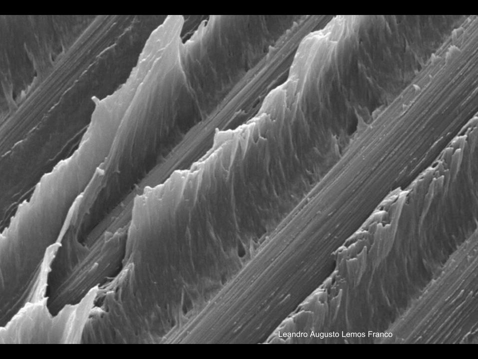
"MEV fractography of a Carbon fiber reinforced Poly Phenilene Sulfide composite after a short bean interlaminar shear strenght test. Surface was coated with a gold thin layer for better conductive. Fractography reveals high plastic matrix deformation and bare fibers."
Author(s): Leandro Augusto Lemos Franco (Leandro Franco)
Affiliation: Instituto de Aeronáutica e Espaço
Leandro Augusto Lemos Franco

The Art Behind the ScienceThe Art Behind the Science
AWARD – AWARD – Life SciencesLife Sciences
SBMM 2009 OUTSTANDING MICROGRAPHSBMM 2009 OUTSTANDING MICROGRAPH
The Art Behind the ScienceThe Art Behind the ScienceAWARD – AWARD – Life SciencesLife Sciences
FIRST PLACEFIRST PLACE
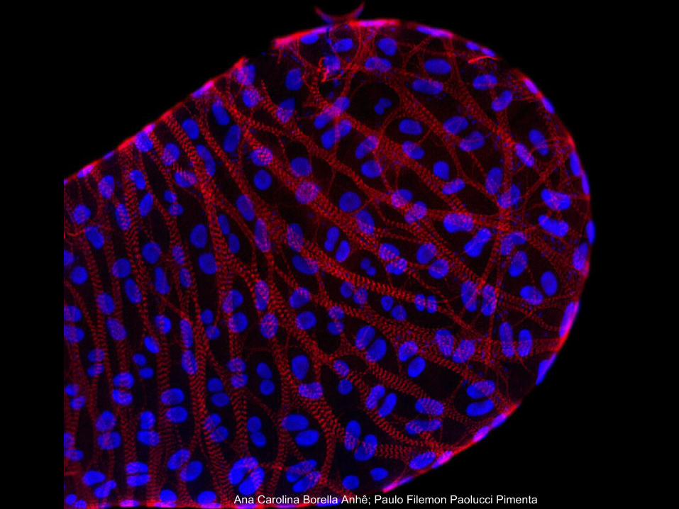
"Fluorescence microscopy of salivary gland of Rhodnius prolixus labeled by rhodamine-phalloidin (red) and DAPI (blue). It is observed the transversal muscle layers covering the entire gland (red) and the two nuclei of each salivary gland cell (blue). Bar= 100μm."
Author(s): Ana Carolina Borella Anhê; Paulo Filemon Paolucci Pimenta (Anhê, A.C.B.; Pimenta, P.F.P.)
Affiliation: CPqRR - FIOCRUZ
Ana Carolina Borella Anhê; Paulo Filemon Paolucci Pimenta
The Art Behind the ScienceThe Art Behind the ScienceAWARD – AWARD – Life SciencesLife Sciences
SECOND PLACESECOND PLACE
"Electron micrograph of mosquito of the genus Haemagogus vector of wild yellow fever. Scanning electron microscopy reveals details of the scales and bristles of the wings of these mosquitoes.“
Author(s): Jacenir Reis dos Santos Mallet (Jacenir Santos-Mallet)
Affiliation: LTL/IOC/FIOCRUZ
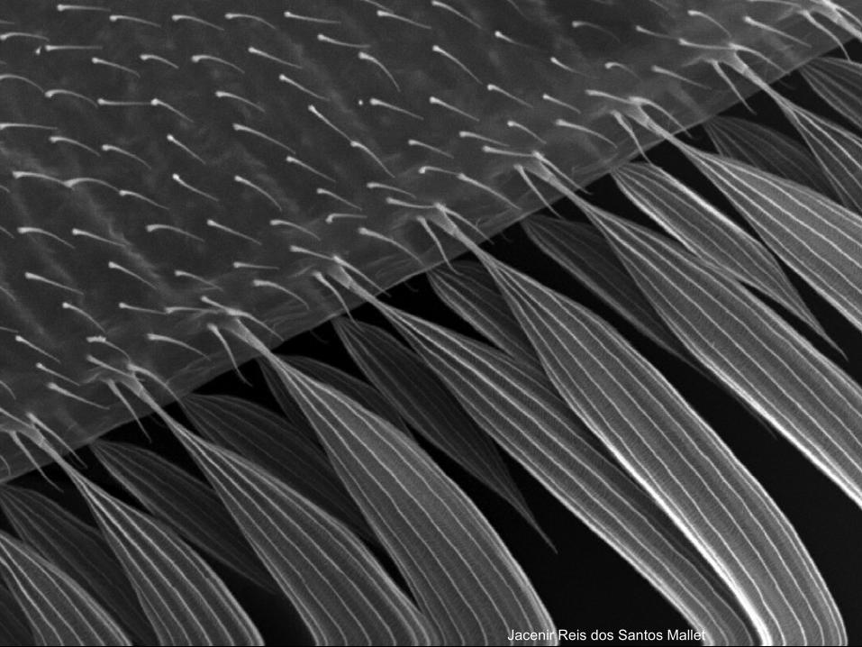
Jacenir Reis dos Santos Mallet
Related Documents