Microfauna associated with amoebic gill disease in sea-farmed Atlantic salmon, Salmo salar L., smolts M L Bermingham and M F Mulcahy Environmental Research Institute, Aquaculture and Fisheries Development Centre, Department of Zoology, Ecology and Plant Science, National University of Ireland, Cork, Ireland Abstract A study of microfauna, associated with pathological changes in the gills of Atlantic salmon, Salmo salar L., was conducted over 2001–2002. Monthly samples of 1 + salmon smolts were taken, protozoan populations were quantified and gill health was assessed histo- logically. Protozoan densities were correlated with pathological changes, in order to determine their possible role in lesions in the gills. The most severe gill tissue changes were observed in summer/autumn and the least in spring. A diverse polyphyletic pro- tozoan community was observed colonizing the gills, including Neoparamoeba sp., other amoebae, scuti- cociliates, Ichthyobodo-like flagellates, trichodinid ciliates and prostomatean ciliates. The earlier gill tissue changes in the gill were not always associated with the presence of these microorganisms, whereas amoebae (other than Neoparamoeba sp.), Ichthyob- odo-like flagellates and trichodinid ciliates correlated with augmenting gill lesions. Neoparamoeba sp. was present, but its abundance did not correlate with the disease. This study suggests that a diversity of pro- tozoans including Ichthyobodo-like flagellates, trich- odinid ciliates and amoebae other than Neoparamoeba sp. are involved in the aetiology of amoebic gill disease in the Irish situation. Keywords: amoebae, amoebic gill disease, Ichthyob- odo-like flagellates, Neoparamoeba sp., scuticocili- ates, trichodinid ciliates. Introduction Amoebic gill disease (AGD) is characterized by focal epithelial hyperplasia of the gills, resulting in fusion of the lamellae, as well as extensive mucus secretion coinciding with an increase in the number of mucous cells (Munday, Foster, Roubal & Lester 1990; Zilberg & Munday 2000; Bermingham & Mulcahy 2004), a decrease in chloride cell densities (Munday et al. 1990; Bermingham & Mulcahy 2004), and the presence of Neoparamoeba sp. (Zilberg & Munday 2000). Neoparamoeba pemaquidensis (Page) (formerly Paramoeba pemaquidensis, Dykova ´, Figueras & Peric 2000), subclass Gymnamoebae (Page 1970), is an endemic, facultative, amphizoic protozoan (Dykova ´ et al. 2000) with a worldwide distribution (Cann & Page 1982). The amoeba is exclusively marine and is found in a free-living, as well as in a parasitic form, in fish (Tan, Nowak & Hodson 2002). In Ireland amoebae in gill sections from AGD affected gills have been shown to give a positive reaction with antiserum specific to Neoparamoeba sp. (Palmer, Carson, Ruttledge, Drinan & Wagner 1997; Bermingham & Mulcahy 2004). However, in a study in Ireland, Neoparamoeba sp. appeared to be out-competed by other amoebae colonizing the gill, and a negative correlation with gill lesions was confirmed (Berming- ham & Mulcahy 2004). Furthermore, simultaneous isolation of Neoparamoeba sp. and other amoebae from the gills of clinically diseased fish has raised the question of the involvement of free-living amoebae other than Neoparamoeba sp. in gill disease (Howard & Carson 1992; Dykova ´ & Novoa 2001; Berming- ham & Mulcahy 2004). In addition many other accompanying organisms have been isolated from affected gills, the most important being histophagous Journal of Fish Diseases 2006, 29, 455–465 Correspondence Dr M L Bermingham, Environmental Research Institute, Aquaculture and Fisheries Development Centre, Department of Zoology, Ecology and Plant Science, National University of Ireland, Cork, Ireland (e-mail: [email protected]) 455 Ó 2006 Blackwell Publishing Ltd

Welcome message from author
This document is posted to help you gain knowledge. Please leave a comment to let me know what you think about it! Share it to your friends and learn new things together.
Transcript
Microfauna associated with amoebic gill disease
in sea-farmed Atlantic salmon, Salmo salar L., smolts
M L Bermingham and M F Mulcahy
Environmental Research Institute, Aquaculture and Fisheries Development Centre, Department of Zoology, Ecology
and Plant Science, National University of Ireland, Cork, Ireland
Abstract
A study of microfauna, associated with pathologicalchanges in the gills of Atlantic salmon, Salmo salar L.,was conducted over 2001–2002. Monthly samples of1+ salmon smolts were taken, protozoan populationswere quantified and gill health was assessed histo-logically. Protozoan densities were correlated withpathological changes, in order to determine theirpossible role in lesions in the gills. The most severegill tissue changes were observed in summer/autumnand the least in spring. A diverse polyphyletic pro-tozoan community was observed colonizing the gills,including Neoparamoeba sp., other amoebae, scuti-cociliates, Ichthyobodo-like flagellates, trichodinidciliates and prostomatean ciliates. The earlier gilltissue changes in the gill were not always associatedwith the presence of these microorganisms, whereasamoebae (other than Neoparamoeba sp.), Ichthyob-odo-like flagellates and trichodinid ciliates correlatedwith augmenting gill lesions. Neoparamoeba sp. waspresent, but its abundance did not correlate with thedisease. This study suggests that a diversity of pro-tozoans including Ichthyobodo-like flagellates, trich-odinid ciliates and amoebae other thanNeoparamoeba sp. are involved in the aetiology ofamoebic gill disease in the Irish situation.
Keywords: amoebae, amoebic gill disease, Ichthyob-odo-like flagellates, Neoparamoeba sp., scuticocili-ates, trichodinid ciliates.
Introduction
Amoebic gill disease (AGD) is characterized byfocal epithelial hyperplasia of the gills, resulting infusion of the lamellae, as well as extensive mucussecretion coinciding with an increase in the numberof mucous cells (Munday, Foster, Roubal & Lester1990; Zilberg & Munday 2000; Bermingham &Mulcahy 2004), a decrease in chloride cell densities(Munday et al. 1990; Bermingham & Mulcahy2004), and the presence of Neoparamoeba sp.(Zilberg & Munday 2000).
Neoparamoeba pemaquidensis (Page) (formerlyParamoeba pemaquidensis, Dykova, Figueras & Peric2000), subclass Gymnamoebae (Page 1970), is anendemic, facultative, amphizoic protozoan (Dykovaet al. 2000) with a worldwide distribution (Cann &Page 1982). The amoeba is exclusively marine and isfound in a free-living, as well as in a parasitic form, infish (Tan, Nowak & Hodson 2002). In Irelandamoebae in gill sections from AGD affected gills havebeen shown to give a positive reaction with antiserumspecific to Neoparamoeba sp. (Palmer, Carson,Ruttledge, Drinan & Wagner 1997; Bermingham& Mulcahy 2004). However, in a study in Ireland,Neoparamoeba sp. appeared to be out-competed byother amoebae colonizing the gill, and a negativecorrelation with gill lesions was confirmed (Berming-ham & Mulcahy 2004). Furthermore, simultaneousisolation of Neoparamoeba sp. and other amoebaefrom the gills of clinically diseased fish has raised thequestion of the involvement of free-living amoebaeother than Neoparamoeba sp. in gill disease (Howard& Carson 1992; Dykova & Novoa 2001; Berming-ham & Mulcahy 2004). In addition many otheraccompanying organisms have been isolated fromaffected gills, the most important being histophagous
Journal of Fish Diseases 2006, 29, 455–465
Correspondence Dr M L Bermingham, Environmental Research
Institute, Aquaculture and Fisheries Development Centre,
Department of Zoology, Ecology and Plant Science, National
University of Ireland, Cork, Ireland
(e-mail: [email protected])
455� 2006
Blackwell Publishing Ltd
scuticociliates (Dykova & Novoa 2001; Berming-ham & Mulcahy 2004).
This study was undertaken in 2001–2002 todetermine the roles played by the various micro-fauna, including Neoparamoeba sp., in AGD inIreland.
Materials and methods
Study site
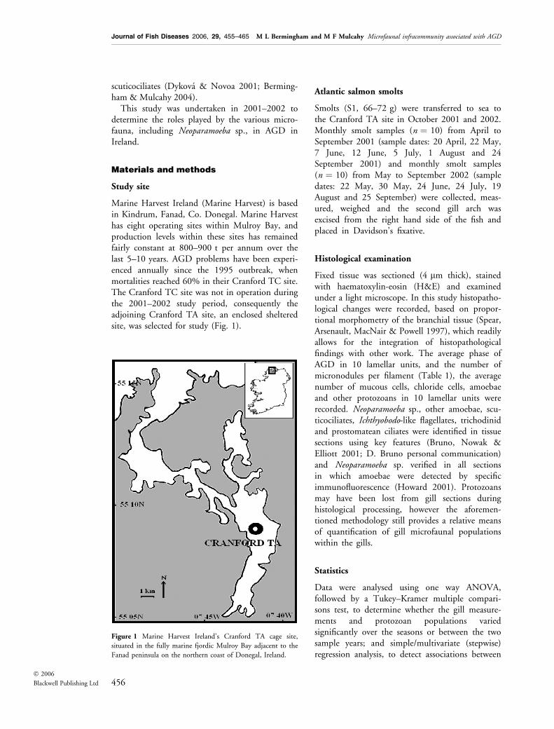
Marine Harvest Ireland (Marine Harvest) is basedin Kindrum, Fanad, Co. Donegal. Marine Harvesthas eight operating sites within Mulroy Bay, andproduction levels within these sites has remainedfairly constant at 800–900 t per annum over thelast 5–10 years. AGD problems have been experi-enced annually since the 1995 outbreak, whenmortalities reached 60% in their Cranford TC site.The Cranford TC site was not in operation duringthe 2001–2002 study period, consequently theadjoining Cranford TA site, an enclosed shelteredsite, was selected for study (Fig. 1).
Atlantic salmon smolts
Smolts (S1, 66–72 g) were transferred to sea tothe Cranford TA site in October 2001 and 2002.Monthly smolt samples (n ¼ 10) from April toSeptember 2001 (sample dates: 20 April, 22 May,7 June, 12 June, 5 July, 1 August and 24September 2001) and monthly smolt samples(n ¼ 10) from May to September 2002 (sampledates: 22 May, 30 May, 24 June, 24 July, 19August and 25 September) were collected, meas-ured, weighed and the second gill arch wasexcised from the right hand side of the fish andplaced in Davidson’s fixative.
Histological examination
Fixed tissue was sectioned (4 lm thick), stainedwith haematoxylin-eosin (H&E) and examinedunder a light microscope. In this study histopatho-logical changes were recorded, based on propor-tional morphometry of the branchial tissue (Spear,Arsenault, MacNair & Powell 1997), which readilyallows for the integration of histopathologicalfindings with other work. The average phase ofAGD in 10 lamellar units, and the number ofmicronodules per filament (Table 1), the averagenumber of mucous cells, chloride cells, amoebaeand other protozoans in 10 lamellar units wererecorded. Neoparamoeba sp., other amoebae, scu-ticociliates, Ichthyobodo-like flagellates, trichodinidand prostomatean ciliates were identified in tissuesections using key features (Bruno, Nowak &Elliott 2001; D. Bruno personal communication)and Neoparamoeba sp. verified in all sectionsin which amoebae were detected by specificimmunofluorescence (Howard 2001). Protozoansmay have been lost from gill sections duringhistological processing, however the aforemen-tioned methodology still provides a relative meansof quantification of gill microfaunal populationswithin the gills.
Statistics
Data were analysed using one way ANOVA,followed by a Tukey–Kramer multiple compari-sons test, to determine whether the gill measure-ments and protozoan populations variedsignificantly over the seasons or between the twosample years; and simple/multivariate (stepwise)regression analysis, to detect associations between
Figure 1 Marine Harvest Ireland’s Cranford TA cage site,
situated in the fully marine fjordic Mulroy Bay adjacent to the
Fanad peninsula on the northern coast of Donegal, Ireland.
456� 2006
Blackwell Publishing Ltd
Journal of Fish Diseases 2006, 29, 455–465 M L Bermingham and M F Mulcahy Microfaunal infracommunity associated with AGD
the gill measurements and protozoan populationsover 2001–2002. Results are given at the 5% levelof significance. The seasons were divided evenlyamong the months: spring: February to April;summer: May to July; autumn: August to Octoberand winter: November to January. All tests wereperformed using the SPSS 11.0 statistical package(SPSS Inc., Chicago, IL, USA).
Results
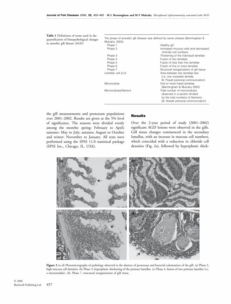
Over the 2-year period of study (2001–2002)significant AGD lesions were observed in the gills.Gill tissue changes commenced in the secondarylamellae, with an increase in mucous cell numbers,which coincided with a reduction in chloride celldensities (Fig. 2a), followed by hyperplastic thick-
Table 1 Definitions of terms used in the
quantification of histopathological changes
in amoebic gill disease (AGD)
The phase of amoebic gill disease was defined by seven phases (Bermingham &
Mulcahy, 2004)
Phase 1 Healthy gill
Phase 2 Increased mucous cells and decreased
chloride cell numbers
Phase 3 Thickening of the individual lamellae
Phase 4 Fusion of two lamellae
Phase 5 Fusion of less than five lamellae
Phase 6 Fusion of five or more lamellae
Phase 7 Structural reorganization of gill tissue
Lamellar unit (LU) Area between two lamellae tips
(i.e. one complete lamella;
M. Powell personal communication)
Micronodule One or more fused lamellae
(Bermingham & Mulcahy 2004)
Micronodules/filament Total number of micronodules
observed in a section divided
by the total numbers of filaments
(B. Nowak personal communication)
(a) (b)
(c) (d)
Figure 2 (a–d) Photomicrographs of pathology observed in the absence of protozoan and bacterial colonisation of the gill. (a) Phase 2,
high mucous cell densities. (b) Phase 3, hyperplastic thickening of the primary lamellae. (c) Phase 4, fusion of two primary lamellae (i.e.
a micronodule). (d). Phase 7, structural reorganization of gill tissue.
457� 2006
Blackwell Publishing Ltd
Journal of Fish Diseases 2006, 29, 455–465 M L Bermingham and M F Mulcahy Microfaunal infracommunity associated with AGD
ening of the secondary lamellae (Fig. 2b). Subse-quently neighbouring lamellae attached to oneanother, forming micronodules (Fig. 2c), thenfused entirely, forming plaques, followed by necro-sis and structural reorganization of gill tissue(Fig. 2d). The observed lesions were not alwaysassociated with the presence of amoebae or othergill parasites (Fig. 2a–d).
Phase of the disease
All phases of the disease were observed over thestudy period. The fish sampled in summer–autumnmanifested the most severe lesions, and those inspring the least (Table 2). In addition, gill tissuechanges were more severe in 2002 than in 2001(Table 3). Amoebae were seen in all phases of thedisease, however, their densities increased withadvancing gill lesions (Table 4). Neoparamoeba sp.was also observed in all phases of the disease, butfailed to correlate with advancing gill lesions. Theassociation of protozoans with gill lesions is shownin Table 4. Ichthyobodo-like flagellate numbersincreased with advancing gill tissue changes butprostomateans correlated negatively with gill lesions.The phase of the disease correlated with micronod-ule, mucous cell, chloride cell, amoeba, Ichthyobodo-like flagellate and prostomatean abundance.
Micronodules
Micronodules (Fig. 2c), consisting of two or morefused lamellae, were observed throughout the studyperiod. The highest micronodule densities wereobserved in autumn and the lowest in spring(Table 2). In addition, higher micronodule densi-ties were observed in 2002 than 2001 (Table 3).Amoeba densities increased with increasing micro-nodule densities in the gill but scuticociliatescorrelated negatively with the number of micro-nodules (Table 4). Numbers of micronodules in thegill associated with the phase of the disease, mucouscells, chloride cells, amoebae, scuticociliates andIchthyobodo-like flagellate numbers (Table 4).
Mucous cells
Mucous cells were either absent, or observed in lownumbers, along unaffected gill filaments. However,high densities were present along hyperplastic gillepithelium and on lamellae adjacent to the hyper-plastic regions (Fig. 2a–d). The highest mucous celldensities were observed in summer–autumn and thelowest in spring (Table 2). Mucous cell densities didnot vary between the two study years. Trichodinidciliate densities increased and prostomatean densitiesdecreased with increasing mucous cell numbers(Table 4). Mucous cell densities correlated with thephase of the disease, micronodules, chloride cells,trichodinid and prostomatean densities (Table 4).
Chloride cells
Chloride cells were abundant along healthy gilllamellae, but were rare along hyperplastic epithe-lium. The highest chloride cell densities wereobserved in spring and the lowest in summer–
Table 2 Significant variance observed over time when analysis of
variance (ANOVA) was conducted on the combined gill
measurements of 2001 (n ¼ 84) & 2002 (n ¼ 60)
Parameter
ANOVA Tukey–Kramer method
F P Season P
Phase 49.1 0.000 Winter–summer 0.000
Winter–autumn 0.000
Spring–summer 0.000
Spring–autumn 0.000
Summer–autumn 0.000
Micronodules 9.8 0.000 Winter–autumn 0.009
Spring–autumn 0.000
Summer–autumn 0.000
Mucous cells 25.2 0.000 Winter–autumn 0.001
Spring–autumn 0.000
Summer–autumn 0.000
Chloride cells 18.9 0.000 Winter–summer 0.000
Winter–autumn 0.000
Spring–summer 0.000
Spring–autumn 0.000
Scuticociliates 4.6 0.004 Winter–spring 0.040
Spring–summer 0.049
Spring–autumn 0.007
Numerator d.f. ¼ 4, denominator d.f. ¼ 138, F ‡ 2.55; P £ 0.05 test
accepted.
Table 3 Significant variance observed when analysis of variance
(ANOVA) was conducted between the gill measurements of
2001 (n ¼ 84) & 2002 (n ¼ 60)
Parameter
ANOVA
F P
Phase 3.9 0.049
Micronodules 11.8 0.001
Chloride cells 16.4 0.000
Amoebae 8.1 0.005
Ichthyobodo-like flagellates 11.8 0.001
Prostomateans 5.0 0.028
Numerator d.f. ¼ 1, denominator d.f. ¼ 141, F ‡ 3.92; P £ 0.05 test
accepted.
458� 2006
Blackwell Publishing Ltd
Journal of Fish Diseases 2006, 29, 455–465 M L Bermingham and M F Mulcahy Microfaunal infracommunity associated with AGD
autumn (Table 2). Furthermore, higher chloridecell densities were observed in 2001 (Table 3).Amoeba, Ichthyobodo-like flagellate and trichodinidabundance increased with decreasing chloride celldensities (Table 4). Chloride cell densities associ-ated with the phase of the disease, micronodule,mucous cell, amoeba, Ichthyobodo-like flagellate,trichodinid ciliate and prostomatean densities(Table 4).
Gill amoebae
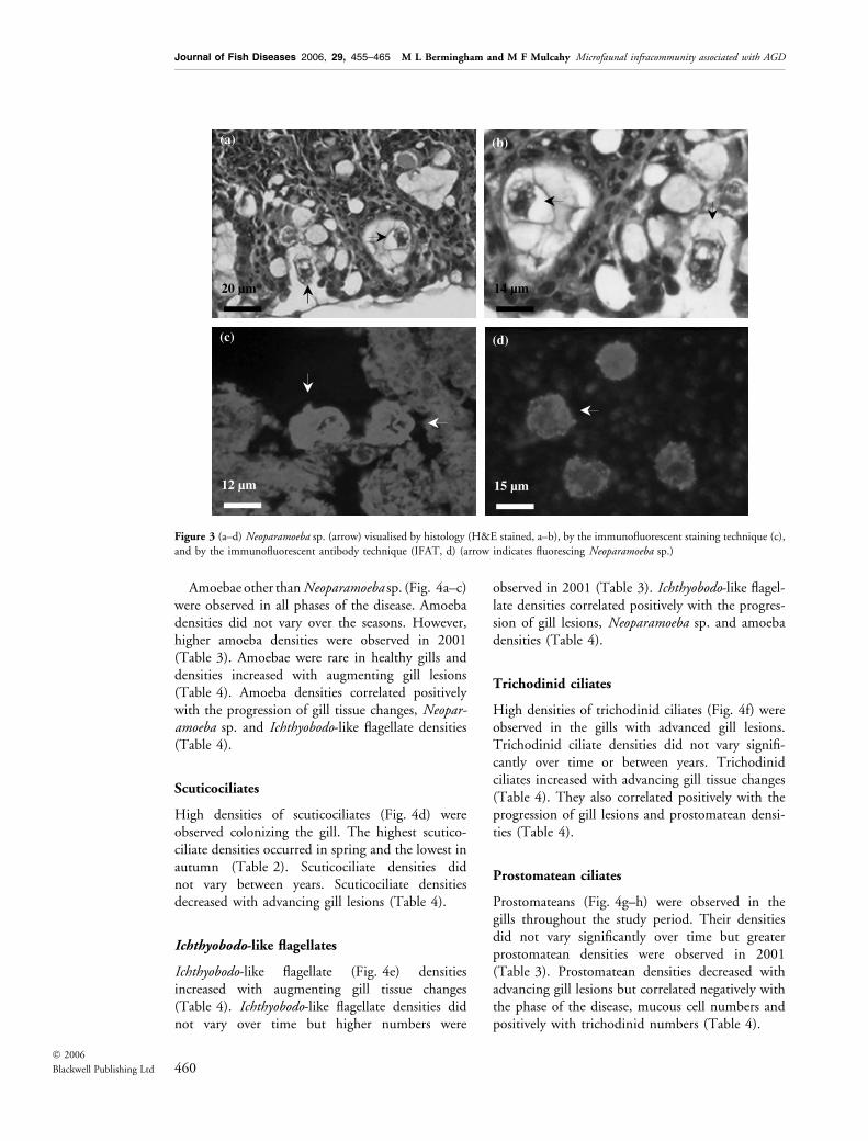
Neoparamoeba sp. (Fig. 3a–d) were observed in allphases of the disease, however they were notgenerally associated with lesions. Neoparamoebasp. densities did not vary over time or betweensample years. Neoparamoeba sp. densities correlatedpositively with amoeba and Ichthyobodo-like flagel-late densities (Table 4).
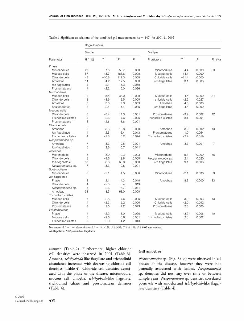
Table 4 Significant associations of the combined gill measurements (n ¼ 142) for 2001 & 2002
Parameter
Regression(s)
Simple Multiple
R 2 (%) T F P Predictors T P R 2 (%)
Phase
Micronodules 29 7.5 55.7 0.000 Micronodules 4.4 0.000 83
Mucous cells 57 13.7 186.6 0.000 Mucous cells 14.1 0.000
Chloride cells 45 )10.6 112.3 0.000 Chloride cells )11.4 0.000
Amoebae 11 4.2 17.5 0.000 Ich-flagellates 3.1 0.003
Ich-flagellates 3 2.1 4.3 0.040
Prostomateans 4 )2.2 5.0 0.026
Micronodules
Mucous cells 19 5.5 33.0 0.000 Mucous cells 4.5 0.000 34
Chloride cells 8 )3.6 12.5 0.000 chloride cells )2.2 0.027
Amoebae 6 3.0 9.3 0.003 Amoebae 4.3 0.000
Scuticociliates 3 )2.1 4.4 0.036 Ich-flagellates )4.5 0.000
Mucous cells
Chloride cells 8 )3.4 11.3 0.001 Prostomateans )3.2 0.002 12
Trichodinid ciliates 5 2.8 7.6 0.006 Trichodinid ciliates 3.4 0.001
Prostomateans 5 )2.6 6.6 0.001
Chloride cells
Amoebae 8 )3.6 12.8 0.000 Amoebae )3.2 0.002 13
Ich-flagellates 4 )2.5 6.4 0.013 Prostomateans 1.9 0.054
Trichodinid ciliates 4 )2.3 5.2 0.024 Trichodinid ciliates )2.4 0.019
Neoparamoeba sp.
Amoebae 7 3.3 10.8 0.001 Amoebae 3.3 0.001 7
Ich-flagellates 5 2.6 6.7 0.011
Amoebae
Micronodules 6 3.0 9.3 0.003 Micronodules 5.3 0.000 46
Chloride cells 8 )3.6 12.8 0.000 Neoparamoeba sp. 2.4 0.020
Ich-flagellates 33 8.3 68.0 0.000 Ich-flagellates 9.1 0.006
Neoparamoeba sp. 7 3.3 10.8 0.001
Scuticociliates
Micronodules 3 )2.1 4.5 0.036 Micronodules )2.1 0.036 3
Ich-flagellates
Phase 3 2.1 4.3 0.040 Amoebae 8.3 0.000 33
Chloride cells 4 )2.5 6.4 0.013
Neoparamoeba sp. 5 2.6 6.7 0.011
Amoebae 33 8.3 68.0 0.000
Trichodinid ciliates
Mucous cells 5 2.8 7.6 0.006 Mucous cells 3.0 0.003 13
Chloride cells 4 )2.3 5.2 0.006 Chloride cells )2.0 0.052
Prostomateans 3 2.0 4.2 0.043 Prostomateans 2.8 0.006
Prostomateans
Phase 4 )2.2 5.0 0.026 Mucous cells )3.2 0.006 10
Mucous cells 5 )2.6 6.6 0.001 Trichodinid ciliates 2.8 0.002
Trichodinid ciliates 3 2.0 4.2 0.043
Numerator d.f. ¼ 1–4, denominator d.f. ¼ 141–138, F ‡ 3.92; T ‡ �1.98; P £ 0.05 test accepted.
Ich-flagellates, Ichthyobodo-like flagellates.
459� 2006
Blackwell Publishing Ltd
Journal of Fish Diseases 2006, 29, 455–465 M L Bermingham and M F Mulcahy Microfaunal infracommunity associated with AGD
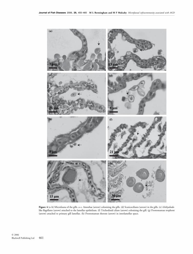
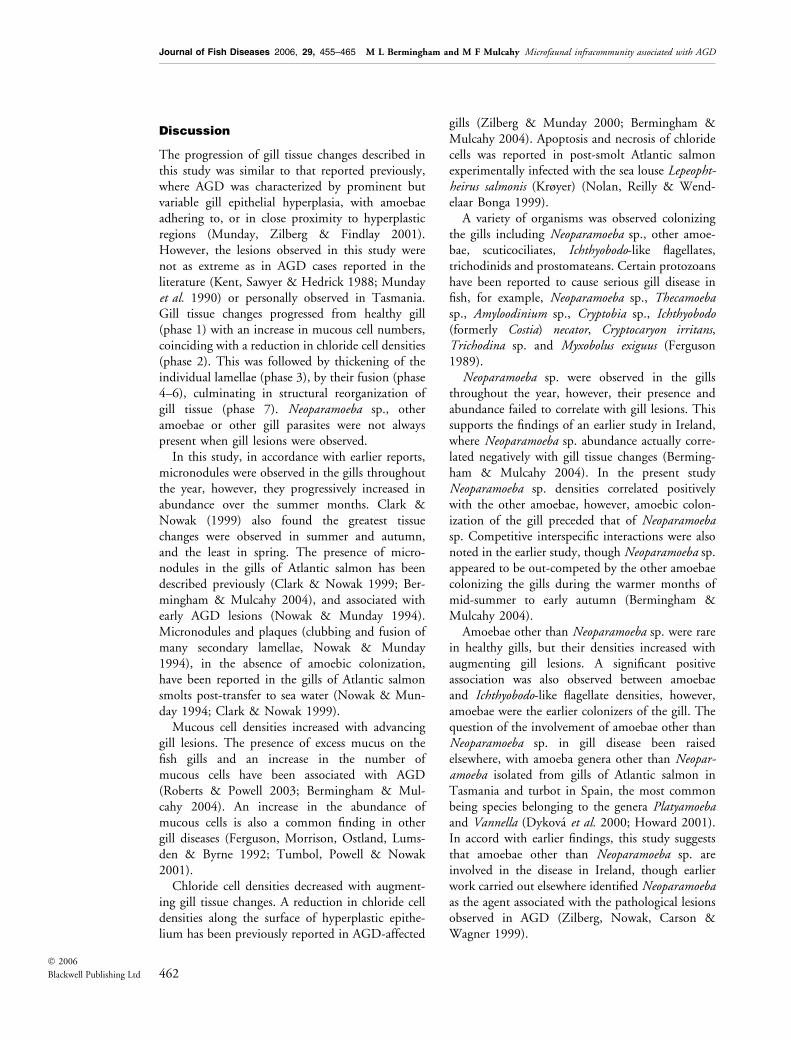
Amoebae other than Neoparamoeba sp. (Fig. 4a–c)were observed in all phases of the disease. Amoebadensities did not vary over the seasons. However,higher amoeba densities were observed in 2001(Table 3). Amoebae were rare in healthy gills anddensities increased with augmenting gill lesions(Table 4). Amoeba densities correlated positivelywith the progression of gill tissue changes, Neopar-amoeba sp. and Ichthyobodo-like flagellate densities(Table 4).
Scuticociliates
High densities of scuticociliates (Fig. 4d) wereobserved colonizing the gill. The highest scutico-ciliate densities occurred in spring and the lowest inautumn (Table 2). Scuticociliate densities didnot vary between years. Scuticociliate densitiesdecreased with advancing gill lesions (Table 4).
Ichthyobodo-like flagellates
Ichthyobodo-like flagellate (Fig. 4e) densitiesincreased with augmenting gill tissue changes(Table 4). Ichthyobodo-like flagellate densities didnot vary over time but higher numbers were
observed in 2001 (Table 3). Ichthyobodo-like flagel-late densities correlated positively with the progres-sion of gill lesions, Neoparamoeba sp. and amoebadensities (Table 4).
Trichodinid ciliates
High densities of trichodinid ciliates (Fig. 4f) wereobserved in the gills with advanced gill lesions.Trichodinid ciliate densities did not vary signifi-cantly over time or between years. Trichodinidciliates increased with advancing gill tissue changes(Table 4). They also correlated positively with theprogression of gill lesions and prostomatean densi-ties (Table 4).
Prostomatean ciliates
Prostomateans (Fig. 4g–h) were observed in thegills throughout the study period. Their densitiesdid not vary significantly over time but greaterprostomatean densities were observed in 2001(Table 3). Prostomatean densities decreased withadvancing gill lesions but correlated negatively withthe phase of the disease, mucous cell numbers andpositively with trichodinid numbers (Table 4).
(a)
20 µm
(c)
12 µm
(d)
15 µm
14 µm
(b)
Figure 3 (a–d) Neoparamoeba sp. (arrow) visualised by histology (H&E stained, a–b), by the immunofluorescent staining technique (c),
and by the immunofluorescent antibody technique (IFAT, d) (arrow indicates fluorescing Neoparamoeba sp.)
460� 2006
Blackwell Publishing Ltd
Journal of Fish Diseases 2006, 29, 455–465 M L Bermingham and M F Mulcahy Microfaunal infracommunity associated with AGD
(c)
26 µm
(d)
8 µm
7 µm
(a)
13 µm
(b)
(e)
5 µm
(f)
21 µm
15 µm 30 µm
(g) (h)
Figure 4 (a–h) Microfauna of the gills. a–c. Amoebae (arrow) colonising the gills. (d) Scuticociliates (arrow) in the gills. (e) Ichthyobodo-
like flagellates (arrow) attached to the lamellar epithelium. (f) Trichodinid ciliate (arrow) colonising the gill. (g) Prostomatean trophont
(arrow) attached to primary gill lamellae. (h) Prostomatean theront (arrow) in interlamellar space.
461� 2006
Blackwell Publishing Ltd
Journal of Fish Diseases 2006, 29, 455–465 M L Bermingham and M F Mulcahy Microfaunal infracommunity associated with AGD
Discussion
The progression of gill tissue changes described inthis study was similar to that reported previously,where AGD was characterized by prominent butvariable gill epithelial hyperplasia, with amoebaeadhering to, or in close proximity to hyperplasticregions (Munday, Zilberg & Findlay 2001).However, the lesions observed in this study werenot as extreme as in AGD cases reported in theliterature (Kent, Sawyer & Hedrick 1988; Mundayet al. 1990) or personally observed in Tasmania.Gill tissue changes progressed from healthy gill(phase 1) with an increase in mucous cell numbers,coinciding with a reduction in chloride cell densities(phase 2). This was followed by thickening of theindividual lamellae (phase 3), by their fusion (phase4–6), culminating in structural reorganization ofgill tissue (phase 7). Neoparamoeba sp., otheramoebae or other gill parasites were not alwayspresent when gill lesions were observed.
In this study, in accordance with earlier reports,micronodules were observed in the gills throughoutthe year, however, they progressively increased inabundance over the summer months. Clark &Nowak (1999) also found the greatest tissuechanges were observed in summer and autumn,and the least in spring. The presence of micro-nodules in the gills of Atlantic salmon has beendescribed previously (Clark & Nowak 1999; Ber-mingham & Mulcahy 2004), and associated withearly AGD lesions (Nowak & Munday 1994).Micronodules and plaques (clubbing and fusion ofmany secondary lamellae, Nowak & Munday1994), in the absence of amoebic colonization,have been reported in the gills of Atlantic salmonsmolts post-transfer to sea water (Nowak & Mun-day 1994; Clark & Nowak 1999).
Mucous cell densities increased with advancinggill lesions. The presence of excess mucus on thefish gills and an increase in the number ofmucous cells have been associated with AGD(Roberts & Powell 2003; Bermingham & Mul-cahy 2004). An increase in the abundance ofmucous cells is also a common finding in othergill diseases (Ferguson, Morrison, Ostland, Lums-den & Byrne 1992; Tumbol, Powell & Nowak2001).
Chloride cell densities decreased with augment-ing gill tissue changes. A reduction in chloride celldensities along the surface of hyperplastic epithe-lium has been previously reported in AGD-affected
gills (Zilberg & Munday 2000; Bermingham &Mulcahy 2004). Apoptosis and necrosis of chloridecells was reported in post-smolt Atlantic salmonexperimentally infected with the sea louse Lepeopht-heirus salmonis (Krøyer) (Nolan, Reilly & Wend-elaar Bonga 1999).
A variety of organisms was observed colonizingthe gills including Neoparamoeba sp., other amoe-bae, scuticociliates, Ichthyobodo-like flagellates,trichodinids and prostomateans. Certain protozoanshave been reported to cause serious gill disease infish, for example, Neoparamoeba sp., Thecamoebasp., Amyloodinium sp., Cryptobia sp., Ichthyobodo(formerly Costia) necator, Cryptocaryon irritans,Trichodina sp. and Myxobolus exiguus (Ferguson1989).
Neoparamoeba sp. were observed in the gillsthroughout the year, however, their presence andabundance failed to correlate with gill lesions. Thissupports the findings of an earlier study in Ireland,where Neoparamoeba sp. abundance actually corre-lated negatively with gill tissue changes (Berming-ham & Mulcahy 2004). In the present studyNeoparamoeba sp. densities correlated positivelywith the other amoebae, however, amoebic colon-ization of the gill preceded that of Neoparamoebasp. Competitive interspecific interactions were alsonoted in the earlier study, though Neoparamoeba sp.appeared to be out-competed by the other amoebaecolonizing the gills during the warmer months ofmid-summer to early autumn (Bermingham &Mulcahy 2004).
Amoebae other than Neoparamoeba sp. were rarein healthy gills, but their densities increased withaugmenting gill lesions. A significant positiveassociation was also observed between amoebaeand Ichthyobodo-like flagellate densities, however,amoebae were the earlier colonizers of the gill. Thequestion of the involvement of amoebae other thanNeoparamoeba sp. in gill disease been raisedelsewhere, with amoeba genera other than Neopar-amoeba isolated from gills of Atlantic salmon inTasmania and turbot in Spain, the most commonbeing species belonging to the genera Platyamoebaand Vannella (Dykova et al. 2000; Howard 2001).In accord with earlier findings, this study suggeststhat amoebae other than Neoparamoeba sp. areinvolved in the disease in Ireland, though earlierwork carried out elsewhere identified Neoparamoebaas the agent associated with the pathological lesionsobserved in AGD (Zilberg, Nowak, Carson &Wagner 1999).
462� 2006
Blackwell Publishing Ltd
Journal of Fish Diseases 2006, 29, 455–465 M L Bermingham and M F Mulcahy Microfaunal infracommunity associated with AGD
Scuticociliates were observed in the gills and theirdensities decreased with augmenting gill lesions.Scuticociliates have been isolated along withNeoparamoeba sp. from the gills of clinicallydiseased Atlantic salmon, turbot, sea bream andsea bass (Dykova & Novoa 2001; Bermingham &Mulcahy 2004). However, it may be that theobserved scuticociliates in this study were merelycommensals, feeding on the microbial flora of thegills, as marine scuticociliates are known to occurabundantly in coastal areas, particularly in eutro-phic mariculture waters (Song 2000).
Trichodinid ciliate densities increased with aug-menting gill tissue changes. Trichodina sp. is themost frequently encountered marine ectoparasiticprotozoan (Lom & Dykova 1992). During the1995 AGD outbreak in Ireland Trichodina sp. wasrecovered with Neoparamoeba sp. in the gills ofcultured Atlantic salmon (Rodger & McArdle1996).
Prostomatean densities peaked in spring andautumn, but decreased as augmenting gill lesionsincreased. The prostomatean, C. irritans has beenreported to cause white punctate lesions in theintegument and gills of marine fish (Lom 1970).The negative association between prostomateansand gill lesions may be indicative of speciessuccession in the gill, as prostomateans appearedas the primary colonizers, but over time werereplaced by amoebae and trichodinid ciliates.
Ichthyobodo-like flagellate densities increased withprogressive gill lesions. A number of Ichthyobodo-like flagellates have been associated with gill damageincluding Ichthyobodo necator, Cryptobia branchialis(Chen) and Piscinoodinium sp. These mastigopho-ran parasites are known to effect fatal epizootics,resulting from gill obstruction and mucus hyperse-cretion (Lom & Lawler 1973).
The observations of the progression of gilltissue changes in this study have, as in earlierfindings, indicated that colonization of the gillsoccurs following micronodule formation (Ber-mingham & Mulcahy 2004). Kent et al. (1988)also suggested that amoebic infection results onlyin gills with pre-existing lesions; and it wasobserved that gill lesions resulting from jellyfishcontact and �clubbing and necrosis syndrome�were rapidly colonized by Neoparamoeba pemaqu-idensis (Handlinger 1991). Nevertheless, Zilberget al. (1999) did observe the attachment ofN. pemaquidensis to healthy gill epithelium. Theamoebae in that situation may have been
extremely virulent, as infection was establishedby cohabitation with AGD infected fish, andthere is some evidence that Neoparamoeba sp.becomes more virulent with sequential passagethrough naıve hosts (Munday et al. 2001).
A sequence of colonization within the gillparasite community as described in this study wasalso evident in an earlier field study, whereNeoparamoeba sp. was out-competed by accom-panying amoebae in gills of Atlantic salmon atcertain times of the year (Bermingham & Mulcahy2004). Natural parasite communities are non-saturated with species, the different parasite speciesrarely co-occur at high densities, and interspecificcompetition does not play a major structuring role.However, when the same community is subjected toenvironmental perturbation, strong interactionoccurs between the species, leading to a reductionin species richness while favouring increased viru-lence, culminating in disease/host death (Leibold,Holyoak, Mouquet, Amarasekare, Chase, Hoopes,Holt, Shurin, Law, Tilman, Loreau & Gonzalez2004).
In conclusion, the microfaunal association withgill lesions in this study was an opportunistic one,involving pathogenic colonization of pre-existingmicronodules by a diverse polyphyletic protozoanarray including Ichthyobodo-like flagellates, trich-odinid ciliates, and amoebae other than Neopar-amoeba sp. The sequence of colonial successionobserved within the parasite community is indicat-ive of a disruption in the natural gill infracommu-nity equilibrium. The absence of associationbetween Neoparamoeba sp. and gill lesions hassuggested that the use of this genus as a diagnosticmarker in the Irish situation may not be justified,indicating that diagnosis of AGD should now bebased on gill lesions in association with amoebae,but not necessarily Neoparamoeba sp.
Acknowledgements
The authors are grateful for study funding fromMarine Harvest Ireland, Fanad, Co. Donegal; helpand advice from Jan Feenstra, Catherine McManus,Nigel Teape, Noreen McConigley and the otherstaff of Marine Harvest; and for an invitation totheir laboratory and for the supply of anti-Neopar-amoeba sp. antibodies from the late Dr BarryMunday, Dr Mark Powell, Dr Dina Zilberg, andDr Marianne Douglas-Helders of the University ofTasmania.
463� 2006
Blackwell Publishing Ltd
Journal of Fish Diseases 2006, 29, 455–465 M L Bermingham and M F Mulcahy Microfaunal infracommunity associated with AGD
References
Bermingham M.L. & Mulcahy M.F. (2004) Environmental risk
factors associated with amoebic gill disease in cultured salmon,
Salmo salar L., smolts in Ireland. Journal of Fish Diseases 27,
555–571.
Bruno D.W., Nowak B.F. & Elliott D.G. (2001) HistopathologyWorkshop: The Identification of Parasites in Fish Tissue Sections.10th EAFP International Conference on Fish and Shellfish
Pathology, 9th–14th September 2001, Dublin.
Cann J.P. & Page F.C. (1982) Fine structure of small free-living
Paramoeba (Amoebida) and taxonomy of the genus. Journal ofthe Marine Biological Association of the United Kingdom 62,
25–43.
Clark A. & Nowak B.F. (1999) Field investigations of amoebic
gill disease in Atlantic salmon, Salmo salar L. Journal of FishDiseases 22, 433–443.
Dykova I. & Novoa B. (2001) Comments on diagnosis of
amoebic gill disease (AGD) in turbot, Scophthalmus maximus.Bulletin of the European Association of Fish Pathologists 21,
40–44.
Dykova I., Figueras A. & Peric Z. (2000) Neoparamoeba (Page
1987): light and electron microscopic observations on six
strains of different origin. Diseases of Aquatic Organisms 43,
217–223.
Ferguson H.W. (1989) Systematic Pathology of Fish: A Text andAtlas of Comparative Tissue Responses in Diseases of Teleosts.Iowa State University Press, Ames, Iowa.
Ferguson H.W., Morrison D., Ostland V.E., Lumsden J. &
Byrne P. (1992) Responses of mucus producing cells in gill
disease of rainbow trout, Oncorhynchus mykiss. Journal ofComparative Pathology 106, 255–265.
Handlinger J. (1991) Normal histology report. In: SummerInvestigation 1989–90: Research and Development ReviewSeminar (ed. by P. Valentine), pp. 157–163. Salmon Enter-
prises of Tasmania Ltd, Dover, Tasmania.
Howard T. (2001) Paramoebiasis of Sea-farmed Salmonids inTasmania: A Study of its Aetiology, Pathogenicity, and Control.PhD Thesis, University of Tasmania, Tasmania.
Howard T. & Carson J. (1992) Studies of amoebae associated
with amoebic gill disease (AGD) in Atlantic salmon. In:
Proceedings of the SALTAS Research and Development ReviewSeminar (ed. by P. Valentine), pp. 101–122. Saltas, Hobart,
Tasmania.
Kent M.L., Sawyer T.K. & Hedrick R.P. (1988) Paramoebapemaquidensis (Sarcmastigophora: Paramoebidae) infesta-
tion of the gills of coho salmon, Oncorhynchus kisutchreared in sea water. Diseases of Aquatic Organisms 5,
163–169.
Leibold M.A., Holyoak M., Mouquet N., Amarasekare P., Chase
J.M., Hoopes M.F., Holt R.D., Shurin J.B., Law R., Tilman
D., Loreau M. & Gonzalez A. (2004) The metacommunity
concept: a framework for multi-scale community ecology.
Ecology Letters 7, 601–613.
Lom J. (1970) Protozoa causing diseases in marine fishes. In:
A Symposium on Diseases of Fishes and Shellfishes (ed. by
S.F. Snieszko), pp. 101–123. Special Publication No. 5,
American Fisheries Society, Washington, DC.
Lom J. & Dykova I. (1992) Protozoan parasites of fishes.
Developments in Aquaculture and Fisheries Science 26, 1–315.
Lom J. & Lawler A.R. (1973) An ultrastructural study on the
mode of attachment in dinoflagellates invading the gills of
Cyprinodontidae. Protistologica 9, 293–309.
Munday B.L., Foster C.K., Roubal F.R. & Lester R.G.J. (1990)
Paramoebic gill infection and associated pathology of Atlantic
salmon, Salmo salar and rainbow trout, Salmo gairdneri in
Tasmania. In: Pathology in Marine Science (ed. by F.O. Perkins
& T.C. Cheng), pp. 215–222. Academic Press, San Diego,
California.
Munday B.L., Zilberg D. & Findlay V. (2001) Gill disease of
marine fish caused by infection with Neoparamoeba pemaqui-densis. Journal of Fish Diseases 24, 497–507.
Nolan D.T., Reilly P. & Wendelaar Bonga S.E. (1999) Infection
with low numbers of the sea louse, Lepeophtheirus salmonisinduces stress-related effects in post-smolt Atlantic salmon,
Salmo salar. Canadian Journal of Fisheries and Aquatic Sciences56, 947–959.
Nowak B.F. & Munday B.L. (1994) Histology of gills of Atlantic
salmon during the first few months following transfer to sea
water. Bulletin of the European Association of Fish Pathologists14, 77–81.
Page F.C. (1970) Two new species of Paramoeba from Maine.
Protozoology 17, 421–427.
Palmer R., Carson J., Ruttledge M., Drinan E. & Wagner T.
(1997) Gill disease associated with Paramoeba, in sea reared
Atlantic salmon in Ireland. Bulletin of the European Associationof Fish Pathologists 17, 112–114.
Roberts S.D. & Powell M.D. (2003) Comparative ionic flux and
gill mucous cell histochemistry: effects of salinity and disease
status in Atlantic salmon, Salmo salar (L.). Comparative Bio-chemistry and Physiology Part A 134, 525–537.
Rodger H.D. & McArdle J.F. (1996) An outbreak of amoebic
gill disease in Ireland. The Veterinary Record 139,
348–349.
Song W. (2000) Morphological and taxonomical studies on
some marine scuticociliates from China Sea, with description
of two new species, Philasterides armtalis sp. n. and Cyclidiumvaribonneti sp. n. (Protozoa: Ciliophora: Scuticociliatida). ActaProtozoologica 39, 295–322.
Spear D.J., Arsenault G., MacNair N. & Powell M.D. (1997)
Branchial lesions associated with intermittent formalin bath
treatment of Atlantic salmon, Salmo salar L., and rainbow
trout, Oncorhynchus mykiss (Walbaum). Journal of Fish Diseases20, 27–33.
Tan C.K.F., Nowak B.F. & Hodson S.L. (2002) Biofouling as a
reservoir of Neoparamoeba pemaquidensis (Page 1970), the
causative agent of amoebic gill disease in Atlantic salmon.
Aquaculture 210, 49–58.
Tumbol R.A., Powell M.D. & Nowak B.F. (2001) Ionic effects
of infection of Ichthyophthirius multifiliis in goldfish. Journal ofAquatic Animal Health 13, 20–26.
Zilberg D. & Munday B. L. (2000) Pathology of experimental
amoebic gill disease in Atlantic salmon, Salmo salar L., and the
effect of pre-maintenance of fish in sea water on the infection.
Journal of Fish Diseases 23, 401–407.
464� 2006
Blackwell Publishing Ltd
Journal of Fish Diseases 2006, 29, 455–465 M L Bermingham and M F Mulcahy Microfaunal infracommunity associated with AGD
Zilberg D., Nowak B., Carson J. & Wagner Y. (1999) Simple
gill smear for diagnosis of amoebic gill disease. Bulletin of theEuropean Association of Fish Pathologists 19, 186–189.
Received: 19 April 2005Revision received: 11 May 2006Accepted: 11 May 2006
465� 2006
Blackwell Publishing Ltd
Journal of Fish Diseases 2006, 29, 455–465 M L Bermingham and M F Mulcahy Microfaunal infracommunity associated with AGD
Related Documents


