MICROFABRICATION IN TISSUE ENGINEERING AND BIOARTIFICIAL ORGANS

Welcome message from author
This document is posted to help you gain knowledge. Please leave a comment to let me know what you think about it! Share it to your friends and learn new things together.
Transcript

MICROFABRICATION IN TISSUE
ENGINEERING AND BIO ARTIFICIAL ORGANS

MICROSYSTEMS
Series Editor Stephen D. Senturia
Massachusetts Institute of Technology
Editorial Board Roger T. Howe, University of California, Berkeley D. Jed Harrison, University of Alberta Hiroyuki Fujita, University of Tokyo Jan-Äke Sehweite, Uppsala University
Books in the Series
MICROMACHINED ULTRASOUND-BASED PROXIMITY SENSORS M.R. Hornung, O. Brand ISBN: 0-7923-8508
BRINGING SCANNING PROBE MICROSCOPY UP TO SPEED S.C. Minne, S.R. Manalis, C.F. Quate ISBN: 0-7923-8466-0
MICROCANTILEVERS FOR ATOMIC FORCE MICROSCOPE DATA STORAGE
B.W. Chui ISBN: 0-7923-8358-3
METHODOLOGY FOR THE MODELING AND SIMULATION OF MICROSYSTEMS
B. Romanowicz ISBN: 0-7923-8306-0
MICROFABRICATION IN TISSUE
ENGINEERING AND BIO ARTIFICIAL ORGANS
Sangeeta Bhatia, M.D., Ph.D.
SPRINGER SCIENCE+BUSINESS MEDIA, LLC

Library of Congress Cataloging-in-Publication Data Bhatia, Sangeeta, 1968 -
Microfabrication in tissue engineering and bioartificial organs / Sangeeta Bhatia
p. cm. — (Microsystems ; mict 5) Includes bibliographical references and index. I S B N 978-1-4613-7386-5 I S B N 978-1-4615-5235-2 (eBook) DOI 10.1007/978-1-4615-5235-2 1. Animal cell biotechnology. 2. Microfabrication. 3. Artificial organs.
4. Tissue culture. I. Title. II Series: Microsystems (series); 5. [ D N L M : 1. Tissue Culture-methods. 2. Artificial Organs.
3. Biocompatible Materials. 4. Biomedical Engineering. 5. Cell Communication. QS 525 B575m 1999] TP248.27 . A53B47 1999 660.6--dc21 D N L M / D L C for Library of Congress 99-35715
CIP Copyright © 1999 by Springer Science+Business Media New York Originally published by Kluwer Academic Publishers in 1999 Softcover reprint of the hardcover 1st edition 1999
A l l rights reserved. No part of this publication may be reproduced, stored in a retrieval system or transmitted in any form or by any means, mechanical, photocopying, recording, or otherwise, without the prior written permission of the publisher,Springer Science+Business Media, L L C .
Printed on acid-free paper.

Editor's Preface
The Microsystems Series has as its goal the creation of an outstanding set of textbooks, references, and monographs on subjects that span the broad field of microsystems. Exceptional PhD dissertations provide a good starting point for such a series, because, unlike monographs by more senior authors, which must compete with other professional duties for attention, the dissertation becomes the sole focus of the author until it is completed. Conversion to book form is then a streamlined process, with final editing and book production completed within a few months. Thus we are able to bring important and timely material into book form at a pace which tracks this rapidly developing field. Our first four books in the series were drawn from the more physics-oriented side of the microsystems field, including such diverse subjects as computer-aided design, atomic-force microscopy, and ultrasonic motion detection. Now, with Sangeeta Bhatia's work, we enter the realm of biology. Her use of artifically structured substrates to encourage the liver cells to form orderly assemblies is a fine example of how microfabrication technology can contribute to cell biology and medicine. I am pleased to be able to add this very new and very interesting work to the Microsystems Series.
Stephen D. Senturia Cambridge MA
Microfabrication in Tissue Engineering and Bioartificial Organs
Foreword
One of the emerging applications of microsystems technology in biology and medicine is in the field of tissue engineering and artificial organs. In order to function, cells need to receive proper signals from their environment. Among these signals are the chemistry and geometry of the extracellular matrix to which cells adhere, and the intercellular communication between homotypic and heterotypic cells. The mechanisms by which various signals mediate their effect on cells is an active area of research in cell biology, and are of utmost importance to tissue engineers who are faced to develop functional tissue substitutes. Microsystems technology provides an unmatched opportunity to scientists and engineers to create complex cellular structures by controlling cell-matrix and cell-cell interactions at a micrometer scale.
The Ph.D. thesis presented in this volume by Dr. Sangeeta Bhatia investigates at a fundamental level the role of heterotypic cell-cell interactions toward the development of a functional liver tissue. The use of microfabrication and surface chemistry techniques enabled Dr. Bhatia to precisely control the heterotypic interactions between hepatocytes and fibroblasts. The results from this study showed, for the first time, that the extent of heterotypic cell-cell interactions determine the level of liverspecific function, and thus, the engineered liver tissue. Furthermore, Dr. Bhatia was able to show that hepatocytes closer to the heterotypic interface differentiated and functioned; whereas, hepatocytes more than several hundred micrometers (i.e., 3 to 4 cell layer) away from the interface lost their functional capacity. The importance of these findings and their utility in the development of a bioartificialliver assist system is described in detail in this thesis.

Vlll Microfabrication in Tissue Engineering and Bioartificial Organs
Dr. Bhatia's work represents a cutting-edge, innovative example of the use of microsystems technology in biology and medicine. Although her work was targeted toward the development of an engineered hepatic tissue, the implications of her work are far-reaching and include many other living systems where the interaction between multiple cell types is important, such as developmental biology, wound healing, and cancer.
Mehmet Toner, Ph.D. Associate Professor Center for Engineering in Medicine Massachusetts General Hospital Harvard Medical School

Abstract
The repair or replacement of damaged tissues using in vitro strategies has focused on manipulation of the cell environment by modulation of cellextracellular matrix interactions, cell-cell interactions, or soluble stimuli. Development of functional tissue substitutes through 'tissue engineering' has been facilitated by the ability to control each of these environmental influences; however, in co-culture systems with two or more cell types, cellcell interactions have been difficult to manipulate precisely. These interactions are important in normal physiology of many organ systems, in embryogenesis where differentiation cues are determined by the local cellular microenvironment, and implicated in the pathophysiology of certain diseases. The ability to spatially control cells at the single cell level using micropatterning would allow the precise manipulation of cell-cell interactions of interest.
We have developed an adaptable method for generating twodimensional, anisotropic model surfaces capable of organizing two different cell types in discrete spatial locations. We have chosen a primary rat hepatocyte/3T3 fibroblast cell system due to its potential clinical significance in bioartificial liver design and also based on widely reported interactions observed in this co-culture model. We have used photolithography to pattern biomolecules (collagen I) on glass which mediates cell adhesion of the first cell type, hepatocytes, followed by nonspecific, serum-mediated attachment of fibroblasts to the remaining unmodified areas. This co-culture technique allowed the manipulation of the initial cellular microenvironment without variation of cell number. Specifically, we were able to control the level of homotypic and heterotypic interactions in co-cultures over a wide range.
x Microfabrication in Tissue Engineering and Bioartificial Organs
Modulation of initial cell-cell interactions was found to have significant effects on liver-specific markers of metabolic, synthetic, and excretory function. In particular, 2 to 3-fold variations in steady-state levels of representative hepatocellular functions were achieved from identical numbers of cells. Furthermore, our results indicated that the use of microfabrication to control cell-cell interactions may allow modulation over the kinetics of functional up-regulation; in fact, micropatterned co-cultures displayed increased levels urea synthesis up to 1 week earlier than randomly distributed, unpatterned co-cultures with the same cellular constituents. Our data indicate that control over cell-cell interactions will allow the control of bulk tissue function based on the local microenvironments.
The mechanisms by which hepatocytes and fibroblasts interact to produce a differentiated hepatocyte phenotype were also investigated. Variations in bulk tissue function were due to spatial heterogeneity in the pattern of induction of hepatocyte differentiation within a hepatocyte population due to interaction with mesenchymal cells. We found that hepatocytes adjacent to the heterotypic expressed increased levels of intracellular albumin (a marker of hepatic synthetic function); whereas, hepatocytes far from the heterotypic interface contained undetectable levels of albumin. Although the actual molecular basis of this signaling was not identified, our experimental results indicated that the source of the observed induction pattern was a tightly cell-associated fibroblast product.
Clinical implementation of a co-culture based, bioartificial liver requires optimization of hepatic function based on fibroblast number and various bioreactor design constraints. To this end, we utilized micro fabrication to achieve a reduction in fibroblast number while preserving the heterotypic interface. We determined that fibroblast number could be reduced by twelve-fold with only a modest reduction in hepatic tissue function. These data were combined with a simple model of oxygen transport and viscous energy losses in a hypothetical multi-unit bioreactor, to determine design criteria for a microfabricated, co-culture based bioartificial liver. This general approach has potential applications in many areas of tissue engineering, implantation biology, and developmental biology, both in the arena of basic science and in the development of cellular therapeutics.

Acknowledgements
I've been lucky to have such an amazing group of people carry me through my graduate school years. One of the only problems with having been so fortunate, is that I'm bound to forget someone- for that, I apologize in advance ...
The center of my training has been Mehmet Toner. I have watched him think and tried to follow his example for almost 6 years. He has continuously shared his time, intellect, creativity, and enthusiasm with me. I thank him for many, many hours of advising (scientific and 'life'), editing, brain-storming, and shaping of our work together. He is a truly generous and talented mentor.
Martin Yarmush and Ron Tompkins are to be thanked most for creating such an interdisciplinary team of talented scientists. Maish, in particular, infused his vision in our surroundings and is to be credited with the creation of such a unique and dynamic atmosphere. It has been an honor to work with Dr. Elizabeth Hay as she served on my thesis committee. I thank her for taking such an active interest in this project and for bringing dimension to this work. I am also grateful to Dr. Marty Schmidt who helped to expose me to the richness of micro fabrication and endured an (over?)exposure to world of cell biology.
Everyone in the lab (the "LSSE") has helped me in some way. Anne Leeds, Pat Meara, and Lynne Stubblefield, with a thousand annoying details. Drs. Jeff Morgan and Livingston Van De Water with their biological perspective. Past and present post-docs and MD fellows with their knowledge and insight- Albert Folch, Brent Foy, Howard Matthews, Fatima Merchant, Prabhas Moghe, Maura Paveo, George Pins, S.B. Rajur, Avi Rotem, Charlie Roth, Steve Reiken, Peter Stefanovich, and Craig Zupke.

xii Microfabrication in Tissue Engineering and Bioartificial Organs
Thanks go to Inne Borel Rinkes for keeping me going in the early years, Francois Berthiaume (a.k.a. "Frenchie") for always knowing the answer or where to get it, Bob Ezzell for his help at East, Greg Russo for imageprocessing nightmares, Joe LeDoux for being a graduate student role model, and Pat Walton, Howard Davis, and Kyong Lee for their influx of energy into the lab. Special HUGE thanks to Ulysses Balis, Kamelia Behnia, Octavio Hurtado, Rob Schoen, and Annie Tsong whose work lies in this document - without their help, I wouldn't be done now. Thanks also go to Kristin Hendricks, Kristin O'Neil, Kealy Ham, Annette MacDonald and Rick Snow for technical and computer support. And, of course, the three musketeers - Will Holmes, Jens Karlsson (or should I say, Professor Jens Karlsson), and Mike Russo, for all of the above and more.
On to MIT. All the faculty, staff and students of Health Sciences and Technology (HST) helped make this experience so valuable. A special thank you to Keiko Oh for making sure my education was paid for, to Ron Smith for making sure I had credit for everything, and to everyone behind the scenes who keep it running smoothly. Also, my heartfelt thank you to Chris Chen, my 'academic twin' who has so enriched this experience every step of the way- grad school, med school, academia... In Mechanical Engineering, thanks go to Leslie Regan for helping with everything from qualifiers to MEGAwomen. At the Microsystems Technology Lab, thanks go to Pat Burkhart and Vicki Diadiuk for helping to coordinate my project, and to Joe DiMaria and Kurt Broderick for their reliability and availability. And at the Public Service Center- my heartfelt thanks to everyone involved with KEY s- Emily Sandberg whose unshakable support has kept KEYs going, Corrie Lathan, Lynn Nelson, Tracy Purinton, Mike Halle, the interns past and present, and all the volunteers- you are an incredible group of people. I have treasured our common passion, have truly enjoyed our work/play together, and learned from each of you.
My family and friends have been my foundation. I'll have to thank each of you in person for enduring graduate school with me. My Dad, the first one to introduce me to bioengineering, has taken an active interest in every experimental detail along the way- has marveled at the frustrating pace of experimental research- and so reveled in every new finding. My Mom and sister, Sujata, have been my glue, my caretakers, and my playmates. Jagesh, has been, at once my scientific peer, my partner, and my very best friend. I'm lucky to have all of you.
Financial support came from the Department of Defense, the American Association of University Women, Harvard-MIT Division of Health Sciences
and Technology, and the Bank of Mom and Dad. Contents

Contents
Contents
Editor's Preface v
Foreword vii
~~ ~
Acknowledgements xi
Contents xiii
List Of Figures xvii
Introduction 1
l.Tissue Engineering 1
2.What Does The Liver Do? 2
3.What Happens When The Liver Fails? 5
4.Hepatic Tissue Engineering 7
5.How Can Hepatocytes Be Stabilized In Vitro? 9
6.How Are Cell-Cell Interactions Important In Vivo? 10
7.Co-Culture 11 7.1 Effects Of Co-Culture On Hepatocyte Morphology And Function _ 13 7.2 Mechanisms Of Induction Of Liver Function In Hepatocytes __ 16
8.Previous Attempts To Control Cell-Cell Interactions 19
9. Micropatterning Of Cells _____________ 21

xiv Microfabrication in Tissue Engineering and Bioartificial Organs
to.Scope Of This Study ______________ 25
Methodology For Fabrication, Characterization, And Analysis Of Micropatterned Co-Cultures 29
1.0verview 29
2.Fabrication Of Micropatterned Co-Cultures 30 2. 1 Microfabrication Of Substrates 30 2.2Surface Modification Of Substrates 31 2.3Cell Culture 31 2.4Cell Culture On Modified Surfaces 32
3.Surface Characterization Of Substrates 33 3. 1 Autofluorescence 33 3.2Profilometry 33 3.3Atomic Force Microscopy (AFM) 33 3.4Indirect Immunofluorescence Of Collagen I 34 3.5lmmunofluorescent Staining 34 3.6lmage Analysis 34
4.Functional Analysis Of Micropatterned Co-Cultures 35 4. 1 Experimental Design 35 4.2Analytical Assays 35 4.3 Immunohistochemistry 37 4.4Functional Bile Duct Staining 37 4.5lmage Acquisition And Analysis 37 4.6Statistics And Data Analysis 38
5.Mechanistic Studies 38 5. 1 Conditioned Media 38 5.2Physical Separation Of Cell Types 39 5.3Agitation 41
6.0ptimization Studies 4t 6. 1 Reduction In Fibroblast Number 41 6.2Randomly Distributed Co-Cultures 43
7.Summary 44
Characterization: Microfabricated Substrates & Co-Cultures 45 1.0verview ____________________ 45
2.Characterization Of Cell-Free Substrates _______ 46
3.Characterization Of Micropatterned Cultures 48
4.Discussion 54
Contents xv
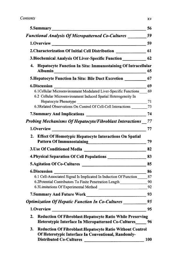
5.Summary ___________________ 56
Functional Analysis Of Micropatterned Co-Cultures 59
1.0verview 59
2.Characterization Of Initial Cell Distribution 61
3.Biochemical Analysis Of Liver-Specific Function 62
4. Hepatocyte Function In Situ: Immunostaining Of Intracellular Albumin 65
5.Hepatocyte Function In Situ: Bile Duct Excretion ____ 67
6.Discussion 69 6. 1 Cellular Microenvironment Modulated Liver-Specific Functions __ 69 6.2 Cellular Microenvironment Induced Spatial Heterogeneity In
Hepatocyte Phenotype 71 6.3Related Observations On Control Of Cell-Cell Interactions 73
7.Summary And Implications 74
Probing Mechanisms Of Hepatocyte/Fibroblast Interactions _77
1.0verview 77
2. Effect Of Homotypic Hepatocyte Interactions On Spatial Pattern Of Immunostaining 79
3.Use Of Conditioned Media 82
4.Physical Separation Of Cell Populations 83
5.Agitation Of Co-Cultures 85
6.Discussion 86 6.1 Cell-Associated Signal Is Implicated In Induction OfFunction __ 87 6.2Potential Contributors To Finite Penetration Length 90 6.3Lirnitations Of Experimental Method 92
7.Summary And Future Work ____________ 93
Optimization Of Hepatic Function In Co-Cultures 95
1.0verview 95
2. Reduction Of Fibroblast: Hepatocyte Ratio While Preserving Heterotypic Interface In Micropatterned Co-Cultures __ 96
3. Reduction Of Fibroblast: Hepatocyte Ratio Without Control Of Heterotypic Interface In Conventional, Randomly-Distributed Co-Cultures 100

XVI Microfabrication in Tissue Engineering and Bioartificial Organs
4. Comparison Of Micropatterned And Randomly-Distributed Co-Cultures 102
5.Discussion ___________________ 105 5. 1 Optimization Of FibroblastHepatocyte Ratio 105 5.2Kinetics Of Up-Regulation Of Hepatic Functions 107 5.3Comparison Between In Vivo And In Vitro Values 109 5.4Design Criteria For Co-Culture-Based Bioreactor 111 5.5Comparison Of Hypothetical Microfabricated Bioreactor To Existing
Technologies 119
6.Summary 120
Conclusions And Outlook 123
l.Summary 123
2.Future Directions 125
References 129
Glossary 141
Index 143

Contents
List of Figures and Tables
Introduction 1 ---------------------------------------Figure 1.1. Liver Lobule. Schematic view. 3
Figure 1.2. Relationships of branches of portal vein (PV), hepatic artery (HA), and bile duct (BD). 3
Figure 1.3. Schematic of Adult Sinusoid. 4
Figure 1.4. Hepatocyte plasma membrane domains and extracellular spaces. 5
Figure 1.5. Development of the human liver in the A. 4th week and B. 5th week of gestation 10
Table 1.1. Cell Types Utilized in Co-Cultures for Stabilization of Rat Hepatocyte Phenotype 11
Table 1.2 Functions Induced in Hepatocytes by Co-Culture __ 15
Figure 1.6. Schematic of Conventional Approaches for Control of Cell-Cell Interaction 20
Methodology for Fabrication, Characterization, and Analysis of Micropatterned Co-Cultures 29
Figure 2.1. Schematic of Process to Generate Micropatterned Co-Cultures 30
Figure 2.2. Schematic of Method for Separation of Cell Populations 39

XVlll Microfabrication in Tissue Engineering and Bioartificial Organs
Figure 2.3. Schematic of Strategy for Minimization of Fibroblast Number. 42
Characterization of Microfabricated Substrates and Co-Cultures 45
Figure 3.1 Characterization of Photoresist. 46
Figure 3.2. Differential Hydrophilicity of Aminosilane Modified Pattern. 47
Figure 3.3. Surface Characterization of Collagen. 48
Figure 3.4. Phase Contrast Micrographs of Micropatterned Heptocytes. 49
Figure 3.5. Micrographs of Micropatterned Hepatocytes and Co-Cultures with 3T3 Fibroblasts. 50
Figure 3.6. Immunofluorescent Staining of Micropatterned Co-Cultures. 51
Figure 3.7. Schematic of Method for Determining, X, Heterotypic Interaction Parameter. 52
Figure 3.8. Average Heterotypic Interaction in Co-Cultures _ 53
Figure 3.9. Distribution of Heterotypic Interactions, X, in Co-Cultures 54
Functional Analysis Of Micropatterned Co-Cultures 59
Figure 4.1. Phase Contrast Micrographs of Micropatterned CoCultures With Varying Heterotypic Interface But Similar Cell Numbers. 61
Figure 4.2. Biochemical Analysis of Micropatterned Cultures and Co-Cultures. 64
Figure 4.3. Kinetics of Immunostaining of Intracellular Albumin in 490 !J.m Island Co-Cultures. 66
Figure 4.4 Comparison of Intracellular Albumin Immunostaining in Various Micropatterns at Day 6. 67
Figure 4.5. Functional Bile Canilicular Staining. 68
Probing The Mechanisms Of Hepatocyte/Fibroblast Interactions ____________________________________________ 77
Figure 5.1. Immunostaining of Intracellular Albumin in Micropatterned Hepatocytes (Only). 80

Contents xix
Figure 5.2. Immunostaining of Intracellular Albumin in Micropatterned Co-Cultures. 81
Figure 5.3. Urea Synthesis in Hepatocytes Treated with Conditioned Media 82
Figure 5.4. Separation of Cell Populations. 84
Figure 5.5. Immunostain of Intracellular Albumin in Separated Cell Populations. 85
Figure 5.6. Immunostain of Intracellular Albumin in Static and Shaken Cultures. 86
Optimization Of Hepatic Function In Co-Cultures 95
Figure 6.1. Phase Contrast Micrograph of Micropatterned Hepatocytes With Reduced Center-to-Center Spacing. 97
Figure 6.2. Phase Contrast Micrograph of Micropatterned Co-Culture with Reduced Spacing. 98
Figure 6.3. Urea Synthesis for Micropatterned Co-Cultures with Reduced Fibroblast:Hepatocyte Ratio and Similar Heterotypic Interface. 99
Figure 6.4. Albumin Secretion for Micropatterned Co-Cultures with Reduced Fibroblast:Hepatocyte Ratio and Similar Heterotypic Interface. 100
Figure 6.5. Urea Synthesis of Randomly-Distributed Co-Cultures with Reduced Fibroblast:Hepatocyte Ratio. 101
Figure 6.5. Albumin Secretion of Randomly-Distributed Co-Cultures with Reduced Fibroblast:Hepatocyte Ratio. 102
Figure 6. 7. Comparison of Micropatterned Co-Cultures to Randomly-Distributed Co-Cultures. 103
Figure 6.7B. Pattern Of Induction Of Urea Synthesis Varies In Fibroblast:Hepatocyte Ratio Of 0.5:1. 104
Figure 6. 7C. Dose Response of Urea Synthesis as a Function of Fibroblast Number in Micropatterned versus Randomly-Distributed Cultures. 104
Figure 6.7D. Kinetics of Up-Regulation of Urea Synthesis __ 105
Figure 6.8. Comparison of Albumin Secretion in Hepatic Tissues In Vitro. 110

xx Microfabrication in Tissue Engineering and Bioartificial Organs
Figure 6.9. Schematic of Hypothetical Bioreactor. Multi-unit bioreactor with cobmmon inlet and outlet. 112
Table 6.1. Effect of Reduced Fibroblast Number on Bioreactor Design 112
Table 6.2. Constants Used in Solution of Transport Equations_ 114
Figure 6.10. Effect of Flow Rate on Oxygen Concentration along Channel Length. 115
Figure 6.11. Effect of Oxygen Tension on Flow Rate versus Cell Surface Area. 116
Figure 6.12. Effect of Channel Height on Shear Stress and Pressure Drop. 118
Table 6.3. Comparison of a hypothetical micro fabricated liver reactor to a spheroid-based, hollow-fiber reactor for replacement of rat liver function. 120
Related Documents


