Journal of Microencapsulation, 2009; 26(4): 315–324 Microencapsulation of a probiotic bacteria with alginate–gelatin and its properties Xiao Yan Li 1 , Xi Guang Chen 1 , Dong Su Cha 2 , Hyun Jin Park 2 and Cheng Sheng Liu 1 1 College of Marine Life Science, Ocean University of China, Qingdao, P. R. China, and 2 The Graduate School of Biotechnology, Korea University, Seoul, Korea Abstract Lactobacillus casei ATCC 393-loaded microcapsules based on alginate and gelatin had been prepared by extrusion method and the product could increase the cell numbers of L. casei ATCC 393 to be 10 7 CFU g 1 in the dry state of microcapsules. The microparticles homogeneously distributed with size of 1.1 0.2 mm. Four kinds of microcapsules (S 1 ,S 2 ,S 3 and S 4 ) exhibited swelling in simulated gastric fluid (SGF) while the beads eroded and disintegrated rapidly in simulated intestinal fluid (SIF). Cells of L. casei ATCC 393 could be continuously released from the microcapsules during simulated gastrointestinal tract (GIT) and the release amounts and speeds in SIF were much higher and faster than that in SGF. Encapsulation in alginate–gelatin microcapsules successfully improved the survival of L. casei ATCC 393 and this approach might be useful in delivery of probiotic cultures as a functional food. Key words: Microcapsule; lactobacillus casei ATCC 393; relative humidity (RH); stability; release Introduction Probiotics have been defined as ‘live microorganisms which, administered in adequate amounts, confer a ben- eficial physiological effect on the host’ 1 . Lactic acid bac- teria (LAB), which are among the most important probiotic microorganisms typically associated with the human gastrointestinal tract (GIT), have many beneficial effects on the human gut flora including immune stimula- tion, cholesterol reduction, inhibition of pathogen growth, maintenance of a healthy gut microflora, prevention of cancer, improvement in lactose utilization, prevention of diarrhoeal diseases or constipation, absorption of calcium and synthesis of vitamins and predigestion of proteins In order to exert positive health effects, viability of pro- biotic bacteria in a product at the point of consumption is an important consideration for their efficacy, as they have to survive during the processing and shelf-life of food and supplements, transit through high acidic conditions of the stomach and enzymes and bile salts in the small intestine. After the LAB pass through the stomach and upper intest- inal tract, LAB should preferably adhere to the epithelium of the intestinal tract and grow 6 . As a guide, the International Dairy Federation has recommended that the bacteria should be active and abundant in the product and be present at least 10 7 CFUg 1 to the date of minimum durability 7 . Unluckily most of the probiotics including LAB lack the ability to survive in a high proportion of the harsh conditions of acidity and bile concentration commonly encountered in the gastrointestinal tract of humans 6 . To improve the survival of LAB, different approaches that increase the resistance of these sensitive microorgan- isms against adverse conditions have been proposed, including appropriate selection of acid- and bile-resistant strains, use of oxygen-impermeable containers, two-step fermentation, stress adaptation, incorporation of micronu- trients such as peptides and amino acids and microencap- sulation 8,9 However, these methods had only a limited success. Therefore, encapsulation of bacterial cells in alginate gels is currently gaining attention to increase viability of probiotic bacteria in acidic products such as yoghurt 10–12 and it is the commonly used technique because this method is very mild and is done at room temperature Address for correspondence: Dr Xi Guang Chen, College of Marine Life Science, Ocean University of China, 5# Yushan Road, Qingdao, P. R. China, 266003. Tel: 86-0532-82032586. Fax: 86-0532-82032586. E-mail: [email protected] (Received 30 Nov 2007; accepted 8 Jul 2008) ISSN 0265-2048 print/ISSN 1464-5246 online ß 2009 Informa UK Ltd DOI: 10.1080/02652040802328685 http://www.informaworld.com/mnc (Received 30 Nov 2007; accepted 8 Jul 2008) ISSN 0265-2048 print/ISSN 1464-5246 online ß 2009 Informa UK Ltd DOI: 10.1080/02652040802328685 http://www.informaworld.com/mnc

Welcome message from author
This document is posted to help you gain knowledge. Please leave a comment to let me know what you think about it! Share it to your friends and learn new things together.
Transcript

Journal of Microencapsulation, 2009; 26(4): 315–324
Microencapsulation of a probiotic bacteria withalginate–gelatin and its properties
Xiao Yan Li1, Xi Guang Chen1, Dong Su Cha2, Hyun Jin Park2 and Cheng Sheng Liu1
1College of Marine Life Science, Ocean University of China, Qingdao, P. R. China, and 2The Graduate Schoolof Biotechnology, Korea University, Seoul, Korea
AbstractLactobacillus casei ATCC 393-loaded microcapsules based on alginate and gelatin had been prepared byextrusion method and the product could increase the cell numbers of L. casei ATCC 393 to be 107 CFU g�1
in the dry state of microcapsules. The microparticles homogeneously distributed with size of 1.1� 0.2 mm.Four kinds of microcapsules (S1, S2, S3 and S4) exhibited swelling in simulated gastric fluid (SGF) while thebeads eroded and disintegrated rapidly in simulated intestinal fluid (SIF). Cells of L. casei ATCC 393 couldbe continuously released from the microcapsules during simulated gastrointestinal tract (GIT) and therelease amounts and speeds in SIF were much higher and faster than that in SGF. Encapsulation inalginate–gelatin microcapsules successfully improved the survival of L. casei ATCC 393 and this approachmight be useful in delivery of probiotic cultures as a functional food.
Key words: Microcapsule; lactobacillus casei ATCC 393; relative humidity (RH); stability; release
Introduction
Probiotics have been defined as ‘live microorganisms
which, administered in adequate amounts, confer a ben-
eficial physiological effect on the host’1. Lactic acid bac-
teria (LAB), which are among the most important
probiotic microorganisms typically associated with the
human gastrointestinal tract (GIT), have many beneficial
effects on the human gut flora including immune stimula-
tion, cholesterol reduction, inhibition of pathogen growth,
maintenance of a healthy gut microflora, prevention of
cancer, improvement in lactose utilization, prevention of
diarrhoeal diseases or constipation, absorption of calcium
and synthesis of vitamins and predigestion of proteins
In order to exert positive health effects, viability of pro-
biotic bacteria in a product at the point of consumption is
an important consideration for their efficacy, as they have
to survive during the processing and shelf-life of food and
supplements, transit through high acidic conditions of the
stomach and enzymes and bile salts in the small intestine.
After the LAB pass through the stomach and upper intest-
inal tract, LAB should preferably adhere to the epithelium
of the intestinal tract and grow6. As a guide, the
International Dairy Federation has recommended that
the bacteria should be active and abundant in the product
and be present at least 107 CFUg�1 to the date of minimum
durability7. Unluckily most of the probiotics including LAB
lack the ability to survive in a high proportion of the harsh
conditions of acidity and bile concentration commonly
encountered in the gastrointestinal tract of humans6.
To improve the survival of LAB, different approaches
that increase the resistance of these sensitive microorgan-
isms against adverse conditions have been proposed,
including appropriate selection of acid- and bile-resistant
strains, use of oxygen-impermeable containers, two-step
fermentation, stress adaptation, incorporation of micronu-
trients such as peptides and amino acids and microencap-
sulation8,9 However, these methods had only a limited
success.
Therefore, encapsulation of bacterial cells in alginate
gels is currently gaining attention to increase viability of
probiotic bacteria in acidic products such as yoghurt10–12
and it is the commonly used technique because this
method is very mild and is done at room temperature
Address for correspondence: Dr Xi Guang Chen, College of Marine Life Science, Ocean University of China, 5# Yushan Road, Qingdao, P. R. China, 266003.Tel: 86-0532-82032586. Fax: 86-0532-82032586. E-mail: [email protected]
(Received 30 Nov 2007; accepted 8 Jul 2008)
ISSN 0265-2048 print/ISSN 1464-5246 online � 2009 Informa UK LtdDOI: 10.1080/02652040802328685 http://www.informaworld.com/mnc
(Received 30 Nov 2007; accepted 8 Jul 2008)
ISSN 0265-2048 print/ISSN 1464-5246 online � 2009 Informa UK LtdDOI: 10.1080/02652040802328685 http://www.informaworld.com/mnc

in aqueous medium by using physiologically acceptable
chemicals. Encapsulation is a process in which the cells
are retained within an encapsulating membrane to reduce
cell injury or cell loss and it has been widely utilized to
protect microorganisms including probiotics during tran-
sit through the human gastro-intestinal tract13. The micro-
bial cells are entrapped within their own secretions
(exopolysaccharides (EPS)) that act as a protective struc-
ture or a capsule, reducing the permeability of material
through the capsule and therefore less exposed to adverse
environmental factors such as gastric acid and bile salts10.
Carbohydrate polymers such as alginate have been
used in various food applications14. Alginate, a natural
polysaccharide found in brown algae, is a linear 1, 4
linked copolymer of �-D-mannuronic acid (M) and
�-L-guluronic acid (G) and has the benefits of being
non-toxic to the cells being immobilized and it is an
accepted food additive. The reversibility of encapsulation,
i.e. solubilizing alginate gel by sequestering calcium ions,
and the possible release of entrapped cells in the human
intestine are other advantages14. However, alginate beads
are not acid resistant and it has been reported that the
beads undergo shrinkage and decreased mechanical
strength during lactic fermentation15.
Gelatin is a protein derived from denatured collagen
that contains high levels of hydroxyproline, proline and
glycine and is useful as a thermally reversible gelling
agent for encapsulation. Gelatin was selected here
because of its excellent membrane-forming ability, bio-
compatibility and non-toxicity. The applicability of gelatin
as a hydrogel matrix is limited because of its low network
rigidity. However, its physical properties can be improved
through the addition of cross-linking agents. Because of
its amphoteric nature, it also is an excellent candidate for
cooperation with anionic polysaccharides such as alginate
and so on12.
The choice of an appropriate drying method is also very
important in the case of LAB, so as to increase their survi-
val rates during dehydration itself and subsequent storage.
Freeze-drying and spray-drying have commonly been
used for the dehydration. Freeze-drying, in particular, is
the most common process for the production of large
amounts of concentrated microbiological cultures.
However, during this process, bacteria are subjected to
adverse conditions, such as low temperature and low
water activity, that produce structural and physiological
injury to the bacterial cells resulting in the loss of viability
of many species16. Spray-drying as a method for preparing
concentrated cultures of microencapsulated cells has
many disadvantages associated with this approach.
A major limitation of spray-drying of probiotic cultures is
the loss of viability which occurs during processing and
storage of the powders. Previous reports have shown,
following spray-drying, probiotic Lactobacillus acidophilus
showed increased sensitivity to lysozyme and NaCl,
indicators of cell wall and cell membrane damage17.
In this paper, alginate and gelatin were used to immo-
bilize Lactobacillus casei ATCC 393 cells and the survival
of L. casei ATCC 393 after drying (at 4�C) was described
since there were only a few mentions in the literature
about lactic acid bacteria encapsulated with alginate and
gelatin. Other characteristics such as the morphology
of beads, effect of cross-linking on the release profile of
L. casei ATCC 393 cells, the swelling and stability of beads
in different media (different pH, different ion intensity)
were also investigated.
Materials and methods
Materials
Sodium alginate, gelatin, calcium chloride and sodium
citrate were purchased from Sigma (St. Louis, MO).
The de Man Rogosa Sharpe (MRS) broth was purchased
from Oxoid (Australia). The simulated gastric juice (SGF)
was prepared by suspending pepsin (10 g 1�1, Sigma,
St. Louis, MO) in saline (0.5%, v/v) and the pH adjusted
to 1.2 with 6 mol l�1 HCl. It was then sterile-filtered
through a membrane (0.22 mm). The simulated intestinal
fluid (SIF) was prepared by dispersing pancreatic enzymes
(Sigma, St. Louis, MO) in sterile sodium phosphate buffer
(pH 6.8) to a final concentration of 10 g 1�1, with 0.5% bile
salts (Oxoid) added. The resulting suspension was sterile-
filtered through a membrane (0.22 mm).
Bacterial strain and culture preparations
L. casei ATCC 393 was used in this study. L. casei ATCC 393
was sub-cultured initially in 20 ml MRS broth at 37�C for
18 h. The resulting cultures were transferred into 20 ml
MRS broth and incubated under the same conditions.
Cultures were harvested by centrifugation at 4500� g at
4�C for 30 min and washed with phosphate buffer saline
(pH 7.4) and collected by centrifugation as above.
The washed bacterial cells were mixed with a MRS broth
for use.
Microencapsulation of microorganisms
L. casei ATCC 393-loaded microcapsules were prepared
with different wall materials (alginate, gelatin) to get sam-
ples (S1, S2, S3 and S4) (shown in Table 1). L. casei ATCC
393 microcapsules were prepared by mixing 5 ml cell
suspensions and 30 ml sterilized wall materials (alginate
solution, sterilized at 121�C for 15 min; gelatin solution,
316 X. Y. Li et al.

sterilized by ultra-high temperature processing (140�C,
5 s)) at 37�C, then the mixture was injected through a
syringe needle into 0.2 mol l�1 calcium chloride solution
at room temperature (25�C) to get S1 and S2 (the distance
between the syringe and the calcium chloride collecting
solution was 10 cm). The beads were allowed to stand for
1 h for hardening before being aseptically transferred to a
sterile flask for storage. S3 and S4 were prepared by sus-
pending S2 in 0.06 mol l�1 sodium citrate for 15 min and
30 min, respectively. Samples were rinsed twice with dis-
tilled water and dried without protective ingredients under
controlled air flow, temperature (4�C) and relative humid-
ity (52� 5%) of the air. The microcapsules could be dried
within 24 h.
Surface morphology and bead size determination
The shape and surface characteristics were determined by
scanning electron microscopy (SEM) using a gold sputter
technique. The microcapsules were vacuum-dried, coated
with gold palladium and observed microscopically.
Particle sizes of four samples (S1, S2, S3 and S4) were mea-
sured by naked eye with a meter and the size of micro-
capsules was studied. In all measurements at least 100
particles were examined.
Effect of relative humidity (RH) on the moisture
resorption of microcapsules
The water resorption experiment was carried out accord-
ing to the following procedures. The dried microcapsules
were kept in a dessicator at a temperature of 25�C and
a relative humidity of 33% (magnesium chloride), 52%
(magnesium nitrate), 75% (ammonium chloride) and
97% (potassium sulphate), respectively. The weight of
microcapsules were determined at the end of 1 week
and compared with the data of freshly prepared dried
microcapsules.
All experiments were done in triplicate. The water
resorption ability of beads was calculated from the
formula:
RS ð%Þ ¼Wt �W0
W0� 100
where W0 was the initial weight of the beads and Wt was
the weight of the microcapsules at equilibrium in different
relative humidity (RH) after 1 week.
Evaluation of the stability of microcapsules
in different media
The stability of four samples (S1, S2, S3 and S4) was
assessed in SGF (pH 1.2), SIF (pH 6.8), respectively, as
described in18. Briefly, dried microcapsules of known
weight (10 g) were placed in a glass vial containing
100 ml of solution and incubated at 37�C with 110 rpm of
shaking for 4 h. The beads were periodically removed and
weighed. The wet weight of the swollen beads was deter-
mined by blotting them with filter paper to remove moist-
ure adhering to the surface, immediately followed by
weighing on an electronic balance and the mean value
was reported. Effect of pH and ion intensity on the stability
of beads was checked with the same method.
All experiments were done in triplicate. The percentage
of swelling of the beads was calculated from the formula:
SW ð%Þ ¼Wt �W0
W0� 100
where W0 was the initial weight of the beads and Wt was
the weight of the swollen beads at equilibrium swelling in
the media.
In vitro release studies (GIT)
To examine the release behaviour of L. casei ATCC 393
from microcapsules in GIT in vitro, samples (S1, S2, S3
and S4, 10 beads) were added to 50 ml SGF (pH 1.2) and
incubated at physiological temperature (37�C) for 2 h and
subsequently transferred into SIF (pH 6.8) for another 4 h.
At specific time intervals (30 min), 2.0 ml aliquots were
removed and assayed OD600 for L. casei ATCC 393 in
triplicate.
Survival of L. casei ATCC 393-loaded
in microcapsules
Non-encapsulated L. casei ATCC 393 were enumerated
in the MRS agar. Peptone water was used to prepare the
Table 1. The wall material, function time of sodium citrate
(0.06 mol l�1) and live amount of L. casei ATCC 393 in different samples
during 1 week.
Sample Wall material Time (min) Live cell amount (CFU g�1)
0 w 1 w
S1 Sodium alginate,
Gelatin (3 : 0, w/w)
– 4.79� 107 2.39� 106
S2 Sodium alginate,
Gelatin (2 : 1, w/w)
0 2.83� 107 6.32� 105
S3 Sodium alginate,
Gelatin (2 : 1, w/w)
15 3.53� 108 4.63� 107
S4 Sodium alginate,
Gelatin (2 : 1, w/w)
30 3.02� 108 3.77� 107
Microencapsulation of a probiotic bacteria with alginate–gelatin and its properties 317
serial dilutions and culture was plated by the pour plate
technique. The plates were incubated at 37�C for 48 h.
To determine the viable counts of the encapsulated
L. casei ATCC 393, 0.1 g of microcapsules were resus-
pended in 10 ml of sodium citrate (0.06 mol l�1) and stir-
red for 45 min using a magnetic stirrer at 37�C. Complete
release of bacteria from the microcapsules in 45 min was
previously assured by comparing the released number of
cells from the microcapsules. The colony forming units
(CFU g�1) were determined by plating on MRS agar plate
and incubating at 37�C for 48 h. The plating procedures
were carried out in triplicate.
Statistical analysis
The assays were performed in triplicate on separate occa-
sions. The data collected in this study were expressed as
the mean value� standard deviation (SD).
Results and discussion
Characteristics of L. casei ATCC 393 microcapsules
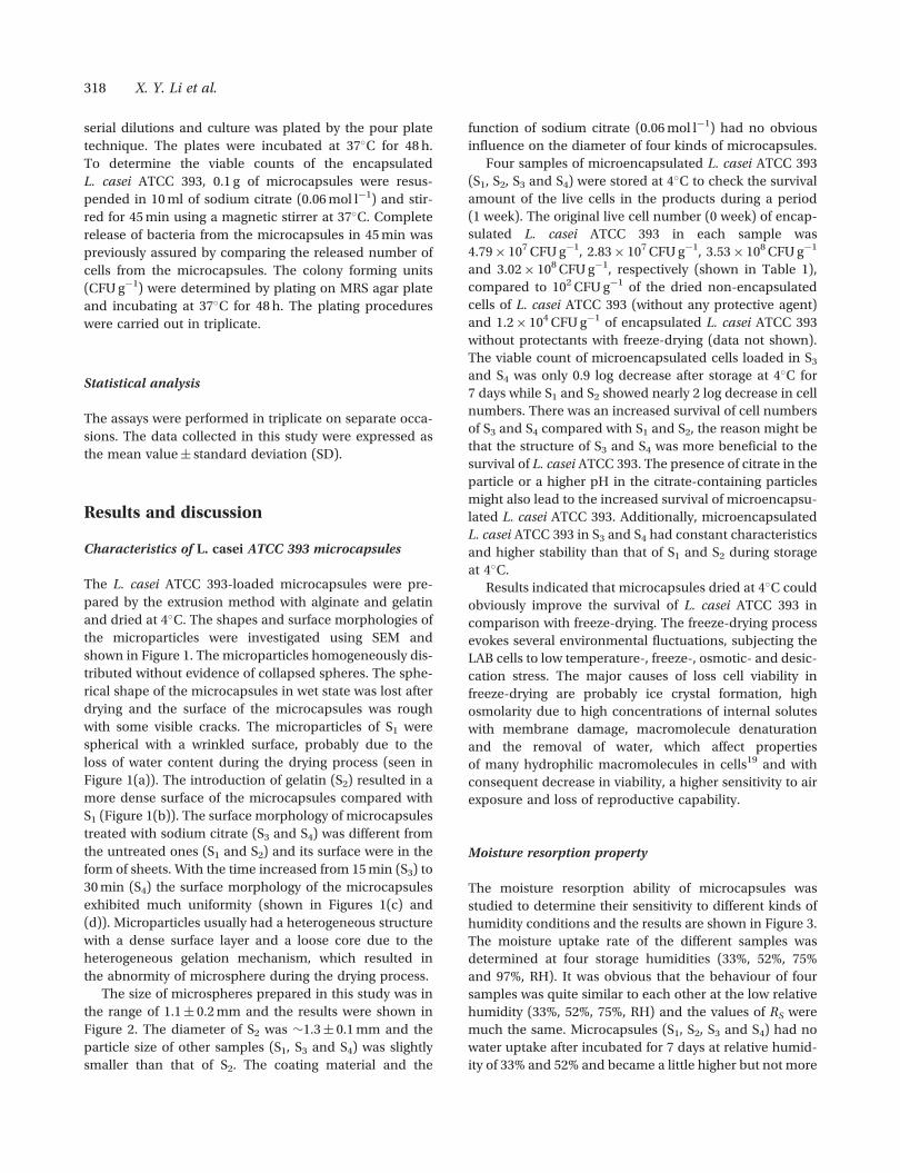
The L. casei ATCC 393-loaded microcapsules were pre-
pared by the extrusion method with alginate and gelatin
and dried at 4�C. The shapes and surface morphologies of
the microparticles were investigated using SEM and
shown in Figure 1. The microparticles homogeneously dis-
tributed without evidence of collapsed spheres. The sphe-
rical shape of the microcapsules in wet state was lost after
drying and the surface of the microcapsules was rough
with some visible cracks. The microparticles of S1 were
spherical with a wrinkled surface, probably due to the
loss of water content during the drying process (seen in
Figure 1(a)). The introduction of gelatin (S2) resulted in a
more dense surface of the microcapsules compared with
S1 (Figure 1(b)). The surface morphology of microcapsules
treated with sodium citrate (S3 and S4) was different from
the untreated ones (S1 and S2) and its surface were in the
form of sheets. With the time increased from 15 min (S3) to
30 min (S4) the surface morphology of the microcapsules
exhibited much uniformity (shown in Figures 1(c) and
(d)). Microparticles usually had a heterogeneous structure
with a dense surface layer and a loose core due to the
heterogeneous gelation mechanism, which resulted in
the abnormity of microsphere during the drying process.
The size of microspheres prepared in this study was in
the range of 1.1� 0.2 mm and the results were shown in
Figure 2. The diameter of S2 was �1.3� 0.1 mm and the
particle size of other samples (S1, S3 and S4) was slightly
smaller than that of S2. The coating material and the
function of sodium citrate (0.06 mol l�1) had no obvious
influence on the diameter of four kinds of microcapsules.
Four samples of microencapsulated L. casei ATCC 393
(S1, S2, S3 and S4) were stored at 4�C to check the survival
amount of the live cells in the products during a period
(1 week). The original live cell number (0 week) of encap-
sulated L. casei ATCC 393 in each sample was
4.79� 107 CFU g�1, 2.83� 107 CFU g�1, 3.53� 108 CFU g�1
and 3.02� 108 CFU g�1, respectively (shown in Table 1),
compared to 102 CFU g�1 of the dried non-encapsulated
cells of L. casei ATCC 393 (without any protective agent)
and 1.2� 104 CFU g�1 of encapsulated L. casei ATCC 393
without protectants with freeze-drying (data not shown).
The viable count of microencapsulated cells loaded in S3
and S4 was only 0.9 log decrease after storage at 4�C for
7 days while S1 and S2 showed nearly 2 log decrease in cell
numbers. There was an increased survival of cell numbers
of S3 and S4 compared with S1 and S2, the reason might be
that the structure of S3 and S4 was more beneficial to the
survival of L. casei ATCC 393. The presence of citrate in the
particle or a higher pH in the citrate-containing particles
might also lead to the increased survival of microencapsu-
lated L. casei ATCC 393. Additionally, microencapsulated
L. casei ATCC 393 in S3 and S4 had constant characteristics
and higher stability than that of S1 and S2 during storage
at 4�C.
Results indicated that microcapsules dried at 4�C could
obviously improve the survival of L. casei ATCC 393 in
comparison with freeze-drying. The freeze-drying process
evokes several environmental fluctuations, subjecting the
LAB cells to low temperature-, freeze-, osmotic- and desic-
cation stress. The major causes of loss cell viability in
freeze-drying are probably ice crystal formation, high
osmolarity due to high concentrations of internal solutes
with membrane damage, macromolecule denaturation
and the removal of water, which affect properties
of many hydrophilic macromolecules in cells19 and with
consequent decrease in viability, a higher sensitivity to air
exposure and loss of reproductive capability.
Moisture resorption property
The moisture resorption ability of microcapsules was
studied to determine their sensitivity to different kinds of
humidity conditions and the results are shown in Figure 3.
The moisture uptake rate of the different samples was
determined at four storage humidities (33%, 52%, 75%
and 97%, RH). It was obvious that the behaviour of four
samples was quite similar to each other at the low relative
humidity (33%, 52%, 75%, RH) and the values of RS were
much the same. Microcapsules (S1, S2, S3 and S4) had no
water uptake after incubated for 7 days at relative humid-
ity of 33% and 52% and became a little higher but not more
318 X. Y. Li et al.

Figure 1. Morphology of alginate and gelatin microspheres. (a) S1; (b) S2; (c) S3; (d) S4.
Figure 3. Moisture resorption ability of microcapsules in different
relative humidity (RH) (data shown were the mean� SD, n¼ 3).
Figure 2. Size distribution analysis of microcapsules (data shown were
the mean� SD, n¼ 3).
Microencapsulation of a probiotic bacteria with alginate–gelatin and its properties 319

than 21% in 1 week when relative humidity increased
to 75%. However the water uptake of S3 and S4 increased
obviously which attained 106.42%, 97.99% compared with
S1 (RS, 31.91%) and S2 (RS, 29.75%) in the same time inter-
val when the relative humidity increased to 97%, perhaps
due to the looser structure of them (S3 and S4). This result
indicated that microcapsules treated with sodium citrate
had a better ability of absorbing water compared with the
untreated ones and the RS value of four samples (S1, S2, S3
and S4) became much higher with the increase of RH
(from 33% to 97%).
Stability of the L. casei ATCC 393-loaded
microcapsules
Stability of microcapsules in SGF and SIF. In order to
obtain data on the behaviour of microcapsules during
simulated gastrointestinal tract, the stability in SGF (pH
1.2) and SIF (pH 6.8) was investigated respectively and
the results are shown in Figure 4. In SGF, the beads
showed swelling without any sign of disintegration
during 4 h. The results presented in Figure 4(a) show
that four samples (S1, S2, S3 and S4) swelled rapidly in
the initial 30 min, after 1 h, the swelling of microcapsules
(S1, S2, S3 and S4) reached the equilibrium without any
erosion and the maximum values were 87.7%, 111.8%,
137.7% and 120.6%, respectively (Figure 4(a)). In SIF,
all the microspheres swelled associating with erosion
which resulted from calcium-alginate cross-linking net-
work were ionized and absorbed water. For microcap-
sules, the maximum water uptake reached after 2.5 h
and after shaking of 4 h, S1 and S2 maintained their
spherical shape with slight erosion, while the treated
ones (S3 and S4) were damaged or even broken into
pieces. Samples (S3 and S4) treated with sodium citrate
swelled rapidly and reached higher SW (1447.7% and
1303.8%) while S1 and S2 exhibited maximum water
uptake of 470.7% and 100.0% in SIF. The results meant
that microcapsules (S1 and S2) without the function of
sodium citrate were much more stable than the treated
ones (S3 and S4) and the introduction of gelatin (S2) in
the system reduced the percentage water uptake of
beads while maintaining their stability compared with
the alginate microcapsules (S1) in SIF.
Effect of pH on the stability of microcapsules. The sta-
bility of microcapsules in disodium hydrogen phosphate-
citric acid buffer with pH 2.4, 3.8, 5.2, 6.4, 7.8, 8.0 were
studied and shown in Figure 5. The results demonstrated
that the beads changed their behaviour when the envir-
onmental pH was altered. When the beads were
immerged in low pH (2.4) their size exhibited swelling
and reached the equilibrium after 1 h (Figure 5(a)).
For pH 3.8, the water uptake of S1, S2, S3 and S4 con-
tinued all the process (Figure 5(b)). With the increase of
pH (from 5.2 to 8.0), the microcapsules exhibited the
same behaviour (first swelled and then began to disin-
tegrate) and carboxyl groups of alginate that were not
cross-linked by Ca2þ or disrupted from calcium-alginate
cross-linking network were ionized and absorbed water,
which resulted in the disintegration of the beads. It was
reported that the disruption of the calcium-alginate gel
matrix occurred fast in phosphate buffer solution with
pH above 5.5 due to the chelating action of phosphate
ions20. The affinity of phosphate for calcium was higher
than that of alginate21. Moreover, the bonds of alginate-
Ca were partially broken during the preparation of S3, S4
and the erosion speed was much rapider than that of S1
and S2. The results showed that microcapsules became
unstable when the environment changed from acidic to
neutral conditions.
Figure 4. Stability of microcapsules in SGF and SIF (data shown were the
mean� SD, n¼ 3): (a) in SGF; (b) in SIF.
320 X. Y. Li et al.

Figure 5. The stability behaviour of microcapsules in different pH conditions (data shown were the mean� SD, n¼ 3): (a) pH 2.4; (b) pH 3.8; (c) pH 5.2;
(d) pH 6.4; (e) pH 7.8; (f) pH 8.0.
Microencapsulation of a probiotic bacteria with alginate–gelatin and its properties 321

Figure 6. Effect of ion intensity of aqueous media on the stability of microparticles (data shown were the mean� SD, n¼ 3): (a) 0.01 mol l�1;
(b) 0.05 mol l�1; (c) 0.1 mol l�1; (d) 0.5 mol l�1; (e) 1 mol l�1.
322 X. Y. Li et al.

Effect of ion intensity on the stability of the
microcapsules. Different concentrations of NaCl (0.01,
0.05, 0.1, 0.5 and 1 mol l�1) were chosen to test its influ-
ence on the stability of four samples (S1, S2, S3 and S4) and
the results are shown in Figure 6.
At the beginning, four kinds of microcapsules (S1, S2,
S3 and S4) absorbed water and swelled continuously in
different ion intensity mediums. After 3 h, the beads
attained maximum swelling, subsequently they began to
show weight loss and dissolve. Samples treated with
sodium citrate (S3 and S4) swelled much more rapidly
and reached higher SW compared with S1 and S2 at the
same ion intensity. As the quantity of NaCl in the
medium increased (from 0.01 to 1 mol l�1) this kind of
difference (the value of SW between the treated ones and
untreated ones) became inconspicuous.
Alginates were hydrophilic and water-soluble anionic
polysaccharides, but the Ca2þ-induced cross-linked beads
of alginate were sufficiently stable in the aqueous media.
When the microcapsules were placed in the buffer system
containing Naþ, the Naþ ions present in the external solu-
tion underwent an ion-exchange process with Ca2þ ions
which were binding with COO– groups mainly in the poly-
guluronate sequences. As a result, the electrostatic repul-
sion among negatively charged –COO– groups increases
which ultimately caused the chain relaxation and
enhances the gel swelling. In the later stage of the swelling
process, the egg-box structure began to loosen and hence
the beads started to disintegrate and lose their weight.
It was the ion exchange process between Naþ and Ca2þ
ions which was supposed to be responsible for the swelling
and subsequent degradation of the beads. In this way, the
ion-exchange process between Naþ ions binding with car-
boxylate groups in polyguluronate and polymannuronate
blocks was ultimately responsible for the swelling and sub-
sequent degradation of the microcapsules22.
In vitro release studies of L. casei ATCC 393 from the
microcapsules
Microcapsule samples (S1, S2, S3 and S4) were treated with
SGF and then with SIF to check the continuously release
characteristics of L. casei ATCC 393 in GIT and the results
are shown in Figure 7. In the first step, the release amounts
of cells were minor in SGF (pH 1.2) from each sample of
the microspheres. After the samples (S1, S2, S3 and S4) were
transferred from SGF to SIF, the larger amounts and faster
release rate of L. casei ATCC 393 cells were found.
The release profile of L. casei ATCC 393 between four
samples had slight differences in the neutral condition.
Compared to the release profile in SIF only (without
prior incubation in SGF) the release of L. casei ATCC 393
from microcapsules in GIT was much faster when the
microcapsules were transferred into SIF (data not
shown). When the beads were treated with SGF, the algi-
nate component underwent acid catalysed hydrolysis and
also the conversion of –COO� groups into –COOH groups,
the electrostatic attraction between Ca2þ and –COO–
groups in ‘egg-box’ junction almost disappears23 and
hence the beads began to disintegrate much more rapidly.
This result indicated that cells of L. casei ATCC 393 could
continuously be released from the microcapsules in GIT
and the release amounts and speeds of L. casei ATCC 393
cells SIF were much higher and faster than that in SGF.
Conclusions
L. casei ATCC 393-loaded microcapsules based on alginate
and gelatin had been prepared by extrusion technology
and the product could increase the live cell numbers to
be 107 CFU g�1 in the dry state of microcapsules.
The microparticles obtained by the extrusion method
homogeneously distributed without evidence of collapsed
spheres and non-aggregated with a size of 1.1� 0.2 mm.
The relative humidity had little effect on the characteristics
of microcapsules (S1, S2, S3 and S4) when it was not more
than 75%. The pH values and ion intensity of solution
affected the swelling behaviour of alginate–gelatin micro-
capsules and the microparticles became unstable and dis-
integrated much rapidly with the increase of pH (from 2.4
to 8.0) and ion intensity (from 0.01 to 1 mol l�1). Cells of
L. casei ATCC 393 could be continuously released from the
microcapsules during GIT and the release amounts and
speeds in SIF (pH 6.8) were much higher and faster than
that in SGF (pH 1.2). In summary, the microencapsulation
method reported in this paper under optimum conditions
proved to be very efficient in increasing the viability of
Figure 7. In vitro release studies of L. casei ATCC 393 from microcap-
sules in GIT (data shown were the mean� SD, n¼ 3).
Microencapsulation of a probiotic bacteria with alginate–gelatin and its properties 323

probiotic bacteria compared to non-encapsulated free
cells. Alginate–gelatin microcapsules might be potentially
used as a safe and protective delivery vehicle for admin-
istering viable probiotic bacteria.
Acknowledgements
The authors are indebted to the financial support from
NSFC (No. 30770582) and the ISTCP (No. 2006DFA33150).
Declaration of interest: The authors report no conflicts of
interest. The authors alone are responsible for the content
and writing of the paper.
References
1. Araya M, Morelli L, Reid G, Sanders ME, Stanton C, Pineiro M, BenEmbarek P. 2002. Guidelines for the evaluation of probiotics in food.Joint FAO/WHO Working Group Report on Drafting Guidelines for theEvaluation of Probiotics in Food, London, Canada, 30 April–1 May.pp. 1–11.
2. Charalampopoulos D, Wang R, Pandiella SS, Webb C. Application ofcereals and cereal components in functional foods: A review. Int JFood Microbiol. 2002;79:131–141.
3. Kaur IP, Chopra K, Saini A. Probiotics: Potential pharmaceuticalapplications. Eur J Pharm Sci. 2002;15:1–9.
4. Shah NP. Probiotics and prebiotics. Agrofoodindustry Hi-Tech. 2004;15:13–16.
5. Reid G, Kim SO, Kohler GA. Selecting testing and understanding pro-biotic microorganisms. FEMS Immunol Med Microbiol. 2006;46:149–157.
6. Chandramouli V, Kailasapathy K, Peiris P, Jones M. An improvedmethod of microencapsulation and its evaluation to protectLactobacillus spp. in simulated gastric conditions. J MicrobiolMethods. 2004;56:27–35.
7. Ouwehand AC, Salminen SJ. The health effects of cultured milkproducts with viable and non-viable bacteria. Int Dairy J. 1998;8:749–758.
8. Anal AK, Singh H. Recent advances in microencapsulation of pro-biotics for industrial applications and targeted delivery. Trends FoodSci Technol. 2007;18:240–251.
9. Kim SJ, Cho SY, Kim SH, Song OJ, Shin IIS, Cha DS, Park HJ. Effect ofmicroencapsulation on viability and other characteristics inLactobacillus acidophilus ATCC 43121. LWT – Food Sci Technol;2008. 41 (3):493–500.
10. Kailasapathy K. Microencapsulation of probiotic bacteria:Technology and potential applications. Curr Issues IntestinalMicrobiol. 2002;3:39–48.
11. Krasaekoopt W, Bhandari B, Deeth H. Evaluation of encapsulationtechniques of probiotics for yogurt. Int Dairy J. 2003;13:3–13.
12. Wenrong S, Griffiths MW. Survival of bifidobacteria in yoghurt andsimulated gastric juice following immobilization in gellan-xanthanbeads. Int J Food Microbiol. 2000;61:17–25.
13. Prakash S, Jones ML. Artificial cell therapy: New strategies for thetherapeutic delivery of live bacteria. J Biomed Biotechnol. 2005;1:44–56.
14. Roy D, Goulet J, Leduy A. Continuous production of lactic acid fromwhey permeate media by free and calcium alginate entrappedLactobacillus helveticus. J Dairy Sci. 1987;70:506–513.
15. Carvalho AS, Silva J, Ho P, Teixeira P, Malcata FX, Gibbs P. Survivalof freeze-dried Lactobacillus plantarum and Lactobacillus rhamno-sus during storage in the presence of protectants. Biotechnol Lett.2002;24:1587–1591.
16. Desmond C, Santon C, Fitzgerald GF, Collins K, Ross RP.Environmental adaptation of probiotic lactobacilli towards improve-ment of performance during spray drying. Intl Dairy J. 2001;11:801–808.
17. Anal AK, Stevens WF. Chitosan-alginate multilayer beads forcontrolled release of ampicillin. Int J Pharm. 2005;290:45–54.
18. Thammavongs B, Corroler D, Panoff JM, Auffray Y, Boutibonnes P.Physiological response of Enterococcus faecalis JH 2-2 to cold shock:Growth at low temperatures and freezing/thawing challenge. LettAppl Microbiol. 1996;23:398–402.
19. Dainty AL, Goulding KH, Robinson PK, Sinpkins I, Trevan MD.Stability of alginate-immobilized algal cells. Biotechnol Bioeng.1986;28:210–216.
20. Liu LS, Liu SQ, Steven YN, Froix M, Ohno T, Heller J.Controlled release of interleukin-2 for tumour immunotherapyusing alginate/chitosan porous microspheres. J Contr Rel. 1997;43:65–74.
21. Bajpai SK, Sharma S. Investigation of swelling/degradation beha-viour of alginate beads crosslinked with Ca2þ and Ba2þ ions. ReactFunct Polym. 2004;59:129–140.
22. Bajpai SK, Saxena SK, Sharma S. Swelling behavior of barium ions-crosslinked bipolymeric sodium alginate-carboxymethyl guar gumblend beads. React Funct Polym. 2006;66:659–666.
324 X. Y. Li et al.
Related Documents