MICROCEPHALY and HYDROCEPHALUS Dr. MUBARAK ABDELRAHMAN MD PEDIATRICS AND CHILD HEALTH Assistant Professor FACULTY OF MEDICINE -JAZAN

Welcome message from author
This document is posted to help you gain knowledge. Please leave a comment to let me know what you think about it! Share it to your friends and learn new things together.
Transcript
MICROCEPHALY and HYDROCEPHALUS
Dr. MUBARAK ABDELRAHMAN
MD PEDIATRICS AND CHILD HEALTH
Assistant Professor
FACULTY OF MEDICINE -JAZAN
The student should be able to:
1. Define and identify causes of micro and macrocephaly.
2. Discus the physiology of CSF production.
3. Classify hydrocephalus and enumerate different causes of it.
4. Describe the clinical presentation, diagnosis and treatment of hydrocephalus.
Objectives:
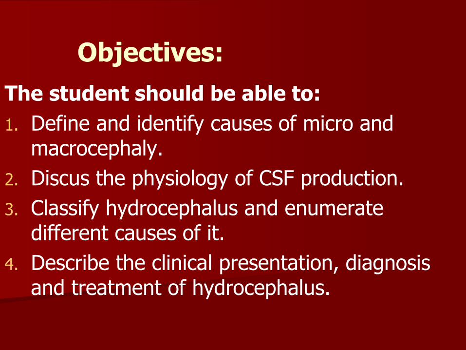
Definition:
- Microcephaly represents a head circumference below the 3rd percentile.
- Macrocephaly represents a head circumference above the 97th percentile.
Microcephaly and Macrocephaly
MICROCEPHALY
Causes:
- Premature closure of skull sutures (craniosynostosis), rare.
- Usually due to small brain due to insult (infectious, toxic, metabolic, vascular) e.g. rubella, CMV, Fetal alcohol syndrome
- Genetic disorder: microcephaly vera (AR), trisomies 21, 18, 13, ….
MACROCEPHALY Causes:
Macrocrania (increased skull thickness) due to
(bone metabolism or hypertrophy of bone marrow secondary to hemolytic anemia).
Hydrocephalus (enlarged ventricles).
Megalencephaly (enlarged brain) due to:
- Disorders causing proliferation of brain tissue (e.g. neurofibromatosis, tuberous sclerosis)
- An accumulation of abnormal metabolic substances (Tay Sach’s, mucopolysaccharidoses).
Hydrocephalus Definition:
- An excessive amount of cerebrospinal fluid (CSF) and is dilated because of increased pressure
PHYSIOLOGY:
• CSF is produced - choroid plexus.
• Circulates through the ventricular system
• Absorbed into the systemic circulation.
Hydrocephalus
CSF production: Choroid plexus - located in the cerebral
ventricles (also in 3rd and 4th). The volume: infants= 50 mL & 150 mL adults. CSF formation continues in raised intracranial
pressure unless extremely high
Hydrocephalus
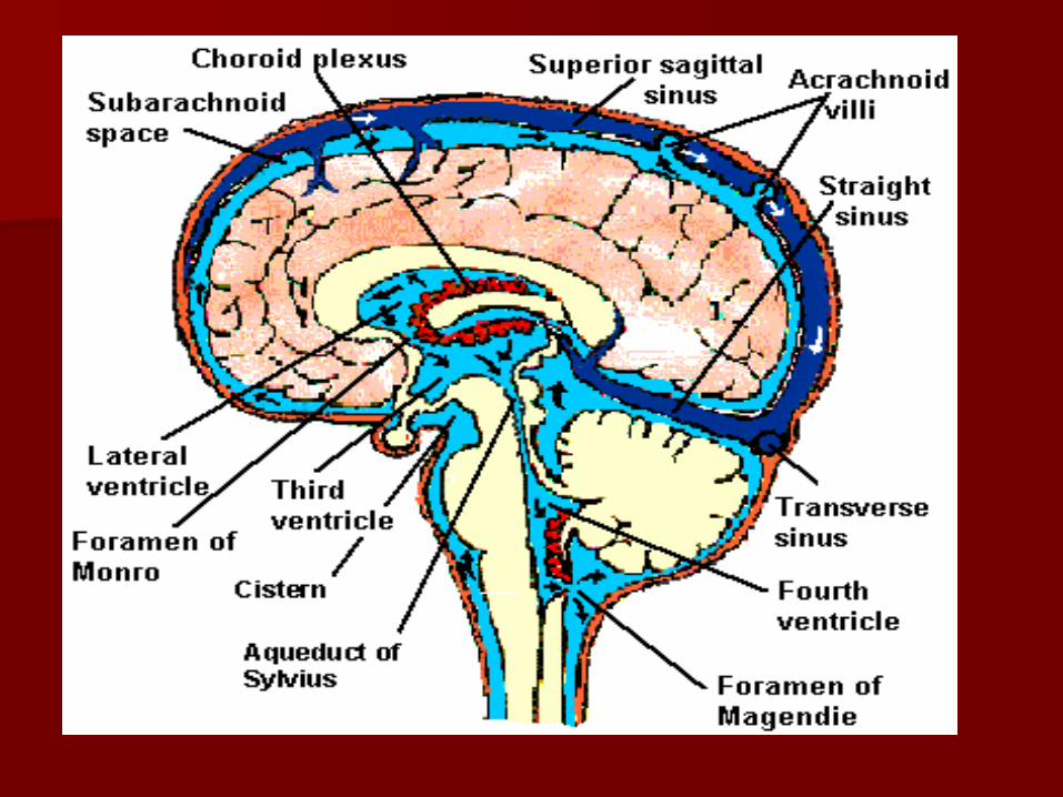
Ventricular system: Lateral ventricles via foramen of Monro to third. Third ventricle via aqueduct of Sylvius to fourth The fourth ventricle via two lateral foramina of
Luschka and one midline foramen of Magendie, to subarachnoid spaces known as cisterns.
CSF is absorbed via arachnoid villi into the sagittal sinus.
hydro
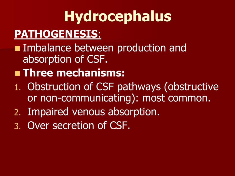
Hydrocephalus PATHOGENESIS:
Imbalance between production and absorption of CSF.
Three mechanisms:
1. Obstruction of CSF pathways (obstructive or non-communicating): most common.
2. Impaired venous absorption.
3. Over secretion of CSF.
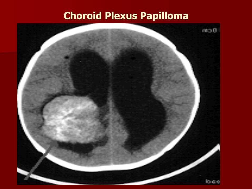
Choroid Plexus Papilloma
Hydrocephalus
Congenital causes: Neural tube defects: Arnold-Chiari
malformation type2.
Isolated aqueduct stenosis (X-linked hydrocephalus).
Vein of Galen malformation (compression of the aqueduct of Sylvius).
Intrauterine infection (aqueduct stenosis).
The Dandy-Walker malformation:
Classic triad:
- Complete or partial agenesis of the vermis.
- Cystic dilation of the fourth ventricle.
- Enlarged posterior fossa.
= Leads to secondary obstruction of the foramina of Luschka and Magendie.
Congenital causes cont.
- CNS infections (Pneumococcal meningitis/ tuberculosis).
- Hemorrhage into the subarachnoid space.
Acquired hydrocephalus:
Hydrocephalus
CLINICAL FEATURES: (Depends on the age, the lesion, the duration and rate of ICP).
Headache is a prominent symptom in older.
Changes in personality and behavior.
Increased ICP (in the posterior fossa often leads to nausea, vomiting and decreased appetite).

CLINICAL FEATURES cont.
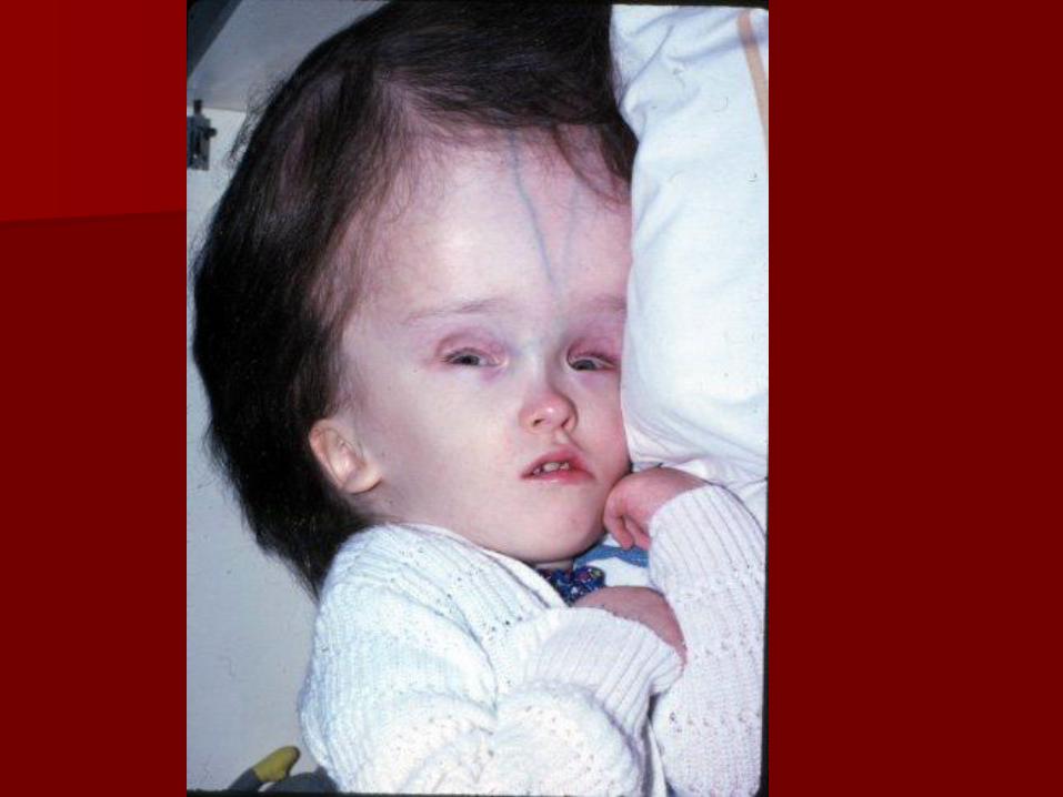
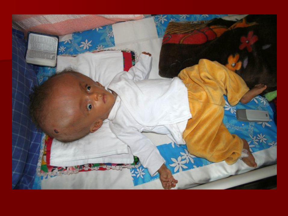
In infants: A large head or rapid increase in the head
size. A bulging anterior fontanel. Sun-setting of the eyes (Pressure on the
midbrain may result in impairment of upward gaze).
Developmental delay.
Poor feeding, irritability, reduced activity and vomiting.
CLINICAL FEATURES cont.
In older children and adults:
Blurred or double vision. Sun-setting of the eyes. Problems with balance, coordination or gait Developmental delay or loss of development. Change in personality
Hydrocephalus
Physical examination ( ICP):
Head size. The anterior fontanel full or distended. The scalp veins dilated and prominent. Setting-sun sign. A cracked pot sound or MacEwen's sign
(indicating separation of the sutures).
Papilledema. Stretching of motor cortex may results in spasticity
of the extremities, especially the legs).
Hydrocephalus
Complications:
Intellectual impairment
Neurological damage.
Problems with the surgical shunt.
Infection at the site of the shunt
HYDROCEPHALUS
Diagnostic Investigations:
Ultrasound of brain (anterior fontanel):
- Shows ventricular enlargement
CT/MRI of head:
- Shows ventricular enlargement, periventricular lucency, narrow/absent sulci ± 4th ventricular enlargement.
HYDROCEPHALUS
Treatment:
Serial Spinal taps.
Surgery- remove obstruction if possible.
Endoscopic third ventriculostomy (ETV)
Shunts.
Acetazolamide (decreases blood flow to choroidal arteries , therefore decreasing CSF production).

Surgical shunts:
Ventriculo Peritoneal Shunt
Ventriculo Pleural Shunt
Ventriculo Atrial Shunt.
Lumboperitoneal shunt.
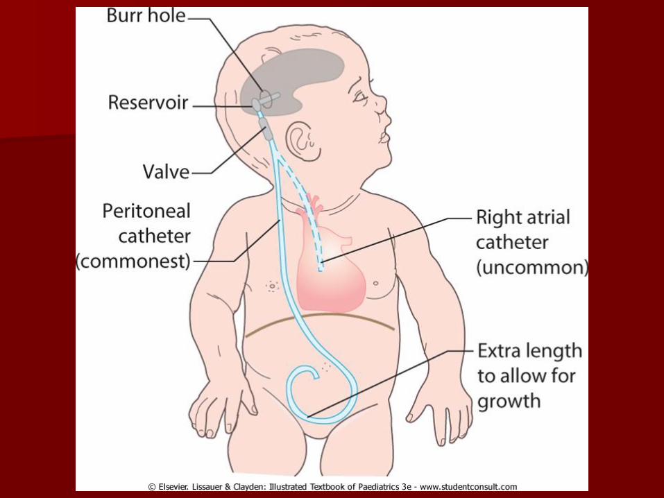
Complications of a Shunt
Shunt blockage.
Shunt infection.
Over drainage/Siphon effect.
Hemorrhage along the tract.
Shunt migration/disconnection.
Seizures.
Related Documents