Microbiology World July – Aug 2014 ISSN 2350 - 8774 www.microbiologyworld.com www.facebook.com/MicrobiologyWorld ~ 1 ~

Welcome message from author
This document is posted to help you gain knowledge. Please leave a comment to let me know what you think about it! Share it to your friends and learn new things together.
Transcript

Microbiology World July – Aug 2014 ISSN 2350 - 8774
www.microbiologyworld.com www.facebook.com/MicrobiologyWorld ~ 1 ~
Microbiology World July – Aug 2014 ISSN 2350 - 8774
www.microbiologyworld.com www.facebook.com/MicrobiologyWorld ~ 2 ~
Chief Editor
Mr. Sagar Aryal
(Founder) Ambassador, iversity
M.Sc. Medical Microbiology
St. Xavier’s College, Nepal
Editors
Mr. Saumyadip Sarkar
ELSEVIER Student Ambassador South Asia 2013 Ph.D Scholar (Human Genetics)
India
Mr. Avishekh Gautam
Ph.D Scholar Hallym University, South Korea
Mr. Manish Thapaliya
Ph.D Scholar China
Mr. Sunil Pandey
ELSEVIER Student Ambassador South Asia 2014 B.Sc. Medical Microbiology
Nobel Medical College, Nepal
Microbiology World July – Aug 2014 ISSN 2350 - 8774
www.microbiologyworld.com www.facebook.com/MicrobiologyWorld ~ 3 ~
Table of Content
Page No.
Ebola Hemorrhagic Fever 4-7
Chemically Programmed Antibodies 8-15
Interview with Dr. Leonard Mermel 16-19
Dye reducing activity of Microorganisms 20-22
Ebola: Most threating virus 23-24
Aromatherapy 25-32
Recent Diagnostic Techniques of
Subclinical Bovine Mastitis 33-38
Cells of Immune System 39-45

Microbiology World July – Aug 2014 ISSN 2350 - 8774
www.microbiologyworld.com www.facebook.com/MicrobiologyWorld ~ 4 ~
Ebola Hemorrhagic Fever
Jeny K. John1, Jobin Jose Kattoor1, Rekha V1, Aswathi P. B1, Aron Jacob2
1 PhD scholars, Indian Veterinary Research Institute, Izatnagar, Bareilly, U.P.
2 M.V.Sc Scholar, Indian Veterinary Research Institute, Izatnagar, Bareilly, U.P
Introduction
Ebola fever is a hemorrhagic fever affecting humans and non human primate species like
monkeys, gorillas and chimpanzees. Ebola hemorrhagic fever is a virus disease caused by virus
of family Filoviridae and genus Ebolavirus. The virus was first identified in 1976 in Democratic
Republic of the Congo near the Ebola River. Occurrence is sporadic in nature. Human disease
was confined in Africa. Disease is zoonotic in nature, bats are considered as the natural
reservoir host.
Causative agent
Ebola virions are linear, negative sense, single stranded RNA with inverse complementary 3'
and 5'termini (Pringle, 2005). Ebola virions are filamentous which appear in the shape of a
shepherd's crook or U or "6", and they may be coiled, toroid, or branched (Kiley et al., 1982).
There are 5 subtypes of Ebola virus: Zaire ebolavirus, Bundibugyo virus, Sudan virus, Taï
Forest virus and Reston virus. Among these, first four are known to cause disease in humans
and restricted in Africa and the fifth one was identified from cynomolgous monkeys from
Philippines.
Transmission
The primates get infection from bats, and humans will acquire infection from infected primates
through handling of diseased animals, through contact with infected body secretions, organs
and blood. Also aerosol route of transmission are reported in non human primates (Jaax et al.,
1995). Ebola-Reston virus mainly spread through aerosol route, which was first identified in a
research facility for primates in Virginia. Human to human transmission are possible through

Microbiology World July – Aug 2014 ISSN 2350 - 8774
www.microbiologyworld.com www.facebook.com/MicrobiologyWorld ~ 5 ~
contact with infected body fluids, organs and use of contaminated needles and syringes in
hospitals/clinics. Semen was also a source of transmission for around 2-3 months.
Outbreaks
Ebola Hemorrhagic fever (HF) has been reported from Democratic Republic of the Congo,
Gabon, Sudan, Ivory Coast, and Uganda, West African countries such as Nigeria, Guinea,
Sierra Leone and Liberia. Ebola – Reston first caused disease in monkeys in a research facility
in the United States and Italy in which monkeys were imported from the Philippines.
Major outbreaks
Year of outbreak Virus type Country
1976 Ebola-Zaire Zaire
1976 Ebola-Sudan Sudan
1979 Ebola-Sudan Sudan
1994 Ebola-Zaire Gabon
1995 Ebola-Zaire Zaire
1996 Ebola-Zaire Gabon
2000-2001 Ebola-Sudan Uganda
2001 -2002 Ebola-Zaire Gabon, Republic of the Congo
2002-2003 Ebola-Zaire Republic of Congo
2004 Ebola-Sudan Sudan
2007 Ebola-Zaire Democratic Republic of the Congo
Dec 2007 -Jan 2008 Ebola-Bundibugyo Uganda
2014 Ebola West Africa

Microbiology World July – Aug 2014 ISSN 2350 - 8774
www.microbiologyworld.com www.facebook.com/MicrobiologyWorld ~ 6 ~
Clinical signs
The incubation period for Ebola virus was around 2 days – 3 week. Case fatality rate was
around 50-90%. Sudden onset of illness and initial clinical signs includes fever, headache,
chills, joint and muscle aches, sore throat, arthritis and weakness, diarrhea, vomiting, and
stomach pain. Later stages hemorrhages from eye, ear, nose, gastrointestinal bleeding, genital
bleeding, and rashes over body surface. Also signs of coma, shock and disseminated
intravascular coagulation. Death occurs mainly due to shock.
Diagnosis
Due to non specific early clinical signs of Ebola hemorrhagic fever, diagnosis was difficult during
the early period. If a case was suspected, immediately isolate the patient and notify to health
authority. The samples suspected of Ebola hemorrhagic fever should be handled only in
biosafety level-4 laboratories. Initial tests include complete blood count, electrolytes, blood
clotting tests, liver function tests. Followed by virus isolation in cell culture, RT-PCR, serological
tests includes antigen-capture enzyme-linked immunosorbent assay (ELISA) testing, IgM
ELISA, indirect fluorescent antibody (IFA). Test should be conducted only in specialized
laboratories with biosafety level 4 facilities.
Treatment
There is no specific antiviral therapy available for Ebola virus, only supportive therapy is
available. Therapy for managing shock includes intravenous fluids and various medicines. Blood
transfusion is required if prolonged bleeding occurs. Balance the patient’s fluids and
electrolytes, maintaining their oxygen status and blood pressure. Treatment should be directed
towards the clinical signs. Shock, hemorrhage, neurological signs, high viremia and pregnancy
confer a poor prognosis.
Prevention
The prevention and control of Ebola HF faces many challenges, due to the lack of exact details
about the reservoir host. Present social and economic situation supports the spread of an
epidemic within health-care facilities. Therefore, health-care providers must be able to recognize
Microbiology World July – Aug 2014 ISSN 2350 - 8774
www.microbiologyworld.com www.facebook.com/MicrobiologyWorld ~ 7 ~
a case of Ebola HF as early as possible. They should be also facility for conducting diagnostic
tests and should adopt practical viral hemorrhagic fever isolation precautions or barrier nursing
techniques. These techniques include the wearing of protective clothing, such as masks, gloves,
gowns, and goggles; the use of infection-control measures, including complete equipment
sterilization and the isolation of Ebola HF patients from contact with unprotected persons. Ebola
virus can stay alive in liquid or dried material for a number of days and also in freezing or
refrigeration conditions. Ebola virus can be inactivated by UV radiation, gamma irradiation,
heating for 60 minutes at 60 °C or boiling for five minutes. The virus is susceptible to sodium
hypochlorite and disinfectants.
Reference
1. Jaax, N. et al. "Transmission of Ebola virus (Zaire strain) to uninfected control monkeys
in a biocontainment laboratory." The Lancet. 346: 1669-1671.
2. Pringle, C. R. (2005). "Order Mononegavirales". In Fauquet, C. M.; Mayo, M. A.;
Maniloff, J.; Desselberger, U.; Ball, L. A. Virus Taxonomy – Eighth Report of the
International Committee on Taxonomy of Viruses. San Diego, US: Elsevier/Academic
Press. pp. 609–614.
3. Kiley, M. P., Bowen, E. T., Eddy, G. A., Isaäcson, M., Johnson, K. M., McCormick, J. B.,
Murphy, F. A., Pattyn, S. R., Peters, D., Prozesky, O. W., Regnery, R. L., Simpson, D. I.,
Slenczka, W., Sureau, P., van der Groen, G., Webb, P. A., Wulff, H. (1982). "Filoviridae:
A taxonomic home for Marburg and Ebola viruses?".Intervirology. 18 (1–2): 24–32.

Microbiology World July – Aug 2014 ISSN 2350 - 8774
www.microbiologyworld.com www.facebook.com/MicrobiologyWorld ~ 8 ~
Chemically programmed antibodies: A next
generation antibody therapeutic
Vikas Gupta*a, Vinod Kumar Singha, Mukesh Bhatta , Vipin kumar Upadhayayb and
Utkarsh kumar tripathib
a-Division of virology, IVRI, Izatnagar, Bareilly, UP(243122)
b-Department of LPM, NDRI, Karnal, Haryana
*Corresponding Author: Phd scholar, Division of virology, I.V.R.I. Izatnagar Bareilly UP.
Email: [email protected]
1. Introduction:
World is facing different type of fatal and chronic disease since ancient time. To overcome these
diseases, demands for therapeutic agents are increasing day by day. A therapeutic agent to be
sufficient effective, it should have some important characteristic such as its site specific action,
sufficient serum half life, low dose with no adverse effect and most importantly economically
attractive. There are myriads of therapeutic agents, among them small therapeutic molecules
and monoclonal antibodies are in trend for the treating of disease. Small therapeutic molecule
have medium to high specificity to target, easily accessible to recessed site, unlimited diversity
and cheap but its serum half life is very less. In compression to small therapeutic molecule,
monoclonal antibodies have high target directed action, tuneable valency, effector function,
long circulatory half life but they have limited diversity , rarely reach to recessed site and costly.
So to get a good therapeutic agent, having long half life, unlimited diversities, target directed
action with high specificity, tuneable valency with effector function and economically attractive,
medicinal chemistry and protein engineering methods blended, yielding a new therapeutic agent
called as chemically programmed antibody. Chemically programmed antibodies are made up of
small therapeutic agents (synthetic agents) and antibodies. It has superior properties in
compression of individual component and target biding is mediated by synthetic agents, so
called chemically programmed antibodies.

Microbiology World July – Aug 2014 ISSN 2350 - 8774
www.microbiologyworld.com www.facebook.com/MicrobiologyWorld ~ 9 ~
2. Architecture of chemically programmed antibodies:
Chemically programmed antibodies are made up of synthetic agents, produced by medicinal
chemistry and antibodies, manufactured by protein engineering methods. So, they have major
two components, in form of antibody component and synthetic component.
2.1. Antibody component:
It includes monoclonal antibodies as whole or its fragment such as Fc or Fab. It acts as carrying
moiety for synthetic component. It provides circulatory half life, bivalency and effecter function of
conventional antibodies and reactive centre for the biding of synthetic component. Antibody
component is produce by reactive immunization and rational designing methods.
Reactive immunization is mean for the development of catalytic antibodies with the help of
reactive immunogens 1-3 diketone and venylenedine hapten with carrier are used as reactive
immunogen. The produce antibody has catalytic activity which mimics aldolase activity and
binding of synthetic component to the reactive centre of catalytic antibody component is
covalent and reversible in nature. There are mainly three types of reactive centre in antibody
component, lysine at paratope of antibody produce through reactive immunization and
selenocystein and cystein reactive centre at C or N terminal of antibody fragment produce
through phage display or native chemical ligation. These reactive centres provide site for the
binding of different synthetic component via reactive group. For conjugation of different
synthetic drugs to monoclonal antibody, require different specific monoclonal antibodies against
each type of target drug while by chemical programming methods, it can be avoided as different
synthetic component equipped with reactive group bind at reactive centre of antibody
component.
Fig: A Fig: B

Microbiology World July – Aug 2014 ISSN 2350 - 8774
www.microbiologyworld.com www.facebook.com/MicrobiologyWorld ~ 10 ~
Fig:1: Comprission between monoclonal antibodies and chemically programmed antibodies to
bind with different type of synthetic component. Fig:A depecting requirement of different types
of monoclonal antibodies for each type of synthetic component, Fig:B depecting same antibody
produce from reactive immunization can be used for different type of synthetic components.
2.2. Synthetic component:
It acts as binding moiety and has three components; pharmacophore, reactive centre and linker
group. Pharmacophore is the main binding moiety for the target site, it may be any peptides,
peptidomimmetics, small or large oligonucleotides and small therapeutic drugs having binding
specificity and affinity to extracellular (membrane bound or secreted) antigens. Reactive group
permits site specific and covalent binding at reactive residue of antibody component. Linker acts
as spacer for pharmacophore and reactive group.
3. Molecular assembly of chemically programmed antibodies:
There are mainly three types of molecular assembly of chemically programmed antibodies; IgG,
Fc and Fab based.
3.1. IgG based: In this type of molecular assembly pharmacophore bind at the paratope of
convention mAb via reactive lysine centre.
3.2. Fc based: It is most common type of assembly in which pharmacophore replaces the Fab
part and bind at C or N terminal with cysteine or selenocysteine reactive centre.
3.3. Fab based: It is a new type of molecular assembly pharmacophore bind with C terminal
selenocystein residue. Fab based chemically antibodies acts as carrier and targeting moiety
simultaneously and they are used as bi-specific chemically programmed antibody.
Fig:2: Different type of molecular assemblies of chemically programmed antibodies.

Microbiology World July – Aug 2014 ISSN 2350 - 8774
www.microbiologyworld.com www.facebook.com/MicrobiologyWorld ~ 11 ~
In bi-specific chemically programmed antibodies, one or both specificities are mediated by
synthetic component to target two epitopes or surface antigens. They have many type of
architecture based on antibody component. A first group among these utilizes two different
monospe-cific synthetic components. These are either conjugated to two identical unique
reactivity centers or to two orthogonal unique reactivity centers. A second group of bispecific
cpAbs utilizes a bispecific synthetic component. Published examples include trifunctional
synthetic components that combine specificities for two different extracellular antigens with a
reactive group. A third group of Fab bispecific cpAbs makes use of antibody components with a
paratope that remains untouched by chemical programming. Conjugating a monospecific
synthetic component to a unique reactivity center in these antibody components affords two
antigen-binding sites; one provided by the antibody component and one provided by the
synthetic component Fab based bispecific abtibodies brings target and effector cells into close
contact to for cytolytic synapse. They may be monovalent bi-specific or divalent bi-specific.
Fig:3: Different form of chemically programmed bispecific antibodies.

Microbiology World July – Aug 2014 ISSN 2350 - 8774
www.microbiologyworld.com www.facebook.com/MicrobiologyWorld ~ 12 ~
4. Targeting agents for chemically programmed antibodies:
Targeting molecule may be membrane bound antigens, integrin molecules and different
miscellaneous molecule. Membrane bound antigen includes gp120 of HIV neuraminidase
glycoproteins of influenza virus chemokine receptors (CCR5) besides these opiodes receptors
and luteinizing hormone receptor can be used. Integrin molecule include several integrins,
which are expressed on tumor cells and tumor endothelial cells, have high-affinity binding sites
for tripeptide motifs, such as RGD (integrin αvβ3, αvβ5, αvβ6, and α5β1) and LDV (integrins
α4β1 and α4β7). These tripeptide motifs are present in extracellular matrix and cell surface
proteins that bind to integrins. As linear or cyclic peptides, they potently antagonize these
interactions and induce apoptosis. Miscellaneous agents includes angiopoietin-2, vascular
endothelial growth factor (VEGF), and placental growth factor-1
5. Characteristic of chemically programmed antibodies:
Antigen recognition by synthetic component
Unlimited chemical diversity
Long circulatory half life
Medium manufacturing cost
Have effector functions
High target binding specificity and affinity
6. Applications of chemically programmed antibodies:
6.1. As a long lasting and potent inhibitors of influenza neuraminidase:
Influenza viruses have two glycoprotein surface spikes, hemagglutinin and neuraminidase.
Hemagglitinin is responsible for bind and fusion of virus on cell surface with help of sialic acid
receptor of cells while neuraminidase is responsible for the release of denova virion from
infected cell and its further spread to other cells. For the control of influenza infection
vaccination plays primary role but due mutations and seasonal incidence, it have some
limitations as vaccine must be read before the influenza season and it is not able to cope up the
new variants of influenza. So along with vaccination, anti influenza therapeutic agents are also

Microbiology World July – Aug 2014 ISSN 2350 - 8774
www.microbiologyworld.com www.facebook.com/MicrobiologyWorld ~ 13 ~
required. Currently, small molecule drugs aim to inhibit neuraminidase are used as therapeutic
agents as neuraminidase catalytic site is resistance to mutation for maintaining the enzymatic
activity. Neuraminidase inhibitors such as oseltamivir and zanamivir are frequently used as anti-
influenza drugs but they have very short life, so frequent doses are required. To avoid frequent
dosing and enhancement of short of serum half life of drug, β-lactam functionalized
neuraminidase inhibitors are conjugated to catalytic aldose monoclonal antibodies 38C2. This
conjugated chemically programmed antibodies bind with catalytic site of neuraminidase and
inhibit the release of newly formed viruses.
Fig:4: Chemically programmed antibody conjugated with Zanamivir antiviral drug.
6.2. As a enhancer of bread and potency neutralization of anti-HIV-1 antibodies and CD4
IgG:
Human immunodeficiency virus-1 is major threat to human being as more than 2 million people
die every year, and more than 33 million individuals are infected worldwide. Highly active
antiretroviral therapy (HAART) is used for therapy of HIV-1but it have so many adverse effect
also. So, development of a potent and broadly acting biological agent as therapeutics might be
a solution. Broadly neutralizing monoclonal antibodies (BNmAbs) that recognize features
conserved across clades of HIV are promising starting points for the development of
immunotherapeutic agents against HIV-1. But their neutralizing capacity is upto 70 to 80%. If a
BNmAb could be modified to inhibit HIV in some different ways, then they can be used as good
therapeutics. HIV-1 entry in side CD4+T cells through receptor mediated mechanism with help

Microbiology World July – Aug 2014 ISSN 2350 - 8774
www.microbiologyworld.com www.facebook.com/MicrobiologyWorld ~ 14 ~
of CD4 receptor along with CCR5 and CXCR4. A promising additional blockade to HIV-1
infection that should complement the targeting of viral proteins is the targeting of host proteins
required for viral entry and replication. A number of small-molecule inhibitors of the HIV-1
coreceptors CCR5 and CXCR4 have been developed and one CCR5-targeting drug has been
approved. CCR5-targeting small molecule, such as meraviroc or aplaviroc is conjugated to
BNmAbs and CD4-IgG, which function as bispecific chemically programmed antibodies. These
bispecific chemically programmed antibodies bind to CCR5 co-receptor and inhibit the entry of
virus in side T cells along with neutralization of HIV-1.
Fig:5: Mechanism of action of chemically programmed antibody conjugated with CCR5
antagonists
6.3. Chemically programmed antibodies as recruiter and activator of T cells:
Biscepicific monoclonal antibodies against CD3 of T cell are conjugated with some receptors
analogues of intergin α4β1 or folate-1 receptor (FOLR1) of malignant tumor cells. Bispecific
CD3 monoclonal antibodies are conjugated with peptidomimetic LLAP2, analogue of integrin
α4β1 binding residue of extracellular matrix bind with integrin α4β1 on tumor cell surface and
recruit the T cells at vicinity of tumor cell. Due to binding of bispecific chemically programmed
antibodies on tumor cell, along with T cells, there is formation of cytolytic synapsis, which
further activate T cell and ultimately lead to cytolysis of malignant tumor cell.

Microbiology World July – Aug 2014 ISSN 2350 - 8774
www.microbiologyworld.com www.facebook.com/MicrobiologyWorld ~ 15 ~
6.4. Chemically programmed antibodies as anti-cancer therapeutics:
Different types of intregin expressed on the cell are responsible for the adhesion, proliferation,
migration differentiation and cell death. Intergin mediated cell-cell binding and cell matrix
attachment with their cognate ligands and expression of these receptors are normal and well
control in normal cells. In contrasts to it, in malignant cells among these intergins, some are
over expressed such as αvβ3 andαvβ5 which are responsible for angiogenesis and matastasis
of maliganant cells. So, these over expressed intergins are the promising target for the
anticancer therapeutics. Anticancer drugs have very short half life and require in high dose, due
high dose they causes cytotoxicity to normal cells also. To avoid this, peptidomimetic drugs
analogue, SCS873 of RGD tri-motif which bind with αvβ3 and αvβ5 intergin, conjugated to the
catalytic aldolase mab 38C2.These conjugated antibodies binds with αvβ3 and αvβ5 intergin
present on tumor cells and Interfere interaction between integrin and extracellular matrix
proteins which leads to Apoptosis of tumor cells and inhibition of angiogenesis.
7. Conclusions:
They are versatile in nature, provide instant immunity, same mAb can be used for different type
of chemical programming. It can be used against two or more antigen at a time via bispecific
chemically programmed antibodies. In last they are economical and less or no toxic.

Microbiology World July – Aug 2014 ISSN 2350 - 8774
www.microbiologyworld.com www.facebook.com/MicrobiologyWorld ~ 16 ~
Interview with Dr. Leonard Mermel
Q) Dr. Leonard Mermel is a well known professor, doctor and researcher
at Brown University. You are well known from the background of
nosocomial infection research. Before knowing more about your research
experiences would like to start off with your early childhood days. How you
used to take medical science during your schooling? How your parents
used to influence you about your higher studies and then going for
research?
Comment: My first interest in becoming a doctor occurred when I was around 10 years of age
seeing my grandmother in a hospital after having a myocardial infarction. I was very close to
her and felt helpless and I told her and my mother then that I want to be a doctor when I grow
up. My family always stressed education, particularly my father who is a told us repeatedly that
receiving a higher education was of the utmost importance. I had great teachers along the way
who served as role models. My interest in clinical and translational research developed during
my infectious diseases fellowship under the tutelage of Dr. Dennis Maki.
Q) You have received multiple awards like Young Investigator award from Society for
Healthcare Epidemiology of America, Teaching awards from The Warren Alpert Medical School
of Brown University and Mentor Scholar award from Society for Healthcare Epidemiology of
America. If you could recall, does these awards gave you zeal to work forth with your research?
Comment: Most certainly, it is an honor and privilege to engage young, bright minds and
mentoring them to have the immense satisfaction in carrying out a successful clinical research
project. There is no greater pleasure as a professor and I feel a strong commitment to such
endeavors.

Microbiology World July – Aug 2014 ISSN 2350 - 8774
www.microbiologyworld.com www.facebook.com/MicrobiologyWorld ~ 17 ~
Q) The techniques used earlier had been more complicated. With the invention of sequencing
techniques, PCR and multiple analysis technique helped people to overcome hurdles of
research nowadays. How you compare the earlier techniques and the current techniques of
research you use.
Comment: Techniques in research should be applied to the question at hand. Knowing which
ones to used reflects collaboration with mentors, mentees and colleagues.
Q) Looking back one of your research published in Infection Control and Hospital Epidemiology
(2010) on identified Methicillin-Resistant Staphylococcus aureus (MRSA) in patients HIV-
infected and hemodialysis patients. In that research findings one in every three individuals have
the prevalence of MRSA in different populations. MRSA ranks 1 in spreading hospital acquired
infection. Does Hospitals can overcome this problem of defeating MRSA with a proper drug?
Comment: Reducing risk of MRSA colonization and infection in hospitalized patients depends
on a strong infection prevention and control program. We have an aggressive program for
MRSA and we have tremendously reduced risk of such infections to our patients over the last 2
decades.
Q) You had been visiting professor at University of Wisconsin Hospital and Clinics, Madison and
Baystate Medical Center, Springfield,MA. How you interact with your students about the
research you carry out? Do you influence your students to think over any complications of
medical research?
Comment: I emphasize that finding a project that is within the scope of the resources and time
frame are critically important as well as finding a mentor who has the time and expertise to
assist in that endeavor.
Q) During the course of your research experience have you ever come up with a bacterium
which you find very hard to disinfect it because of its high resistance (apart from MRSA)?

Microbiology World July – Aug 2014 ISSN 2350 - 8774
www.microbiologyworld.com www.facebook.com/MicrobiologyWorld ~ 18 ~
Resistance of bacterium is acquired based on the resistance genes present in their plasmid.
Have you come up with any research technique to reduce copy numbers of the plasmid of those
bacteria?
Comment: My research has not focused on reducing plasmid transmission of resistance genes.
However, I have collaborated on research looking at novel anti-infective compounds such as
using catheters coated with 5-fluorocytosine as well as more recently looking at novel
compounds with antimicrobial properties to use as antimicrobial lock solutions to prevent or treat
catheter infections.
Q) Highlight one more important and spectacular research of yours, ‘Infection Prevention and
Control during Prolonged Human Space Travel’ published in Clinical Infectious Diseases (2012).
Prolonged space flight and microgravity can provide unique advantages to germs specially
Salmonella or Pneumococcus outbreak. We would like to know how encouraging was that
journey in solving the entire mystery behind microbial advantage in space.
Comment: It is my great fortune to work with bright individuals at Johnson Space Center and
discuss prevention of infections during prolonged human space flight. This is a most interesting
issue as microgravity of space travel presents tremendous challenges and in some cases novel
solutions to mitigate such risk.
Q) Being a professor, researcher and doctor you might have faced varied controversies during
the course of your research. How you used to overcome them and stayed focused in your work?
This will definitely provide a better understanding and will grow motivation among young
researchers and students.
Comment: Controversy abounds in science. It is important to try to remain objective and open-
minded to other points of view, even if in conflict with one’s own research findings. History will
ultimately reveal what was the right direction, but at the time, it may be hard to know which is
why replication of scientific findings by other investigators is essential in the scientific endeavor.

Microbiology World July – Aug 2014 ISSN 2350 - 8774
www.microbiologyworld.com www.facebook.com/MicrobiologyWorld ~ 19 ~
Q) While concluding your journey of research, we would like to know your personal message
towards young researchers and students of medical microbiology.
Comment: I hope I am not concluding my journey but continuing down the same rewarding
path as I have done over the last quarter century. My message is find a good mentor, be
patient, humble, forward-thinking, keep up with the medical literature and acknowledge those
who help you along the way.
Q) There is obvious a wonderful happy life behind research which help in focusing any work.
Thereby would like to know about your life apart from research.
Comments: I have many other interests, I enjoy biking, hiking, surfing, photography, music, and
I have a moderate-sized vegetable garden and numerous fruit trees all of which I planted,
manage, and harvest.
Interview Taken By:
Saumyadip Sarkar
Science Communicator and Reviewer,
Microbiology World,
www.microbiologyworld.com

Microbiology World July – Aug 2014 ISSN 2350 - 8774
www.microbiologyworld.com www.facebook.com/MicrobiologyWorld ~ 20 ~
Dye reducing activity of Microorganisms
Mr. Harshwardhan M. Shrungare
Technical assistant,
Microbiology Research lab,
R. A. College, Washim
Introduction:
It is prove that water is life but now a day that life is threatening due to the advancement in
industrialization. The increase in demand of chemical dyes, fertilizers also increases the
manufacturing and its supply by many industries. During this manufacturing gallons of dyes and
chemicals are incorporated into the river that can cause damage to the environment. Pollutants
mainly include acids, bases, toxic organic and inorganic dissolved solids and colors and their
sources are varying. Textile industry is one of them which extensively use synthetic chemicals
for dye production. Fortunately microbes have ability to reduce these dyes and hence an
attempt is made in Microbiology Research Lab, R. A. College, Washim as a part of project.
Consequences of dyes and chemical in rivers:
Textile industries generate high volume of waste water and dyes. About 10,000 different dyes
and pigments produced annually and about 10% are lost in river via waste water of industry.
The strong color of this waste water is most serious problem because it is the most undesirable
character of water. The disposal of such waste water containing dyes into the river causes
damage to the environment. Generally dye can be describe as, a colored compound that has an
affinity to the substrate to which it is being applied. Dyes affect the photosynthetic activity in
aquatic habitat; it is toxic to aquatic life and causes dangerous effect on all living system. Some
physicochemical methods are available for water treatment including ozonation, photo-
oxidation, adsorption, activated carbon, froth flotation etc. but these techniques creates
secondary disposal problem and are expensive.

Microbiology World July – Aug 2014 ISSN 2350 - 8774
www.microbiologyworld.com www.facebook.com/MicrobiologyWorld ~ 21 ~
Significance of bioremediation of dyes:
Considering above drawbacks, the concept of bioremediation of textile effluent has gain much
attention. The process of microbial de-colorization is eco-friendly, cost competitive and effective
method for effluent treatment. Hence an experiment was carried out to check the efficiency of
the process.
Experimental procedure and results:
In the performed experiment, bacteria such as Pseudomonas and Bacillus have been used to
degrade azo dyes from industrial effluent. Micro-organisms from rhizospheric soil is grown on
nutrient agar modified with effluent and after incubation microbes which decolorizes or grown on
medium are collected and proceed for further application. Two types of cultures were prepared
from the growth obtained, one is free culture and other one is immobilized cell culture. Both
cultures were mixed with industrial effluent and after 6-7 days it was found that the effluent is
decolorizes. The maximum dye de-colorization ability of free and immobilized cells were
analyzed and it was found that immobilized cell culture have maximum activity. Further, cultural
and morphological analysis was also done and the cultures were confirmed as Bacillus,
Clostridium and Pseudomonas for textile effluent de-colorization.
References:
1. Caldwell, B., and M. P. Bryant. 1966. Medium without rumen fluid for nonselective
enumeration and isolation of rumen bacteria. Appl. Microbiol. 14:794-801.
2. Cerniglia, C. E., J. P. Freeman, W. Franklin, and L. D. Pack. 1982. Metabolism of azo
dyes derived from benzidine, 3,3'- dimethylbenzidine and 3,3'-dimethoxybenzidine to
potentially carcinogenic aromatic amines by intestinal bacteria. Carcinogenesis 3:1255-
1260.

Microbiology World July – Aug 2014 ISSN 2350 - 8774
www.microbiologyworld.com www.facebook.com/MicrobiologyWorld ~ 22 ~
3. G. McMullan, C. Meehan, A. Conneely, N. Kirby, T. Robinson, P. Nigam, I. Banat, R.
Marchant, W. Smyth 2000. Microbial decolourisation and degradation of textile dyes.
Applied Microbiology and Biotechnology July 2001, Volume 56, Issue 1-2, pp 81-87.
4. King-Thom Chung* and S. Edward Stevens Jr. 1993. Degradation azo dyes by
environmental microorganisms and helminthes. Environmental Toxicology and
Chemistry Volume 12, Issue 11, pages 2121–2132,
5. Tim Robinson, Geoff McMullan, Roger Marchant, Poonam Nigam 2000. Remediation of
dyes in textile effluent: a critical review on current treatment technologies with a
proposed alternative. Bioresource Technology Volume 77, Issue 3, May 2001, Pages
247–255.

Microbiology World July – Aug 2014 ISSN 2350 - 8774
www.microbiologyworld.com www.facebook.com/MicrobiologyWorld ~ 23 ~
Ebola: most threating virus
Anu Shree Varshney
Now a day everyone was hearing about Ebola virus which was spread all over Sierra Leone,
Liberia, Guinea and Nigeria. You know how this virus is spread, are you safe from the infection
of this virus? If you don’t know about this then what are the symptoms of this virus? How can
you protect yourself not getting infected with this virus? The biggest question is your country
your area is safe from Ebola virus? And most important whether you exactly know what was it?
When someone talk about a Ebola virus people take it lightly by saying “It is not prevailing in our
country , so there is no point of discussing it ” There is no awareness program for it i.e. what it is
, what are the symptoms , if you got infected what you should do first ?
Ebola is also called as Ebola haemorrhagic
fever. Ebola first appeared in 1976 in 2
simultaneous outbreaks, in Nzara, Sudan, and
in Yambuku,. The latter was in a village situated
near the Ebola River, from which the disease
takes its name. Genus Ebolavirus is 1 of 3
members of the Filoviridae family (filovirus),
along with genus Marburgvirus and genus Cuevavirus. Genus Ebolavirus comprises 5 distinct
species.Ebola is a spread from infected person to healthy person. Ebola enter into the human
population through the animal which are at higher risk i.e. monkey, fruits bat, chimpanzee,
gorillas. So it is advisable not to handle the infected animal as the secretion of these animal i.e.
urine, saliva and when a person come in contact with the blood of these animal get infected with
the Ebola virus. When the secretion of the infected person come in contact with the healthy
person it transfer the virus in the healthy person this is how this virus spread. Health workers
are at higher risk of getting this infection if they are not protection equipment like gloves etc
when treating with Ebola infected patient. Burial ceremonies in which mourners have direct
contact with the body of the deceased person can also play a role in the transmission of Ebola.
Microbiology World July – Aug 2014 ISSN 2350 - 8774
www.microbiologyworld.com www.facebook.com/MicrobiologyWorld ~ 24 ~
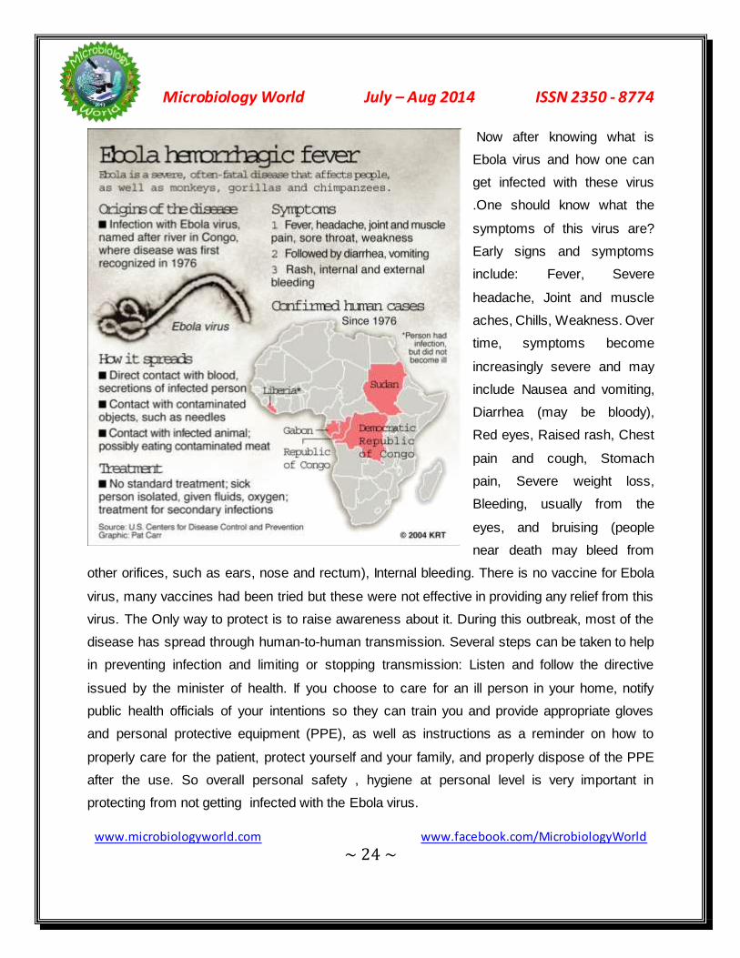
Now after knowing what is
Ebola virus and how one can
get infected with these virus
.One should know what the
symptoms of this virus are?
Early signs and symptoms
include: Fever, Severe
headache, Joint and muscle
aches, Chills, Weakness. Over
time, symptoms become
increasingly severe and may
include Nausea and vomiting,
Diarrhea (may be bloody),
Red eyes, Raised rash, Chest
pain and cough, Stomach
pain, Severe weight loss,
Bleeding, usually from the
eyes, and bruising (people
near death may bleed from
other orifices, such as ears, nose and rectum), Internal bleeding. There is no vaccine for Ebola
virus, many vaccines had been tried but these were not effective in providing any relief from this
virus. The Only way to protect is to raise awareness about it. During this outbreak, most of the
disease has spread through human-to-human transmission. Several steps can be taken to help
in preventing infection and limiting or stopping transmission: Listen and follow the directive
issued by the minister of health. If you choose to care for an ill person in your home, notify
public health officials of your intentions so they can train you and provide appropriate gloves
and personal protective equipment (PPE), as well as instructions as a reminder on how to
properly care for the patient, protect yourself and your family, and properly dispose of the PPE
after the use. So overall personal safety , hygiene at personal level is very important in
protecting from not getting infected with the Ebola virus.

Microbiology World July – Aug 2014 ISSN 2350 - 8774
www.microbiologyworld.com www.facebook.com/MicrobiologyWorld ~ 25 ~
Aromatherapy
*Hasnain Nangyal1, Upvan Bhushan2, Ammara Nawaz3
1Department of Botany, Hazara University, Mansehra
2Department of Botany, University of Jammu, India
3Department of Zoology, Punjab University, Lahore
Aromatherapy
Aromatherapy is composed of two Greek letters, Aroma (fragrance) and therapy (treatment).It
means that treatment with different fragrances.
Does aromatherapy really work?
It is use of aromatic compounds like
essential oils that derived from plant
sources that help to promote the
physiological and physical well-being. It is
used as a source of alternative medicine
in form of concentrated essential oils.
These essential oils are extracted from
different parts of plants like flower,
leaves, seed and twigs etc. Essential oils does not means that it have some nutritional type
values, but it means that they are extracted from plants that have aromatic and volatile
components that may contain antibiotics, vitamins, to some extant hormones and antiseptics.
Fragrances of these essential oils have been used to reduce the mental stress and help to
relieve the tension since many centuries. Further research and experimentation, now it is
proved that these essential oils have been play its role in reduce the dandruff and enhance the
healing properties. Most commonly used essential oils are lavender, rose, orange and sand
wood oils. So aromatherapy massage helps to reduce the anxiety and depression

Microbiology World July – Aug 2014 ISSN 2350 - 8774
www.microbiologyworld.com www.facebook.com/MicrobiologyWorld ~ 26 ~
History of Aromatherapy
History of aromatherapy is as old as history of
human being. History of aromatherapy is
started from circa (460-377 BC) era that first
physician of Egypt and had belief that illness
was caused by supernatural forces.So for the
treatment of such supernatural forces different therapies recommended, and aromatherapy is
one of them. More than 200 herbs had studied by Hippocrates that used for source of essential
oils in aromatherapy.
Over 2000 years later such therapies was employed in Egypt in form of bath, massage and
even embalming bodies. Between 18th and the 25th Dynasty (1539-657 BC), the Egyptians
continued to refine the use of aromatics in incense, medicine, cosmetics, and finally perfumes.
Similarly in 1928 this therapy is introduced in France by a chemist, René Maurice Gattefossé. In
France this therapy got importance when Rene become interested in healing proprieties of
essential oils that extracted from different plant sources. He uses the technique of distillation
with the help of which, he extract the aromatic compounds in form of essential oils from the
plants that have wonderful smell. Then a question arouses in His mind how the smell of these
aromatic compounds helps in relaxation of body and mind. To find out the question of this
answer, accidently his hand burnt during the laboratory work then he used the lavender oil for
relieving the pain it was amazing for him within few minutes sensation of burning was gone.
With the passage of time and advancement in science and technology, Aromatherapy get more
importance in 20th century and more working will be continued on aromatherapy.
Why we use Aromatherapy
Basic purpose of aromatherapy is to keep the mind and body in good state. It helps to promote
the sensory experiences of massage and relieve the mental tension. The fragrance of aromatic
compounds helps to maintain the outlook of limbic system that helps to regulate the mental
Microbiology World July – Aug 2014 ISSN 2350 - 8774
www.microbiologyworld.com www.facebook.com/MicrobiologyWorld ~ 27 ~
stress. Aromatic compounds that obtain from the different plant sources help to regulate the
emotional condition and mental stress where the synthetic medicines currently fail.
Essential oils
These are the extracted material obtain
from the different parts of plants by
different methods. Basic component of
these essential oils are aromatic
compounds and its derivates that have
healing properties. Essential oils are
volatile in nature that evaporated at room temperature that left a specific fragrance. Essential
oils are differing in chemical compositions that are found in oils of fat and fatty acids.
Components of Essential oils
There are following the important components that found in Essential oils.
Terpenes, Phenols, Ketones, Ether, Ester, Aldehyde, Alcohols
Terpenes: There are two types of terpenes; 1) Monoterpenes & 2) Sesquiterpenes
Monoterpenes: These compounds are found all most in all essential oils and have 10 carbon
atom structures with at least one double bond. In the presence of air such compounds are
readily oxidized.
Sesquiterpenes: Such compounds have almost 15 carbon atom structures that are more
complex. It is found in German Chamomile.
Phenols: Mostly the derivatives of phenols are found in essential oils such as thymol, eugenol
and carvacrol. Such oils have antiseptic and disinfectant properties.
Ketones: Such compounds are found in hyssop, eucalyptus and rosemary oils. The essential
oils that have such ketonic compounds must be avoided to use during pregnancy.
Microbiology World July – Aug 2014 ISSN 2350 - 8774
www.microbiologyworld.com www.facebook.com/MicrobiologyWorld ~ 28 ~
Ethers: Such compounds are found in tarragon and basil oils.
Esters: The essential oils that have such ester compounds have fruity smell and have sedative,
antibacterial and antifungal properties.
Aldehyde: Such compounds are found in citrus fruits and lemon oils. Such essential oils have
anti-fungal, anti-inflammatory, disinfectant, sedative yet uplifting therapeutic qualities.
Alcohols: It has two types; monoalcohols and Sesquialcohols
Monoalcohols: Such compounds are found in juniper oil and tea tree oils
Sesquialcohols: Such compounds are rarely found in essential oils such as chamomile oils
Extraction of Essential oils: Following the techniques is used for extraction of essential oils
from plants.
Distillation: Plant material is soaked and after that process of distillation is done. The process
of distillation may be steam distillation and hydro distillation. In case of steam distillation steam
is passed through condenser then liquefies the oils. While in case of hydro distillation plant
materials is soaked and make a broth. But this method is not suitable because material is
overcooked. This method is suitable for extraction of oils from seeds or stem.
Solvent extraction: Different organic solvents are used for extraction of oils like Linden
Blossom and Jasmine
Resinoids: these are prepared from the dead organic plants that basically have hydrocarbons
play its role for extraction of essential oil from plant source.
Mode of action of Aromatherapy
Total of the 15% of air that we inhaled is enter in nose where olfactory receptors transport
odors’ to apart of the brain called the limbic system that connected to our mood and emotion. In
case of aromatherapy essential oils have specific odor due to chemicals that unlock the
emotions .These small molecules are absorbed in blood through lungs where it diffuse to

Microbiology World July – Aug 2014 ISSN 2350 - 8774
www.microbiologyworld.com www.facebook.com/MicrobiologyWorld ~ 29 ~
tissues .The aroma from the oils sends a message to limbic system which is center of
controlling the emotions, memory and sexual arousal.
Fight and Flight hormones involve in Aromatherapy
Epinephrine and nor epinephrine hormones are called normally fight and flight hormones that
play a crucial role for survival of life. During the emotional and physiological stress, over
production of these hormones weaken the immune system by reducing the T helper cell and
inhibit the natural killer cells. Now a day's research on aromatherapy may leading to this aspect
the persons who treated by aromatherapy boost up their immune system during the depression
and stress conditions. Regular treatment of aromatherapy and massage with essential oils helps
to break the depression cycle and boost up the immune system.
Applications of Aromatheorapy
Several different methods are adopted to use the essential oils in aromatherapy.
Full-body baths: 5 to 10 drops of essential oils are mixed in tub of water either hot or cold.
Hand or foot baths: 2 to 3 drops of essential oils are mixed in tub of water either hot or cold
and then soaked hand or feet in this water for 10 to 20 minutes.

Microbiology World July – Aug 2014 ISSN 2350 - 8774
www.microbiologyworld.com www.facebook.com/MicrobiologyWorld ~ 30 ~
Inhalations: This is apply in treatment of nasal problems such as sinus or allergic reactions.
Small amount of water boil and then add 2 to 3 drops of essential oils and inhale its steam.
Diffusion: In this technique special type of nebulizer used that help to disperse small droplets of
essential oil into the air. This is recommended to break down the depression cycle.
Massage: In this technique essential oils mixed with other carrier oils e.g wheat germ, avocado,
olive, safflower, grape seed, or Soya bean oil. A ratio that is commonly recommended is 2.5–5%
essential oil to 95–97.5% carrier oil.
Essential oils that commonly used: some of important essential oils presented there that mostly
commonly used
Roman chamomile oil;
It is helpful in treatment of skin diseases, menstrual pain and depression.
Peppermint oil;
It helps to relax the stomach muscles and gastrointestinal tract. Its acts as
an anti-inflammatory, antiseptic, and antimicrobial that make effective in
treatment of cold and flu symptoms.
Rosemary oil;
It helps in treatment of muscular, as well as low blood pressure,
gastrointestinal problems and headaches.
Lemon oil;
It acts as anti-stress and anti-depressant.

Microbiology World July – Aug 2014 ISSN 2350 - 8774
www.microbiologyworld.com www.facebook.com/MicrobiologyWorld ~ 31 ~
Eucalyptus oil;
It helps to aid respiratory system ailments by enhancing deep
breathing
Lavender oil;
Aroma therapists use it to treat respiratory problems, abdominal cramps,
depression, insomnia, tension-related problems, burns, sun-damaged skin,
and various types of skin infections.
Rose oil;
Good for headache, nervous tension, stress related conditions, insomnia,
and nausea. Use against broken capillaries, dry skin, and poor circulation.
Jasmine oil;
Relaxing and intoxicating. Jasmine is valued in skincare in aiding dry,
sensitive and irritated skin
Side Effects of Aromatheorapy
We should never take the essential oils orally as recommended by professional aroma
therapeutics. Aromatherapy can induce side effects, such as rash, headache, liver and nerve
damage, as well as harm to the fetus. Oils that have high phenolic contents such as cinnamon
can cause skin irritation. Dilute oil with water or a base massages oil (such as almond or

Microbiology World July – Aug 2014 ISSN 2350 - 8774
www.microbiologyworld.com www.facebook.com/MicrobiologyWorld ~ 32 ~
sesame oil) before applying to your skin, and avoid using near your eyes. In addition, essential
oils are highly volatile and flame able so they should never be used near an open flame.
Precautions
Some essential oils directly apply to the skin in form of perfume. Oils of orange and peppermint
cause irritation to the skin if applied in concentrated form. During the massage essential oils are
mixed with carrier or vegetable oils. A final precaution is to avoid taking essential oils internally
without a consultation with a physician or naturopathist. Citrus-based essential oils, including
bitter and sweet orange, lime, lemon, grapefruit, and tangerine, are phototoxic, and exposure to
direct sunlight should be avoided for at least four hours after their application. Before using
essential oils on the skin, individuals should perform a skin patch test by applying a small
amount of the diluted oil behind the wrist and covering it with a bandage or cloth for up to 12
hours.

Microbiology World July – Aug 2014 ISSN 2350 - 8774
www.microbiologyworld.com www.facebook.com/MicrobiologyWorld ~ 33 ~
Recent Diagnostic Techniques of Subclinical
Bovine Mastitis
Aron Jacob, Tshering Dolma, Rekha V, Aswathy P B and Jeny K John
Indian Veterinary Research Institute, Izatnagar, Bareilly, Uttar Pradesh.
Introduction
Mastitis remains the most common and costly disease of dairy cattle (Oviedo- Boyso et al.,
2007). Treatment and control of mastitis is difficult due to the multiple causative agents i.e.,
more than 200 different organisms have been recorded in scientific literatures as being causes
of bovine mastitis. Mastitis results when pathogenic bacteria are able to gain entrance to the
udder, overcome the cows’ immune defences, establish an infection and produce inflammation
of udder secretory tissue. Contagious pathogens like Streptococcus agalactiae and
Staphylococcus aureus reside primarily in the udder of infected cows. Transmission is limited
only at the time of milking process. Environmental pathogens are common inhabitants of the
cow’s environment. The transmission can occur at any time including milking time, between
milkings, during dry period and prior to first calving in heifers.
Recent diagnostic techniques for detection of subclinical mastitis
Subclinical mastitis is difficult to detect due to the absence of any visible indications and
requires the availability of a rapid screening test for early disease detection (Viguier et al.,
2009).
Somatic Cell Count
The test measures increase in somatic cells in milk. SCC at quarter level gives better detection
performance than measuring SCC at cow level (Mollenhorst, H., 2010). Somatic cell counters
operate on the principle of optical fluorescence where ethidium bromide or propidium iodide
used stain nuclear DNA and the fluorescent signal generated is used to estimate the SCC in
milk. Esterase-catalyzed enzymatic reaction assay is also useful to determine the SCC.

Microbiology World July – Aug 2014 ISSN 2350 - 8774
www.microbiologyworld.com www.facebook.com/MicrobiologyWorld ~ 34 ~
Electrical Conductivity
This test measures the increase in conductance in milk caused by the elevation in levels of ions
such as sodium, potassium, calcium, magnesium and chloride during inflammation. Janzekovic
et al., (2009) indicated that the conductivity in individual quarters was <5.5 mS/cm for healthy
udders and >6.5 mS/cm for subclinical udders.
NAGase test
According to Pyrola et al., (2003) estimation of endogenous enzyme N- acetyl-β-D-
glucosaminidase is the most accurate of the indirect tests. It is an intracellular lysosomal
enzyme from neutrophils and epithelial cells. Production month, breed, lactation stage and milk
yield significantly affects NAGase activity. Compared to primiparous animals activity is higher for
multiparous animals. NAGase activity is higher at beginning of lactation and in late lactation.
Colorimetric and fluorometric assays have been developed to measure the elevated
concentration levels of these enzymes in milk (Larsen, 2005).Chagunda et al., 2005 states that
in mastitis NAGase tend to increase before the day of diagnosis and drop after treatment. The
enzyme activity tends to increase about eight days before diagnosis and more rapidly than
somatic cell count. So NAGase activity could be effectively utilized for diagnosis of mastitis.
Normal animals: 43.47nmol/ml mastitic: 69.42nmol/ml.
LDH estimation
Lactose dehydrogenase (endogenous) activity tends to increase 8 days before diagnosis and
more rapidly compared to somatic cell count. Compared to NAGase and SCC, LDH activity has
highest change in mastitis; SCC has the lowest (Chagunda et al., 2005). In healthy animals LDH
activity in milk is 485.94±13.66 IU/l, where as subclinical mastitis cases it is 1524.04±111.74
IU/l. The activity of LDH, NAGase and SCC could be used as early indicators of mastitis.
Production month, lactation stage and milk yield significantly affects LDH activity. Age at first
calving and parity didn’t have significant effect on enzyme activity. Compared to primiparous
animals’ activity is higher for multiparous animals. LDH activity is higher at beginning of lactation
and in late lactation than in mid lactation. Mohammadian (2011), states that the mean activity of
(LDH) was higher in milk from mastitic udders than in milk from healthy udders.

Microbiology World July – Aug 2014 ISSN 2350 - 8774
www.microbiologyworld.com www.facebook.com/MicrobiologyWorld ~ 35 ~
Spectrophotometry, colorimetric LDH quantification assay, ELISA are the tests used for
estimating LDH activity in milk.
L (+) lactate estimation
Lactate concentration may increase in a series of secretory, post secretory, physiological and
pathological reasons, still it can be used as a supportive parameter for diagnosis of subclinical
mastitis, and special attention must be given to physiological factors. Davis, et al., (2004)
described a rapid increase in lactate concentration during mastitis. Lactate can be used as
supportive parameter for mastitis detection (Grabowski et al., 2005). Milk sample should be
fixed properly in trichloroacetic acid to avoid post secretory lactate increase. Lactate is oxidized
to NAD+ and pyruvate and NADPH produced is measured photometrically. L+ Lactate content
in milk healthy: 0•1mM, subclinical: 0•7 to 1•5 mM, clinical: 3•3 mM. Bacteria also produce
lactate but it is D type rather than L type. Lactate together with pyruvate is used to estimate
bacterial count in milk.
Lactose estimation
In mastitis lactose content reduces to 2.5-2.83 %. According to Solverod (2005) a somatic cell
counts of more than 400000/ml corresponds with reduction in lactose content in same fraction
possibly because of damage of epithelium. Lactose content in milk can be used as indicator of
subclinical mastitis (Sharif et al., 2007).
Acute phase proteins - Milk Amyloid A
Lahtolainen, T., (2004) found that in experimental udder infections had a promising potential for
production of milk amyloid A as an indicator. A local synthesis of Milk amyloid A, a milk specific
acute phase protein in inflamed udder has been reported (Jacobsen et al., 2005). Petersen, et
al, 2005 stated that even though there is no overall difference in diagnostic performance the
somatic cell count and milk amyloid A have different diagnostic potential that could be depend
on factors such as nature of causative agents, milk kinetics during inflammation and degree of
tissue damage. M-SAA concentrations for healthy cows and cows with clinical mastitis were
Microbiology World July – Aug 2014 ISSN 2350 - 8774
www.microbiologyworld.com www.facebook.com/MicrobiologyWorld ~ 36 ~
reported also by Haghkhah et al., (2009) 9.90 and 105.12 μg/ml, respectively. MAA as a
potential physiological marker of subclinical mastitis (Gerandi, et al., 2009)
Diagnosis of mastitis by benzoic acid and sodium carbonate
Ayaz et al., (2005) described a method to diagnose mastitis using benzene sulfonic acid and
sodium carbonate solution as somatic cell count increase in mastitis sodium carbonate liquefy
DNA of these cells and release in solution to form jelly like substance with benzene sulfonic
acid. This test could be the cheapest and easy test to diagnose subclinical mastitis.
Cytokines analysis
TNFα, IL-1β, IL-6, IL-8 have been found to be released in bacterial mastitis For cytokine
analysis milk should be centrifuged at 2500 rpm for 20 min at 4°C to get fat free cell free milk
extract and should be kept at -30° C until assayed using ELISA or flurescencoptical method. In
a study conducted by Winter et al., 2005, found that during experimental challenge with
Staphylococcus epidermidis to ovine udders showed elevation of IL-8 in infected gland at 2hr
and peak at 8hr – 24 hr and will remain for 10wks and IL-1β transiently elevated at 1 and 2 day.
Conclusion
The challenge in treatment and control of mastitis is mainly due to presence of multiple
etiological agents and development of resistance to antimicrobial agent. Timely diagnosis and
prompt treatment can reduce the losses to the livestock economy by this disease syndrome.
Since the disease is of public health significance diagnosis of mastitis at the earliest is a
requisite for human health too.
References
1. Ayaz , M. M. and Akhtar, M (2005) Diagnosis of mastitis by benzoic acid and sodium
carbonate on pregnant and lactating mastitic animals. Mastitis in Dairy production
Current knowledge and future solutions.

Microbiology World July – Aug 2014 ISSN 2350 - 8774
www.microbiologyworld.com www.facebook.com/MicrobiologyWorld ~ 37 ~
2. Chagunda, M. G. G., Larsen, T., Bjerring, M. and Ingvartsen K. L. 2005 Changes in
lactate dehydrogenase, N- acetyl β D- glucosaminidase and somatic cell count in
relation to mastitis development in dairy cows. Mastitis in Dairy production Current
knowledge and future solutions.
3. Davis, S.R., Farr. V.C., Prosser C.G., Nicholas, G.D., Turner S.A., Lee, J. and Hart A.L.
2004. Milk L-lactate concentration is increased during mastitis. J.Dairy Res.78, p.175-
181.
4. Gerandi, G., Bernadini, D., Elica, A.C., Ferrari, V., Iob, L. and Segato, S. 2009. Use of
serum amyloid A and milk amyloid A in diagnosis of subclinical mastitis in dairy cows. J.
Dairy Res 76(4):p 411-17
5. Grabowski, N. Th., Redetzky, R.,Sulzer A., Hamann, J. and Kliein, G. 2005. Using milk
L(+) lactate as a diagnostic tool to detect mastitis in early lactation. Mastitis in Dairy
production Current knowledge and future solutions.
6. Haghkhah, M., Nafizi, S. and Jahromi, A. G. 2010. Evaluation of milk haptoglobin and
amyloid A in high producing dairy cattle with clinical and subclinical mastitis in Shiraz. J.
Comp. Clin. Pathol. Dec 2010.19 (6) p. 547-552.
7. Jacobsen,S., Niewold T.A., Kornalijnslijper, E., Toussaint M.J.M. and Gruyi E. 2005
Kinetics of local and systemic isoforms of serum amyloid A in bovine mastitic milk. Vet.
Immunol. Immunopathol.104, p 389-399
8. Janzekovic, M., Brus, M., Mursec, B., Vinis, P., Stajnko, D. and Cus F., (2009). Mastitis
detection based on electric conductivity of milk. J. Achievements in Materials and
Manufacturing Engg. 34 (1)
9. Lahtolainen, T., Rontved, C. and Pyrola, S. 2004 Serum amyloid A and TNFα in serum
and milk during experimental endotoxin mastitis. Vet. Res. 35 p 651-659
10. Larsen, T., 2005. Determination of lactate dehydrogenase (LDH) activity in milk by a
fluorometric assay. J. Dairy Res. 72, 209.
11. Mohammadian, B., 2011. The effect of subclinical mastitis on lactate dehydrogenase in
dairy cows International J. of Anim. and Vet. Advances 3(3): 161-163
12. Mollenhorst, H.; van der Tol, P.P.J.; Hogeveen, H. Somatic cell count assessment at the
quarter or cow milking level. J. Dairy Sci. 2010, 93, 3358-3364.

Microbiology World July – Aug 2014 ISSN 2350 - 8774
www.microbiologyworld.com www.facebook.com/MicrobiologyWorld ~ 38 ~
13. Oviedo- Boyso, J., Valdez- Alarcon, J.J., Cajero- Juarez, M., and Baizabal-Auirre,
V.M.(2007) Innate immune response of bovine mammary gland to pathogenic bacteria,
responsible for mastitis. J.Infect., 54(4):399-409
14. Petersen, H. H., Gardner I.A., Rossito, P., Larsen H.D. Heegard, P.M.H.( 2005) Milk
amyloid A concentration and somatic cell count in diagnosis of bovine mastitis. Mastitis
in Dairy production Current knowledge and future solutions.
15. Pyrola , S. 2003, Indicators of inflammation in the diagnosis of mastitis. Vet. Res. 34 p :
565–578
16. Sharif, A., Ahamad T., Bilal M.Q., Yusuf ,A. and Muhammad,G. 2007. Effect of severiety
of subclinical mastitis on somatic cell count and lactose content of buffalo milk. Pak.Vet.
J., 27(3) 142-144
17. Solverod L., Simonsen, S., Waldmann, A. and Ropstad, E.(2005) Variation in somatic
cell count and milk components in fraction collected quarter milk samples. Mastitis in
Dairy production Current knowledge and future solutions.
18. Viguier, C., Arora, S., Gilmartin, N., Welbeck, K., O'Kennedy, R., 2009. Mastitis
detection: Current trends and future perspectives. Trends Biotechnol. 27,486.
19. Winter, P., Fuch, K. and Schilcher, F. 2005. Host response reaction in lactating ovine
udder during experimental challenge with Staphylococcus epidermidis. Mastitis in Dairy
production Current knowledge and future solutions
Microbiology World July – Aug 2014 ISSN 2350 - 8774
www.microbiologyworld.com www.facebook.com/MicrobiologyWorld ~ 39 ~
Cells of Immune System
Mr. Shaikh Rajesh Ali
Assistant professor, Dept. of Microbiology,
Acharya Prafulla Chandra College, New Barrackpore, Kol-131
The lymphocytes are the cells that define the adaptive immune system as they are the only with
the attributes of specificity, diversity, memory, and self/non-self discrimination. Lymphocytes are
part of group of cells collectively known as white blood cells or leukocytes that participate in the
immune response. Leukocytes, together with the red blood cells (erythrocytes) and platelets,
are the blood cells. Hematopoiesis is the process of formation and differentiation of blood cells.
All blood cells arise from a type of cell called the hematopoietic stem cell (HSC). Stem cells
differentiate into one or more cell types (i.e., they can be unipotent or pluripotent), and have the
property of selfrenewing. In humans, hematopoiesis begins in the embryonic yolk sac in the first
weeks of development. In the third month of development, HSC migrate from the yolk sac to
fetal liver and then to the spleen. These two organs are the responsible of hematopoiesis from
the 3rd to the 7th month of development. After that, the bone marrow takes the place as the
hematopoietic organ.
Early in hematopoiesis, a HSC differentiate into one of two pathways, giving rise to a lymphoid
stem cell or a myeloid stem cell. The lymphoid stem cell gives rise to B, T and NK cells through
the differentiation of progenitor cells specific for each lineage.
The myeloid stem cell generates progenitors for the red blood cells, the platelets and for all the
other leukocytes, which are the granulocytic cells (neutrophils, eosinophils, basophils),
monocytes, mast cells, and dendritic cells.
Differentiation of progenitors into specialized cells is controlled by hematopoietic cytokines or
growth factors. A family of cytokines known as colony-stimulating factors (CSF) is important to
induce the differentiation of the different hematopoietic cell lines. There are different types of

Microbiology World July – Aug 2014 ISSN 2350 - 8774
www.microbiologyworld.com www.facebook.com/MicrobiologyWorld ~ 40 ~
CSFs: multilineage CSF or interleukin-3, macrophage CSF, granulocyte CSF, granulocyte-
macrophage CSF.
The cytokine that regulates the production of red blood cells is from a different family and is
called erythropoietin. Thus, the commitment of a progenitor to a particular lineage depends on
the expression of specific receptors on the cell membrane for particular cytokines.

Microbiology World July – Aug 2014 ISSN 2350 - 8774
www.microbiologyworld.com www.facebook.com/MicrobiologyWorld ~ 41 ~
Hematopoiesis is exquisitely regulated to maintain a constant number of cells in a given time.
This is accomplished by controlling the expression of cytokines and cytokine receptors, and by a
process known as programmed cell death, in which the cell actively participate in its own death.
The morphological changes seen in programmed cell death are known as apoptosis.
The use of HSCs has proved to be an important tool in medicine. Transplantation of HSCs is
used in patients with genetically immunodeficiencies to provide a functional immune system.
Also, in some genetically determined anemias, transplantation of HSC is use to replace the non-
functional red blood cells progenitors. More common is perhaps its use in patients with leukemia
or in patients subjected to chemotherapy and radiation, which have destroyed the hematopoietic
system.
An effective immune response involves two major groups of cells: T lymphocytes and antigen-
presenting cells. Lymphocytes are one of many types of white blood cells produced in the bone
marrow by the process of hematopoiesis. Lymphocytes leave the bone marrow, circulate in the
blood and lymphatic systems, and reside in various lymphoid organs. Because they produce
and display antigen binding cell-surface receptors, lymphocytes mediate the defining
immunologic attributes of specificity, diversity, memory, and self/nonself recognition. The two
major populations of lymphocytes are B lymphocytes (B cells) and T lymphocytes (T cells).
B Lymphocytes:
B lymphocytes mature within the bone marrow; when they leave it, each expresses a unique
antigen-binding receptor
on its membrane. This
antigen-binding or B-cell
receptor is a membrane-
bound antibody
molecule. Antibodies are
glycoproteins that consist

Microbiology World July – Aug 2014 ISSN 2350 - 8774
www.microbiologyworld.com www.facebook.com/MicrobiologyWorld ~ 42 ~
of two identical heavy polypeptide chains and two identical light polypeptide chains. Each heavy
chain is joined with a light chain by disulfide bonds, and additional disulfide bonds hold the two
pairs together. The amino-terminal ends of the pairs of heavy and light chains form a cleft within
which antigen binds. When a naive B cell (one that has not previously encountered antigen) first
encounters the antigen that matches its membrane bound antibody, the binding of the antigen to
the antibody causes the cell to divide rapidly; its progeny differentiate into memory B cells and
effector B cells called plasma cells. Memory B cells have a longer life span than naive cells, and
they express the same membrane-bound antibody as their parent B cell. Plasma cells produce
the antibody in a form that can be secreted and have little or no membrane-bound antibody.
Although plasma cells live for only a few days, they secrete enormous amounts of antibody
during this time. It has been estimated that a single plasma cell can secrete more than 2000
molecules of antibody per second. Secreted antibodies are the major effector molecules of
humoral immunity.
T Lymphocytes:
T lymphocytes also arise in the bone marrow. Unlike B cells, which mature within the bone
marrow, T cells migrate to the thymus gland to mature. During its maturation within the thymus,
the T cell comes to express a unique antigen-binding molecule, called the T-cell receptor, on its
membrane.Unlike membrane-bound antibodies on B cells,which can recognize antigen alone,
T-cell receptors can recognize only antigen that is bound to cell-membrane proteins called
major histocompatibility complex (MHC) molecules. MHC molecules that function in this
recognition event,which is termed “antigen presentation,” are polymorphic (genetically diverse)
glycoproteins found on cell membranes. There are two major types of MHC molecules: Class I
MHC molecules, which are expressed by nearly all nucleated cells of vertebrate species, consist
of a heavy chain linked to a small invariant protein called β2-microglobulin. Class II MHC
molecules, which consist of an alpha and a beta glycoprotein chain, are expressed only by
antigen-presenting cells.When a naive T cell encounters antigen combined with a MHC
molecule on a cell, the T cell proliferates and differentiates into memory T cells and various
effector T cells. There are two well-defined subpopulations of T cells: T helper (TH) and T
cytotoxic (TC) cells.Although a third type of T cell, called a T suppressor (TS) cell, has been
Microbiology World July – Aug 2014 ISSN 2350 - 8774
www.microbiologyworld.com www.facebook.com/MicrobiologyWorld ~ 43 ~
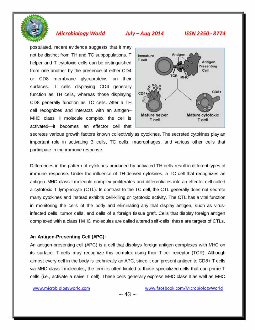
postulated, recent evidence suggests that it may
not be distinct from TH and TC subpopulations. T
helper and T cytotoxic cells can be distinguished
from one another by the presence of either CD4
or CD8 membrane glycoproteins on their
surfaces. T cells displaying CD4 generally
function as TH cells, whereas those displaying
CD8 generally function as TC cells. After a TH
cell recognizes and interacts with an antigen–
MHC class II molecule complex, the cell is
activated—it becomes an effector cell that
secretes various growth factors known collectively as cytokines. The secreted cytokines play an
important role in activating B cells, TC cells, macrophages, and various other cells that
participate in the immune response.
Differences in the pattern of cytokines produced by activated TH cells result in different types of
immune response. Under the influence of TH-derived cytokines, a TC cell that recognizes an
antigen–MHC class I molecule complex proliferates and differentiates into an effector cell called
a cytotoxic T lymphocyte (CTL). In contrast to the TC cell, the CTL generally does not secrete
many cytokines and instead exhibits cell-killing or cytotoxic activity. The CTL has a vital function
in monitoring the cells of the body and eliminating any that display antigen, such as virus-
infected cells, tumor cells, and cells of a foreign tissue graft. Cells that display foreign antigen
complexed with a class I MHC molecules are called altered self-cells; these are targets of CTLs.
An Antigen-Presenting Cell (APC):
An antigen-presenting cell (APC) is a cell that displays foreign antigen complexes with MHC on
its surface. T-cells may recognize this complex using their T-cell receptor (TCR). Although
almost every cell in the body is technically an APC, since it can present antigen to CD8+ T cells
via MHC class I molecules, the term is often limited to those specialized cells that can prime T
cells (i.e., activate a naive T cell). These cells generally express MHC class II as well as MHC

Microbiology World July – Aug 2014 ISSN 2350 - 8774
www.microbiologyworld.com www.facebook.com/MicrobiologyWorld ~ 44 ~
class I molecules, and can stimulate CD4+ ("helper") T cells as well as CD8+ ("cytotoxic") T
cells. To help distinguish between the two types of APCs, those that express MHC class II
molecules are often called professional antigen-presenting cells.
The APCs are very efficient at
phagocytosis, which allows them to present
exogenous as well as internal antigens.
After dendritic cells or macrophages
swallow pathogens, they usually migrate to
the lymph nodes, where most T cells are.
They do this chemotactically: chemokines
that flow in the blood and lymph vessels
"draw" the APCs to the lymph nodes.
During the migration, DCs or macrophages
undergo a process of maturation, like they lose most of their ability to further swallow
pathogens, and they develop an increased ability to communicate with T cells. Enzymes within
the cell digest the swallowed pathogen into smaller pieces containing epitopes, which are then
presented to T cells using MHC class II. After interaction with TCR an additional co-stimulatory
signal is then produced by antigen presenting cell, leading to activation of TH cell and release
different types of cytokines.
Natural Killer (NK) Cell:
A small group of lymphocytes, call null cell, in the peripheral blood do not express the
membrane molecules and receptors that distinguish T and B cell lineage. These cells fail to
produce immunoglobulin. Because these cells do not produce any antigen binding receptor
(ABC receptor), they lack precise immunologic specificity and memory. Most members of null
cell population are large, granular lymphocyte called natural killer (NK) cell.

Microbiology World July – Aug 2014 ISSN 2350 - 8774
www.microbiologyworld.com www.facebook.com/MicrobiologyWorld ~ 45 ~
NK cell were shown to play an important role
in host defense against tumor cell and
against some virus infected cell. NK cell can
recognize the potential target in two different
ways. In some cases, an NK cell employs
NK cell receptors to distinguish
abnormalities, which recognize the tumor
cell or virus infected cell by abnormal cell’s
MHC class I molecules. Another way in
which NK cell recognize potential target cells depends upon the fact that the tumor cell or virus
infected cell display antigen against which immune system produce anti-tumor or antiviral
antibodies and which bind to the abnormal cell surface. Because NK cell express CD16, a
membrane receptor for the carboxyl-terminal end of IgG molecule, called Fc region, they can
attached to these antibodies and subsequently destroy the target cell. This process of cell
destruction is called antibody dependent cell mediated cytotoxicity (ADCC).
Microbiology World July – Aug 2014 ISSN 2350 - 8774
www.microbiologyworld.com www.facebook.com/MicrobiologyWorld ~ 46 ~
You can also send your articles to
or
Selected ones will be published in
our next issue of Sept-Oct 2014.
Thanks,
Microbiology World Team
Related Documents