Microbiology Induction for Neurosurgery trainees Kavita Sethi Consultant Microbiologist LTHT

Microbiology Induction for Neurosurgery trainees Kavita Sethi Consultant Microbiologist LTHT.
Dec 17, 2015
Welcome message from author
This document is posted to help you gain knowledge. Please leave a comment to let me know what you think about it! Share it to your friends and learn new things together.
Transcript

Microbiology Induction for Neurosurgery trainees
Kavita Sethi
Consultant Microbiologist
LTHT
LTHT Microbiology
Contact Details
Duty Microbiologist for 0113-392-3962 or -8580
interpretative amp clinical advice
Ext 25034 if need to contact myself
(Contact via switchboard for urgent queries)
For laboratory assistance within normal working hours (eg
urgent samples) 0113-392-3499
Results will NOT normally be given out by telephone if the result is already available on the results server
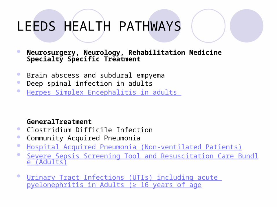
LEEDS HEALTH PATHWAYS
Neurosurgery Neurology Rehabilitation MedicineSpecialty Specific Treatment
Brain abscess and subdural empyema Deep spinal infection in adults Herpes Simplex Encephalitis in adults
GeneralTreatment Clostridium Difficile Infection Community Acquired Pneumonia Hospital Acquired Pneumonia (Non-ventilated Patients) Severe Sepsis Screening Tool and Resuscitation Care Bundle (Adults) Urinary Tract Infections (UTIs) including acute pyelonephritis
in Adults (ge 16 years of age
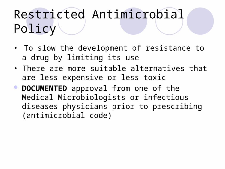
Restricted Antimicrobial Policy
bull To slow the development of resistance to a drug by limiting its use
bull There are more suitable alternatives that are less expensive or less toxic
DOCUMENTED approval from one of the Medical Microbiologists or infectious diseases physicians prior to prescribing (antimicrobial code)
How do you evaluate infection
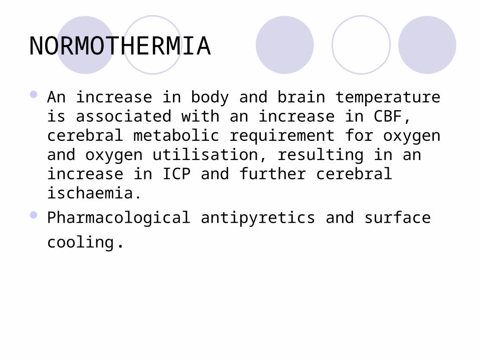
NORMOTHERMIA
An increase in body and brain temperature is associated with an increase in CBF cerebral metabolic requirement for oxygen and oxygen utilisation resulting in an increase in ICP and further cerebral ischaemia
Pharmacological antipyretics and surface cooling
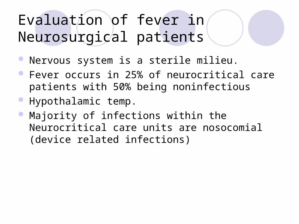
Evaluation of fever in Neurosurgical patients
Nervous system is a sterile milieu Fever occurs in 25 of neurocritical care patients with
50 being noninfectious Hypothalamic temp Majority of infections within the Neurocritical care units
are nosocomial (device related infections)
0
5
10
15
20
25
30
-6 -4 -2 0 2 4 6 8 10 12Day
WB
C (
x 10
9 L
)
0
50
100
150
200
250
CR
P
(mg
L)
WBC
CRP
surgery
Total white cell count and C-reactive protein
Risk Factors
Admission for more than 48 hours Mechanical ventilation Trauma Vascular catheterisation Urinary catheterisation Stress ulcer prophylaxis
EPIC 1995
Nosocomial infections on NITU
Respiratory infection Early Late
Bacteremia GI infection UTIs
Neurosurgical device related infections
Haemophilus influenzae Staphylococcus aureus Streptococcus pneumoniae
Pseudomonas Coliforms Acinetobacter MRSA
Clostridium difficile Asymptomatic bacteruria in
catheterised patients
Coagulase negative Staphylococcus Pacnes Staphylococcus aureus
Postoperative bacterial meningitis
Low overall incidence Variables
Clean or clean-contaminated Antibiotic prophylaxis
Aetiology Aerobic GNB (60-70)
Ecoli Kpneumoniae Paeruginosa Acinetobacter spp Saureus Spneumoniae (dural defects CSF otorrhoea or
rhinorrhoea) REMEMBER ASEPTIC MENINGITIS
Pathogenesis
What is on the skin goes in the wound Most cases are a result of surgical wound infection Independent risk factors GCS lt 10 Emergency surgery CSF leakage External CSF drainage
Diagnosis of bacterial meningitis post-neurosurgery
Useful criteria Less helpful criteria
high fever CSF glucose
new neurological deficit CSF protein
active CSF leak type of operation
CSF leukocytosis presence of foreign material
blood leukocytosis steroid use
altered mental status
neck stiffness
headache nausea
Ross et al Journal of Neurosurgery 1988 69 669-74
Diagnosis of Nosocomial meningitis
Challenging Clinical
Indistinguishable from community-acquired meningitis May be difficult to distinguish from neurological signs symptoms
of underlying disease or associated with post-op period LP must be performed to confirm diagnosis (CT first to
establish safety) CSF parameters may be altered due to surgery itself especially
in the presence of SAH CSF leucocytosis not infrequent
Antibiotics which achieve adequate CSF levels (guided by gram stain and culture)
Surgical management of wound infection persistent CSF leak
CSF Penetration of antibiotics
Excellent Ceftazidime Meropenem Metronidazole Rifampicin Chloramphenicol
Useless Macrolides Aminoglycosides Clindamycin
Additional points
Duration of therapy Saureus ndash 2 weeks AGNB ndash up to 3 weeks
Vancomycin penetrates poorly into CSF and patient may fail to respond to systemic therapy Implant an Ommaya reservoir and instill vancomycin
directly into ventricles every 3rd day
Postoperative aseptic meningitis
Thought to be the result of irritation caused either by blood degradation products introduced into SAS during surgery
Indistinguishable from postoperative bacterial meningitis (clinical amp CSF cell count and chemistry) CSF lactate to distinguish
Approach Empirical antibiotic therapy If CSF sterile ndash discontinue antibiotics Responds favourably to high dose corticosteroids
Empiric therapy
1st line therapy Cefotaxime 2-3g qds
Confirmed Paeruginosa or recent broad spectrum antibiotic
Ceftazidime 2g tds
Suspected or confirmed ESBL-producing Enterobacteriaceae or Acinetobacter spp
Meropenem 2g tds
MSSA Flucloxacillin 2-3g qds
MRSA Vancomycin PLUS
Rifampicin
1g bd
600mg bd
Antibiotic Prophylaxis for Basilar Skull Fracture Meta Analysis
12 studies 1241 patients 58 received antibiotics
Antibiotics did not prevent meningitis RR 115 (068 - 194) p=068
CSF leakage subset RR 134 (075 - 241) p=036
Brain abscess
Hematogenous spread from extracranial site Can arise from direct spread from mastoid and sinus infections Corticomedullary junction Frontal and parietal lobes most common Posterior fossa lt5
Overall mortality rate has ranged from 0 to 24 Prognosis the rapidity of progression before hospitalization mental status onadmission
Bacteriology
1048698 Streptococci (70)1048698mixed infections (30 to 60) 1048698 Streptococcus milleri group 1048698 oral cavity appendix and female genital tract 1048698 otopharyngeal infections 1048698 IE
1048698Staphylococcus aureus 10 to 15 1048698 cranial trauma 1048698 IE Neurosurgical procedure
Contd
Bacteroides and Prevotella in 20 to 40 1048698 mixed infection Enteric gram-negative bacilli (Proteus species
Escherichia coli Klebsiella and Pseudomonas) in 23 to 33
1048698 otitic infection 1048698 Septicemia 1048698 neurosurgical procedures 1048698 immunocompromised Rare pathogens Nocardia Mycobacterium tuberculosis Listeria
monocytogenes
Brain abscess
Blood cultures should be obtained when diagnosis is suspected
Lumbar puncture (LP) should be deferred in any case for which brain abscess is suspected because of the potential for CNS
herniation and low likelihood of positive cultures
Pus collected in a sterile universal container (NOT SWAB) should be sent to Microbiology for urgent microscopy culture and sensitivity
Cerebritis and abscess
Early cerebritis ndash (3-5 days) Late Cerebritis -(4 to 14 days)
Early Capsule Stage (Begins at 2 weeks following initial infection)
Late capsule stage
Initial approach to the patient witha suspected brain abscess
Contrast CT or MRI should be performed If single or multiple ring-enhancing lesions are found the patient
should taken urgently to surgery 1048698 All lesions gt 25 cm in diameter should be excised or
stereotactically aspirated 1048698 For abscesses in the early cerebritis stage or when the
abscesses are lt 25 cm in diameter the largest lesionshould be aspirated for diagnosis and organism identification
Antimicrobial therapy
empirical antimicrobial therapy should be initiated on the basis of the patientrsquos predisposing conditions and the presumed
pathogenesis of abscess formation
Otitis mediamastoiditissinusitis Cefotaxime Metronidazole
Post neurosurgical trauma add anti-staphylococcal cover
Use Ceftazidime as the 3rd generation cephalosporin if Pseudomonas aeruginosa is suspected
Brain Abscess - Surgical treatment
significant mass effect exerted by lesion proximity to ventricle poor neurological condition Inability to obtain weekly CT scans In patient undergoing medical treatment
Intervention if neurological deterioration occurs anatomic progression of abscess towards ventricles or after 2 weeks of therapy if abscess is enlarged Also consider if there is no decrease in abscess size by 4 weeks of treatment
Shunt infections
CSF shunts become infected by various routesbull Organisms directly colonize the shunt usually at the time of surgerybull Organisms reach the CSF and the shunt via haematogenous spreadbull Organisms travel along the shunt by retrograde spread (uncommon)
Coagulase-negative Staphylococci are isolated most commonly Production of extracellular slime has been reported as being important in the pathogenesis of shunt infections
Clinical features
Variable Fever Signs of raised ICP Evidence of shunt malfunction Distal shunt infections can present with peritonitis
Shunt infections
CSF from shunt ( before antimicrobial therapy) SHUNT REMOVAL Proximal catheter valve or shunt reservoir distal
catheter in three separate containers Intrathecal +- systemic antibiotic Shunt replacement once the CSF sterile
Clinical Suspicion of VP Shunt Infection
Mild Ventriculitis Severe Ventriculitis and wound infection
POSITIVE Gram Positive Gram Negative MER if any risk factors for resistant GNBs
Repeat CSF in 48-72 Hours Monitor Response
Shunt replacement when CSF sterile at the end of therapy (see duration of antibiotics under treatment section)
Consult Microbiology to discuss alternatives if documented Penicillin allergy or multi-resistant gram negative organism
IVCTX + IT GEN
SHUNT REMOVAL AND EVD
IT VAN
CSF Gram Strain amp Culture
Await Cultures as gram stain usually negative
IV antibiotics and drainage of pus with shunt removal
EVD-related ventriculitis
Incidence between 10-17 Risk
Lowest risk 1st 4 days that EVD is in-situ Rises over next 10 days Falls thereafter
Incidence not decreased by exchanging EVD at regular intervals
Aetiology CNS (predominant)
Diagnostic approach
Patient with EVD with symptomsand signs of ventriculitis
CSF sample
+ Gram stain or+ culture
- Gram stain and culture
2nd sample
+ Gram stain or + culture (sameAs 1st)
Treat No treatment
Additional points
Collect CSF from EVD itself or the Ommaya reservoir not the drainage bag
Treat CNS infections with intraventricular Vancomycin for 5-7 days Product license does not cover this route Not validated by clinical trials Guarantees max concentrations of vancomycin at the site of
infection Avoids systemic toxicity Cheaper than systemic therapy No need to monitor levels
Hand Hygiene

LTHT Microbiology
Contact Details
Duty Microbiologist for 0113-392-3962 or -8580
interpretative amp clinical advice
Ext 25034 if need to contact myself
(Contact via switchboard for urgent queries)
For laboratory assistance within normal working hours (eg
urgent samples) 0113-392-3499
Results will NOT normally be given out by telephone if the result is already available on the results server
LEEDS HEALTH PATHWAYS
Neurosurgery Neurology Rehabilitation MedicineSpecialty Specific Treatment
Brain abscess and subdural empyema Deep spinal infection in adults Herpes Simplex Encephalitis in adults
GeneralTreatment Clostridium Difficile Infection Community Acquired Pneumonia Hospital Acquired Pneumonia (Non-ventilated Patients) Severe Sepsis Screening Tool and Resuscitation Care Bundle (Adults) Urinary Tract Infections (UTIs) including acute pyelonephritis
in Adults (ge 16 years of age
Restricted Antimicrobial Policy
bull To slow the development of resistance to a drug by limiting its use
bull There are more suitable alternatives that are less expensive or less toxic
DOCUMENTED approval from one of the Medical Microbiologists or infectious diseases physicians prior to prescribing (antimicrobial code)
How do you evaluate infection
NORMOTHERMIA
An increase in body and brain temperature is associated with an increase in CBF cerebral metabolic requirement for oxygen and oxygen utilisation resulting in an increase in ICP and further cerebral ischaemia
Pharmacological antipyretics and surface cooling
Evaluation of fever in Neurosurgical patients
Nervous system is a sterile milieu Fever occurs in 25 of neurocritical care patients with
50 being noninfectious Hypothalamic temp Majority of infections within the Neurocritical care units
are nosocomial (device related infections)
0
5
10
15
20
25
30
-6 -4 -2 0 2 4 6 8 10 12Day
WB
C (
x 10
9 L
)
0
50
100
150
200
250
CR
P
(mg
L)
WBC
CRP
surgery
Total white cell count and C-reactive protein
Risk Factors
Admission for more than 48 hours Mechanical ventilation Trauma Vascular catheterisation Urinary catheterisation Stress ulcer prophylaxis
EPIC 1995
Nosocomial infections on NITU
Respiratory infection Early Late
Bacteremia GI infection UTIs
Neurosurgical device related infections
Haemophilus influenzae Staphylococcus aureus Streptococcus pneumoniae
Pseudomonas Coliforms Acinetobacter MRSA
Clostridium difficile Asymptomatic bacteruria in
catheterised patients
Coagulase negative Staphylococcus Pacnes Staphylococcus aureus
Postoperative bacterial meningitis
Low overall incidence Variables
Clean or clean-contaminated Antibiotic prophylaxis
Aetiology Aerobic GNB (60-70)
Ecoli Kpneumoniae Paeruginosa Acinetobacter spp Saureus Spneumoniae (dural defects CSF otorrhoea or
rhinorrhoea) REMEMBER ASEPTIC MENINGITIS
Pathogenesis
What is on the skin goes in the wound Most cases are a result of surgical wound infection Independent risk factors GCS lt 10 Emergency surgery CSF leakage External CSF drainage
Diagnosis of bacterial meningitis post-neurosurgery
Useful criteria Less helpful criteria
high fever CSF glucose
new neurological deficit CSF protein
active CSF leak type of operation
CSF leukocytosis presence of foreign material
blood leukocytosis steroid use
altered mental status
neck stiffness
headache nausea
Ross et al Journal of Neurosurgery 1988 69 669-74
Diagnosis of Nosocomial meningitis
Challenging Clinical
Indistinguishable from community-acquired meningitis May be difficult to distinguish from neurological signs symptoms
of underlying disease or associated with post-op period LP must be performed to confirm diagnosis (CT first to
establish safety) CSF parameters may be altered due to surgery itself especially
in the presence of SAH CSF leucocytosis not infrequent
Antibiotics which achieve adequate CSF levels (guided by gram stain and culture)
Surgical management of wound infection persistent CSF leak
CSF Penetration of antibiotics
Excellent Ceftazidime Meropenem Metronidazole Rifampicin Chloramphenicol
Useless Macrolides Aminoglycosides Clindamycin
Additional points
Duration of therapy Saureus ndash 2 weeks AGNB ndash up to 3 weeks
Vancomycin penetrates poorly into CSF and patient may fail to respond to systemic therapy Implant an Ommaya reservoir and instill vancomycin
directly into ventricles every 3rd day
Postoperative aseptic meningitis
Thought to be the result of irritation caused either by blood degradation products introduced into SAS during surgery
Indistinguishable from postoperative bacterial meningitis (clinical amp CSF cell count and chemistry) CSF lactate to distinguish
Approach Empirical antibiotic therapy If CSF sterile ndash discontinue antibiotics Responds favourably to high dose corticosteroids
Empiric therapy
1st line therapy Cefotaxime 2-3g qds
Confirmed Paeruginosa or recent broad spectrum antibiotic
Ceftazidime 2g tds
Suspected or confirmed ESBL-producing Enterobacteriaceae or Acinetobacter spp
Meropenem 2g tds
MSSA Flucloxacillin 2-3g qds
MRSA Vancomycin PLUS
Rifampicin
1g bd
600mg bd
Antibiotic Prophylaxis for Basilar Skull Fracture Meta Analysis
12 studies 1241 patients 58 received antibiotics
Antibiotics did not prevent meningitis RR 115 (068 - 194) p=068
CSF leakage subset RR 134 (075 - 241) p=036
Brain abscess
Hematogenous spread from extracranial site Can arise from direct spread from mastoid and sinus infections Corticomedullary junction Frontal and parietal lobes most common Posterior fossa lt5
Overall mortality rate has ranged from 0 to 24 Prognosis the rapidity of progression before hospitalization mental status onadmission
Bacteriology
1048698 Streptococci (70)1048698mixed infections (30 to 60) 1048698 Streptococcus milleri group 1048698 oral cavity appendix and female genital tract 1048698 otopharyngeal infections 1048698 IE
1048698Staphylococcus aureus 10 to 15 1048698 cranial trauma 1048698 IE Neurosurgical procedure
Contd
Bacteroides and Prevotella in 20 to 40 1048698 mixed infection Enteric gram-negative bacilli (Proteus species
Escherichia coli Klebsiella and Pseudomonas) in 23 to 33
1048698 otitic infection 1048698 Septicemia 1048698 neurosurgical procedures 1048698 immunocompromised Rare pathogens Nocardia Mycobacterium tuberculosis Listeria
monocytogenes
Brain abscess
Blood cultures should be obtained when diagnosis is suspected
Lumbar puncture (LP) should be deferred in any case for which brain abscess is suspected because of the potential for CNS
herniation and low likelihood of positive cultures
Pus collected in a sterile universal container (NOT SWAB) should be sent to Microbiology for urgent microscopy culture and sensitivity
Cerebritis and abscess
Early cerebritis ndash (3-5 days) Late Cerebritis -(4 to 14 days)
Early Capsule Stage (Begins at 2 weeks following initial infection)
Late capsule stage
Initial approach to the patient witha suspected brain abscess
Contrast CT or MRI should be performed If single or multiple ring-enhancing lesions are found the patient
should taken urgently to surgery 1048698 All lesions gt 25 cm in diameter should be excised or
stereotactically aspirated 1048698 For abscesses in the early cerebritis stage or when the
abscesses are lt 25 cm in diameter the largest lesionshould be aspirated for diagnosis and organism identification
Antimicrobial therapy
empirical antimicrobial therapy should be initiated on the basis of the patientrsquos predisposing conditions and the presumed
pathogenesis of abscess formation
Otitis mediamastoiditissinusitis Cefotaxime Metronidazole
Post neurosurgical trauma add anti-staphylococcal cover
Use Ceftazidime as the 3rd generation cephalosporin if Pseudomonas aeruginosa is suspected
Brain Abscess - Surgical treatment
significant mass effect exerted by lesion proximity to ventricle poor neurological condition Inability to obtain weekly CT scans In patient undergoing medical treatment
Intervention if neurological deterioration occurs anatomic progression of abscess towards ventricles or after 2 weeks of therapy if abscess is enlarged Also consider if there is no decrease in abscess size by 4 weeks of treatment
Shunt infections
CSF shunts become infected by various routesbull Organisms directly colonize the shunt usually at the time of surgerybull Organisms reach the CSF and the shunt via haematogenous spreadbull Organisms travel along the shunt by retrograde spread (uncommon)
Coagulase-negative Staphylococci are isolated most commonly Production of extracellular slime has been reported as being important in the pathogenesis of shunt infections
Clinical features
Variable Fever Signs of raised ICP Evidence of shunt malfunction Distal shunt infections can present with peritonitis
Shunt infections
CSF from shunt ( before antimicrobial therapy) SHUNT REMOVAL Proximal catheter valve or shunt reservoir distal
catheter in three separate containers Intrathecal +- systemic antibiotic Shunt replacement once the CSF sterile
Clinical Suspicion of VP Shunt Infection
Mild Ventriculitis Severe Ventriculitis and wound infection
POSITIVE Gram Positive Gram Negative MER if any risk factors for resistant GNBs
Repeat CSF in 48-72 Hours Monitor Response
Shunt replacement when CSF sterile at the end of therapy (see duration of antibiotics under treatment section)
Consult Microbiology to discuss alternatives if documented Penicillin allergy or multi-resistant gram negative organism
IVCTX + IT GEN
SHUNT REMOVAL AND EVD
IT VAN
CSF Gram Strain amp Culture
Await Cultures as gram stain usually negative
IV antibiotics and drainage of pus with shunt removal
EVD-related ventriculitis
Incidence between 10-17 Risk
Lowest risk 1st 4 days that EVD is in-situ Rises over next 10 days Falls thereafter
Incidence not decreased by exchanging EVD at regular intervals
Aetiology CNS (predominant)
Diagnostic approach
Patient with EVD with symptomsand signs of ventriculitis
CSF sample
+ Gram stain or+ culture
- Gram stain and culture
2nd sample
+ Gram stain or + culture (sameAs 1st)
Treat No treatment
Additional points
Collect CSF from EVD itself or the Ommaya reservoir not the drainage bag
Treat CNS infections with intraventricular Vancomycin for 5-7 days Product license does not cover this route Not validated by clinical trials Guarantees max concentrations of vancomycin at the site of
infection Avoids systemic toxicity Cheaper than systemic therapy No need to monitor levels
Hand Hygiene
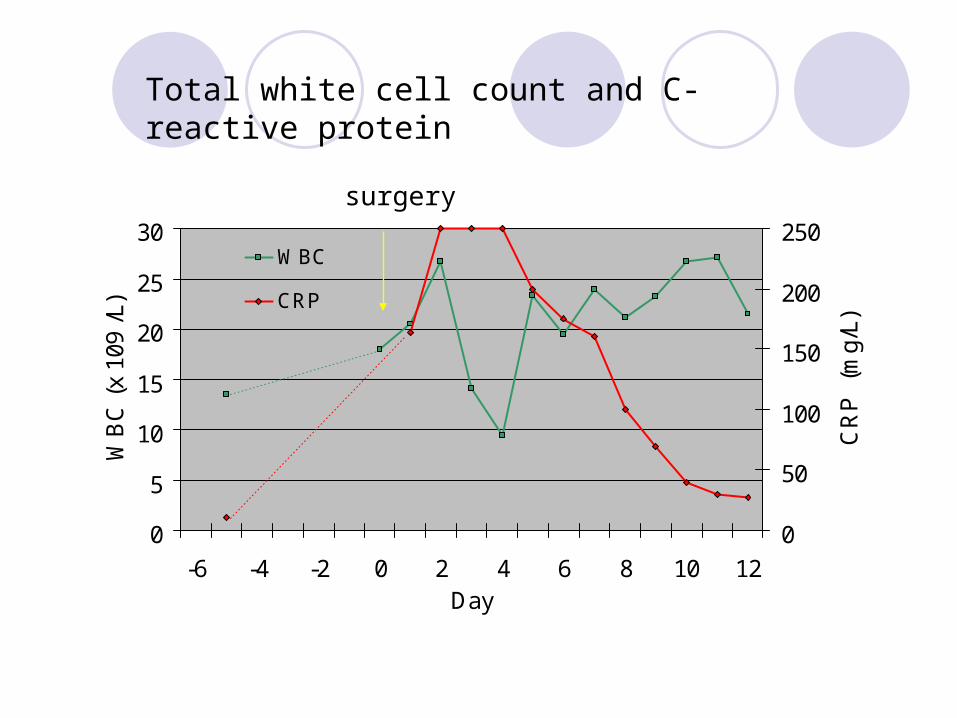
0
5
10
15
20
25
30
-6 -4 -2 0 2 4 6 8 10 12Day
WB
C (
x 10
9 L
)
0
50
100
150
200
250
CR
P
(mg
L)
WBC
CRP
surgery
Total white cell count and C-reactive protein
Risk Factors
Admission for more than 48 hours Mechanical ventilation Trauma Vascular catheterisation Urinary catheterisation Stress ulcer prophylaxis
EPIC 1995
Nosocomial infections on NITU
Respiratory infection Early Late
Bacteremia GI infection UTIs
Neurosurgical device related infections
Haemophilus influenzae Staphylococcus aureus Streptococcus pneumoniae
Pseudomonas Coliforms Acinetobacter MRSA
Clostridium difficile Asymptomatic bacteruria in
catheterised patients
Coagulase negative Staphylococcus Pacnes Staphylococcus aureus
Postoperative bacterial meningitis
Low overall incidence Variables
Clean or clean-contaminated Antibiotic prophylaxis
Aetiology Aerobic GNB (60-70)
Ecoli Kpneumoniae Paeruginosa Acinetobacter spp Saureus Spneumoniae (dural defects CSF otorrhoea or
rhinorrhoea) REMEMBER ASEPTIC MENINGITIS
Pathogenesis
What is on the skin goes in the wound Most cases are a result of surgical wound infection Independent risk factors GCS lt 10 Emergency surgery CSF leakage External CSF drainage
Diagnosis of bacterial meningitis post-neurosurgery
Useful criteria Less helpful criteria
high fever CSF glucose
new neurological deficit CSF protein
active CSF leak type of operation
CSF leukocytosis presence of foreign material
blood leukocytosis steroid use
altered mental status
neck stiffness
headache nausea
Ross et al Journal of Neurosurgery 1988 69 669-74
Diagnosis of Nosocomial meningitis
Challenging Clinical
Indistinguishable from community-acquired meningitis May be difficult to distinguish from neurological signs symptoms
of underlying disease or associated with post-op period LP must be performed to confirm diagnosis (CT first to
establish safety) CSF parameters may be altered due to surgery itself especially
in the presence of SAH CSF leucocytosis not infrequent
Antibiotics which achieve adequate CSF levels (guided by gram stain and culture)
Surgical management of wound infection persistent CSF leak
CSF Penetration of antibiotics
Excellent Ceftazidime Meropenem Metronidazole Rifampicin Chloramphenicol
Useless Macrolides Aminoglycosides Clindamycin
Additional points
Duration of therapy Saureus ndash 2 weeks AGNB ndash up to 3 weeks
Vancomycin penetrates poorly into CSF and patient may fail to respond to systemic therapy Implant an Ommaya reservoir and instill vancomycin
directly into ventricles every 3rd day
Postoperative aseptic meningitis
Thought to be the result of irritation caused either by blood degradation products introduced into SAS during surgery
Indistinguishable from postoperative bacterial meningitis (clinical amp CSF cell count and chemistry) CSF lactate to distinguish
Approach Empirical antibiotic therapy If CSF sterile ndash discontinue antibiotics Responds favourably to high dose corticosteroids
Empiric therapy
1st line therapy Cefotaxime 2-3g qds
Confirmed Paeruginosa or recent broad spectrum antibiotic
Ceftazidime 2g tds
Suspected or confirmed ESBL-producing Enterobacteriaceae or Acinetobacter spp
Meropenem 2g tds
MSSA Flucloxacillin 2-3g qds
MRSA Vancomycin PLUS
Rifampicin
1g bd
600mg bd
Antibiotic Prophylaxis for Basilar Skull Fracture Meta Analysis
12 studies 1241 patients 58 received antibiotics
Antibiotics did not prevent meningitis RR 115 (068 - 194) p=068
CSF leakage subset RR 134 (075 - 241) p=036
Brain abscess
Hematogenous spread from extracranial site Can arise from direct spread from mastoid and sinus infections Corticomedullary junction Frontal and parietal lobes most common Posterior fossa lt5
Overall mortality rate has ranged from 0 to 24 Prognosis the rapidity of progression before hospitalization mental status onadmission
Bacteriology
1048698 Streptococci (70)1048698mixed infections (30 to 60) 1048698 Streptococcus milleri group 1048698 oral cavity appendix and female genital tract 1048698 otopharyngeal infections 1048698 IE
1048698Staphylococcus aureus 10 to 15 1048698 cranial trauma 1048698 IE Neurosurgical procedure
Contd
Bacteroides and Prevotella in 20 to 40 1048698 mixed infection Enteric gram-negative bacilli (Proteus species
Escherichia coli Klebsiella and Pseudomonas) in 23 to 33
1048698 otitic infection 1048698 Septicemia 1048698 neurosurgical procedures 1048698 immunocompromised Rare pathogens Nocardia Mycobacterium tuberculosis Listeria
monocytogenes
Brain abscess
Blood cultures should be obtained when diagnosis is suspected
Lumbar puncture (LP) should be deferred in any case for which brain abscess is suspected because of the potential for CNS
herniation and low likelihood of positive cultures
Pus collected in a sterile universal container (NOT SWAB) should be sent to Microbiology for urgent microscopy culture and sensitivity
Cerebritis and abscess
Early cerebritis ndash (3-5 days) Late Cerebritis -(4 to 14 days)
Early Capsule Stage (Begins at 2 weeks following initial infection)
Late capsule stage
Initial approach to the patient witha suspected brain abscess
Contrast CT or MRI should be performed If single or multiple ring-enhancing lesions are found the patient
should taken urgently to surgery 1048698 All lesions gt 25 cm in diameter should be excised or
stereotactically aspirated 1048698 For abscesses in the early cerebritis stage or when the
abscesses are lt 25 cm in diameter the largest lesionshould be aspirated for diagnosis and organism identification
Antimicrobial therapy
empirical antimicrobial therapy should be initiated on the basis of the patientrsquos predisposing conditions and the presumed
pathogenesis of abscess formation
Otitis mediamastoiditissinusitis Cefotaxime Metronidazole
Post neurosurgical trauma add anti-staphylococcal cover
Use Ceftazidime as the 3rd generation cephalosporin if Pseudomonas aeruginosa is suspected
Brain Abscess - Surgical treatment
significant mass effect exerted by lesion proximity to ventricle poor neurological condition Inability to obtain weekly CT scans In patient undergoing medical treatment
Intervention if neurological deterioration occurs anatomic progression of abscess towards ventricles or after 2 weeks of therapy if abscess is enlarged Also consider if there is no decrease in abscess size by 4 weeks of treatment
Shunt infections
CSF shunts become infected by various routesbull Organisms directly colonize the shunt usually at the time of surgerybull Organisms reach the CSF and the shunt via haematogenous spreadbull Organisms travel along the shunt by retrograde spread (uncommon)
Coagulase-negative Staphylococci are isolated most commonly Production of extracellular slime has been reported as being important in the pathogenesis of shunt infections
Clinical features
Variable Fever Signs of raised ICP Evidence of shunt malfunction Distal shunt infections can present with peritonitis
Shunt infections
CSF from shunt ( before antimicrobial therapy) SHUNT REMOVAL Proximal catheter valve or shunt reservoir distal
catheter in three separate containers Intrathecal +- systemic antibiotic Shunt replacement once the CSF sterile
Clinical Suspicion of VP Shunt Infection
Mild Ventriculitis Severe Ventriculitis and wound infection
POSITIVE Gram Positive Gram Negative MER if any risk factors for resistant GNBs
Repeat CSF in 48-72 Hours Monitor Response
Shunt replacement when CSF sterile at the end of therapy (see duration of antibiotics under treatment section)
Consult Microbiology to discuss alternatives if documented Penicillin allergy or multi-resistant gram negative organism
IVCTX + IT GEN
SHUNT REMOVAL AND EVD
IT VAN
CSF Gram Strain amp Culture
Await Cultures as gram stain usually negative
IV antibiotics and drainage of pus with shunt removal
EVD-related ventriculitis
Incidence between 10-17 Risk
Lowest risk 1st 4 days that EVD is in-situ Rises over next 10 days Falls thereafter
Incidence not decreased by exchanging EVD at regular intervals
Aetiology CNS (predominant)
Diagnostic approach
Patient with EVD with symptomsand signs of ventriculitis
CSF sample
+ Gram stain or+ culture
- Gram stain and culture
2nd sample
+ Gram stain or + culture (sameAs 1st)
Treat No treatment
Additional points
Collect CSF from EVD itself or the Ommaya reservoir not the drainage bag
Treat CNS infections with intraventricular Vancomycin for 5-7 days Product license does not cover this route Not validated by clinical trials Guarantees max concentrations of vancomycin at the site of
infection Avoids systemic toxicity Cheaper than systemic therapy No need to monitor levels
Hand Hygiene
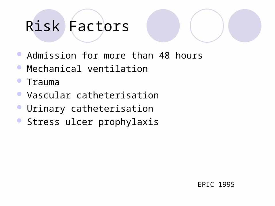
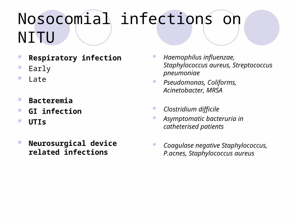
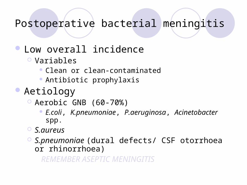
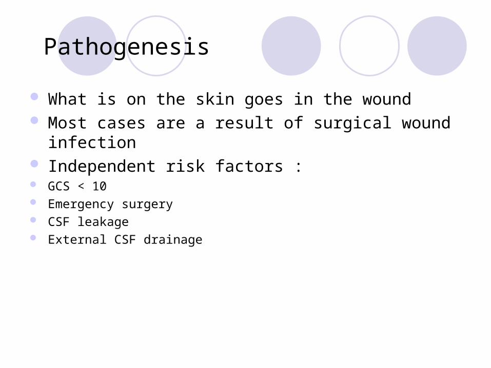
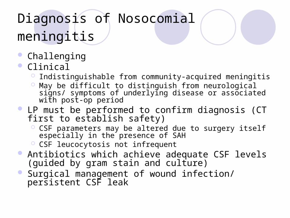
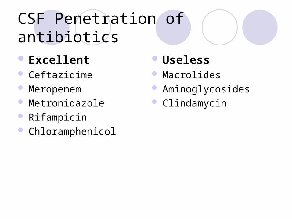
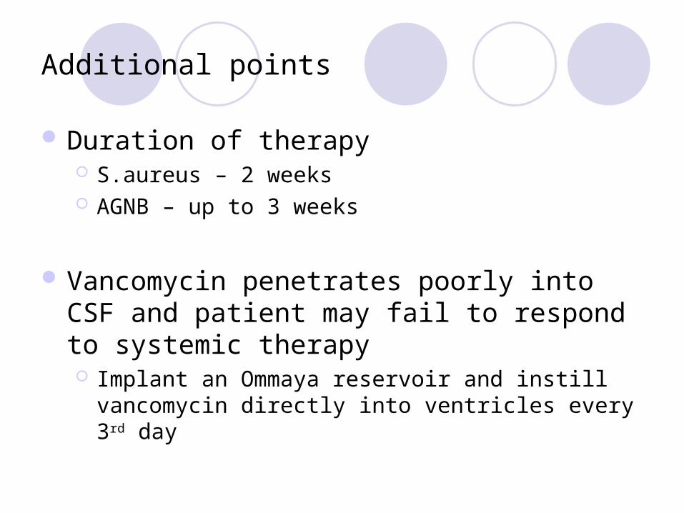
Additional points
Duration of therapy Saureus ndash 2 weeks AGNB ndash up to 3 weeks
Vancomycin penetrates poorly into CSF and patient may fail to respond to systemic therapy Implant an Ommaya reservoir and instill vancomycin
directly into ventricles every 3rd day
Postoperative aseptic meningitis
Thought to be the result of irritation caused either by blood degradation products introduced into SAS during surgery
Indistinguishable from postoperative bacterial meningitis (clinical amp CSF cell count and chemistry) CSF lactate to distinguish
Approach Empirical antibiotic therapy If CSF sterile ndash discontinue antibiotics Responds favourably to high dose corticosteroids
Empiric therapy
1st line therapy Cefotaxime 2-3g qds
Confirmed Paeruginosa or recent broad spectrum antibiotic
Ceftazidime 2g tds
Suspected or confirmed ESBL-producing Enterobacteriaceae or Acinetobacter spp
Meropenem 2g tds
MSSA Flucloxacillin 2-3g qds
MRSA Vancomycin PLUS
Rifampicin
1g bd
600mg bd
Antibiotic Prophylaxis for Basilar Skull Fracture Meta Analysis
12 studies 1241 patients 58 received antibiotics
Antibiotics did not prevent meningitis RR 115 (068 - 194) p=068
CSF leakage subset RR 134 (075 - 241) p=036
Brain abscess
Hematogenous spread from extracranial site Can arise from direct spread from mastoid and sinus infections Corticomedullary junction Frontal and parietal lobes most common Posterior fossa lt5
Overall mortality rate has ranged from 0 to 24 Prognosis the rapidity of progression before hospitalization mental status onadmission
Bacteriology
1048698 Streptococci (70)1048698mixed infections (30 to 60) 1048698 Streptococcus milleri group 1048698 oral cavity appendix and female genital tract 1048698 otopharyngeal infections 1048698 IE
1048698Staphylococcus aureus 10 to 15 1048698 cranial trauma 1048698 IE Neurosurgical procedure
Contd
Bacteroides and Prevotella in 20 to 40 1048698 mixed infection Enteric gram-negative bacilli (Proteus species
Escherichia coli Klebsiella and Pseudomonas) in 23 to 33
1048698 otitic infection 1048698 Septicemia 1048698 neurosurgical procedures 1048698 immunocompromised Rare pathogens Nocardia Mycobacterium tuberculosis Listeria
monocytogenes
Brain abscess
Blood cultures should be obtained when diagnosis is suspected
Lumbar puncture (LP) should be deferred in any case for which brain abscess is suspected because of the potential for CNS
herniation and low likelihood of positive cultures
Pus collected in a sterile universal container (NOT SWAB) should be sent to Microbiology for urgent microscopy culture and sensitivity
Cerebritis and abscess
Early cerebritis ndash (3-5 days) Late Cerebritis -(4 to 14 days)
Early Capsule Stage (Begins at 2 weeks following initial infection)
Late capsule stage
Initial approach to the patient witha suspected brain abscess
Contrast CT or MRI should be performed If single or multiple ring-enhancing lesions are found the patient
should taken urgently to surgery 1048698 All lesions gt 25 cm in diameter should be excised or
stereotactically aspirated 1048698 For abscesses in the early cerebritis stage or when the
abscesses are lt 25 cm in diameter the largest lesionshould be aspirated for diagnosis and organism identification
Antimicrobial therapy
empirical antimicrobial therapy should be initiated on the basis of the patientrsquos predisposing conditions and the presumed
pathogenesis of abscess formation
Otitis mediamastoiditissinusitis Cefotaxime Metronidazole
Post neurosurgical trauma add anti-staphylococcal cover
Use Ceftazidime as the 3rd generation cephalosporin if Pseudomonas aeruginosa is suspected
Brain Abscess - Surgical treatment
significant mass effect exerted by lesion proximity to ventricle poor neurological condition Inability to obtain weekly CT scans In patient undergoing medical treatment
Intervention if neurological deterioration occurs anatomic progression of abscess towards ventricles or after 2 weeks of therapy if abscess is enlarged Also consider if there is no decrease in abscess size by 4 weeks of treatment
Shunt infections
CSF shunts become infected by various routesbull Organisms directly colonize the shunt usually at the time of surgerybull Organisms reach the CSF and the shunt via haematogenous spreadbull Organisms travel along the shunt by retrograde spread (uncommon)
Coagulase-negative Staphylococci are isolated most commonly Production of extracellular slime has been reported as being important in the pathogenesis of shunt infections
Clinical features
Variable Fever Signs of raised ICP Evidence of shunt malfunction Distal shunt infections can present with peritonitis
Shunt infections
CSF from shunt ( before antimicrobial therapy) SHUNT REMOVAL Proximal catheter valve or shunt reservoir distal
catheter in three separate containers Intrathecal +- systemic antibiotic Shunt replacement once the CSF sterile
Clinical Suspicion of VP Shunt Infection
Mild Ventriculitis Severe Ventriculitis and wound infection
POSITIVE Gram Positive Gram Negative MER if any risk factors for resistant GNBs
Repeat CSF in 48-72 Hours Monitor Response
Shunt replacement when CSF sterile at the end of therapy (see duration of antibiotics under treatment section)
Consult Microbiology to discuss alternatives if documented Penicillin allergy or multi-resistant gram negative organism
IVCTX + IT GEN
SHUNT REMOVAL AND EVD
IT VAN
CSF Gram Strain amp Culture
Await Cultures as gram stain usually negative
IV antibiotics and drainage of pus with shunt removal
EVD-related ventriculitis
Incidence between 10-17 Risk
Lowest risk 1st 4 days that EVD is in-situ Rises over next 10 days Falls thereafter
Incidence not decreased by exchanging EVD at regular intervals
Aetiology CNS (predominant)
Diagnostic approach
Patient with EVD with symptomsand signs of ventriculitis
CSF sample
+ Gram stain or+ culture
- Gram stain and culture
2nd sample
+ Gram stain or + culture (sameAs 1st)
Treat No treatment
Additional points
Collect CSF from EVD itself or the Ommaya reservoir not the drainage bag
Treat CNS infections with intraventricular Vancomycin for 5-7 days Product license does not cover this route Not validated by clinical trials Guarantees max concentrations of vancomycin at the site of
infection Avoids systemic toxicity Cheaper than systemic therapy No need to monitor levels
Hand Hygiene
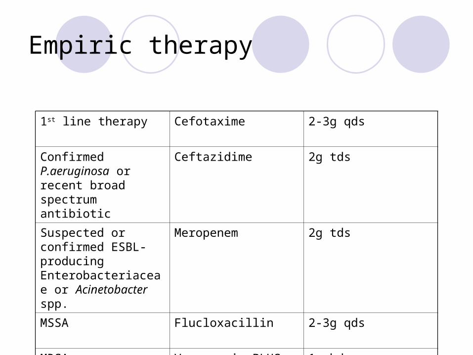
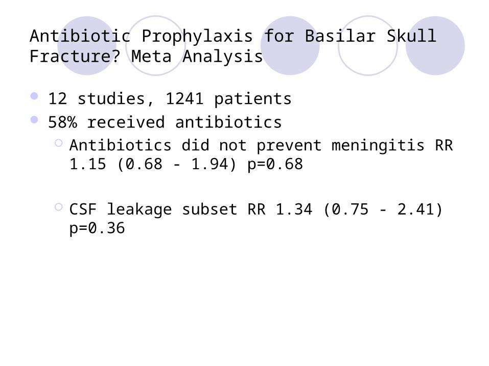
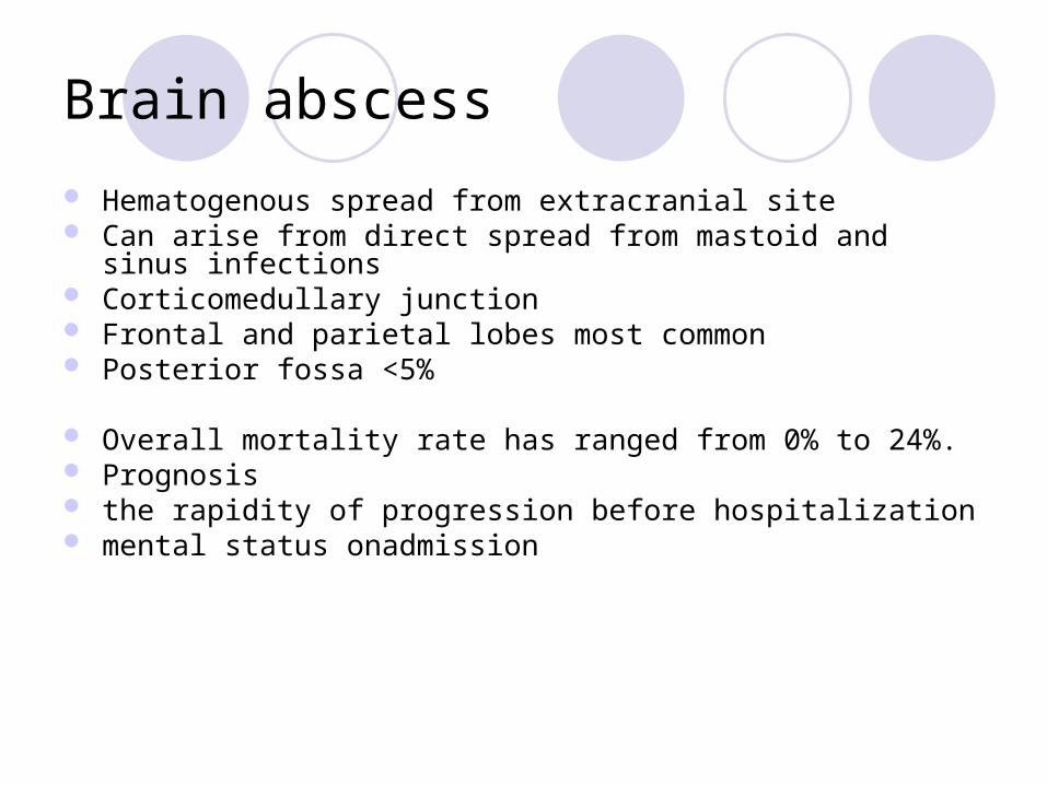
Brain abscess
Hematogenous spread from extracranial site Can arise from direct spread from mastoid and sinus infections Corticomedullary junction Frontal and parietal lobes most common Posterior fossa lt5
Overall mortality rate has ranged from 0 to 24 Prognosis the rapidity of progression before hospitalization mental status onadmission
Bacteriology
1048698 Streptococci (70)1048698mixed infections (30 to 60) 1048698 Streptococcus milleri group 1048698 oral cavity appendix and female genital tract 1048698 otopharyngeal infections 1048698 IE
1048698Staphylococcus aureus 10 to 15 1048698 cranial trauma 1048698 IE Neurosurgical procedure
Contd
Bacteroides and Prevotella in 20 to 40 1048698 mixed infection Enteric gram-negative bacilli (Proteus species
Escherichia coli Klebsiella and Pseudomonas) in 23 to 33
1048698 otitic infection 1048698 Septicemia 1048698 neurosurgical procedures 1048698 immunocompromised Rare pathogens Nocardia Mycobacterium tuberculosis Listeria
monocytogenes
Brain abscess
Blood cultures should be obtained when diagnosis is suspected
Lumbar puncture (LP) should be deferred in any case for which brain abscess is suspected because of the potential for CNS
herniation and low likelihood of positive cultures
Pus collected in a sterile universal container (NOT SWAB) should be sent to Microbiology for urgent microscopy culture and sensitivity
Cerebritis and abscess
Early cerebritis ndash (3-5 days) Late Cerebritis -(4 to 14 days)
Early Capsule Stage (Begins at 2 weeks following initial infection)
Late capsule stage
Initial approach to the patient witha suspected brain abscess
Contrast CT or MRI should be performed If single or multiple ring-enhancing lesions are found the patient
should taken urgently to surgery 1048698 All lesions gt 25 cm in diameter should be excised or
stereotactically aspirated 1048698 For abscesses in the early cerebritis stage or when the
abscesses are lt 25 cm in diameter the largest lesionshould be aspirated for diagnosis and organism identification
Antimicrobial therapy
empirical antimicrobial therapy should be initiated on the basis of the patientrsquos predisposing conditions and the presumed
pathogenesis of abscess formation
Otitis mediamastoiditissinusitis Cefotaxime Metronidazole
Post neurosurgical trauma add anti-staphylococcal cover
Use Ceftazidime as the 3rd generation cephalosporin if Pseudomonas aeruginosa is suspected
Brain Abscess - Surgical treatment
significant mass effect exerted by lesion proximity to ventricle poor neurological condition Inability to obtain weekly CT scans In patient undergoing medical treatment
Intervention if neurological deterioration occurs anatomic progression of abscess towards ventricles or after 2 weeks of therapy if abscess is enlarged Also consider if there is no decrease in abscess size by 4 weeks of treatment
Shunt infections
CSF shunts become infected by various routesbull Organisms directly colonize the shunt usually at the time of surgerybull Organisms reach the CSF and the shunt via haematogenous spreadbull Organisms travel along the shunt by retrograde spread (uncommon)
Coagulase-negative Staphylococci are isolated most commonly Production of extracellular slime has been reported as being important in the pathogenesis of shunt infections
Clinical features
Variable Fever Signs of raised ICP Evidence of shunt malfunction Distal shunt infections can present with peritonitis
Shunt infections
CSF from shunt ( before antimicrobial therapy) SHUNT REMOVAL Proximal catheter valve or shunt reservoir distal
catheter in three separate containers Intrathecal +- systemic antibiotic Shunt replacement once the CSF sterile
Clinical Suspicion of VP Shunt Infection
Mild Ventriculitis Severe Ventriculitis and wound infection
POSITIVE Gram Positive Gram Negative MER if any risk factors for resistant GNBs
Repeat CSF in 48-72 Hours Monitor Response
Shunt replacement when CSF sterile at the end of therapy (see duration of antibiotics under treatment section)
Consult Microbiology to discuss alternatives if documented Penicillin allergy or multi-resistant gram negative organism
IVCTX + IT GEN
SHUNT REMOVAL AND EVD
IT VAN
CSF Gram Strain amp Culture
Await Cultures as gram stain usually negative
IV antibiotics and drainage of pus with shunt removal
EVD-related ventriculitis
Incidence between 10-17 Risk
Lowest risk 1st 4 days that EVD is in-situ Rises over next 10 days Falls thereafter
Incidence not decreased by exchanging EVD at regular intervals
Aetiology CNS (predominant)
Diagnostic approach
Patient with EVD with symptomsand signs of ventriculitis
CSF sample
+ Gram stain or+ culture
- Gram stain and culture
2nd sample
+ Gram stain or + culture (sameAs 1st)
Treat No treatment
Additional points
Collect CSF from EVD itself or the Ommaya reservoir not the drainage bag
Treat CNS infections with intraventricular Vancomycin for 5-7 days Product license does not cover this route Not validated by clinical trials Guarantees max concentrations of vancomycin at the site of
infection Avoids systemic toxicity Cheaper than systemic therapy No need to monitor levels
Hand Hygiene
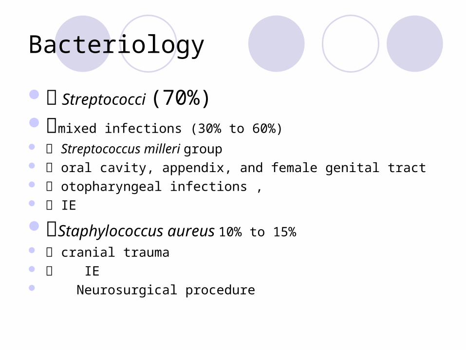
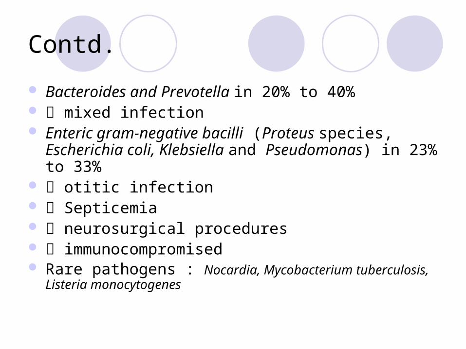
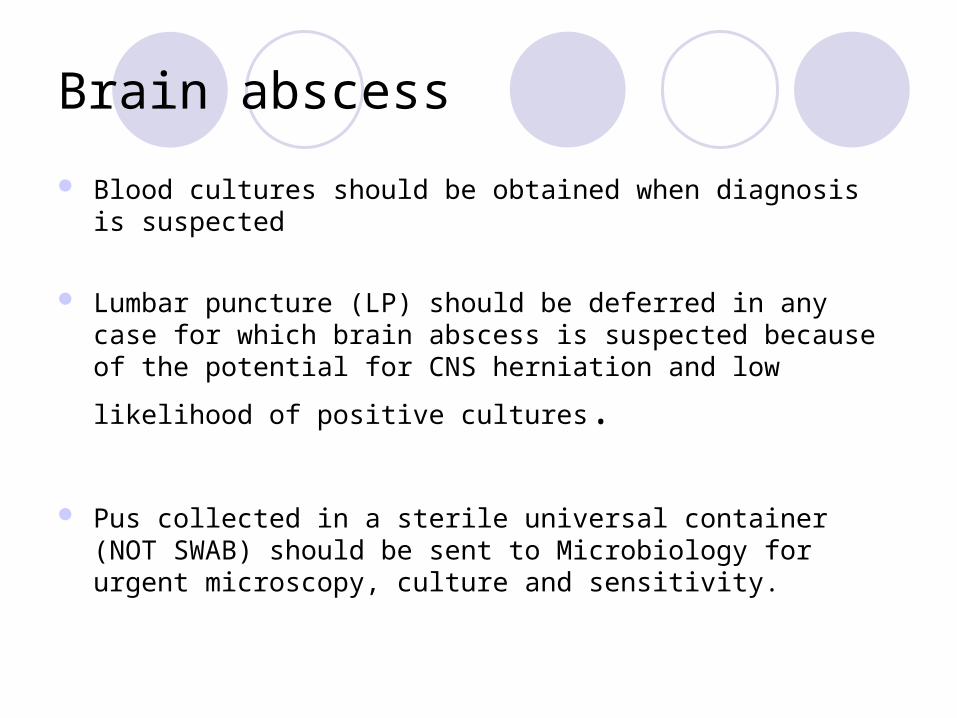
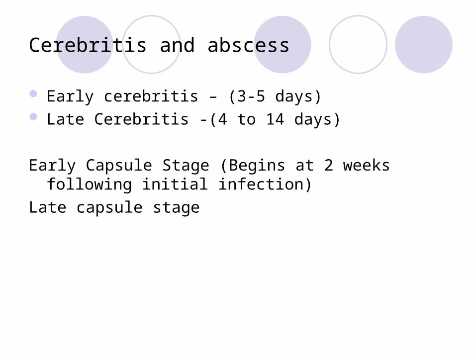
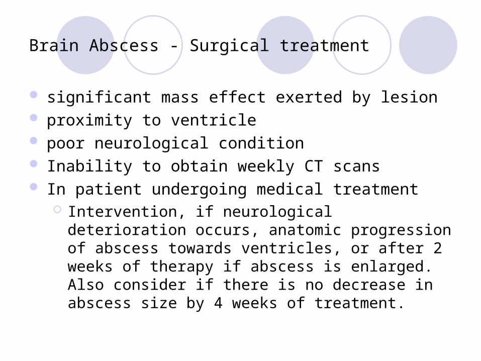

Shunt infections
CSF shunts become infected by various routesbull Organisms directly colonize the shunt usually at the time of surgerybull Organisms reach the CSF and the shunt via haematogenous spreadbull Organisms travel along the shunt by retrograde spread (uncommon)
Coagulase-negative Staphylococci are isolated most commonly Production of extracellular slime has been reported as being important in the pathogenesis of shunt infections
Clinical features
Variable Fever Signs of raised ICP Evidence of shunt malfunction Distal shunt infections can present with peritonitis
Shunt infections
CSF from shunt ( before antimicrobial therapy) SHUNT REMOVAL Proximal catheter valve or shunt reservoir distal
catheter in three separate containers Intrathecal +- systemic antibiotic Shunt replacement once the CSF sterile
Clinical Suspicion of VP Shunt Infection
Mild Ventriculitis Severe Ventriculitis and wound infection
POSITIVE Gram Positive Gram Negative MER if any risk factors for resistant GNBs
Repeat CSF in 48-72 Hours Monitor Response
Shunt replacement when CSF sterile at the end of therapy (see duration of antibiotics under treatment section)
Consult Microbiology to discuss alternatives if documented Penicillin allergy or multi-resistant gram negative organism
IVCTX + IT GEN
SHUNT REMOVAL AND EVD
IT VAN
CSF Gram Strain amp Culture
Await Cultures as gram stain usually negative
IV antibiotics and drainage of pus with shunt removal
EVD-related ventriculitis
Incidence between 10-17 Risk
Lowest risk 1st 4 days that EVD is in-situ Rises over next 10 days Falls thereafter
Incidence not decreased by exchanging EVD at regular intervals
Aetiology CNS (predominant)
Diagnostic approach
Patient with EVD with symptomsand signs of ventriculitis
CSF sample
+ Gram stain or+ culture
- Gram stain and culture
2nd sample
+ Gram stain or + culture (sameAs 1st)
Treat No treatment
Additional points
Collect CSF from EVD itself or the Ommaya reservoir not the drainage bag
Treat CNS infections with intraventricular Vancomycin for 5-7 days Product license does not cover this route Not validated by clinical trials Guarantees max concentrations of vancomycin at the site of
infection Avoids systemic toxicity Cheaper than systemic therapy No need to monitor levels
Hand Hygiene
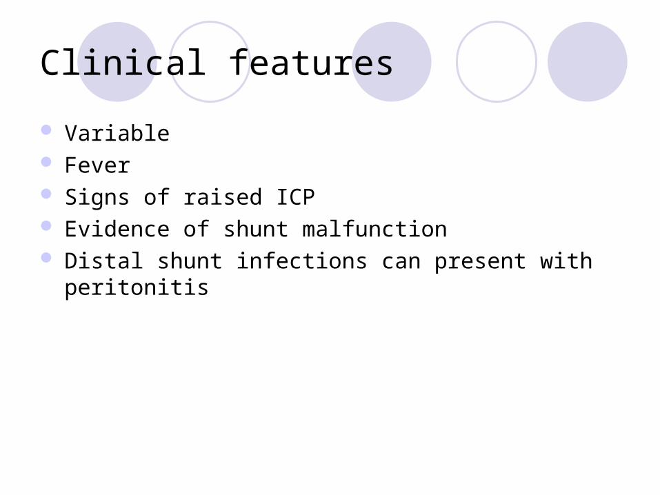
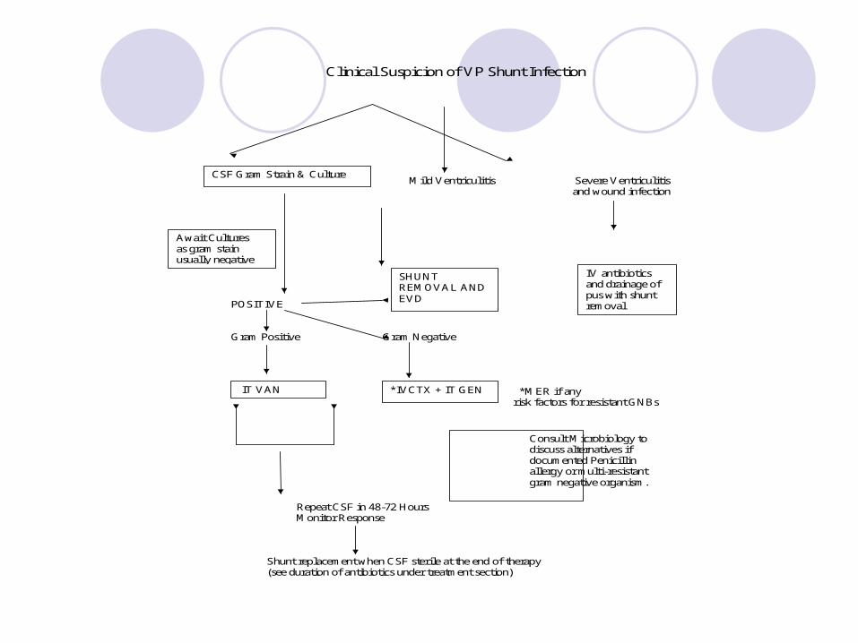
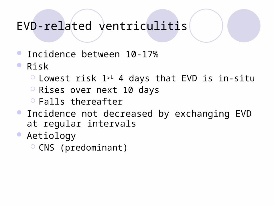
Hand Hygiene
Related Documents