Int. J. Mol. Sci. 2014, 15, 12027-12060; doi:10.3390/ijms150712027 International Journal of Molecular Sciences ISSN 1422-0067 www.mdpi.com/journal/ijms Review Microbial-Catalyzed Biotransformation of Multifunctional Triterpenoids Derived from Phytonutrients Syed Adnan Ali Shah 1,2, *, Huey Ling Tan 3, *, Sadia Sultan 1,2, *, Muhammad Afifi Bin Mohd Faridz 1,2 , Mohamad Azlan Bin Mohd Shah 1,2 , Sharifah Nurfazilah 1,2 and Munawar Hussain 4 1 Faculty of Pharmacy, Universiti Teknologi MARA (UiTM), Puncak Alam Campus, 42300 Bandar Puncak Alam, Selangor Darul Ehsan, Malaysia; E-Mails: [email protected] (M.A.B.M.F.); [email protected] (M.A.B.M.S.); [email protected] (S.N.) 2 Atta-ur-Rahman Institute for Natural Products Discovery (AuRIns), Level 9, FF3, Universiti Teknologi MARA (UiTM), Puncak Alam Campus, 42300 Bandar Puncak Alam, Selangor Darul Ehsan, Malaysia 3 Faculty of Chemical Engineering, Universiti Teknologi MARA (UiTM), 40450 Shah Alam, Selangor Darul Ehsan, Malaysia 4 Department of Basic Sciences, DHA Suffa University, Off, Khayaban-e-Tufail, Phase VII (Extension), DHA, Karachi 75500, Pakistan; E-Mail: [email protected] * Authors to whom correspondence should be addressed; E-Mails: [email protected] or [email protected] (S.A.A.S.); [email protected] (H.L.T.); [email protected] (S.S.) Tel.: +603-3258-4616 (S.A.A.S.); +603-5543-6310 (H.L.T.); +603-3258-4614 (S.S.); Fax: +603-3258-4602 (S.A.A.S.). Received: 22 May 2014 / in revised form: 12 June 2014 / Accepted: 26 June 2014 / Published: 7 July 2014 Abstract: Microbial-catalyzed biotransformations have considerable potential for the generation of an enormous variety of structurally diversified organic compounds, especially natural products with complex structures like triterpenoids. They offer efficient and economical ways to produce semi-synthetic analogues and novel lead molecules. Microorganisms such as bacteria and fungi could catalyze chemo-, regio- and stereospecific hydroxylations of diverse triterpenoid substrates that are extremely difficult to produce by chemical routes. During recent years, considerable research has been performed on the microbial transformation of bioactive triterpenoids, in order to obtain biologically active OPEN ACCESS

Welcome message from author
This document is posted to help you gain knowledge. Please leave a comment to let me know what you think about it! Share it to your friends and learn new things together.
Transcript
Int. J. Mol. Sci. 2014, 15, 12027-12060; doi:10.3390/ijms150712027
International Journal of
Molecular Sciences ISSN 1422-0067
www.mdpi.com/journal/ijms
Review
Microbial-Catalyzed Biotransformation of Multifunctional Triterpenoids Derived from Phytonutrients
Syed Adnan Ali Shah 1,2,*, Huey Ling Tan 3,*, Sadia Sultan 1,2,*,
Muhammad Afifi Bin Mohd Faridz 1,2, Mohamad Azlan Bin Mohd Shah 1,2,
Sharifah Nurfazilah 1,2 and Munawar Hussain 4
1 Faculty of Pharmacy, Universiti Teknologi MARA (UiTM), Puncak Alam Campus,
42300 Bandar Puncak Alam, Selangor Darul Ehsan, Malaysia;
E-Mails: [email protected] (M.A.B.M.F.); [email protected] (M.A.B.M.S.);
[email protected] (S.N.) 2 Atta-ur-Rahman Institute for Natural Products Discovery (AuRIns), Level 9, FF3,
Universiti Teknologi MARA (UiTM), Puncak Alam Campus, 42300 Bandar Puncak Alam,
Selangor Darul Ehsan, Malaysia 3 Faculty of Chemical Engineering, Universiti Teknologi MARA (UiTM), 40450 Shah Alam,
Selangor Darul Ehsan, Malaysia 4 Department of Basic Sciences, DHA Suffa University, Off, Khayaban-e-Tufail, Phase VII (Extension),
DHA, Karachi 75500, Pakistan; E-Mail: [email protected]
* Authors to whom correspondence should be addressed;
E-Mails: [email protected] or [email protected] (S.A.A.S.);
[email protected] (H.L.T.); [email protected] (S.S.)
Tel.: +603-3258-4616 (S.A.A.S.); +603-5543-6310 (H.L.T.); +603-3258-4614 (S.S.);
Fax: +603-3258-4602 (S.A.A.S.).
Received: 22 May 2014 / in revised form: 12 June 2014 / Accepted: 26 June 2014 /
Published: 7 July 2014
Abstract: Microbial-catalyzed biotransformations have considerable potential for the
generation of an enormous variety of structurally diversified organic compounds,
especially natural products with complex structures like triterpenoids. They offer efficient
and economical ways to produce semi-synthetic analogues and novel lead molecules.
Microorganisms such as bacteria and fungi could catalyze chemo-, regio- and stereospecific
hydroxylations of diverse triterpenoid substrates that are extremely difficult to produce by
chemical routes. During recent years, considerable research has been performed on the
microbial transformation of bioactive triterpenoids, in order to obtain biologically active
OPEN ACCESS
Int. J. Mol. Sci. 2014, 15 12028
molecules with diverse structures features. This article reviews the microbial modifications
of tetranortriterpenoids, tetracyclic triterpenoids and pentacyclic triterpenoids.
Keywords: microbial transformation; tetranortriterpenoids; tetracyclic triterpenoids;
pentacyclic triterpenoids; biocatalysis
1. Introduction
Natural products extracted from plants, marine sources and microorganisms constitute a rich source
of diverse scaffolds for drug discovery. Often they form the backbone of innovative drug discovery
programs. They can either be directly used as drugs to treat various diseases, or used as a valuable
starting material (“lead”) for drug discovery process. From the 1940s until 2010, 65% of antibacterial
and 41% of anticancer small molecule drugs developed were either natural products or semi-synthetic
derivatives of natural products [1–4]. Structural diversification of multifunctional natural products is
often required to improve their solubility, reduce toxicity, or enhance efficacy. Chemical conversions
may provide abundant products, but are limited by regio- and stereoselectivity constraints. Moreover,
multi-step chemical reactions often result in low overall yield of the final products. However,
biocatalytic reactions are well-established “green” techniques for carrying out high chemo-, regio- and
stereoselective functionalization of various sensitive and complex molecules under mild reaction
conditions and hence are much more attractive for drug development process [5–8]. Biocatalysis, using
multi-enzyme systems of fungi, bacteria, and cultured plant suspension cells has the advantage of
producing compounds with high selectivity and efficiency under mild conditions. Therefore, biological
systems are widely used in the pharmaceutical industry [9–21]. The use of microorganisms
such as bacteria and fungi as a biocatalytic system imitates the mamalian metabolism to perform
selective transformation reactions and improve the economically and ecologically friendly
microbial transformations [22–24]. A number of filamentous fungi are known to perform complex
biotransformations that are difficult to achieve by chemical means [22–26]. Many microorganisms,
especially certain filamentous fungi, have the ability to transform terpenoids chemo-, regio- and
stereoselectively. The fungal-mediated oxidation of terpene under mild conditions appears as an
attractive alternative as compared to the traditional chemical methods, have an elevated chemo-, regio-
and enantioselectivity, and do not generate toxic waste products, and the products obtained can be
labeled as “natural” source [5]. Microbial cell-mediated transformations have been extensively used
for in vitro in vitro drug metabolic studies. Moreover, microbial transformations can also provide
better yields of the metabolites with high selectivity for toxicological and biological studies [22]. Fungi
also provide additional advantage in performing reactions similar to mammalian transformations [24].
Triterpenes are plant-derived natural compounds built-up from six isoprene units (C5H8), while
triterpenoids consist of both the basic triterpene skeleton and their derivatives that contain oxygen
moiety. The simplest triterpene with skeletal structure that forms basis for complex triterpenoids is
squalene (C30) [8]. It forms the precursor for a structurally diverse group of natural products
that display nearly 200 distinct skeletons. These products have been studied for their antiviral
(anti-HIV), antineoplastic, anti-inflammatory, anti-ulcerogenic, antimicrobial, anti-plasmodial, hepato-
Int. J. Mol. Sci. 2014, 15 12029
and cardio-protective, analgesic, anti-mycotic, and immunomodulatory effects [1,2,6–8]. They are
routinely found in numerous medicinal plants. They are also excellent starting material for the
synthesis of many fine chemicals due to their homogeneous carbon skeleton [1]. Microbial cell-based
transformations of triterpenoids have been developed primarily in the past two decades to produce novel
lead molecules, new pharmaceuticals, and agrochemical compounds [7,8,27–31]. Some synthetic
oleanane triterpenoids derived from microbial transformation act as multifunctional drugs that regulate
the activity of entire networks [32–36]. Microbial biocatalysis has already been proven as powerful
tool in the generation of structural diversity in triterpenoid skeletons for future structure-activity
relationship studies [5–8].
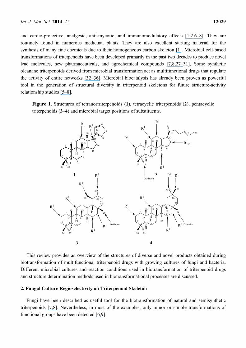
Figure 1. Structures of tetranortriterpenoids (1), tetracyclic triterpenoids (2), pentacyclic
triterpenoids (3–4) and microbial target positions of substituents.
This review provides an overview of the structures of diverse and novel products obtained during
biotransformation of multifunctional triterpenoid drugs with growing cultures of fungi and bacteria.
Different microbial cultures and reaction conditions used in biotransformation of triterpenoid drugs
and structure determination methods used in biotransformational processes are discussed.
2. Fungal Culture Regioselectivity on Triterpenoid Skeleton
Fungi have been described as useful tool for the biotransformation of natural and semisynthetic
triterpenoids [7,8]. Nevertheless, in most of the examples, only minor or simple transformations of
functional groups have been detected [6,9].
1
H
O
H
R1R1
4
H
H
H
1
3 5 74
6
810
1113
15
16
17
18
19
20
21
22
23
29 28
30
12
3
H
H
H
R1 R1
R1
R1
R1R1
R1
R1
R1
R1
1
3 5 76
810
11
13
4
1
3 5 76
810
4
29
20
30
21
22
2815
1718
25 26
27
24 23
Oxidation
25 26
2021
22
29
30
28
24 23
11
27Oxidation
R1R1
H
H
R1
R1
1
3 5 76
810
4
19 18
29 28
11
30
25 26
27
15
17
20
21
R1
R1
Oxidation
R1
2
Int. J. Mol. Sci. 2014, 15 12030
Microbial cell cultures are capable of performing specific chemical transformations in triterpenoids,
such as rearrangement, hydroxylation, oxidation, reduction, hydrolysis, epimerization and isomerization,
with high regio- and stereoselectivity as shown in Figure 1 [6–8]. In Figures 2–9, we can see a
variation in biocatalytic system introduce regio-selectivity among 12β- or 17β-hydroxylation in
limonoids skeletons [23]. Cunninghamella elegans AS 3.1207 also transforms steroidal saponins into
pregnenolones, S. racemosum AS 3.264 converts paeoniflorin into albiflorin [26]. Cunninghamella
blakesleeana NRRL 1369 performs complicated rearrangement of tetracyclic triterpenoids into novel
ranunculane framework, which reveals the biocatalytic potential of microorganisms in diversification
and promoting structural transformation [22–26].
3. Microbial-Catalyzed Biotransformation of Tetranortriterpenoids
Limonoids (1), chemically classified as tetranortriterpenoids, are metabolically modified triterpenes
having an intact 4,4,8-trimethyl-17-furanylsteroid precursor skeleton (basic limonoid). In some cases,
the skeleton is further rearranged and highly oxygenated, creating a structural diversity. They are
known to possess anti-cancer, anti-malarial, anti-HIV, antimicrobial and several other pharmacological
activities. Azadiradione (5), epoxyazadiradione (14), gedunin (23) and their derivatives fall under this
group. The strong antifeedant properties along with anti-plasmoidal, anti-HIV, and anti-inflammatory
activities of azadiradione and epoxyazadiradione have attracted the attention of synthetic chemists in
the last two decades [23]. In particular, gedunin (23) is a well-studied anti-malarial, anti-carcinogenic
and antiulcerogenic agent.
Thulasiram et al. developed a highly efficient fungi-mediated bioconversion for the 12β- and
17β-hydroxylation of the basic limonoid family of compounds (Figure 2). The fungal system
belonging to the genera of Mucor (National Collection of Industrial Microorganisms or NCIM, Pune,
catalogue no. 881 and abbreviated as M881) efficiently transformed azadiradione (5), epoxyazadiradione
(14), gedunin (23) and their derivatives (8, 11, 16, 18 and 21) into corresponding 12β- and/or
17β-hydroxy derivatives. These microbial-catalyzed stereo- and regioselective hydroxylation of
limonoid skeleton was highly efficient in introducing chemically sensitive functional moieties [23].
Azadiradione (5), a limonoid isolated from the fruit of Azadirachta indica (Neem). The fungus
Mucor sp. M881 regioselectively transformed Azadiradione (5), an isolated limonoid from the fruit of
Azadirachta indica (Neem), to 17β-hydroxyazadiradione (6) and 12β-hydroxyazadiradione (7) (Figure 2).
To further investigate the substrate specificity of the organism, the biotransformation was studied with
seven natural or semi-synthetic derivatives of azadiradione (8, 11, 14, 16, 18, 21 and 23) (see Figures 3–9).
Fungi-mediated biocatalysis of 1,2-dihydroazadiradione (8) and 1,2α-epoxyazadiradione (11) resulted
in regioselective hydroxylation at C-17β- and C-12β-sites (9, 10, 12 and 13) with excellent
yields. These biotransformation reactions are shown in Figures 3 and 4. Fermentation of 14,
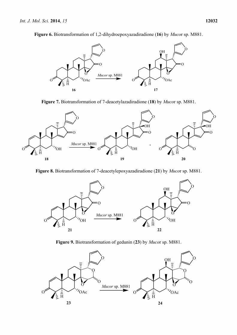
15β-epoxyazadiradione (14) and 1,2-dihydroepoxyazadiradione (16) with Mucor sp. M881
(see Figures 5 and 6) afforded regioselective hydroxylation specifically at the C-12β site (15 and 17).
The organism also hydroxylated gedunin (23, expanded D ring as six membered lactone) and produced
12β-hydroxy gedunin (24) as the sole biotransformed product (Figure 9) [23].
Similarly, the Mucor sp. M881 was able to hydroxylate 7-deacetylepoxyazadiradione (21) at the
12β-position (22) (Figure 8). Nevertheless, when 7-deacetylazadiradione (18, nimbocinol) was used

Int. J. Mol. Sci. 2014, 15 12031
as a substrate (Figure 7), this compound was hydroxylated at the 17β-position to produce
17β-hydroxynimbocinol (19) along with another product 7-oxo-17β-hydroxynimbocinol (20) as
metabolites. These results seems to indicate that the two functional groups (epoxy group in ring D and
acetate in ring B) are critical in producing different products (17β-hydroxy:12β-hydroxy). All these
metabolites (6–24) were characterised by detailed 1H and 13C NMR spectroscopy. The location of the
hydroxyl functionalities were deduced on the basis of the heteronuclear multiple bond connectivity
(HMBC) interactions whereas orientation of OH groups was deduced on the basis of Nuclear
Overhauser Effect spectroscopy (NOESY) correlations [23].
Figure 2. Biotransformation of azadiradione (5) by Mucor sp. M881.
Figure 3. Biotransformation of 1,2-dihydroazadiradione (8) by Mucor sp. M881.
Figure 4. Biotransformation of 1,2α-epoxyazadiradione (11) by Mucor sp. M881.
Figure 5. Biotransformation of 14,15β-epoxyazadiradione (14) by Mucor sp. M881.
5H
O OAc
O
O
6H
O OAc
O
O
7H
O OAc
O
O
OH
OH
Mucor sp. M881
8
HO OAc
O
O
9
HO OAc
O
O
10
HO OAc
O
O
OH
OH
Mucor sp. M881
11
HO OAc
O
O
12
HO OAc
O
O
13
HO OAc
O
O
OH
OH
Mucor sp. M881
O O O
14
HO OAc
O
O
15
HO OAc
O
OOH
Mucor sp. M881O O
Int. J. Mol. Sci. 2014, 15 12032
Figure 6. Biotransformation of 1,2-dihydroepoxyazadiradione (16) by Mucor sp. M881.
Figure 7. Biotransformation of 7-deacetylazadiradione (18) by Mucor sp. M881.
Figure 8. Biotransformation of 7-deacetylepoxyazadiradione (21) by Mucor sp. M881.
Figure 9. Biotransformation of gedunin (23) by Mucor sp. M881.
16
HO OAc
O
O
17
HO OAc
O
OOH
Mucor sp. M881O O
18
HO OH
O
O
19
HO OH
O
O
Mucor sp. M881
OH
20
HO
O
O
OH
O
21H
O OH
O
O
22H
O OH
O
OOH
Mucor sp. M881O O
23
HO OAc
O
Mucor sp. M881O
O
O
24
HO OAc
O
O
O
O
OH
Int. J. Mol. Sci. 2014, 15 12033
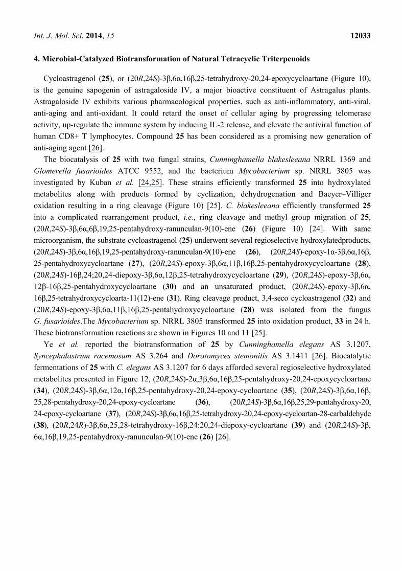
4. Microbial-Catalyzed Biotransformation of Natural Tetracyclic Triterpenoids
Cycloastragenol (25), or (20R,24S)-3β,6α,16β,25-tetrahydroxy-20,24-epoxycycloartane (Figure 10),
is the genuine sapogenin of astragaloside IV, a major bioactive constituent of Astragalus plants.
Astragaloside IV exhibits various pharmacological properties, such as anti-inflammatory, anti-viral,
anti-aging and anti-oxidant. It could retard the onset of cellular aging by progressing telomerase
activity, up-regulate the immune system by inducing IL-2 release, and elevate the antiviral function of
human CD8+ T lymphocytes. Compound 25 has been considered as a promising new generation of
anti-aging agent [26].
The biocatalysis of 25 with two fungal strains, Cunninghamella blakesleeana NRRL 1369 and
Glomerella fusarioides ATCC 9552, and the bacterium Mycobacterium sp. NRRL 3805 was
investigated by Kuban et al. [24,25]. These strains efficiently transformed 25 into hydroxylated
metabolites along with products formed by cyclization, dehydrogenation and Baeyer–Villiger
oxidation resulting in a ring cleavage (Figure 10) [25]. C. blakesleeana efficiently transformed 25
into a complicated rearrangement product, i.e., ring cleavage and methyl group migration of 25,
(20R,24S)-3β,6α,6β,19,25-pentahydroxy-ranunculan-9(10)-ene (26) (Figure 10) [24]. With same
microorganism, the substrate cycloastragenol (25) underwent several regioselective hydroxylatedproducts,
(20R,24S)-3β,6α,16β,19,25-pentahydroxy-ranunculan-9(10)-ene (26), (20R,24S)-epoxy-1α-3β,6α,16β,
25-pentahydroxycycloartane (27), (20R,24S)-epoxy-3β,6α,11β,16β,25-pentahydroxycycloartane (28),
(20R,24S)-16β,24;20,24-diepoxy-3β,6α,12β,25-tetrahydroxycycloartane (29), (20R,24S)-epoxy-3β,6α,
12β-16β,25-pentahydroxycycloartane (30) and an unsaturated product, (20R,24S)-epoxy-3β,6α,16β,25-tetrahydroxycycloarta-11(12)-ene (31). Ring cleavage product, 3,4-seco cycloastragenol (32) and
(20R,24S)-epoxy-3β,6α,11β,16β,25-pentahydroxycycloartane (28) was isolated from the fungus
G. fusarioides.The Mycobacterium sp. NRRL 3805 transformed 25 into oxidation product, 33 in 24 h.
These biotransformation reactions are shown in Figures 10 and 11 [25].
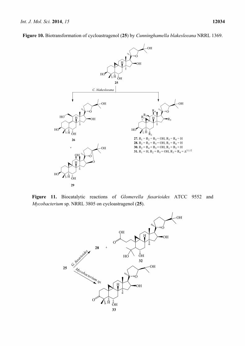
Ye et al. reported the biotransformation of 25 by Cunninghamella elegans AS 3.1207,
Syncephalastrum racemosum AS 3.264 and Doratomyces stemonitis AS 3.1411 [26]. Biocatalytic
fermentations of 25 with C. elegans AS 3.1207 for 6 days afforded several regioselective hydroxylated
metabolites presented in Figure 12, (20R,24S)-2α,3β,6α,16β,25-pentahydroxy-20,24-epoxycycloartane
(34), (20R,24S)-3β,6α,12α,16β,25-pentahydroxy-20,24-epoxy-cycloartane (35), (20R,24S)-3β,6α,16β,
25,28-pentahydroxy-20,24-epoxy-cycloartane (36), (20R,24S)-3β,6α,16β,25,29-pentahydroxy-20,
24-epoxy-cycloartane (37), (20R,24S)-3β,6α,16β,25-tetrahydroxy-20,24-epoxy-cycloartan-28-carbaldehyde
(38), (20R,24R)-3β,6α,25,28-tetrahydroxy-16β,24:20,24-diepoxy-cycloartane (39) and (20R,24S)-3β,
6α,16β,19,25-pentahydroxy-ranunculan-9(10)-ene (26) [26].
Int. J. Mol. Sci. 2014, 15 12034
Figure 10. Biotransformation of cycloastragenol (25) by Cunninghamella blakesleeana NRRL 1369.
Figure 11. Biocatalytic reactions of Glomerella fusarioides ATCC 9552 and
Mycobacterium sp. NRRL 3805 on cycloastragenol (25).
HO
25OH
O
OH
OH
HO
26
OH
O
OH
OHHO
H
H
H
H
C. blakesleeana
HO
27. R1 = R2 = R5 = OH, R3 = R4 = H28. R2 = R3 = R5 = OH, R1 = R4 = H30. R2 = R4 = R5 = OH, R1 = R3 = H31. R1 = H, R2 = R5 = OH, R3 = R4 = 11,12
R2
O
OH
R5H
H
R1
HOOH
O
OH
OH
H
29
OH
R3
R4
G. fusa
rioide
s
32OH
O
OH
OHH
HO
OH
O
28
25 Mycobacterium sp.
33OH
O
OH
OHH
HO
Int. J. Mol. Sci. 2014, 15 12035
Figure 12. Biocatalytic reactions of Cunninghamella elegans AS 3.1207 on
cycloastragenol (25).
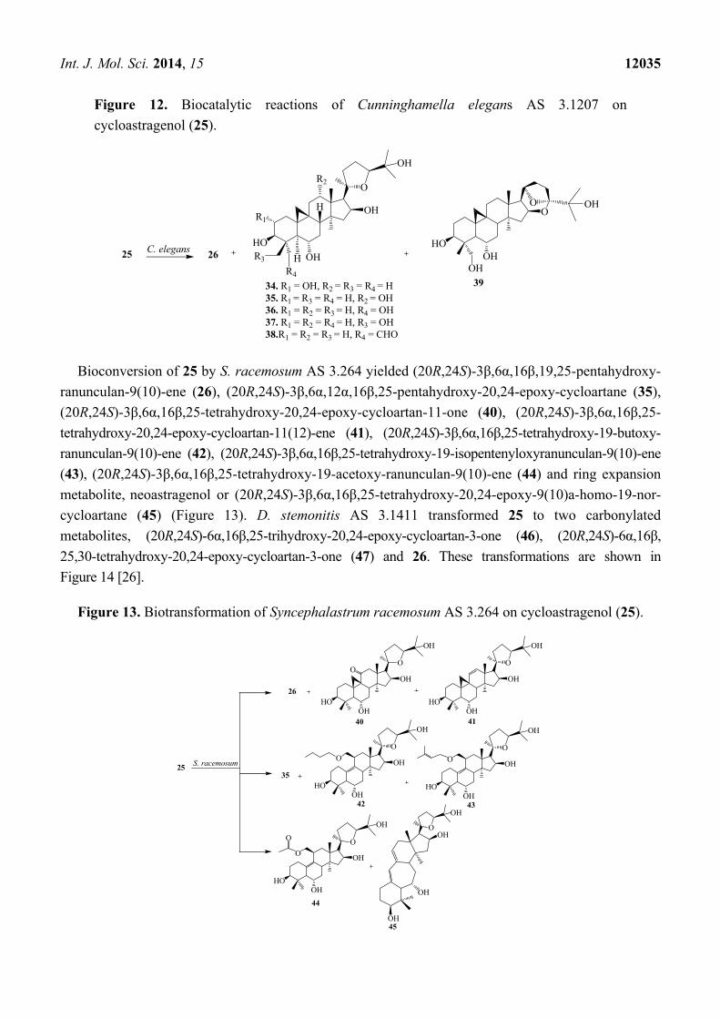
Bioconversion of 25 by S. racemosum AS 3.264 yielded (20R,24S)-3β,6α,16β,19,25-pentahydroxy-
ranunculan-9(10)-ene (26), (20R,24S)-3β,6α,12α,16β,25-pentahydroxy-20,24-epoxy-cycloartane (35),
(20R,24S)-3β,6α,16β,25-tetrahydroxy-20,24-epoxy-cycloartan-11-one (40), (20R,24S)-3β,6α,16β,25-
tetrahydroxy-20,24-epoxy-cycloartan-11(12)-ene (41), (20R,24S)-3β,6α,16β,25-tetrahydroxy-19-butoxy-
ranunculan-9(10)-ene (42), (20R,24S)-3β,6α,16β,25-tetrahydroxy-19-isopentenyloxyranunculan-9(10)-ene
(43), (20R,24S)-3β,6α,16β,25-tetrahydroxy-19-acetoxy-ranunculan-9(10)-ene (44) and ring expansion
metabolite, neoastragenol or (20R,24S)-3β,6α,16β,25-tetrahydroxy-20,24-epoxy-9(10)a-homo-19-nor-
cycloartane (45) (Figure 13). D. stemonitis AS 3.1411 transformed 25 to two carbonylated
metabolites, (20R,24S)-6α,16β,25-trihydroxy-20,24-epoxy-cycloartan-3-one (46), (20R,24S)-6α,16β,
25,30-tetrahydroxy-20,24-epoxy-cycloartan-3-one (47) and 26. These transformations are shown in
Figure 14 [26].
Figure 13. Biotransformation of Syncephalastrum racemosum AS 3.264 on cycloastragenol (25).
HO
OH
O
OH
OHH
H26C. elegans HOOH
OH
OO OH
39
25
R1
R2
R3
R4
34. R1 = OH, R2 = R3 = R4 = H35. R1 = R3 = R4 = H, R2 = OH36. R1 = R2 = R3 = H, R4 = OH37. R1 = R2 = R4 = H, R3 = OH38.R1 = R2 = R3 = H, R4 = CHO
26HO
40OH
O
OH
OH
HOOH
O
OH
OH
HOOH
O
OH
OH
41
35
42
O
O
HOOH
O
OH
OH
43
O
HOOH
O
OH
OH
44
O
OO
OH
OH
OH
OH
45
S. racemosum25
Int. J. Mol. Sci. 2014, 15 12036
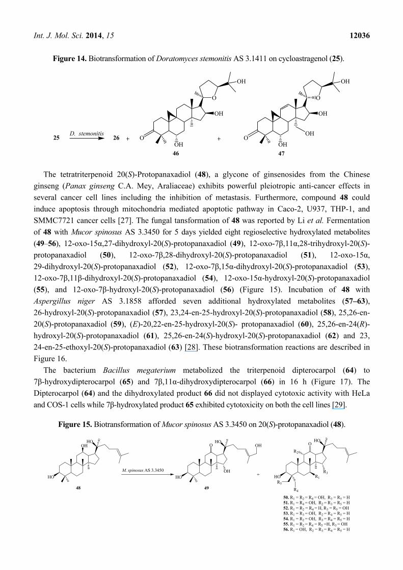
Figure 14. Biotransformation of Doratomyces stemonitis AS 3.1411 on cycloastragenol (25).
The tetratriterpenoid 20(S)-Protopanaxadiol (48), a glycone of ginsenosides from the Chinese
ginseng (Panax ginseng C.A. Mey, Araliaceae) exhibits powerful pleiotropic anti-cancer effects in
several cancer cell lines including the inhibition of metastasis. Furthermore, compound 48 could
induce apoptosis through mitochondria mediated apoptotic pathway in Caco-2, U937, THP-1, and
SMMC7721 cancer cells [27]. The fungal tansformation of 48 was reported by Li et al. Fermentation
of 48 with Mucor spinosus AS 3.3450 for 5 days yielded eight regioselective hydroxylated metabolites
(49–56), 12-oxo-15α,27-dihydroxyl-20(S)-protopanaxadiol (49), 12-oxo-7β,11α,28-trihydroxyl-20(S)-
protopanaxadiol (50), 12-oxo-7β,28-dihydroxyl-20(S)-protopanaxadiol (51), 12-oxo-15α,
29-dihydroxyl-20(S)-protopanaxadiol (52), 12-oxo-7β,15α-dihydroxyl-20(S)-protopanaxadiol (53),
12-oxo-7β,11β-dihydroxyl-20(S)-protopanaxadiol (54), 12-oxo-15α-hydroxyl-20(S)-protopanaxadiol
(55), and 12-oxo-7β-hydroxyl-20(S)-protopanaxadiol (56) (Figure 15). Incubation of 48 with
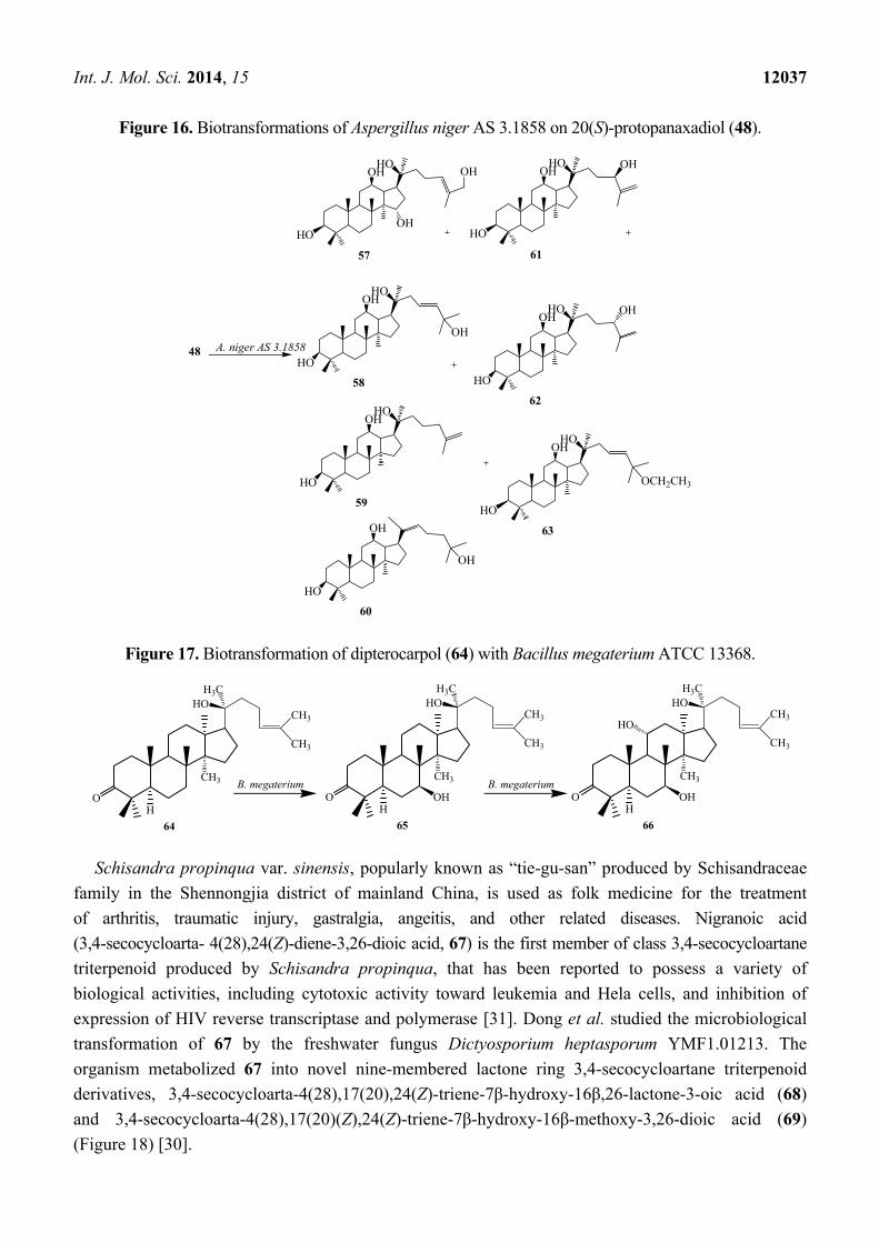
Aspergillus niger AS 3.1858 afforded seven additional hydroxylated metabolites (57–63),
26-hydroxyl-20(S)-protopanaxadiol (57), 23,24-en-25-hydroxyl-20(S)-protopanaxadiol (58), 25,26-en-
20(S)-protopanaxadiol (59), (E)-20,22-en-25-hydroxyl-20(S)- protopanaxadiol (60), 25,26-en-24(R)-
hydroxyl-20(S)-protopanaxadiol (61), 25,26-en-24(S)-hydroxyl-20(S)-protopanaxadiol (62) and 23,
24-en-25-ethoxyl-20(S)-protopanaxadiol (63) [28]. These biotransformation reactions are described in
Figure 16.
The bacterium Bacillus megaterium metabolized the triterpenoid dipterocarpol (64) to
7β-hydroxydipterocarpol (65) and 7β,11α-dihydroxydipterocarpol (66) in 16 h (Figure 17). The
Dipterocarpol (64) and the dihydroxylated product 66 did not displayed cytotoxic activity with HeLa
and COS-1 cells while 7β-hydroxylated product 65 exhibited cytotoxicity on both the cell lines [29].
Figure 15. Biotransformation of Mucor spinosus AS 3.3450 on 20(S)-protopanaxadiol (48).
25
46
OH
O
OH
OH
OH
O
OH
OH
47
OOH
O26D. stemonitis
M. spinosus AS 3.3450
HO
OHHO
48
HO
HO
49
O
OH
OH
HO
HO
R1
O
R2
R3
R5
R4
50. R1 = R2 = R4 = OH, R3 = R5 = H51. R1 = R4 = OH, R2 = R3 = R5 = H52. R1 = R2 = R4 = H, R3 = R5 = OH53. R1 = R3 = OH, R2 = R4 = R5 = H54. R1 = R2 = OH, R3 = R4 = R5 = H55. R1 = R2 = R4 = R5 =H, R3 = OH 56. R1 = OH, R2 = R3 = R4 = R5 = H
Int. J. Mol. Sci. 2014, 15 12037
Figure 16. Biotransformations of Aspergillus niger AS 3.1858 on 20(S)-protopanaxadiol (48).
Figure 17. Biotransformation of dipterocarpol (64) with Bacillus megaterium ATCC 13368.
Schisandra propinqua var. sinensis, popularly known as “tie-gu-san” produced by Schisandraceae
family in the Shennongjia district of mainland China, is used as folk medicine for the treatment
of arthritis, traumatic injury, gastralgia, angeitis, and other related diseases. Nigranoic acid
(3,4-secocycloarta- 4(28),24(Z)-diene-3,26-dioic acid, 67) is the first member of class 3,4-secocycloartane
triterpenoid produced by Schisandra propinqua, that has been reported to possess a variety of
biological activities, including cytotoxic activity toward leukemia and Hela cells, and inhibition of
expression of HIV reverse transcriptase and polymerase [31]. Dong et al. studied the microbiological
transformation of 67 by the freshwater fungus Dictyosporium heptasporum YMF1.01213. The
organism metabolized 67 into novel nine-membered lactone ring 3,4-secocycloartane triterpenoid
derivatives, 3,4-secocycloarta-4(28),17(20),24(Z)-triene-7β-hydroxy-16β,26-lactone-3-oic acid (68)
and 3,4-secocycloarta-4(28),17(20)(Z),24(Z)-triene-7β-hydroxy-16β-methoxy-3,26-dioic acid (69)
(Figure 18) [30].
A. niger AS 3.185848
HO
HO
57
HO
HO
61
HO
HO
58 HO
HO
62
HO
HO
59HO
HO
63
HO
60
OH
OHOH OHOH
OH
OHOH
OH
OH
OH
OCH2CH3
OH
OH
64
HO
CH3
CH3
CH3
H3CHO
65
HO
CH3
CH3
CH3
H3CHO
OH
66
HO
CH3
CH3
CH3
H3CHO
OH
HO
B. megaterium B. megaterium
Int. J. Mol. Sci. 2014, 15 12038
Dong et al. reported the bioconversion of 67 by cultures of Trichoderma sp. JY-1. The fungus
yielded 15α,16α-dihydroxy-3,4-secocyloarta-4(28),17(20),17(E),24(E)-triene-3,26-dioic acid (70) and
16α,20α-dihydroxy-18 (1317β) abeo-3,4-secocyloarta-4 (28),12(13),24(Z)-triene-3,26-dioic acid
(71). Substrate 67 and its transformed products 70 and 71 displayed weak anti-HIV activity with EC50
values of 10.5, 8.8 and 7.6 mg/mL, and therapeutic index values (CC50/EC50) of 8.48, 9.12 and 10.1,
respectively (Figure 19) [31].
Figure 18. Microbiological transformation of the triterpene nigranoic acid (67) by the
freshwater fungus Dictyosporium heptasporum.
Figure 19. Metabolism of 67 by Trichoderma sp. JY-1 culture.
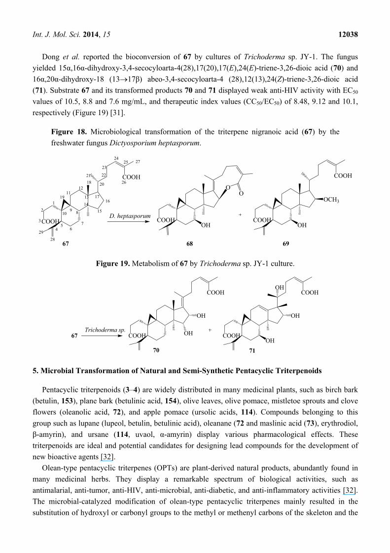
5. Microbial Transformation of Natural and Semi-Synthetic Pentacyclic Triterpenoids
Pentacyclic triterpenoids (3–4) are widely distributed in many medicinal plants, such as birch bark
(betulin, 153), plane bark (betulinic acid, 154), olive leaves, olive pomace, mistletoe sprouts and clove
flowers (oleanolic acid, 72), and apple pomace (ursolic acids, 114). Compounds belonging to this
group such as lupane (lupeol, betulin, betulinic acid), oleanane (72 and maslinic acid (73), erythrodiol,
β-amyrin), and ursane (114, uvaol, α-amyrin) display various pharmacological effects. These
triterpenoids are ideal and potential candidates for designing lead compounds for the development of
new bioactive agents [32].
Olean-type pentacyclic triterpenes (OPTs) are plant-derived natural products, abundantly found in
many medicinal herbs. They display a remarkable spectrum of biological activities, such as
antimalarial, anti-tumor, anti-HIV, anti-microbial, anti-diabetic, and anti-inflammatory activities [32].
The microbial-catalyzed modification of olean-type pentacyclic triterpenes mainly resulted in the
substitution of hydroxyl or carbonyl groups to the methyl or methenyl carbons of the skeleton and the
COOH
COOH
1
2
3
45
67
8910
1112
13
14
15
1617
18
19
20
21 22
23
2425
26
27
28
29
COOHOH
OO
COOH
COOH
OCH3
OH
D. heptasporum
67 68 69
COOH
COOHOH
OH
Trichoderma sp.COOH
COOH
OH
OH
OH
67
70 71
Int. J. Mol. Sci. 2014, 15 12039
formation of corresponding glycosides [32]. The presence of such functional moieties, especially at C-3,
C-28, and C-30, seems to contribute to the biological activities of pentacyclic triterpenoids [17,35,36].
Oleanolic acid (3β-hydroxyolean-12-en-28-oic acid, 72) is a natural pentacyclic triterpenoid
compounds widely present in the form of free acid or aglycone of triterpenoid saponins. It is usually
found in high concentrations in olive-pomace oil [32]. Some oleanolic acids have been reported to be
antimalarial, antitumor, hepatoprotective, anti-HIV, and skin protective. They seem to possess
α-glucosidase inhibitory activity.
Figure 20. Microbial transformation of oleanolic acid (72) by Furarium lini NRRL-68751
and Colletotrichum lini AS3.4486.
Several biotransformations of oleanolic acid (72) with different microorganisms have been reported
so far. Choudhary et al. investigated the metabolism of oleanolic acid (72) with Fusarium lini
NRRL-68751 and reported the production of two oxidative metabolites, maslinic acid
(2α,3β-dihydroxyolean-12-en-28-oic acid, 73) and 2α,3β,11β-trihydroxyolean-12-en-28-oic acid (74)
(Figure 20). These metabolites show potent inhibition of α-glucosidase activity. Hua et al. reported the
metabolism of 72 with Colletotrichum lini AS3.4486 which resulted in one polar metabolite,
15α-hydroxyl-oleanolic acid (75) (Figure 20) [17,33].
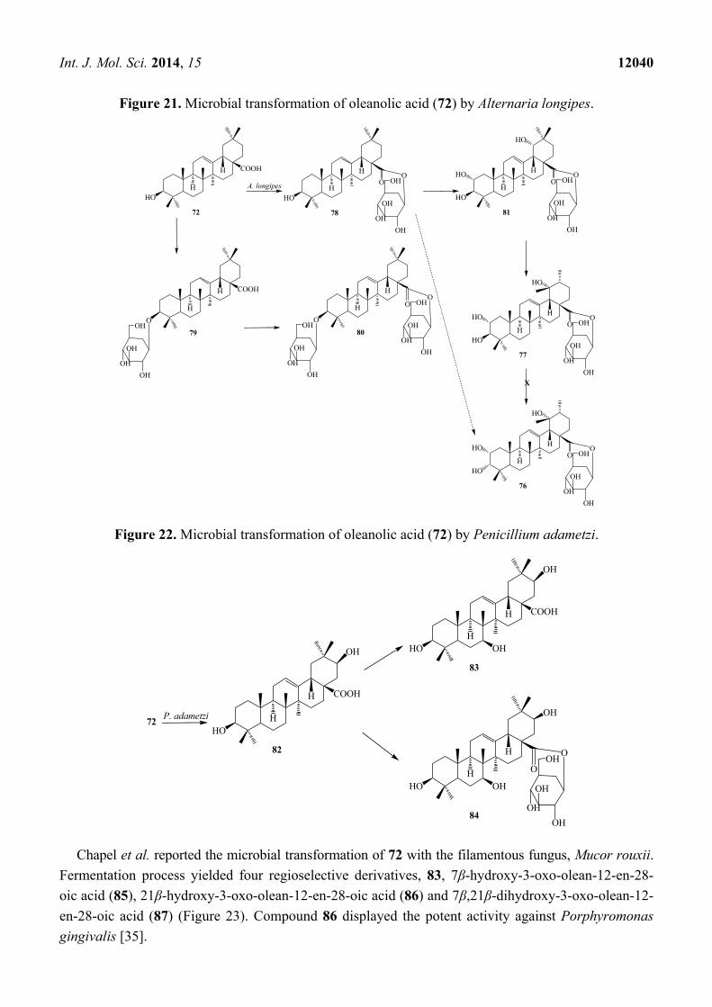
Liu et al. reported microbial transformation of 72 with Alternaria longipes and Penicillium
adametzi. The fungus Alternaria longipes converted 72 to six different regioselective hydroxylated
products, 2α,3α,19α-trihydroxy-ursolic acid-28-O-β-d-glucopyranoside (76), 2α,3β,19α-trihydroxy-ursolic
acid-28-O-β-d-glucopyranoside (77), oleanolic acid 28-O-β-d-glucopyranosyl ester (78), oleanolic
acid-3-O-β-d-glucopyranoside (79), 3-O-(β-d-glucopyranosyl)-oleanolic acid-28-O-β-d-glucopyranoside
(80), 2α,3β,19α-trihydroxy-oleanolic acid-28-O-β-d-glucopyranoside (81), while cultures of
Penicillium adametzi transformed the 72 to 21β-hydroxyl oleanolic acid (82), 7α,21β-dihydroxyl
oleanolic acid (83) and 21β-hydroxyl oleanolic acid-28-O-β-d-glucopyranoside (84). These
fermentation reactions are presented in Figures 21 and 22. Compounds 79 and 82–84 exhibited
stronger cytotoxic activities against Hela cell lines than the substrate 72 [34].
HO
COOHF. li
ni
72
H
H
HO
COOH
73
H
H
HO
COOH
74
H
HHO HO
HO
HO
COOH
H
HC. lini
75
OH
Int. J. Mol. Sci. 2014, 15 12040
Figure 21. Microbial transformation of oleanolic acid (72) by Alternaria longipes.
Figure 22. Microbial transformation of oleanolic acid (72) by Penicillium adametzi.
Chapel et al. reported the microbial transformation of 72 with the filamentous fungus, Mucor rouxii.
Fermentation process yielded four regioselective derivatives, 83, 7β-hydroxy-3-oxo-olean-12-en-28-
oic acid (85), 21β-hydroxy-3-oxo-olean-12-en-28-oic acid (86) and 7β,21β-dihydroxy-3-oxo-olean-12-
en-28-oic acid (87) (Figure 23). Compound 86 displayed the potent activity against Porphyromonas
gingivalis [35].
HO
COOH
72
H
H
A. longipes
HO
78
H
H
O
OH
OH
OH
OHO
HO
81
H
H
O
OH
OH
OH
OHOHO
HO
COOH
79
H
H
OH
OH
OH
OHO
80
H
H
OH
OH
OH
OHO
OH
OH
OH
OHO
O
HO
77
H
H
O
OH
OH
OH
OHOHO
HO
HO
76
H
H
O
OH
OH
OH
OHOHO
HO
X
72P. adametzi
HO
COOH
82
H
H
OH HO
COOH
83
H
H
OH
OH
HOH
H
OH
OH
OH
OH
OHO
O
OH
84
Int. J. Mol. Sci. 2014, 15 12041
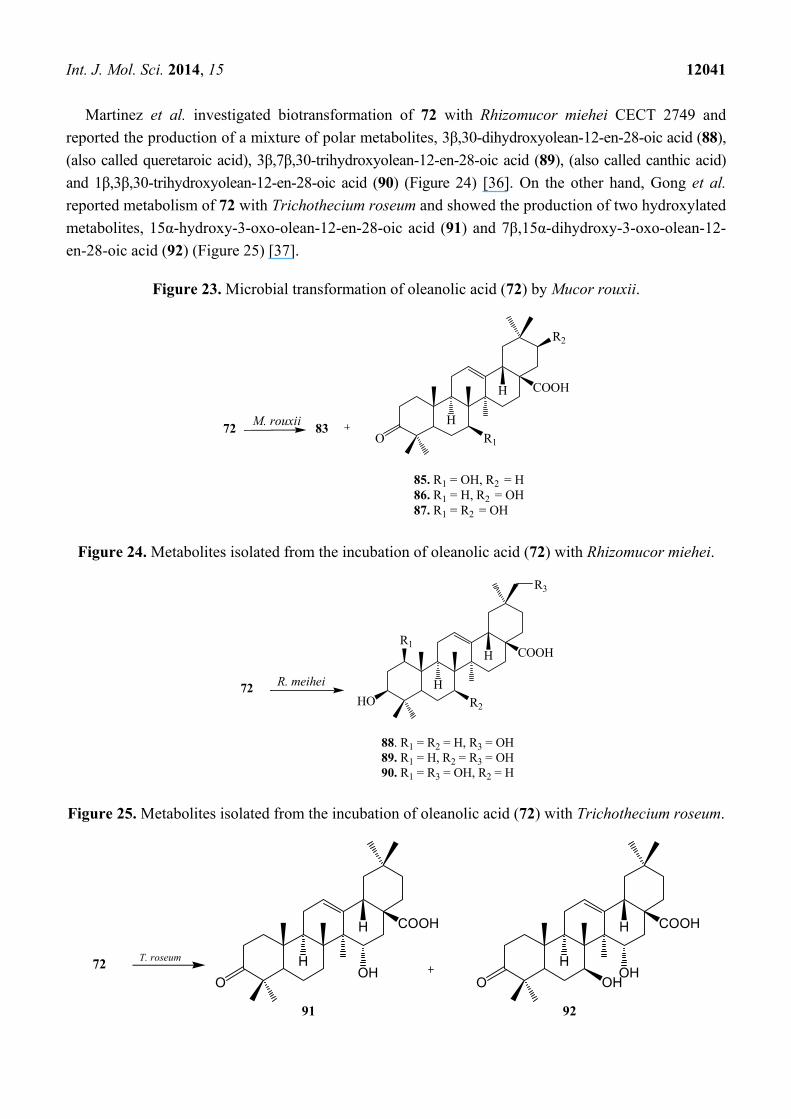
Martinez et al. investigated biotransformation of 72 with Rhizomucor miehei CECT 2749 and
reported the production of a mixture of polar metabolites, 3β,30-dihydroxyolean-12-en-28-oic acid (88),
(also called queretaroic acid), 3β,7β,30-trihydroxyolean-12-en-28-oic acid (89), (also called canthic acid)
and 1β,3β,30-trihydroxyolean-12-en-28-oic acid (90) (Figure 24) [36]. On the other hand, Gong et al.
reported metabolism of 72 with Trichothecium roseum and showed the production of two hydroxylated
metabolites, 15α-hydroxy-3-oxo-olean-12-en-28-oic acid (91) and 7β,15α-dihydroxy-3-oxo-olean-12-
en-28-oic acid (92) (Figure 25) [37].
Figure 23. Microbial transformation of oleanolic acid (72) by Mucor rouxii.
Figure 24. Metabolites isolated from the incubation of oleanolic acid (72) with Rhizomucor miehei.
Figure 25. Metabolites isolated from the incubation of oleanolic acid (72) with Trichothecium roseum.
72M. rouxii
83
COOH
85. R1 = OH, R2 = H86. R1 = H, R2 = OH87. R1 = R2 = OH
H
H
O R1
R2
R. meihei72HO
COOH
H
HR1
R3
R2
88. R1 = R2 = H, R3 = OH89. R1 = H, R2 = R3 = OH90. R1 = R3 = OH, R2 = H
72
COOH
91
H
H COOH
92
H
H
T. roseum
OHO O OH
OH
Int. J. Mol. Sci. 2014, 15 12042
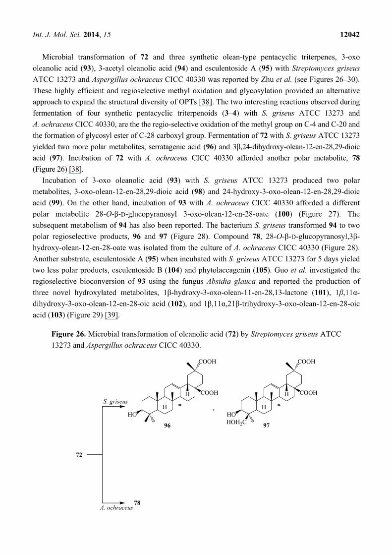
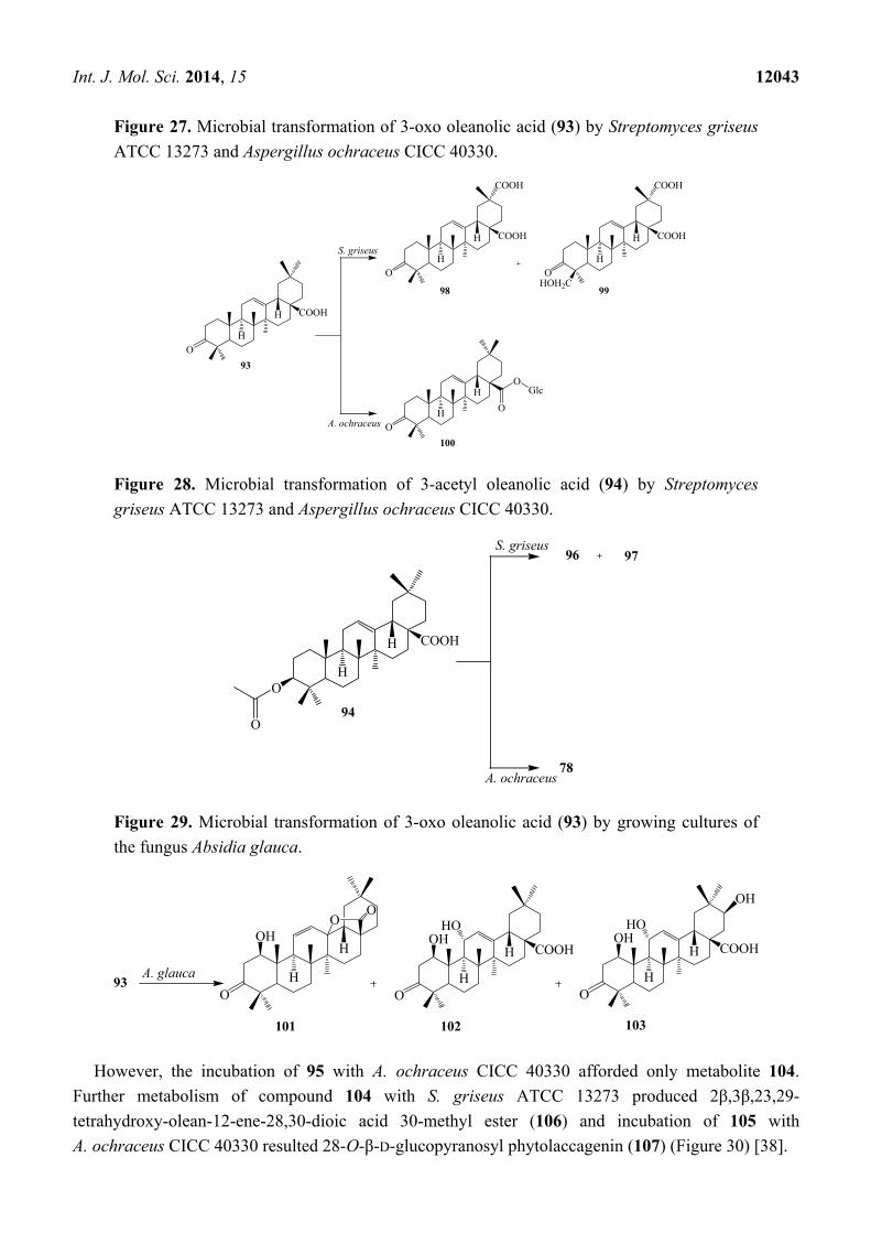
Microbial transformation of 72 and three synthetic olean-type pentacyclic triterpenes, 3-oxo
oleanolic acid (93), 3-acetyl oleanolic acid (94) and esculentoside A (95) with Streptomyces griseus
ATCC 13273 and Aspergillus ochraceus CICC 40330 was reported by Zhu et al. (see Figures 26–30).
These highly efficient and regioselective methyl oxidation and glycosylation provided an alternative
approach to expand the structural diversity of OPTs [38]. The two interesting reactions observed during
fermentation of four synthetic pentacyclic triterpenoids (3–4) with S. griseus ATCC 13273 and
A. ochraceus CICC 40330, are the the regio-selective oxidation of the methyl group on C-4 and C-20 and
the formation of glycosyl ester of C-28 carboxyl group. Fermentation of 72 with S. griseus ATCC 13273
yielded two more polar metabolites, serratagenic acid (96) and 3β,24-dihydroxy-olean-12-en-28,29-dioic
acid (97). Incubation of 72 with A. ochraceus CICC 40330 afforded another polar metabolite, 78
(Figure 26) [38].
Incubation of 3-oxo oleanolic acid (93) with S. griseus ATCC 13273 produced two polar
metabolites, 3-oxo-olean-12-en-28,29-dioic acid (98) and 24-hydroxy-3-oxo-olean-12-en-28,29-dioic
acid (99). On the other hand, incubation of 93 with A. ochraceus CICC 40330 afforded a different
polar metabolite 28-O-β-D-glucopyranosyl 3-oxo-olean-12-en-28-oate (100) (Figure 27). The
subsequent metabolism of 94 has also been reported. The bacterium S. griseus transformed 94 to two
polar regioselective products, 96 and 97 (Figure 28). Compound 78, 28-O-β-D-glucopyranosyl,3β-
hydroxy-olean-12-en-28-oate was isolated from the culture of A. ochraceus CICC 40330 (Figure 28).
Another substrate, esculentoside A (95) when incubated with S. griseus ATCC 13273 for 5 days yieled
two less polar products, esculentoside B (104) and phytolaccagenin (105). Guo et al. investigated the
regioselective bioconversion of 93 using the fungus Absidia glauca and reported the production of
three novel hydroxylated metabolites, 1β-hydroxy-3-oxo-olean-11-en-28,13-lactone (101), 1β,11α-
dihydroxy-3-oxo-olean-12-en-28-oic acid (102), and 1β,11α,21β-trihydroxy-3-oxo-olean-12-en-28-oic
acid (103) (Figure 29) [39].
Figure 26. Microbial transformation of oleanolic acid (72) by Streptomyces griseus ATCC
13273 and Aspergillus ochraceus CICC 40330.
72
HO
COOH
COOH
96
H
H
HOHOH2C
COOH
COOH
97
H
H
78
S. griseus
A. ochraceus
Int. J. Mol. Sci. 2014, 15 12043
Figure 27. Microbial transformation of 3-oxo oleanolic acid (93) by Streptomyces griseus
ATCC 13273 and Aspergillus ochraceus CICC 40330.
Figure 28. Microbial transformation of 3-acetyl oleanolic acid (94) by Streptomyces
griseus ATCC 13273 and Aspergillus ochraceus CICC 40330.
Figure 29. Microbial transformation of 3-oxo oleanolic acid (93) by growing cultures of
the fungus Absidia glauca.
However, the incubation of 95 with A. ochraceus CICC 40330 afforded only metabolite 104.
Further metabolism of compound 104 with S. griseus ATCC 13273 produced 2β,3β,23,29-
tetrahydroxy-olean-12-ene-28,30-dioic acid 30-methyl ester (106) and incubation of 105 with
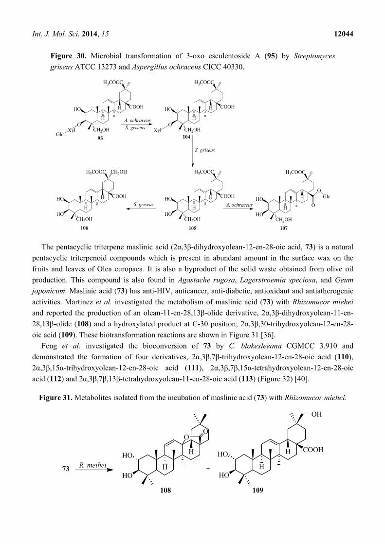
A. ochraceus CICC 40330 resulted 28-O-β-D-glucopyranosyl phytolaccagenin (107) (Figure 30) [38].
COOH
93
H
H
COOH
COOH
98
H
H
HOH2C
COOH
COOH
99
H
H
100
H
H
O
OGlc
S. griseus
A. ochraceus
O
O O
O
96 97
78
S. griseus
A. ochraceus
COOH
94
H
H
O
O
93A. glauca
COOH
H
H
OH
H
OO
101
O
OH
102
OHHO
COOH
H
H
O
103
OHHO
OH
Int. J. Mol. Sci. 2014, 15 12044
Figure 30. Microbial transformation of 3-oxo esculentoside A (95) by Streptomyces
griseus ATCC 13273 and Aspergillus ochraceus CICC 40330.
The pentacyclic triterpene maslinic acid (2α,3β-dihydroxyolean-12-en-28-oic acid, 73) is a natural
pentacyclic triterpenoid compounds which is present in abundant amount in the surface wax on the
fruits and leaves of Olea europaea. It is also a byproduct of the solid waste obtained from olive oil
production. This compound is also found in Agastache rugosa, Lagerstroemia speciosa, and Geum
japonicum. Maslinic acid (73) has anti-HIV, anticancer, anti-diabetic, antioxidant and antiatherogenic
activities. Martinez et al. investigated the metabolism of maslinic acid (73) with Rhizomucor miehei
and reported the production of an olean-11-en-28,13β-olide derivative, 2α,3β-dihydroxyolean-11-en-
28,13β-olide (108) and a hydroxylated product at C-30 position; 2α,3β,30-trihydroxyolean-12-en-28-
oic acid (109). These biotransformation reactions are shown in Figure 31 [36].
Feng et al. investigated the bioconversion of 73 by C. blakesleeana CGMCC 3.910 and
demonstrated the formation of four derivatives, 2α,3β,7β-trihydroxyolean-12-en-28-oic acid (110),
2α,3β,15α-trihydroxyolean-12-en-28-oic acid (111), 2α,3β,7β,15α-tetrahydroxyolean-12-en-28-oic
acid (112) and 2α,3β,7β,13β-tetrahydroxyolean-11-en-28-oic acid (113) (Figure 32) [40].
Figure 31. Metabolites isolated from the incubation of maslinic acid (73) with Rhizomucor miehei.
COOH
CH2OH
95
H
H
H3COOC
O
HO
XylGlc
COOH
CH2OH104
H
H
H3COOC
O
HO
Xyl
COOH
CH2OH
105
H
H
H3COOC
HO
HO
CH2OH
107
H
H
H3COOC
HO
HOCOOH
CH2OH
CH2OH
106
H
H
H3COOC
HO
HOO
OGlc
A. ochraceusS. griseus
S. griseus
S. griseus A. ochraceus
R. meihei73HO
H
H
OO
HO
COOH
109
H
HHO
OH
108
HO
Int. J. Mol. Sci. 2014, 15 12045
Figure 32. Microbial transformation of maslinic acid (73) by Cunninghamella blakesleeana.
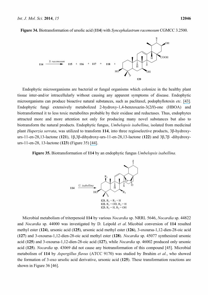
Ursolic acid (3β-hydroxy-urs-12-en-28-oic acid, UA, 114), a pentacyclic triterpene acid, exists
abundantly in many medicinal plants including sage, rosemary, thyme and lavender [1,41,42]. It
displays a remarkable spectrum of biological activities, such as anti-inflammatory activity, anti-allergic
activity, antibacterial activity, anti-mutagenicity, hepatoprotective activity, rantimalarial and anti-tumor
activity. In addition, UA is also used to induce apoptosis in human liver cancer cell lines, to enhance
the cellular immune system and pancreatic β-cell function and to inhibit invasion [1,41].
Biotransformation of 114 by the filamentous fungus Syncephalastrum racemosum (Cohn) Schroter
AS 3.264 was reported by Huang et al. They reported the regioselective hydroxylation, carbonylation,
and condensation reactions. Bioconversion of 114 by S. racemosum afforded five metabolites, 3β,21β-
dihydroxy-urs-11-en-28-oic acid-13-lactone (115), 3β,7β,21β-trihydroxy-urs-11-en-28-oic acid-13-
lactone (116), 1β,3β-dihydroxy-urs-12-en-21-one-28-oic acid (117), 1β,3β,21β-trihydroxy-urs-12-en-
28-oic acid (118) and 11,26-epoxy-3β,21β-dihydroxyurs-12-en-28-oic acid (119) (Figure 33).
Compound 117 showed moderate PTP1B inhibitory activity [41].
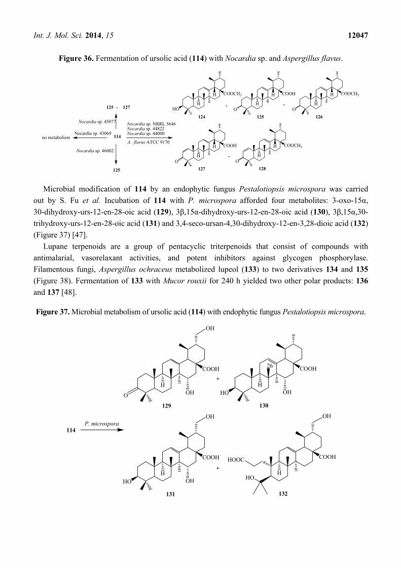
Fu et al. conducted microbial transformation of 114 with filamentous fungus Syncephalastrum
racemosum CGMCC 3.2500 (see Figure 34) and showed the formation of five polar metabolites 115, 116,
117, 118 and 3β,7β,21β-trihydroxy-urs-12-en-28-oic acid (120). Metabolites 118 and 120 exhibited
anti-HCV activity [42].
Figure 33. Fermentation of ursolic acid (114) with Syncephalastrum racemosum (Cohn)
Schroter AS 3.264.
73HO
COOH
113
H
HHO
OH
OH
C. blakesleeana HO
COOH
H
HHO
R1R2
110. R1 = OH, R2 = H111. R1 = H, R2 = OH112. R1 = R2 = OH
HO
COOH
HO
S. racemosum
114
115. R = H116. R = OH
HO
COOH
118
HO
COOH
117
HO
O COOH
119
R
H
H
H
O
O
OH O
H
H
OH
OH
OH
H
H H
H
OH
Int. J. Mol. Sci. 2014, 15 12046
Figure 34. Biotransformation of ursolic acid (114) with Syncephalastrum racemosum CGMCC 3.2500.
Endophytic microorganisms are bacterial or fungal organisms which colonize in the healthy plant
tissue inter-and/or intracellularly without causing any apparent symptoms of disease. Endophytic
microorganisms can produce bioactive natural substances, such as paclitaxel, podophyllotoxin etc. [43].
Endophytic fungi extensively metabolized 2-hydroxy-1,4-benzoxazin-3(2H)-one (HBOA) and
biotransformed it to less toxic metabolites probable by their oxidase and reductases. Thus, endophytes
attracted more and more attention not only for producing many novel substances but also to
biotransform the natural products. Endophytic fungus, Umbelopsis isabellina, isolated from medicinal
plant Huperzia serrata, was utilized to transform 114, into three regioselective products, 3β-hydroxy-
urs-11-en-28,13-lactone (121), 1β,3β-dihydroxy-urs-11-en-28,13-lactone (122) and 3β,7β -dihydroxy-
urs-11-en-28, 13-lactone (123) (Figure 35) [44].
Figure 35. Biotransformation of 114 by an endophytic fungus Umbelopsis isabellina.
Microbial metabolism of triterpenoid 114 by various Nocardia sp. NRRL 5646, Nocardia sp. 44822
and Nocardia sp. 44000 was investigated by D. Leipold et al. Micobial conversion of 114 resulted
methyl ester (124), ursonic acid (125), ursonic acid methyl ester (126), 3-oxoursa-1,12-dien-28-oic acid
(127) and 3-oxoursa-1,12-dien-28-oic acid methyl ester (128). Nocardia sp. 45077 synthesized ursonic
acid (125) and 3-oxoursa-1,12-dien-28-oic acid (127), while Nocardia sp. 46002 produced only ursonic
acid (125). Nocardia sp. 43069 did not cause any biotransformation of this compound [45]. Microbial
metabolism of 114 by Aspergillus flavus (ATCC 9170) was studied by Ibrahim et al., who showed
the formation of 3-oxo ursolic acid derivative, ursonic acid (125). These transformation reactions are
shown in Figure 36 [46].
S. racemosum114 115 118117
HO
COOH
120
H
H
OH
116
OH
114
HO
H
O
OU. isabellina
R1
R2
121. R1 = R2 = H122. R1 = OH, R2 = H123. R1 = H, R2 = OH
Int. J. Mol. Sci. 2014, 15 12047
Figure 36. Fermentation of ursolic acid (114) with Nocardia sp. and Aspergillus flavus.
Microbial modification of 114 by an endophytic fungus Pestalotiopsis microspora was carried
out by S. Fu et al. Incubation of 114 with P. microspora afforded four metabolites: 3-oxo-15α,
30-dihydroxy-urs-12-en-28-oic acid (129), 3β,15α-dihydroxy-urs-12-en-28-oic acid (130), 3β,15α,30-
trihydroxy-urs-12-en-28-oic acid (131) and 3,4-seco-ursan-4,30-dihydroxy-12-en-3,28-dioic acid (132)
(Figure 37) [47].
Lupane terpenoids are a group of pentacyclic triterpenoids that consist of compounds with
antimalarial, vasorelaxant activities, and potent inhibitors against glycogen phosphorylase.
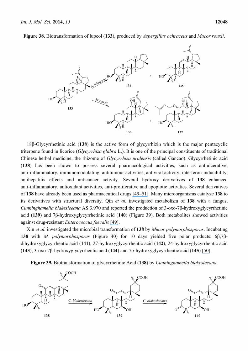
Filamentous fungi, Aspergillus ochraceus metabolized lupeol (133) to two derivatives 134 and 135
(Figure 38). Fermentation of 133 with Mucor rouxii for 240 h yielded two other polar products: 136
and 137 [48].
Figure 37. Microbial metabolism of ursolic acid (114) with endophytic fungus Pestalotiopsis microspora.
HO
COOCH3
Nocardia sp. 45077
114
124
H
H COOH
125
H
H
O
COOCH3
126
H
H
O
COOH
127
H
H
O
COOCH3
128
H
H
O
Nocardia sp. NRRL 5646Nocardia sp. 44822Nocardia sp. 44000
125 127
Nocardia sp. 46002
125
Nocardia sp. 43069no metabolism
A. flavus ATCC 9170
114
COOH
129
HHO
COOH
130
26
H
HO
COOH
131
HHO
COOH
132
H
OH
OHO OH
OH
OH
OH
HOOC
P. microspora
Int. J. Mol. Sci. 2014, 15 12048
Figure 38. Biotransformation of lupeol (133), produced by Aspergillus ochraceus and Mucor rouxii.
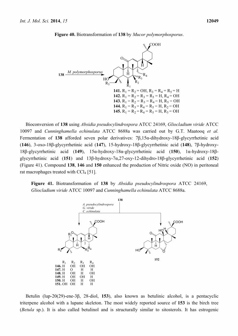
18β-Glycyrrhetinic acid (138) is the active form of glycyrrhizin which is the major pentacyclic
triterpene found in licorice (Glycyrrhiza glabra L.). It is one of the principal constituents of traditional
Chinese herbal medicine, the rhizome of Glycyrrhiza uralensis (called Gancao). Glycyrrhetinic acid
(138) has been shown to possess several pharmacological activities, such as antiulcerative,
anti-inflammatory, immunomodulating, antitumour activities, antiviral activity, interferon-inducibility,
antihepatitis effects and anticancer activity. Several hydroxy derivatives of 138 enhanced
anti-inflammatory, antioxidant activities, anti-proliferative and apoptotic activities. Several derivatives
of 138 have already been used as pharmaceutical drugs [49–51]. Many microorganisms catalyze 138 to
its derivatives with structural diversity. Qin et al. investigated metabolism of 138 with a fungus,
Cunninghamella blakesleeana AS 3.970 and reported the production of 3-oxo-7β-hydroxyglycyrrhetinic
acid (139) and 7β-hydroxyglycyrrhetinic acid (140) (Figure 39). Both metabolites showed activities
against drug-resistant Enterococcus faecalis [49].
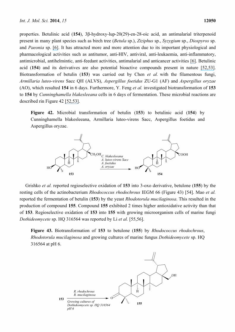
Xin et al. investigated the microbial transformation of 138 by Mucor polymorphosporus. Incubating
138 with M. polymorphosporus (Figure 40) for 10 days yielded five polar products: 6β,7β-
dihydroxyglycyrrhentic acid (141), 27-hydroxyglycyrrhentic acid (142), 24-hydroxyglycyrrhentic acid
(143), 3-oxo-7β-hydroxyglycyrrhentic acid (144) and 7α-hydroxyglycyrrhentic acid (145) [50].
Figure 39. Biotransformation of glycyrrhetinic Acid (138) by Cunninghamella blakesleeana.
HO
133
H
HH HO
134
H
H
HO
136
H
H
H
HO
135
H
H
H
HO
137
H
H
H
H
H H
OH OH
O
H
A.ochraceus
M. rouxii
138
H
H
COOH
C. blakesleeanaHO
O
139
H
H
COOH
HO
O
140
H
H
COOH
O
OH OHO
C. blakesleeana
Int. J. Mol. Sci. 2014, 15 12049
Figure 40. Biotransformation of 138 by Mucor polymorphosporus.
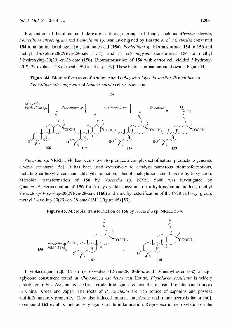
Bioconversion of 138 using Absidia pseudocylindrospora ATCC 24169, Gliocladium viride ATCC
10097 and Cunninghamella echinulata ATCC 8688a was carried out by G.T. Maatooq et al.
Fermentation of 138 afforded seven polar derivatives: 7β,15α-dihydroxy-18β-glycyrrhetinic acid
(146), 3-oxo-18β-glycyrrhetinic acid (147), 15-hydroxy-18β-glycyrrhetinic acid (148), 7β-hydroxy-
18β-glycyrrhetinic acid (149), 15α-hydroxy-18α-glycyrrhetinic acid (150), 1α-hydroxy-18β-
glycyrrhetinic acid (151) and 13β-hydroxy-7α,27-oxy-12-dihydro-18β-glycyrrhetinic acid (152)
(Figure 41). Compound 138, 146 and 150 enhanced the production of Nitric oxide (NO) in peritoneal
rat macrophages treated with CCl4 [51].
Figure 41. Biotransformation of 138 by Absidia pseudocylindrospora ATCC 24169,
Gliocladium viride ATCC 10097 and Cunninghamella echinulata ATCC 8688a.
Betulin (lup-20(29)-ene-3β, 28-diol, 153), also known as betulinic alcohol, is a pentacyclic
triterpene alcohol with a lupane skeleton. The most widely reported source of 153 is the birch tree
(Betula sp.). It is also called betulinol and is structurally similar to sitosterols. It has estrogenic
138M. polymorphosporus H
H
COOH
HO
O
R1
R2
R3R4
R5
141. R1 = R2 = OH, R3 = R4 = R5 = H142. R1 = R2 = R3 = R5 = H, R4 = OH143. R1 = R2 = R3 = R4 = H, R5 = OH144. R1 = R3 = R4 = R5 = H, R2 = OH145. R1 = R2 = R4 = R5 = H, R3 = OH
138
H
H
COOH
R2
O
R3
R1
R4
146. H OH OH OH147. H O H H148. H OH H OH149. H OH OH H150. H OH H OH151. OH OH H H
152
H
H
COOH
HO
OHO
O
A. pseudocylindrospora G. viride C. echinulata
R1 R2 R3 R4
Int. J. Mol. Sci. 2014, 15 12050
properties. Betulinic acid (154), 3β-hydroxy-lup-20(29)-en-28-oic acid, an antimalarial triterpenoid
present in many plant species such as birch tree (Betula sp.), Ziziphus sp., Syzygium sp., Diospyros sp.
and Paeonia sp. [6]. It has attracted more and more attention due to its important physiological and
pharmacological activities such as antitumor, anti-HIV, antiviral, anti-leukaemia, anti-inflammatory,
antimicrobial, antihelmintic, anti-feedant activities, antimalarial and anticancer activities [6]. Betulinic
acid (154) and its derivatives are also potential bioactive compounds present in nature [52,53].
Biotransformation of betulin (153) was carried out by Chen et al. with the filamentous fungi,
Armillaria luteo-virens Sacc QH (ALVS), Aspergillus foetidus ZU-G1 (AF) and Aspergillus oryzae
(AO), which resulted 154 in 6 days. Furthermore, Y. Feng et al. investigated biotransformation of 153
to 154 by Cunninghamella blakesleeana cells in 6 days of fermentation. These microbial reactions are
described rin Figure 42 [52,53].
Figure 42. Microbial transformation of betulin (153) to betulinic acid (154) by
Cunninghamella blakesleeana, Armillaria luteo-virens Sacc, Aspergillus foetidus and
Aspergillus oryzae.
Grishko et al. reported regioselective oxidation of 153 into 3-oxo derivative, betulone (155) by the
resting cells of the actinobacterium Rhodococcus rhodochrous IEGM 66 (Figure 43) [54]. Mao et al.
reported the fermentation of betulin (153) by the yeast Rhodotorula mucilaginosa. This resulted in the
production of compound 155. Compound 155 exhibited 2 times higher antioxidative activity than that
of 153. Regioselective oxidation of 153 into 155 with growing microorganism cells of marine fungi
Dothideomycete sp. HQ 316564 was reported by Li et al. [55,56].
Figure 43. Biotransformation of 153 to betulone (155) by Rhodococcus rhodochrous,
Rhodotorula mucilaginosa and growing cultures of marine fungus Dothideomycete sp. HQ
316564 at pH 6.
CH2OH
153
H
HO
COOH
154
H
HO
C. blakesleeanaA. luteo-virens Sacc A. foetidus A. oryzae
153155
HR. rhodochrous R. mucilaginosa
O
OH
Growing cultures of Dothideomycete sp. HQ 316564pH 6
Int. J. Mol. Sci. 2014, 15 12051
Preparation of betulinic acid derivatives through groups of fungi, such as Mycelia sterilia,
Penicillium citreonigrum and Penicillium sp. was investigated by Baratto et al. M. sterilia converted
154 to an antimalarial agent [6], betulonic acid (156), Penicillium sp. biotransformed 154 to 156 and
methyl 3-oxolup-20(29)-en-28-oate (157), and P. citreonigrum transformed 156 to methyl
3-hydroxylup-20(29)-en-28-oate (158). Biotranformation of 156 with carrot cell yielded 3-hydroxy-
(20R)-29-oxolupan-28-oic acid (159) in 14 days [57]. These biotransformations are shown in Figure 44.
Figure 44. Biotransformation of betulonic acid (154) with Mycelia sterilia, Penicillium sp.
Penicillium citreonigrum and Daucus carota cells suspension.
Nocardia sp. NRRL 5646 has been shown to produce a complex set of natural products to generate
diverse structures [58]. It has been used extensively to catalyze numerous biotransformations,
including carboxylic acid and aldehyde reduction, phenol methylation, and flavone hydroxylation.
Microbial transformation of 156 by Nocardia sp. NRRL 5646 was investigated by
Qian et al. Fermentation of 156 for 6 days yielded asymmetric α-hydroxylation product, methyl
2α-acetoxy-3-oxo-lup-20(29)-en-28-oate (160) and a methyl esterification of the C-28 carboxyl group,
methyl 3-oxo-lup-20(29)-en-28-oate (161) (Figure 45) [59].
Figure 45. Microbial transformation of 156 by Nocardia sp. NRRL 5646.
Phytolaccagenin (2β,3β,23-trihydroxy-olean-12-ene-28,30-dioic acid 30-methyl ester, 162), a major
aglycone constituent found in rPhytolacca esculenta van Houtte. Phytolacca esculenta is widely
distributed in East Asia and is used as a crude drug against edema, theumatism, bronchitis and tumors
in China, Korea and Japan. The roots of P. esculenta are rich source of saponins and possess
anti-inflammatory properties. They also induced immune interferons and tumor necrosis factor [60].
Compound 162 exhibits high activity against acute inflammation. Regiospecific hydroxylation on the
154
COOH
156
HO
COOCH3
HO
COOCH3
HHO
COOCH3
HHO
OH
157 158 159
M. steriliaPenicillium sp. Penicillium sp. P. citreonigrum D. carota
Nocardia sp. NRRL 5646156
COOCH3
161
HO
COOCH3
160
HO
AcO
Int. J. Mol. Sci. 2014, 15 12052
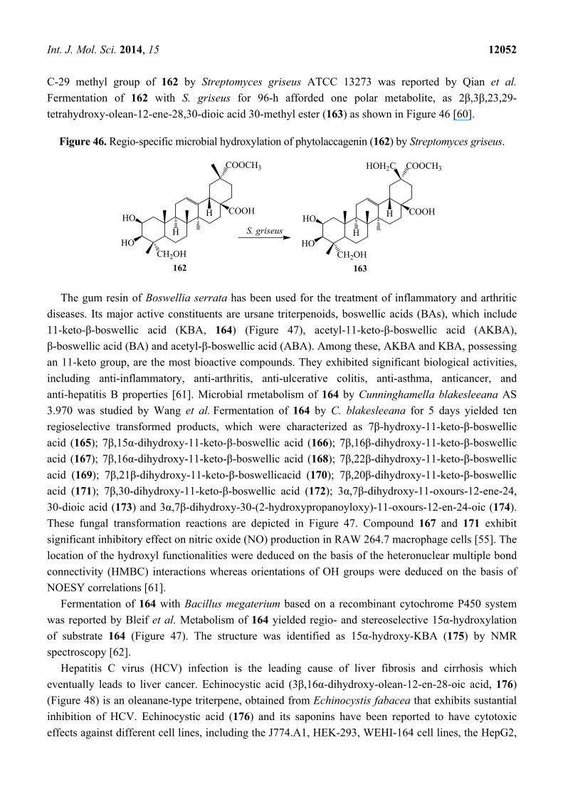
C-29 methyl group of 162 by Streptomyces griseus ATCC 13273 was reported by Qian et al.
Fermentation of 162 with S. griseus for 96-h afforded one polar metabolite, as 2β,3β,23,29-
tetrahydroxy-olean-12-ene-28,30-dioic acid 30-methyl ester (163) as shown in Figure 46 [60].
Figure 46. Regio-specific microbial hydroxylation of phytolaccagenin (162) by Streptomyces griseus.
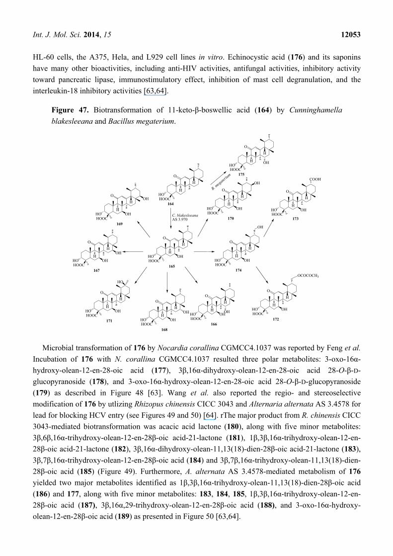
The gum resin of Boswellia serrata has been used for the treatment of inflammatory and arthritic
diseases. Its major active constituents are ursane triterpenoids, boswellic acids (BAs), which include
11-keto-β-boswellic acid (KBA, 164) (Figure 47), acetyl-11-keto-β-boswellic acid (AKBA),
β-boswellic acid (BA) and acetyl-β-boswellic acid (ABA). Among these, AKBA and KBA, possessing
an 11-keto group, are the most bioactive compounds. They exhibited significant biological activities,
including anti-inflammatory, anti-arthritis, anti-ulcerative colitis, anti-asthma, anticancer, and
anti-hepatitis B properties [61]. Microbial rmetabolism of 164 by Cunninghamella blakesleeana AS
3.970 was studied by Wang et al. Fermentation of 164 by C. blakesleeana for 5 days yielded ten
regioselective transformed products, which were characterized as 7β-hydroxy-11-keto-β-boswellic
acid (165); 7β,15α-dihydroxy-11-keto-β-boswellic acid (166); 7β,16β-dihydroxy-11-keto-β-boswellic
acid (167); 7β,16α-dihydroxy-11-keto-β-boswellic acid (168); 7β,22β-dihydroxy-11-keto-β-boswellic
acid (169); 7β,21β-dihydroxy-11-keto-β-boswellicacid (170); 7β,20β-dihydroxy-11-keto-β-boswellic
acid (171); 7β,30-dihydroxy-11-keto-β-boswellic acid (172); 3α,7β-dihydroxy-11-oxours-12-ene-24,
30-dioic acid (173) and 3α,7β-dihydroxy-30-(2-hydroxypropanoyloxy)-11-oxours-12-en-24-oic (174).
These fungal transformation reactions are depicted in Figure 47. Compound 167 and 171 exhibit
significant inhibitory effect on nitric oxide (NO) production in RAW 264.7 macrophage cells [55]. The
location of the hydroxyl functionalities were deduced on the basis of the heteronuclear multiple bond
connectivity (HMBC) interactions whereas orientations of OH groups were deduced on the basis of
NOESY correlations [61].
Fermentation of 164 with Bacillus megaterium based on a recombinant cytochrome P450 system
was reported by Bleif et al. Metabolism of 164 yielded regio- and stereoselective 15α-hydroxylation
of substrate 164 (Figure 47). The structure was identified as 15α-hydroxy-KBA (175) by NMR
spectroscopy [62].
Hepatitis C virus (HCV) infection is the leading cause of liver fibrosis and cirrhosis which
eventually leads to liver cancer. Echinocystic acid (3β,16α-dihydroxy-olean-12-en-28-oic acid, 176)
(Figure 48) is an oleanane-type triterpene, obtained from Echinocystis fabacea that exhibits sustantial
inhibition of HCV. Echinocystic acid (176) and its saponins have been reported to have cytotoxic
effects against different cell lines, including the J774.A1, HEK-293, WEHI-164 cell lines, the HepG2,
HO
COOH
COOCH3
CH2OH
162
H
HHO
HO
COOH
COOCH3
CH2OH
163
H
H
HOH2C
HOS. griseus
Int. J. Mol. Sci. 2014, 15 12053
HL-60 cells, the A375, Hela, and L929 cell lines in vitro. Echinocystic acid (176) and its saponins
have many other bioactivities, including anti-HIV activities, antifungal activities, inhibitory activity
toward pancreatic lipase, immunostimulatory effect, inhibition of mast cell degranulation, and the
interleukin-18 inhibitory activities [63,64].
Figure 47. Biotransformation of 11-keto-β-boswellic acid (164) by Cunninghamella
blakesleeana and Bacillus megaterium.
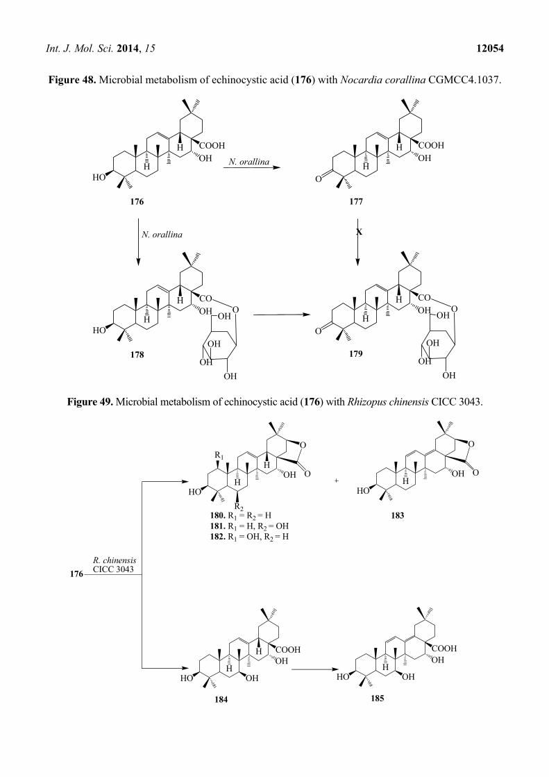
Microbial transformation of 176 by Nocardia corallina CGMCC4.1037 was reported by Feng et al.
Incubation of 176 with N. corallina CGMCC4.1037 resulted three polar metabolites: 3-oxo-16α-
hydroxy-olean-12-en-28-oic acid (177), 3β,16α-dihydroxy-olean-12-en-28-oic acid 28-O-β-D-
glucopyranoside (178), and 3-oxo-16α-hydroxy-olean-12-en-28-oic acid 28-O-β-D-glucopyranoside
(179) as described in Figure 48 [63]. Wang et al. also reported the regio- and stereoselective
modification of 176 by utlizing Rhizopus chinensis CICC 3043 and Alternaria alternata AS 3.4578 for
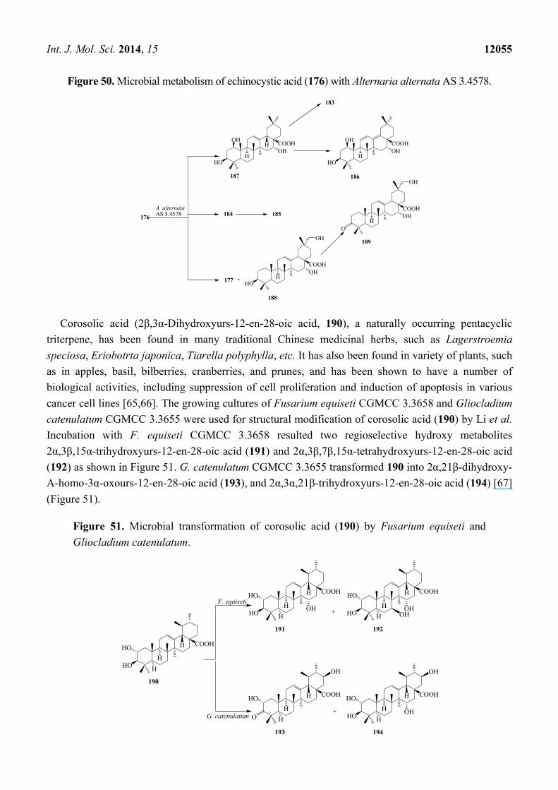
lead for blocking HCV entry (see Figures 49 and 50) [64]. rThe major product from R. chinensis CICC
3043-mediated biotransformation was acacic acid lactone (180), along with five minor metabolites:
3β,6β,16α-trihydroxy-olean-12-en-28β-oic acid-21-lactone (181), 1β,3β,16α-trihydroxy-olean-12-en-
28β-oic acid-21-lactone (182), 3β,16α-dihydroxy-olean-11,13(18)-dien-28β-oic acid-21-lactone (183),
3β,7β,16α-trihydroxy-olean-12-en-28β-oic acid (184) and 3β,7β,16α-trihydroxy-olean-11,13(18)-dien-
28β-oic acid (185) (Figure 49). Furthermore, A. alternata AS 3.4578-mediated metabolism of 176
yielded two major metabolites identified as 1β,3β,16α-trihydroxy-olean-11,13(18)-dien-28β-oic acid
(186) and 177, along with five minor metabolites: 183, 184, 185, 1β,3β,16α-trihydroxy-olean-12-en-
28β-oic acid (187), 3β,16α,29-trihydroxy-olean-12-en-28β-oic acid (188), and 3-oxo-16α-hydroxy-
olean-12-en-28β-oic acid (189) as presented in Figure 50 [63,64].
HOHOOC
164
H
H
C. blakesleeana AS 3.970
O
HOHOOC
165
H
H
O
OH
HOHOOC
169
H
H
O
OHHOHOOC
170
H
H
O
OH
OH
OH
HOHOOC
167
H
H
O
OH HOHOOC
174
H
H
O
OH
OH
OH
HOHOOC
COOH
H
H
O
OH
HOHOOC
171
H
H
O
OH
HOHOOC
168
H
H
O
OHHOHOOC
166
H
H
O
OHHOHOOC
172
H
H
O
OH
HO
OH OH
OCOCOCH3
173
HOHOOC
175
H
H
O
OH
B. meg
aterium
Int. J. Mol. Sci. 2014, 15 12054
Figure 48. Microbial metabolism of echinocystic acid (176) with Nocardia corallina CGMCC4.1037.
Figure 49. Microbial metabolism of echinocystic acid (176) with Rhizopus chinensis CICC 3043.
HO
COOH
176
H
H
N. orallinaOH
COOH
177
H
HOH
HO
CO
178
H
HOH
O
OH
OH
OH
OHO
CO
179
H
HOH
OH
OH
OH
OHO
X
O
N. orallina
176
HOH
HOH
O
O
HO
183
HOH
O
O
HO
COOH
184
H
HOH
OH HO
COOH
185
HOH
OH
R. chinensis CICC 3043
180. R1 = R2 = H181. R1 = H, R2 = OH182. R1 = OH, R2 = H
R2
R1
Int. J. Mol. Sci. 2014, 15 12055
Figure 50. Microbial metabolism of echinocystic acid (176) with Alternaria alternata AS 3.4578.
Corosolic acid (2β,3α-Dihydroxyurs-12-en-28-oic acid, 190), a naturally occurring pentacyclic
triterpene, has been found in many traditional Chinese medicinal herbs, such as Lagerstroemia
speciosa, Eriobotrta japonica, Tiarella polyphylla, etc. It has also been found in variety of plants, such
as in apples, basil, bilberries, cranberries, and prunes, and has been shown to have a number of
biological activities, including suppression of cell proliferation and induction of apoptosis in various
cancer cell lines [65,66]. The growing cultures of Fusarium equiseti CGMCC 3.3658 and Gliocladium
catenulatum CGMCC 3.3655 were used for structural modification of corosolic acid (190) by Li et al.
Incubation with F. equiseti CGMCC 3.3658 resulted two regioselective hydroxy metabolites
2α,3β,15α-trihydroxyurs-12-en-28-oic acid (191) and 2α,3β,7β,15α-tetrahydroxyurs-12-en-28-oic acid
(192) as shown in Figure 51. G. catenulatum CGMCC 3.3655 transformed 190 into 2α,21β-dihydroxy-
A-homo-3α-oxours-12-en-28-oic acid (193), and 2α,3α,21β-trihydroxyurs-12-en-28-oic acid (194) [67]
(Figure 51).
Figure 51. Microbial transformation of corosolic acid (190) by Fusarium equiseti and
Gliocladium catenulatum.
176
177HO
COOH
188
HOH
A. alternata AS 3.4578
HO
COOH
187
H
HOH
OH
HO
COOH
186
HOH
OH
183
185
OH
COOH
189
HOH
OH
O
184
F. equiseti
HO
COOH
190
H
HHO
H
G. catenulatum
HO
COOH
191
H
HHO
HOH
HO
COOH
192
H
HHO
HOH
OH
COOH
193
H
HHO
HO
OH
HO
COOH
194
H
HHO
HOH
OH
Int. J. Mol. Sci. 2014, 15 12056
6. Concluding Remarks and Future Aspects
In summary, microbial transformations are attractive alternative tools for the preparation of
bioactive complex triterpenoids, which might be difficult to prepare by conventional chemical
routes. They can produce commercially valuable pharmaceuticals for the biorefineries and novel
lead molecules towards the development of new drug candidates. The transformation of triterpenoid
skeleton through microorganisms in cell cultures exploited regioselective hydroxylations mainly in
rings A, B, C, D, E and C-23, C-24, C-29 and C-30 methyl groups, oxidation of C-28 methyl moiety
and reduction of C-3 alcohol group, ketones and C=C bond at C-11 and C-12 positions. These
modified triterpenoid drugs are currently favored when compared to their natural counterparts due to
several therapeutic advantages. Moreover, microbial-catalyzed biotransformations in association with
conventional organic synthesis can provide novel routes for the development of new drugs and drug
candidates. A number of optimization techniques such as medium, temperature, agitation, pH, etc.,
have to be established for microbial transformations to be successful and viable. Strain improvement
by conventional methods or by genetic engineering identification of alternate biosynthetic routes via
microorganisms that have not yet been exploited, new fermentation techniques and optimizing the
production facilities will cut the manufacturing cost in future and allow the biotransformation
processes to be more competitive to the current synthetic and isolation protocols.
Acknowledgments
Syed Adnan Ali Shah and Huey Ling Tan would like to acknowledge the Ministry of Higher
Education (MOHE) for financial support under the Fundamental Research Grant Scheme (FRGS) with
reference numbers 600-RMI/FRGS 5/3 (12/2012) and Research Acculturation Grant Scheme (RAGS)
with reference number 600-RMI/RAGS 5/3 (21/2012). The authors would also like to acknowledge
Universiti Teknologi MARA for the financial support under the Cumulative Impact Factor Initiative
(CIFI) Grant Scheme with reference number UiTM 600-RMI/DANA 5/3/CIFI (117/2013) and the
Principal Investigator Support Initiative (PSI) Grant Scheme with reference number UiTM 600-
RMI/DANA 5/3/PSI (251/2013).
Author Contributions
All authors equally contributed.
Conflicts of Interest
The authors declare no conflict of interest.
References
1. Salvador, J.A.R.; Moreira, V.M.; Gonçalves, B.M.F.; Lealab, A.S.; Jing, Y. Ursane-type pentacyclic
triterpenoids as useful platforms to discover anticancer drugs. Nat. Prod. Rep. 2012, 29, 1463–1469.
2. Bauer, A.; Brönstrup, M. Industrial natural product chemistry for drug discovery and
development. Nat. Prod. Rep. 2014, 31, 35–60.
Int. J. Mol. Sci. 2014, 15 12057
3. Newman, D.J.; Cragg, G.M. Natural products as sources of new drugs over the 30 years from
1981 to 2010. J. Nat. Prod. 2012, 75, 311–335.
4. Morrison, K.C.; Hergenrother, P.J. Natural products as starting points for the synthesis of
complex and diverse compounds. Nat. Prod. Rep. 2014, 31, 6–14.
5. Bicas, J.L.; Dionìsio, A.P.; Pastore, G.M. Bio-oxidation of terpenes: An approach for the flavor
industry. Chem. Rev. 2009, 109, 4518–4531.
6. Parshikov, I.A.; Netrusov, A.I.; Sutherland, J.B. Microbial transformation of antimalarial
terpenoids. Biotechnol. Adv. 2012, 30, 1516–1523.
7. Parra, A.; Rivas, F.; Garcia-Granados, A.; Martinez, A. Microbial transformation of triterpenoids.
Mini Rev. Org. Chem. 2009, 6, 307–320.
8. Muffler, K.; Leipold, D.; Schellera, M.; Haas, C.; Steingroewer, J.; Bley, T.; Neuhaus, H.E.; Mirata, M.A.;
Schrader, J.; Ulber, R. Biotransformation of triterpenes. Process Biochem. 2011, 46, 1–15.
9. Shah, S.A.A.; Sultan, S.; Hassan, N.B.; Muhammad, F.K.B.; Faridz, M.A.B.M.; Hussain, F.B.M.;
Hussain, M.; Adnan, H.S. Biotransformation of 17α-ethynyl substituted steroidal drugs with
microbial and plant cell cultures: A review. Steroids 2013, 78, 1312–1324.
10. Shah, S.A.A.; Sultan, S.; Zaimi, M. Biotransformation of tissue-specific hormone tibolone with
fungal culture Trichothecium roseum. J. Mol. Struct. 2013, 1042, 118–122.
11. Shah, S.A.A.; Sultan, S.; Adnan, H.S. A Whole-cell biocatalysis application of steroidal drugs.
Orient. J. Chem. 2013, 29, 389–403.
12. Azam, S.S.; Uddin, R.; Shah, S.A.A.; Zaheer-ul-Haq. Molecular docking studies of potent
inhibitors of tyrosinase and α-glucosidase. Med. Chem. Res. 2012, 21, 1677–1683.
13. Shah, S.A.A.; Sultan, S.; Adnan, H.S. Solid phase microbial transformation of cortexolone and prolyl
endopeptidase inhibitory activity of the transformed products. Int. J. Pharm. Pharm. Sci. 2011, 3, 1–6.
14. Choudhary, M.I.; Shah, S.A.A.; Atta-ur-Rahman; Khan, S.N.; Khan, M.T.H. Alpha-glucosidase
and tyrosinase inhibitors from fungal hydroxylation of tibolone. Steroids 2010, 75, 956–966.
15. Choudhary, M.I.; Shah, S.A.A.; Atta-ur-Rahman. Microbial transformation of anabolic steroids.
Nat. Prod. Res. 2008, 22, 1289–1296.
16. Choudhary, M.I.; Batool, I.; Shah, S.A.A.; Khan, S.N.; Atta-ur-Rahman. Microbial transformation
of oleanolic acid by Fusarium lini and α-glucosidase inhibitory activity of its transformed
products. Nat. Prod. Res. 2008, 22, 489–494.
17. Atta-ur-Rahman; Choudhary, M.I.; Basha, F.Z.; Abbas, G.; Khan, S.N.; Shah, S.A.A. Science at
the interface of chemistry and biology: Discoveries of α-glucosidase inhibitors and antiglycation
agents. Pure Appl. Chem. 2007, 79, 2263–2267.
18. Choudhary, M.I.; Yousuf, S.; Samreen; Shah, S.A.A.; Ahmed, S.; Atta-ur-Rahman.
Biotransformation of physalin H and antileishmanial activity of transformed product.
Chem. Pharm. Bull. 2006, 54, 927–930.
19. Choudhary, M.I.; Shah, S.A.A.; Sami, A.; Ajaz, A.; Shaheen, F.; Atta-ur-Rahman. Fungal
metabolites of (E)-guggulsterone and their antibacterial and radical-scavenging activities.
Chem. Biodivers. 2005, 2, 516–524.
20. Choudhary, M.I.; Batool, I.; Shah, S.A.A.; Nawaz, S.A.; Atta-ur-Rahman. Microbial hydroxylation
of pregnenolone derivative and cholinesterase inhibitory activity. Chem. Pharm. Bull. 2005, 53,
1455–1459.
Int. J. Mol. Sci. 2014, 15 12058
21. Choudhary, M.I.; Shah, S.A.A.; Musharraf, S.G.; Shaheen, F.; Atta-ur-Rahman. Microbial
transformation of dehydroepiandrosterone. Nat. Prod. Res. 2003, 17, 215–220.
22. Paludo, C.R.; da Silva-Junior, E.A.; Santos, R.A.; Pupo, M.T.; Emery, F.S.; Furtado, N.A.J.C.
Microbial transformation of β-lapachone to its glycosides by Cunninghamella elegans ATCC
10028b. Phytochem. Lett. 2013, 6, 657–661.
23. Haldar, S.; Kolet, S.P.; Thulasiram, H.V. Biocatalysis: fungi mediated novel and selective 12β- or
17β-hydroxylation on the basic limonoid skeleton. Green Chem. 2013, 15, 1311–1317.
24. Kuban, M.; Öngen, G.; Bedir, E. Biotransformation of cycloastragenol by Cunninghamella
blakesleeana NRRL 1369 resulting in a novel framework. Org. Lett. 2010, 12, 4252–4255.
25. Kuban, M.; Öngen, G.; Khan, I.A.; Bedir, E. Microbial transformation of cycloastragenol.
Phytochemistry 2013, 88, 99–104.
26. Yang, W.; Ye, M.; Huang, F.; He, W.; Guo, D. Biocatalysis of cycloastragenol by filamentous
fungi to produce unexpected triterpenes. Adv. Synth. Catal. 2012, 354, 527–539.
27. Li, H.; Ye, M.; Guo, H.; Tian, Y.; Zhang, J.; Zhou, J.; Hub, Y.; Guo, D. Biotransformation of
20(S)-protopanaxadiol by Mucor spinosus. Phytochemistry 2009, 70, 1416–1420.
28. Chen, G.; Yang, X.; Li, J.; Ge, H.; Song, Y.; Ren, J. Biotransformation of 20(S)-protopanaxadiol
by Aspergillus niger AS 3.1858. Fitoterapia 2013, 91, 256–260.
29. Schmitz, D.; Zapp, J.; Bernhardt, R. Hydroxylation of the triterpenoid dipterocarpol with
CYP106A2 from Bacillus megaterium. FEBS J. 2012, 279, 1663–1674.
30. Sun, R.; Song, H.C.; Yang, Y.H.; Yang, P.; Yang, D.Y.; Shen, K.Z.; Xu, Y.B.; Gao, Y.X.;
Chen, Y.G.; Dong, J.Y. Microbiological transformation of the triterpene nigranoic acid by the
freshwater fungus Dictyosporium heptasporum. J. Asian Nat. Prod. Res. 2013, 15, 433–440.
31. Yang, Y.H.; Sun, R.; Song, H.C.; Xu, Y.B.; Yang, P.; Yang, D.Y.; Shen, Z.K.; Wanga, A.R.;
Chen, Y.G.; Dong, J.Y. Microbial transformation of the triterpene nigranoic acid in Trichoderma sp.
Phytochem. Lett. 2012, 5, 123–127.
32. Jäger, S.; Trojan, H.; Kopp, T.; Laszczyk, M.N.; Scheffler, A. Pentacyclic triterpene distribution in
various plants–rich sources for a new group of multi-potent plant extracts. Molecules 2009, 14,
2016–2031.
33. Hua, S.; Ting, L.; Yan-Jun, S.; Li-Ming, Z.; Min, W. Preparation and crystal structure of
15α-hydroxyl-oleanolic acid. Chin. J. Struct. Chem. 2010, 29, 1798–1801.
34. Liu, D.L.; Liu, Y.; Qiu, F.; Gao, Y.; Zhang, J.Z. Biotransformation of oleanolic acid by Alternaria
longipes and Penicillium Adametzi. J. Asian Nat. Prod. Res. 2011, 13, 160–167.
35. Capel, C.S.; de Souza, A.C.D.; de Carvalho, T.C.; de Sousa, J.P.B.; Ambrósio, S.R.; Martins, C.H.G.;
Cunha, W.R.; Galán, R.H.; Furtado, N.A.J.C. Biotransformation using Mucor rouxii for the
production of oleanolic acid derivatives and their antimicrobial activity against oral pathogens.
J. Ind. Microbiol. Biotechnol. 2011, 38, 1493–1498.
36. Martinez, A.; Rivas, F.; Perojil, A.; Parra, A.; Garcia-Granados, A.; Fernandez-Vivas, A.
Biotransformation of oleanolic and maslinic acids by Rhizomucor miehei. Phytochemistry 2013,
94, 229–237.
37. Gong, T.; Zheng, L.; Zhen, X.; He, H.; Zhu, H.; Zhu, P. Microbial transformation of oleanolic
acid by Trichothecium roseum. J. Asian Nat. Prod. Res. 2014, 16, 383–386.
Int. J. Mol. Sci. 2014, 15 12059
38. Zhu, Y.Y.; Qian, L.W.; Zhang, J.; Liu, J.H.; Yu, B.Y. New approaches to the structural
modification of olean-type pentacylic triterpenes via microbial oxidation and glycosylation.
Tetrahedron 2011, 67, 4206–4211.
39. Guo, N.; Zhao, Y.; Fang, W. Biotransformation of 3-oxo-oleanolic acid by Absidia glauca.
Planta Medica 2010, 76, 1904–1907.
40. Feng, X.; Luan, J.; Guo, F.F.; Li, D.P.; Chu, Z.Y. Microbial transformation of maslinic acid by
Cunninghamella blakesleana. J. Mol. Catal. B Enzym. 2012, 82, 127–130.
41. Huang, F.; Yang, W.; Ye, F.; Tian, J.; Hu, H.; Feng, L.; Guo, D.; Ye, M. Microbial transformation
of ursolic acid by Syncephalastrum racemosum (Cohn) Schroter AS 3.264. Phytochemistry 2012,
82, 56–60.
42. Fu, S.; Yang, J.; Cui, J.; Sun, D.A. Biotransformation of ursolic acid by Syncephalastrum
racemosum CGMCC 3.2500 and anti-HCV activity. Fitoterapia 2013, 86, 123–128.
43. Nair, D.N.; Padmavathy, S. Impact of endophytic microorganisms on plants, environment and
humans. Sci. World J. 2014, 1–11. doi:10.1155/2014/250693.
44. Fu, S.; Yang, J.; Cui, J.; Feng, X.; Sun, D.A. Biotransformation of ursolic acid by an endophytic
fungus from medicinal plant Huperzia serrata. Chem. Pharm. Bull. 2011, 59, 1180—1182.
45. Leipold, D.; Wünscha, G.; Schmidt, M.; Bart, H.; Bley, T.; Neuhausd, H.E.; Bergmanne, H.;
Richlinge, E.; Mufflera, K.; Ulbera, R. Biosynthesis of ursolic acid derivatives by microbial
metabolism of ursolic acid with Nocardia sp. Strains-Proposal of new biosynthetic pathways.
Process Biochem. 2010, 45, 1043–1051.
46. Ibrahim, A.; Khalifa, S.I.; Khafagi, I.; Youssef, D.T.; Khan, S.; Mesbah, M.; Khan, I. Microbial
metabolism of biologically active secondary metabolites from Nerium oleander L. Chem Pharm Bull.
2008, 56, 1253–1258.
47. Fu, S.; Yang, J.; Cui, J.; Meng, Q.; Feng, X.; Sun, D. Multihydroxylation of ursolic acid by
Pestalotiopsis microspora isolated from the medicinal plant Huperzia serrata. Fitoterapia 2011,
82, 1057–1061.
48. De Carvalho, T.C.; Polizeli, A.M.; Turatti, I.C.C.; de Carvalho M.E.S.C.E.; Ambrósio, S.R.;
Crotti, A.E.M.; de Figueiredo, U.S.; Vieira, P.C.; Furtado, N.A.J.C. Screening of filamentous
fungi to identify biocatalysts for lupeol biotransformation. Molecules 2010, 15, 6140–6151.
49. Yu-Juan, Q.; Bing, F.; Xin-Bo, S.; Wen-Bin, Z.; He-Shui, Y.; Li-Li, Z.; Li-Yan, Y.; Bai-Ping, M.A.
Biotransformation of glycyrrhetinic acid by Cunninghamella blakesleeana. Chin. J. Nat. Med.
2010, 8, 373–381.
50. Xin, X.J.; Yang, G.; Gou, Z.P.; Yao, J.H.; Lana, R.; Ma, X.C. Structural determination of
two newtriterpenoids biotransformed from glycyrrhetinic acid by Mucor polymorphosporus.
Magn. Reson. Chem. 2010, 48, 164–167.
51. Maatooq, G.T.; Marzouk, A.M.; Gray, A.I.; Rosazza, J.P. Bioactive microbial metabolites from
glycyrrhetinic acid. Phytochemistry 2010, 71, 262–270.
52. Qi-he, C.; Jing, L.; Hai-feng, Z.; Guo-qing, H.; Ming-liang, F. The betulinic acid production from
betulin through biotransformation by fungi. Enzym. Microb. Technol. 2009, 45, 175–180.
53. Feng, Y.; Li, M.; Liu, J.; Xu, T.Y.; Fang, R.S.; Chen, Q.H.; He, G.Q. A novel one-step
microbial transformation of betulin to betulinic acid catalysed by Cunninghamella blakesleeana.
Food Chem. 2013, 136, 73–79.
Int. J. Mol. Sci. 2014, 15 12060
54. Grishko, V.V.; Tarasova, E.V.; Ivshina, I.B. Biotransformation of betulin to betulone by growing
and resting cells of the actinobacterium Rhodococcus rhodochrous IEGM 66. Process Biochem.
2013, 48, 1640–1644.
55. Mao, D.B.; Feng, Y.Q.; Bai, Y.H.; Xu, C.P. Novel biotransformation of betulin to produce
betulone by Rhodotorula Mucilaginosa. J. Taiwan Inst. Chem. Eng. 2012, 43, 825–829.
56. Liu, H.; Lei, X.L.; Li, N.; Zong, M.H. Highly regioselective synthesis of betulone from betulin by
growing cultures of marine fungus Dothideomycete sp. HQ 316564. J. Mol. Catal. B Enzym.
2013, 88, 32–35.
57. Baratto, L.C.; Porsani, M.V.; Pimentel, I.C.; Netto, A.B.P.; Paschke, R.; Oliveira, B.H. Preparation
of betulinic acid derivatives by chemical and biotransformation methods and determination of
cytotoxicity against selected cancer cell lines. Eur. J. Med. Chem. 2013, 68, 121–131.
58. Zhang, J.; Cheng, Z.H.; Yu, B.Y.; Cordell, G.A.; Qiu, S.X. Novel biotransformation of pentacyclic
triterpenoids acids by Nocardia sp. NRRL 5646. Tetrahedron Lett. 2005, 46, 2337–2340.
59. Qian, L.W.; Zhang, J.; Liu, J.H.; Yu, B.Y. Direct microbial-catalyzed asymmetric α-hydroxylation
of betulonic acid by Nocardia sp. NRRL 5646. Tetrahedron Lett. 2009, 50, 2193–2195.
60. Qian, L.; Zhang, J.; Liu, J.; Yu, B. Regio-specific microbial hydroxylation of phytolaccagenin by
Streptomyces griseus ATCC 13273. Chin. J. Chem. 2009, 27, 1434–1436.
61. Wang, Y.; Sun, Y.; Wang, C.; Huo, X.; Liu, P.; Wang, C.; Zhang, B.; Zhan, L.; Zhang, H.; Deng, S.;
et al. Biotransformation of 11-keto-b-boswellic acid by Cunninghamella blakesleana. Phytochemistry
2013, 96, 330–336.
62. Bleif, S.; Hannemann, F.; Zapp, J.; Hartmann, D.; Jauch, J.; Bernhardt, R. A new Bacillus megaterium
whole-cell catalyst for the hydroxylation of the pentacyclic triterpene 11-keto-beta-boswellic acid
(KBA) based on a recombinant cytochrome P450 system. Appl. Microbiol. Biotechnol. 2012,
doi:10.1007/s00253-011-3467-0.
63. Feng, X.; Zou, Z.; Fu, S.; Sun, L.; Su, Z.; Sun, D. Microbial oxidation and glucosidation of
echinocystic acid by Nocardia coralline. J. Mol. Catal. B Enzym. 2010, 66, 219–223.
64. Wang, H.; Wang, Q.; Xiao, S.; Yu, F.; Ye, M.; Zheng, Y.X.; Zhao, C.K.; Sun, D.; Zhang, L.H.;
Zhou, D.M. Elucidation of the pharmacophore of echinocystic acid, a new lead for blocking HCV
entry. Eur. J. Med. Chem. 2013, 64, 160–168.
65. Nho, K.J.; Chun, J.M.; Kim, H.K. Corosolic acid induces apoptotic cell death in human lung
adenocarcinoma A549 cells in vitro. Food Chem. Toxicol. 2013, 56, 8–17.
66. Kim, J.; Kim, Y.H.; Song, G.; Kim, D.; Jeong, Y.; Liu, K.; Chung, Y.; Oh, S. Ursolic acid and its
natural derivative corosolic acid suppress the proliferation of APC-mutated colon cancer cells
through promotion of β-catenin degradation. Food Chem. Toxicol. 2014, 67, 87–95.
67. Li, P.; Feng, X.; Chu, Z.; Guo, F.; Zhang, Z. Microbial transformation of corosolic acid by
Fusarium equiseti and Gliocladium catenulatum. J. Asian Nat. Prod. Res. 2013, 15, 789–808.
© 2014 by the authors; licensee MDPI, Basel, Switzerland. This article is an open access article
distributed under the terms and conditions of the Creative Commons Attribution license
(http://creativecommons.org/licenses/by/3.0/).
Related Documents


