MICROBATS APPEAR TO HAVE ADULT HIPPOCAMPAL NEUROGENESIS, BUT POST-CAPTURE STRESS CAUSES A RAPID DECLINE IN THE NUMBER OF NEURONS EXPRESSING DOUBLECORTIN R. CHAWANA, a A. ALAGAILI, b N. PATZKE, a M. A. SPOCTER, a,c O. B. MOHAMMED, b C. KASWERA, d E. GILISSEN, e,f,g N. C. BENNETT, h A. O. IHUNWO a AND P. R. MANGER a * a School of Anatomical Sciences, Faculty of Health Sciences, University of the Witwatersrand, 7 York Road, Parktown, 2193 Johannesburg, South Africa b KSU Mammals Research Chair, Department of Zoology, College of Sciences, King Saud University, Box 2455, Riyadh 11451, Saudi Arabia c Department of Anatomy, Des Moines University, Des Moines, Iowa, USA d Faculte ´ des Sciences, University of Kisangani, B.P 1232 Kisangani, Congo e Department of African Zoology, Royal Museum for Central Africa, Leuvensesteenweg 13, B-3080 Tervuren, Belgium f Laboratory of Histology and Neuropathology, Universite ´ Libre de Bruxelles, 1070 Brussels, Belgium g Department of Anthropology, University of Arkansas, Fayetteville, AR 72701, USA h Department of Zoology and Entomology, University of Pretoria, Pretoria 0002, South Africa Abstract—A previous study investigating potential adult hippocampal neurogenesis in microchiropteran bats failed to reveal a strong presence of this neural trait. As microchir- opterans have a high field metabolic rate and a small body mass, it is possible that capture/handling stress may lead to a decrease in the detectable presence of adult hippocam- pal neurogenesis. Here we looked for evidence of adult hip- pocampal neurogenesis using immunohistochemical techniques for the endogenous marker doublecortin (DCX) in 10 species of microchiropterans euthanized and perfu- sion fixed at specific time points following capture. Our results reveal that when euthanized and perfused within 15 min of capture, abundant putative adult hippocampal neurogenesis could be detected using DCX immunohisto- chemistry. Between 15 and 30 min post-capture, the detect- able levels of DCX dropped dramatically and after 30 min post-capture, immunohistochemistry for DCX could not reveal any significant evidence of putative adult hippocam- pal neurogenesis. Thus, as with all other mammals studied to date apart from cetaceans, bats, including both microchir- opterans and megachiropterans, appear to exhibit substan- tial levels of adult hippocampal neurogenesis. The present study underscores the concept that, as with laboratory experiments, studies conducted on wild-caught animals need to be cognizant of the fact that acute stress (capture/ handling) may induce major changes in the appearance of specific neural traits. Ó 2014 IBRO. Published by Elsevier Ltd. All rights reserved. Key words: adult neurogenesis, doublecortin, Chiroptera, free-living animals, capture stress, hippocampus. INTRODUCTION Studies on adult neurogenesis in free-living mammals are becoming more numerous due to the need to understand this biological process in relation to normal life-history parameters (Amrein et al., 2004, 2011; Bartkowska et al., 2008, 2010; Epp et al., 2009; Kempermann, 2012; Cavegn et al., 2013; Chawana et al., 2013; Patzke et al. 2013a,b). The investigation of free-living mammals may provide a broader understanding of the dynamics and mechanisms influencing adult neurogene- sis of species in their natural habitat and ultimately reveal potential reasons for the presence of adult neurogenesis in the mammalian brain. Free living mammals are subject to a number of pressures such as predation, foraging and varying weather patterns, all of which are factors that may influence the process of adult neurogenesis (Kempermann, 2012). While working on wild-caught mammals has the potential advantage to reveal aspects of interest to a broad understanding of adult neurogenesis, the capture of these animals from their natural environments may be considered to be an acute stressor that is difficult to control and unpredictable. While chemical capture of wild animals (using dart guns) appears to lower blood glucocorticoid levels, physical restraint and translocation leads to significant increases in the stress-related release of glucocorticoids (e.g. Widmaier and Kunz, 1993; Morton et al., 1995). In terms of adult neurogenesis, the effect of acute stress has been observed to lead to a reduction in hippocampal neurogenesis in a range of lab- oratory-kept species (Gould et al., 1998; Tanapat et al., 2001; Falconer and Galea, 2003; Kim et al., 2004; Dagyte et al., 2009; Hulshof et al., 2012), although in rats the reduction in the number of proliferating cells was http://dx.doi.org/10.1016/j.neuroscience.2014.07.063 0306-4522/Ó 2014 IBRO. Published by Elsevier Ltd. All rights reserved. * Corresponding author. Tel: +27-11-717-2497; fax: +27-11-717- 2422. E-mail address: [email protected] (P. R. Manger). Abbreviations: BSA, bovine serum albumin; DAB, diaminobenzidine; DCX, doublecortin; DCX+, doublecortin immunopositive; GCL, granular cell layer; NRS, normal rabbit serum; PB, phosphate buffer; SVZ, subventricular zone. Neuroscience 277 (2014) 724–733 724

Welcome message from author
This document is posted to help you gain knowledge. Please leave a comment to let me know what you think about it! Share it to your friends and learn new things together.
Transcript
Neuroscience 277 (2014) 724–733
MICROBATS APPEAR TO HAVE ADULT HIPPOCAMPALNEUROGENESIS, BUT POST-CAPTURE STRESS CAUSES A RAPIDDECLINE IN THE NUMBER OF NEURONS EXPRESSING DOUBLECORTIN
R. CHAWANA, a A. ALAGAILI, b N. PATZKE, a
M. A. SPOCTER, a,c O. B. MOHAMMED, b C. KASWERA, d
E. GILISSEN, e,f,g N. C. BENNETT, h A. O. IHUNWO a ANDP. R. MANGER a*
aSchool of Anatomical Sciences, Faculty of Health Sciences,
University of the Witwatersrand, 7 York Road, Parktown, 2193
Johannesburg, South Africa
bKSU Mammals Research Chair, Department of Zoology, College
of Sciences, King Saud University, Box 2455, Riyadh 11451,
Saudi Arabia
cDepartment of Anatomy, Des Moines University, Des Moines,
Iowa, USA
dFaculte des Sciences, University of Kisangani, B.P 1232
Kisangani, Congo
eDepartment of African Zoology, Royal Museum for Central
Africa, Leuvensesteenweg 13, B-3080 Tervuren, BelgiumfLaboratory of Histology and Neuropathology, Universite Libre
de Bruxelles, 1070 Brussels, Belgium
gDepartment of Anthropology, University of Arkansas,
Fayetteville, AR 72701, USA
hDepartment of Zoology and Entomology, University of
Pretoria, Pretoria 0002, South Africa
Abstract—A previous study investigating potential adult
hippocampal neurogenesis in microchiropteran bats failed
to reveal a strong presence of this neural trait. As microchir-
opterans have a high field metabolic rate and a small body
mass, it is possible that capture/handling stress may lead
to a decrease in the detectable presence of adult hippocam-
pal neurogenesis. Here we looked for evidence of adult hip-
pocampal neurogenesis using immunohistochemical
techniques for the endogenous marker doublecortin (DCX)
in 10 species of microchiropterans euthanized and perfu-
sion fixed at specific time points following capture. Our
results reveal that when euthanized and perfused within
15 min of capture, abundant putative adult hippocampal
neurogenesis could be detected using DCX immunohisto-
chemistry. Between 15 and 30 min post-capture, the detect-
able levels of DCX dropped dramatically and after 30 min
post-capture, immunohistochemistry for DCX could not
reveal any significant evidence of putative adult hippocam-
pal neurogenesis. Thus, as with all other mammals studied
to date apart from cetaceans, bats, including both microchir-
http://dx.doi.org/10.1016/j.neuroscience.2014.07.0630306-4522/� 2014 IBRO. Published by Elsevier Ltd. All rights reserved.
*Corresponding author. Tel: +27-11-717-2497; fax: +27-11-717-2422.
E-mail address: [email protected] (P. R. Manger).Abbreviations: BSA, bovine serum albumin; DAB, diaminobenzidine;DCX, doublecortin; DCX+, doublecortin immunopositive; GCL,granular cell layer; NRS, normal rabbit serum; PB, phosphate buffer;SVZ, subventricular zone.
724
opterans and megachiropterans, appear to exhibit substan-
tial levels of adult hippocampal neurogenesis. The present
study underscores the concept that, as with laboratory
experiments, studies conducted on wild-caught animals
need to be cognizant of the fact that acute stress (capture/
handling) may induce major changes in the appearance of
specific neural traits. � 2014 IBRO. Published by Elsevier
Ltd. All rights reserved.
Key words: adult neurogenesis, doublecortin, Chiroptera,
free-living animals, capture stress, hippocampus.
INTRODUCTION
Studies on adult neurogenesis in free-living mammals are
becoming more numerous due to the need to understand
this biological process in relation to normal life-history
parameters (Amrein et al., 2004, 2011; Bartkowska
et al., 2008, 2010; Epp et al., 2009; Kempermann,
2012; Cavegn et al., 2013; Chawana et al., 2013;
Patzke et al. 2013a,b). The investigation of free-living
mammals may provide a broader understanding of the
dynamics and mechanisms influencing adult neurogene-
sis of species in their natural habitat and ultimately reveal
potential reasons for the presence of adult neurogenesis
in the mammalian brain. Free living mammals are subject
to a number of pressures such as predation, foraging
and varying weather patterns, all of which are factors
that may influence the process of adult neurogenesis
(Kempermann, 2012).
While working on wild-caught mammals has the
potential advantage to reveal aspects of interest to a
broad understanding of adult neurogenesis, the capture
of these animals from their natural environments may
be considered to be an acute stressor that is difficult to
control and unpredictable. While chemical capture of
wild animals (using dart guns) appears to lower blood
glucocorticoid levels, physical restraint and translocation
leads to significant increases in the stress-related
release of glucocorticoids (e.g. Widmaier and Kunz,
1993; Morton et al., 1995). In terms of adult neurogenesis,
the effect of acute stress has been observed to lead to a
reduction in hippocampal neurogenesis in a range of lab-
oratory-kept species (Gould et al., 1998; Tanapat et al.,
2001; Falconer and Galea, 2003; Kim et al., 2004;
Dagyte et al., 2009; Hulshof et al., 2012), although in rats
the reduction in the number of proliferating cells was
R. Chawana et al. / Neuroscience 277 (2014) 724–733 725
observed to occur within 2 h of the acute stressor and
recovery to baseline levels within 24 h post exposure
(Heine et al., 2004).
An earlier study of adult neurogenesis in
microchiropterans led to the conclusion that the
hippocampus of the species studied had absent to low
rates of adult neurogenesis (Amrein et al., 2007). While
possible reasons for the absence of adult hippocampal
neurogenesis were raised, it appears that no specific con-
clusion was reached. One issue that was not raised by
Amrein et al. (2007) was whether the stress of capture/
handling of these small mammals may have had an
important role in the lack of detectable adult hippocampal
neurogenesis. While Amrein et al. (2007) state the bats
were ‘‘perfused rapidly after trapping’’, no estimate of
the time that elapsed between trapping and perfusion
was provided, thus it is possible that capture stress could
pose a serious methodological problem; however, this
does not explain the absence of adult hippocampal neuro-
genesis in the three neotropical bat species obtained from
breeding colonies located in Germany, but again, no infor-
mation regarding the handling of these bats prior to perfu-
sion was provided. Given that microchiropterans have
generally lower basal metabolic rates compared to other
mammals of similar size (Austad and Fischer, 1991;
Neuweiler, 2000), but active or field metabolic rates signif-
icantly higher than other mammals and even birds
(Neuweiler, 2000), it is possible that even a short period
of stress, in the range of minutes, related to capture and
handling may have a major effect on the expression of
proteins in the microchiropteran brain, and in the case
of Amrein et al. (2007) may have led to a false-negative
report of the absence of adult hippocampal neurogenesis
in the bat species studied, a finding that is becoming
entrenched in the neurogenesis literature (e.g. Bonfanti
and Peretto, 2011; Powers, 2013). Given this potential
confound in the study of Amrein et al. (2007), we sought
to analyze the relationship between capture stress and
adult hippocampal neurogenesis in wild-caught microchir-
opterans using immunohistochemistry for the doublecor-
tin protein (DCX), an endogenous marker of putative
adult hippocampal neurogenesis (Kempermann, 2012;
Patzke et al., 2013b).
EXPERIMENTAL PROCEDURES
In the current study we examined 36 brains from 10
microchiropteran species including Miniopterusschreibersii (n= 2) captured from a wild population in
Gauteng, South Africa, Cardioderma cor (n= 2),
Chaerophon pumilus (n= 2), Coleura afra (n= 2),
Hipposideros commersoni (n= 2), and Triaenops
persicus (n= 2) captured from wild populations in
coastal Kenya, Hipposideros fuliganosas (n= 2) and
Nycteris macrotis (n= 2) captured from wild
populations in the Yoko Forest near Kisangani,
Democratic Republic of the Congo, and Pipistrelluskuhlii (n= 2) and Asellia tridens (n= 18) captured from
wild populations near Unizah, Saudi Arabia. All animals
were adults, as judged from epiphyseal closure of the
metacarpophalangeal joints (Anthony, 1988). Appropriate
permissions to trap and euthanize the bats were obtained
from the Gauteng Department of Nature Conservation,
South Africa, the Kenya National Museums, Kenya, the
University of Kisangani, DR Congo, and the Saudi Wildlife
Authority, Saudi Arabia. All animals were treated and
used in accordance with the University of the Witwaters-
rand Animal Ethics Committee Guidelines (clearance
number 2008/36/1) which parallel those of the NIH for
the care and use of animals in scientific experiments. All
bats were euthanized (overdose of sodium pentobarbital,
100 mg/kg, i.p.) and perfused through the left ventricle
with 0.9% saline, followed by 4% paraformaldehyde in
0.1 M phosphate buffer (PB, pH 7.4) at various times fol-
lowing capture. For H. fuliganosas and T. persicus, the
animals were perfusion fixed within 15 min of capture.
For M. schreibersii, C. cor and C. pumilus, the specimens
were fixed between 15 and 30 min of capture, and for
C. afra, H. commersoni, N. macrotis and P. kuhlii, the
specimens were fixed within an hour of capture. ForA. tridens,two animals were sacrificed and perfused at each of the
following time points (in minutes) post-capture: 10, 15,
20, 30, 60, 120, 180, 240 and 300. Following perfusion,
the brains were removed and post-fixed in 4% paraformal-
dehyde in 0.1 M PB overnight, cryoprotected in 30%
sucrose in 0.1 M PB at 4 �C and stored in an antifreeze
solution at �20 �C until sectioning and histological pro-
cessing. Before sectioning, the brains were divided into
two halves along the mid-sagittal fissure and the tissue
was allowed to equilibrate in 30% sucrose in 0.1 M PB
at 4 �C. The specimens were cryosectioned in the sagittal
plane into 50-lm-thick sections. A one in three series of
sections was stained for Nissl substance (Cresyl Violet)
to reveal cytoarchitectural features, and immunostained
at two different dilutions of the primary antibody to DCX
(1:300 and 1:600) to reveal immature neurons.
In the current study we used immunolabeling of DCX,
an endogenous marker of putative immature neurons, to
ascertain the potential presence or absence of adult
neurogenesis. While DCX immunopositive neurons
away from the hippocampus may not relate to adult
neurogenesis in these regions, such as the piriform
cortex (Klempin et al., 2011), it has been established that
DCX immunolabeling of granule cells of the dentate gyrus
is a good proxy for the presence of adult hippocampal
neurogenesis (Rao and Shetty, 2004; Couillard-Despres
et al., 2005). The presence of DCX is also thought to
reflect cumulative adult hippocampal neurogenesis over
a period of 2 weeks to 6 months, although this period is
species specific (Rao and Shetty, 2004; Kohler et al.,
2011). Thus, lack of DCX staining should be a reliable
indicator of the absence of adult hippocampal neurogene-
sis (Patzke et al., 2013b) or of a perturbation in the matu-
ration process of newly generated neurons.
Free floating sections were incubated in a 1.6% H2O2,
49.2% methanol, 49.2% 0.1 M PB solution, for 30 min to
reduce endogenous peroxidase activity, which was
followed by three 10-min rinses in 0.1 M PB. To block
non-specific binding sites the sections were then pre-
incubated for 2 h, at room temperature, in blocking
buffer (3% normal rabbit serum – NRS, 2% bovine
serum albumin, BSA, and 0.25% Triton X-100 in
726 R. Chawana et al. / Neuroscience 277 (2014) 724–733
0.1 M PB). Thereafter, the sections were incubated for
48 h at 4 �C in the primary antibody solution (1:300 and
1:600, goat anti-doublecortin, DCX, SC-18 Santa Cruz
Biotech, Santa Cruz, California, USA) under gentle
agitation. The primary antibody incubation was followed
by three 10-min rinses in 0.1 M PB and the sections
were then incubated in a secondary antibody solution
(1:1000 dilution of biotinylated rabbit anti-goat IgG, BA
5000, Vector Labs, Burlingame, California, USA, in 3%
NRS and 2% BSA in 0.1 M PB) for 2 h at room
temperature. This was followed by three 10-min rinses
in 0.1 M PB, after which sections were incubated for 1 h
in an avidin–biotin solution (1:125; Vector Labs),
followed by three 10-min rinses in 0.1 M PB. Sections
were then placed in 1 ml of a solution containing
0.025% diaminobenzidine (DAB) in 0.1 M PB for 5 min,
followed by the addition of 3.3 ll of 30% hydrogen
peroxide per 1 ml of DAB solution. Chromatic
precipitation was visually monitored under a low power
stereomicroscope. Staining continued until such time
that the background stain was at a level that would
allow for accurate architectonic matching to the Nissl
sections without obscuring the immunopositive
structures. Development was arrested by placing
sections in 0.1 M PB for 10 min, followed by two more
10-min rinses in this solution. Sections were then
mounted on 0.5% gelatine-coated glass slides, dried
overnight, dehydrated in a graded series of alcohols,
cleared in xylene and coverslipped with Depex. To
ensure non-specific staining of the immunohistochemical
protocol, we ran tests on sections where we omitted the
primary antibody, and sections where we omitted the
secondary antibody. In both cases no staining was
observed. It was not possible to undertake Western blot
control testing due to the nature of the collection of the
tissue from wild populations. Staining patterns of DCX
were observed using low power stereomicroscope and
digital photomicrographs were captured using Zeiss
Axioshop and Axiovision software (Carl Zeiss
Microscopy GmbH, Jena, Germany). No pixilation
adjustments or manipulation of the captured images
was undertaken, except for the adjustment of contrast,
brightness, and levels using Adobe Photoshop 7.
For quantifying DCX immunopositive cells, a modified
unbiased stereological procedure was used as described
previously (Malberg et al., 2000; Segi-Nishida et al., 2008;
Noori and Fornal, 2011). All sections stained with the
1:300 dilution of DCX were coded to ensure that the anal-
ysis was performed by a blinded observer (M.A.S) and
immunopositive DCX cells were counted at 40� magnifi-
cation in the subgranular zone of the left hippocampus
of all specimens using an Olympus BX-60 light micro-
scope equipped with a video camera. Cells were included
if the cells lay within, or touched, the subgranular zone.
The subgranular zone was defined as the area from one
cell diameter within the granular cell layer (GCL) from
the hilus-GCL border and two cell diameters below the
hilus-GCL border (Eriksson et al., 1998). Cells were
excluded if the cell was more than two cell diameters from
the GCL, focusing through the thickness of the section
(optical dissector principle, see Gundersen et al., 1988;
West, 1993; Coggeshall and Lekan, 1996) to avoid errors
due to oversampling. Every section was counted through-
out the hippocampus and the sum was multiplied by 3 (as
we used a one in three series, see above) to provide an
estimate of the total number of immunopositive DCX cells
in the entire left hippocampus (Table 1).
Statistical analysis was done using STATA software
package version 13.1 (StataCorp. 2013. Stata Statistical
Software: Release 13. College Station, TX: StataCorp
LP). Given our sample sizes, we performed non-
parametric tests (Mann–Whitney tests) to compare
hippocampal DCX cell counts of animals from different
perfusion delay time groups (those perfused within
15 min of capture and those perfused after 15 min of
capture). In addition we undertook Spearman’s
correlation test to measure the strength of correlation of
cell counts obtained in animals belonging to a particular
time group.
RESULTS
Doublecortin immunopositive (DCX+) cells in themicrochiropteran hippocampus
Of the 10 microchiropteran species examined in the
current study, we found DCX+ cells clearly present in
the subgranular zone of the dentate gyrus of three
species (A. tridens, H. fuliganosas and T. persicus)which were perfused within 15 min of capture, partially
present in three species (M. schreibersii, C. cor, C.pumilus) which were perfused between 15 and 30 min
of capture, and low to absent in four species (C. afra, H.commersoni, N. macrotis and P. kuhlii) all of which were
perfused between 30 and 60 min post-capture (Figs. 1–
3; Table 1). The number of DCX-labeled cells in the
hippocampus for all animals perfused more than 15 min
(n= 28) after capture declined nine times
(median = 536 cells and range = 345–3294 cells) when
compared to those perfused within 15 min of capture
(n= 8; median = 4787 cells and range = 1380–6945
cells) (Mann–Whitney test z = 3.08 and p= 0.0001). In
addition, the species where DCX+ cells were observed,
the subgranular zone at the base of the granule cell
layer was populated by immunopositive cells that had a
small soma size with dendritic processes that extended
through the granule cell layer to ramify into the
molecular layer. Furthermore, the mossy fibers that
emanate from these cells were also observed with DCX
immunohistochemistry, indicating that they are likely to
be in the process of becoming functionally integrated
into the hippocampal circuitry (Fig. 2B). The morphology
of the DCX+ cells, when present, in the dentate gyrus
of the microchiropterans was similar to that seen in
other mammals studied with the same technique.
In the A. tridens time series (Table 1), DCX+ cells
were readily observed at the 10 min post-capture time
point (Figs. 2B, 3B and 4A), showing the full range of
normal morphology of these immature neurons,
including the presence of DCX+ mossy fibers. By
15 min post-capture, the number of DCX+ cells was
dramatically reduced (to around 20% of the pre-15 min
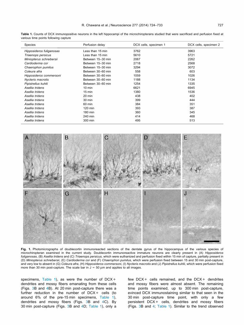
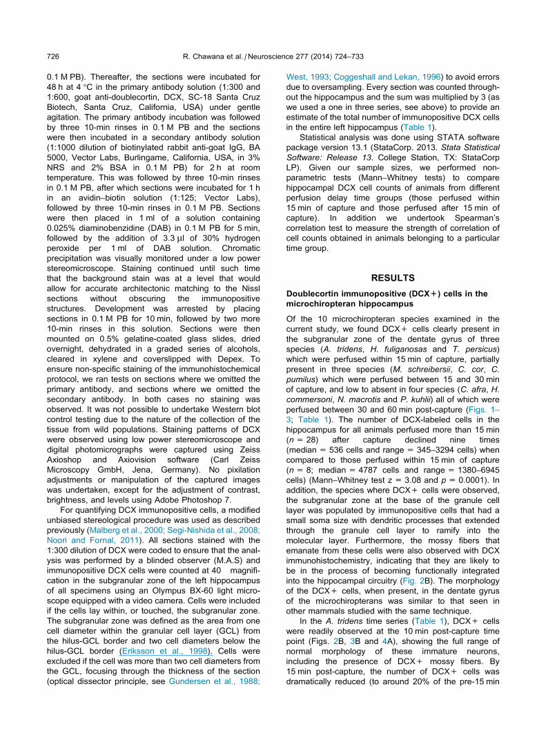
Table 1. Counts of DCX immunopositive neurons in the left hippocampi of the microchiropterans studied that were sacrificed and perfusion fixed at
various time points following capture
Species Perfusion delay DCX cells, specimen 1 DCX cells, specimen 2
Hipposideros fuliganosas Less than 15 min 3762 3963
Triaenops persicus Less than 15 min 5610 5721
Miniopterus schreibersii Between 15–30 min 2067 2262
Cardioderma cor Between 15–30 min 2718 2568
Chaerophon pumilus Between 15–30 min 3294 3072
Coleura afra Between 30–60 min 558 603
Hipposideros commersoni Between 30–60 min 1059 1026
Nycteris macrotis Between 30–60 min 1188 1134
Pipistrellus kuhlii Between 30–60 min 1254 1335
Asellia tridens 10 min 6621 6945
Asellia tridens 15 min 1380 1536
Asellia tridens 20 min 438 402
Asellia tridens 30 min 399 444
Asellia tridens 60 min 384 351
Asellia tridens 120 min 393 387
Asellia tridens 180 min 360 345
Asellia tridens 240 min 414 468
Asellia tridens 300 min 495 513
Fig. 1. Photomicrographs of doublecortin immunoreacted sections of the dentate gyrus of the hippocampus of the various species of
microchiropteran examined in the current study. Doublecortin immunoreactive immature neurons are clearly present in (A) Hipposiderosfuliganosas, (B) Asellia tridens and (C) Triaenops persicus, which were euthanized and perfusion fixed within 15 min of capture, partially present in
(D) Miniopterus schreibersii, (E) Cardioderma cor and (F) Chaerophon pumilus, which were perfusion fixed between 15 and 30 min post-capture,
and very low to absent in (G) Coleura afra, (H) Hipposideros commersoni, (I) Nycteris macrotis and (J) Pipistrellus kuhlii, which were perfusion fixed
more than 30 min post-capture. The scale bar in J = 50 lm and applies to all images.
R. Chawana et al. / Neuroscience 277 (2014) 724–733 727
specimens, Table 1), as were the number of DCX+
dendrites and mossy fibers emanating from these cells
(Figs. 3B and 4B). At 20 min post-capture there was a
further reduction in the number of DCX+ cells (to
around 6% of the pre-15 min specimens, Table 1),
dendrites and mossy fibers (Figs. 3B and 4C). By
30 min post-capture (Figs. 3B and 4D; Table 1), only a
few DCX+ cells remained, and the DCX+ dendrites
and mossy fibers were almost absent. The remaining
time points examined, up to 300 min post-capture,
evinced DCX immunostaining similar to that seen in the
30 min post-capture time point, with only a few
persistent DCX+ cells, dendrites and mossy fibers
(Figs. 3B and 4; Table 1). Similar to the trend observed
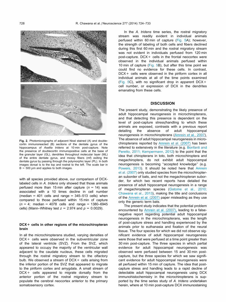
Fig. 2. Photomicrographs of adjacent Nissl stained (A) and double-
cortin immunoreacted (B) sections of the dentate gyrus of the
hippocampus of Asellia tridens at 10 min post-capture. Note
the presence of doublecortin immunopositive cells at the base of
the granular layer (GL), dendrites throughout molecular layer (ML)
of the entire dentate gyrus, and mossy fibers (mf) exiting the
dentate gyrus by passing through the polymorphic layer (PL). In both
images dorsal is to the top and rostral to the left. The scale bar in
B = 500 lm and applies to both images.
728 R. Chawana et al. / Neuroscience 277 (2014) 724–733
with all species provided above, our comparison of DCX-
labeled cells in A. tridens only showed that those animals
perfused more than 15 min after capture (n=14) was
associated with a 10 times decline in cell number
(median = 401 cells and range= 345–513 cells) when
compared to those perfused within 15 min of capture
(n=4; median = 4078 cells and range= 1380–6945
cells) (Mann–Whitney test z=2.974 and p= 0.0029).
DCX+ cells in other regions of the microchiropteranbrain
In all the microchiropterans studied, varying densities of
DCX+ cells were observed in the subventricular zone
of the lateral ventricle (SVZ). From the SVZ, which
appeared to occupy the majority of the ventricular wall
adjacent to the caudate nucleus, these cells migrated
through the rostral migratory stream to the olfactory
bulb. We observed a stream of DCX+ cells arising from
the inferior portion of the SVZ that appeared to migrate
to the piriform cortex and amygdala. A small stream of
DCX+ cells appeared to migrate dorsally from the
anterior portion of the rostral migratory stream to
populate the cerebral neocortex anterior to the primary
somatosensory cortex.
In the A. tridens time series, the rostral migratory
stream was readily evident in individual animals
perfused within 60 min of capture (Fig. 5A); however,
the strength of labeling of both cells and fibers declined
during this first 60 min and the rostral migratory stream
was not evident in individuals perfused from 120 min
post-capture. DCX+ cells in the frontal neocortex were
observed in the individual animals perfused within
10 min of capture (Fig. 5B), but after this time point we
could find no evidence for these cells. In contrast,
DCX+ cells were observed in the piriform cortex in all
individual animals at all of the time points examined
(Fig. 5C), with no significant drop in apparent DCX+
cell number, or expression of DCX in the dendrites
emanating from these cells.
DISCUSSION
The present study, demonstrating the likely presence of
adult hippocampal neurogenesis in microchiropterans,
and that detecting this presence is dependent on the
level of post-capture stress/handling to which these
animals are exposed, contrasts with a previous report
detailing the absence of adult hippocampal
neurogenesis in microchiropterans (Amrein et al., 2007).
The absence of adult hippocampal neurogenesis in micro-
chiropterans reported by Amrein et al. (2007) has been
referred to extensively in the literature (e.g. Bonfanti and
Peretto, 2011; Kempermann, 2012) to the point that the
idea that chiropterans in toto, both microchiroptera and
megachiroptera, do not exhibit adult hippocampal
neurogenesis is becoming ‘‘accepted knowledge’’ (e.g.
Powers, 2013). It should be noted here that Amrein
et al. (2007) only studied species from the microchiropter-
an suborder of bats, and not the megachiropteran subor-
der, for which two recent reports have detailed the
presence of adult hippocampal neurogenesis in a range
of megachiropteran species (Gatome et al., 2010;
Chawana et al., 2013), making the title and conclusions
of the Amrein et al. (2007) paper misleading as they use
only the generic term bats.
The present study indicates that the potential problem
encountered by Amrein et al. (2007), leading to a false-
negative report regarding potential adult hippocampal
neurogenesis in the microchiropterans, was the length
of post-capture stress and handling experienced by the
animals prior to euthanasia and fixation of the neural
tissue. The four species for which we did not observe sig-
nificant evidence of adult hippocampal neurogenesis
were those that were perfused at a time point greater than
30 min post-capture. The three species in which partial
evidence for adult hippocampal neurogenesis was
observed were perfused between 15 and 30 min post-
capture, but the three species for which we saw signifi-
cant evidence for adult hippocampal neurogenesis were
all perfused within 15 min of capture. The idea that post-
capture stress and handling leads to a rapid decline of
detectable adult hippocampal neurogenesis using DCX
immunohistochemistry in the microchiropterans is sup-
ported by the time series study of A. tridens undertaken
herein, where at 10 min post-capture DCX immunostaining
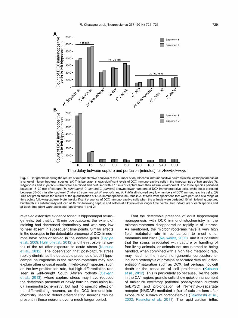
Fig. 3. Bar graphs showing the results of our quantitative analysis of the number of doublecortin immunopositive neurons in the left hippocampus of
a range of microchiropteran species. (A) This bar graph shows significant levels of DCX immunoreactive cells in the hippocampus of two species (H.fuliganosas and T. persicus) that were sacrificed and perfused within 15 min of capture from their natural environment. The three species perfused
between 15–30 min of capture (M. schrebersii, C. cor and C. pumilus) showed lower numbers of DCX immunoreactive cells, while those perfused
between 30–60 min after capture (C. afra, H. commersoni, N. macrotis and P. kuhlii) all showed very low numbers of DCX immunoreactive cells. (B)
This bar graph shows the results of the quantification of DCX immunopositive neurons in A. tridens from specimens that were perfused at a range of
time points following capture. Note the significant presence of DCX immunoreactive cells when the animals were perfused 10 min following capture,
but that this is substantially reduced at 15 min following capture and settles at a low level for longer time points. Two individuals of each species and
at each time point were assessed (specimens 1 and 2).
R. Chawana et al. / Neuroscience 277 (2014) 724–733 729
revealed extensive evidence for adult hippocampal neuro-
genesis, but that by 15 min post-capture, the extent of
staining had decreased dramatically and was very low
to near absent in subsequent time points. Similar effects
in the decrease in the detectable presence of DCX in neu-
rons have been observed in the dentate gyrus (Dagyte
et al., 2009; Hulshof et al., 2012) and the retrosplenial cor-
tex of the rat after exposure to acute stress (Kutsuna
et al., 2012). The observation that post-capture stress
rapidly diminishes the detectable presence of adult hippo-
campal neurogenesis in the microchiropterans may also
explain other unusual results in field-caught species, such
as the low proliferation rate, but high differentiation rate
seen in wild-caught South African rodents (Cavegn
et al., 2013), where capture stress may have reduced
the detectable presence of newly born neurons using Ki-
67 immunohistochemistry, but had no specific effect on
the differentiating neurons, as the DCX immunohisto-
chemistry used to detect differentiating neurons can be
present in these neurons over a much longer period.
That the detectable presence of adult hippocampal
neurogenesis with DCX immunohistochemistry in the
microchiropterans disappeared so rapidly is of interest.
As mentioned, the microchiropterans have a very high
field metabolic rate in comparison to most other
mammals and birds (Neuweiler, 2000), and it is possible
that the stress associated with capture or handling of
free-living animals, or animals not accustomed to being
handled, when combined with a high field metabolic rate,
may lead to the rapid non-genomic corticosterone-
induced proteolysis of proteins associated with cell differ-
entiation/maturation such as DCX, but perhaps not cell
death or the cessation of cell proliferation (Kutsuna
et al., 2012). This is particularly so because, like the cells
in the CA1 region, granule cells show quick enhancement
of miniature excitatory potential post-synaptic currents
(mEPSC) and prolongation of N-methyl-D-aspartate
receptor (NMDAR)-mediated influx of calcium ions after
exposure to a wave of corticosteroids (Takahashi et al.,
2002; Pasricha et al., 2011). The rapid calcium influx
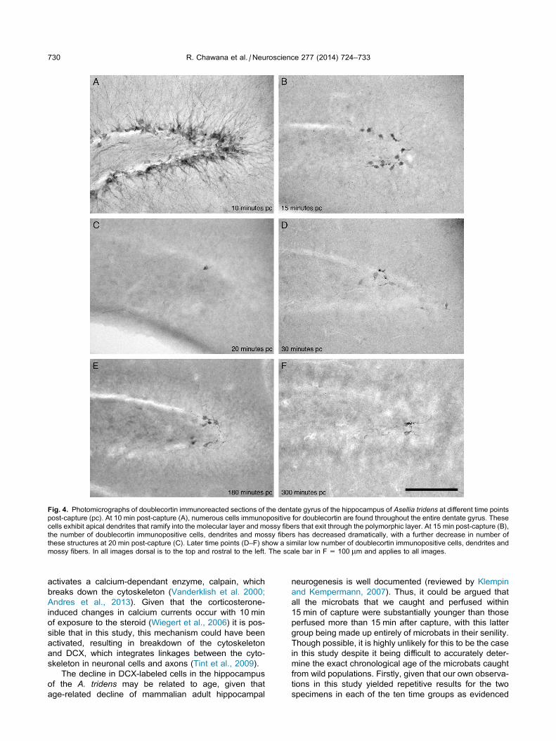
Fig. 4. Photomicrographs of doublecortin immunoreacted sections of the dentate gyrus of the hippocampus of Asellia tridens at different time points
post-capture (pc). At 10 min post-capture (A), numerous cells immunopositive for doublecortin are found throughout the entire dentate gyrus. These
cells exhibit apical dendrites that ramify into the molecular layer and mossy fibers that exit through the polymorphic layer. At 15 min post-capture (B),
the number of doublecortin immunopositive cells, dendrites and mossy fibers has decreased dramatically, with a further decrease in number of
these structures at 20 min post-capture (C). Later time points (D–F) show a similar low number of doublecortin immunopositive cells, dendrites and
mossy fibers. In all images dorsal is to the top and rostral to the left. The scale bar in F = 100 lm and applies to all images.
730 R. Chawana et al. / Neuroscience 277 (2014) 724–733
activates a calcium-dependant enzyme, calpain, which
breaks down the cytoskeleton (Vanderklish et al. 2000;
Andres et al., 2013). Given that the corticosterone-
induced changes in calcium currents occur with 10 min
of exposure to the steroid (Wiegert et al., 2006) it is pos-
sible that in this study, this mechanism could have been
activated, resulting in breakdown of the cytoskeleton
and DCX, which integrates linkages between the cyto-
skeleton in neuronal cells and axons (Tint et al., 2009).
The decline in DCX-labeled cells in the hippocampus
of the A. tridens may be related to age, given that
age-related decline of mammalian adult hippocampal
neurogenesis is well documented (reviewed by Klempin
and Kempermann, 2007). Thus, it could be argued that
all the microbats that we caught and perfused within
15 min of capture were substantially younger than those
perfused more than 15 min after capture, with this latter
group being made up entirely of microbats in their senility.
Though possible, it is highly unlikely for this to be the case
in this study despite it being difficult to accurately deter-
mine the exact chronological age of the microbats caught
from wild populations. Firstly, given that our own observa-
tions in this study yielded repetitive results for the two
specimens in each of the ten time groups as evidenced
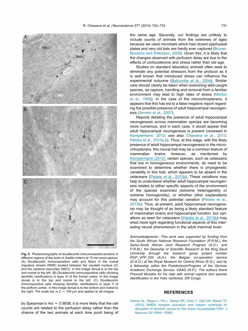
Fig. 5. Photomicrographs of doublecortin immunoreacted sections in
different regions of the brain of Asellia tridens at 10 min post-capture.
(A) Doublecortin immunopositive cells and fibers in the rostral
migratory stream (RMS) located between the caudate nucleus (C)
and the cerebral neocortex (NEO). In this image dorsal is to the top
and rostral to the left. (B) Doublecortin immunopositive cells showing
dendritic ramifications in layer III of the frontal cortex. In this image
dorsal is to the top and rostral to the left. (C) Doublecortin
immunopositive cells showing dendritic ramifications in layer II of
the piriform cortex. In this image dorsal is to the bottom and rostral to
the right. The scale bar in C = 100 lm and applies to all images.
R. Chawana et al. / Neuroscience 277 (2014) 724–733 731
by Spearman’s rho = 0.9938, it is more likely that the cell
counts are related to the perfusion delay rather than the
chance of the two animals at each time point being of
the same age. Secondly, our findings are unlikely to
include counts of animals from the extremes of ages
because we used microbats which had closed epiphyseal
plates and very old bats are hardly ever captured (Brunet-
Rossinni and Wilkinson, 2009). Given this, it is likely that
the changes observed with perfusion delay are due to the
effects of corticosterone and stress rather than old age.
Studies on standard laboratory animals often seek to
eliminate any potential stressors from the protocol as it
is well known that introduced stress can influence the
experimental outcome (Balcombe et al., 2004). Similar
care should clearly be taken when examining wild-caught
species, as capture, handling and removal from a familiar
environment may lead to high rates of stress (Morton
et al., 1995). In the case of the microchiropterans, it
appears that this has led to a false-negative report regard-
ing the possible presence of adult hippocampal neurogen-
esis (Amrein et al., 2007).
Reports detailing the presence of adult hippocampal
neurogenesis across mammalian species are becoming
more numerous, and in each case, it would appear that
adult hippocampal neurogenesis is present (reviewed in
Kempermann, 2012; see also Chawana et al., 2013;
Patzke et al., 2013a,b). Thus, at this stage, with the likely
presence of adult hippocampal neurogenesis in the micro-
chiropterans, this neural trait may be a common feature of
mammalian brains; however, as mentioned by
Kempermann (2012), certain species, such as cetaceans
that live in homogeneous environments, do need to be
examined to determine whether there is phylogenetic
variability in this trait, which appears to be absent in the
cetaceans (Patzke et al., 2013b). These variations may
help to understand whether adult hippocampal neurogen-
esis relates to either specific aspects of the environment
of the species examined (extreme heterogeneity or
extreme homogeneity), or whether other explanations
may account for this potential variation (Patzke et al.,
2013b). Thus, at present, adult hippocampal neurogene-
sis may be thought of as being a likely standard feature
of mammalian brains and hippocampal function, but vari-
ations as seen for cetaceans (Patzke et al., 2013b) may
shed more light regarding functional aspects of this inter-
esting neural phenomenon in the adult mammal brain.
Acknowledgments—This work was supported by funding from
the South African National Research Foundation (P.R.M.), the
Swiss-South African Joint Research Program (A.O.I. and
P.R.M.), the Deanship of Scientific Research at the King Saud
University through the research group project number
RGP_VPP_020 (A.A.), the Belgian co-operation service
(D.G.D.) at the Royal Museum for Central Africa (E.G.), and by
a fellowship within the Postdoctoral-Program of the German
Academic Exchange Service, DAAD (N.P.). The authors thank
Prescott Musaba for his help with animal capture and species
identification in the Yoko rainforest, DR Congo.
REFERENCES
Andres AL, Regev L, Phi L, Seese RR, Chen Y, Gall CM, Baram TZ
(2013) NMDA receptor activation and calpain contribute to
disruption of dendritic spines by the stress neuropeptide CRH. J
Neurosci 33:16945–16960.
732 R. Chawana et al. / Neuroscience 277 (2014) 724–733
Amrein I, Slomianka L, Poletaeva II, Bologova NV, Lipp HP (2004)
Marked species and age-dependent differences in cell
proliferation and neurogenesis in the hippocampus of wild-living
rodents. Hippocampus 14:1000–1010.
Amrein I, Dechmann DK, Winter Y, Lipp HP (2007) Absent or low rate
of adult neurogenesis in the hippocampus of bats (Chiroptera).
PLoS One 2:e455.
Amrein I, Isler K, Lipp HP (2011) Comparing adult hippocampal
neurogenesis in mammalian species and orders: influence of
chronological age and life history stage. Eur J Neurosci
34:978–987.
Anthony ELP (1988) Age determination in bats. In: Kunz TH, editor.
Ecological and behavioral methods for the study of
bats. Washington [DC]: Smithsonian Institution Press. p. 47–58.
Austad SN, Fischer KE (1991) Mammalian aging, metabolism, and
ecology – evidence from the bats and marsupials. J Gerontol
46:B47–B53.
Balcombe JP, Barnard ND, Sandusky C (2004) Laboratory routines
cause animal stress. Contemp Top Lab Anim Sci 43:42–51.
Bartkowska K, Djavadian RL, Taylor JR, Turlejski K (2008)
Generation, recruitment and death of brain cells throughout the
life cycle of Sorex shrews (Lipotyphla). Eur J Neurosci
27:1710–1721.
Bartkowska K, Turlejski K, Grabiec M, Ghazaryan A, Yavruoyan E,
Djavadian RL (2010) Adult neurogenesis in the hedgehog
(Erinaceus concolor) and mole (Talpa europaea). Brain Behav
Evol 76:128–143.
Bonfanti L, Peretto P (2011) Adult neurogenesis in mammals – a
theme with many variations. Eur J Neurosci 34:930–950.
Brunet-Rossinni AK, Wilkinson GS (2009) Methods for age estimation
and the study of senescence in bats. In: Kunz TH, Parsons S,
editors. Ecological and behavioral methods for the study of
bats. Baltimore: Johns Hopkins University Press. p. 315–325.
Cavegn N, van Dijk RM, Menges D, Brettschneider H, Phalanndwa
M, Chimimba CT, Isler K, Lipp HP, Slomianka L, Amrein I (2013)
Habitat-specific shaping of proliferation and neuronal
differentiation in adult hippocampal neurogenesis of wild
rodents. Front Neurosci 7:59.
Chawana R, Patzke N, Kaswera C, Gilissen E, Ihunwo AO, Manger
PR (2013) Adult neurogenesis in eight megachiropteran species.
Neuroscience 244:159–172.
Coggeshall RE, Lekan HA (1996) Methods for determining numbers
of cells and synapses: a case for more uniform standards of
review. J Comp Neurol 364:6–15.
Couillard-Despres S, Winner B, Schaubeck S, Aigner R, Vroemen M,
Weidner N, Bogdahn U, Winkler J, Kuhn HG, Aigner L (2005)
Doublecortin expression levels in adult brain reflect neurogenesis.
Eur J Neurosci 21:1–14.
Dagyte G, van der Zee EA, Postema F, Luiten PGM, den Boer JA,
Trentani A, Meerlo P (2009) Chronic but not acute foot-shock
stress leads to temporary suppression of cell proliferation in rat
hippocampus. Neuroscience 162:904–913.
Epp JR, Barker JM, Galea LA (2009) Running wild: neurogenesis in
the hippocampus across the lifespan in wild and laboratory-bred
Norway rats. Hippocampus 19:1040–1049.
Eriksson PS, Perfilieva E, Bjork-Eriksson E, Alborn A, Nordborg C,
Peterson DA, Gage FH (1998) Neurogenesis in the adult human
hippocampus. Nat Med 4:1313–1317.
Falconer EM, Galea LA (2003) Sex differences in cell proliferation,
cell death and defensive behavior following acute predator odor
stress in adult rats. Brain Res 975:22–36.
Gatome CW, Mwangi DK, Lipp HP, Amrein I (2010) Hippocampal
neurogenesis and cortical cellular plasticity in Wahlberg’s
epauletted fruit bat: a qualitative and quantitative study. Brain
Behav Evol 76:116–127.
Gould E, Tanapat P, McEwen BS, Flugge G, Fuch E (1998)
Proliferation of granule cell precursors in the dentate gyrus of
adult monkeys is diminished by stress. Proc Natl Acad Sci U S A
9:3168–3171.
Gundersen HJ, Bagger P, Bendtsen TF, Evans SM, Korbo L,
Marcussen N, Moller A, Nielsen K, Nyengaard JR, Pakkenberg
B, Sorensen FB, Vesterby A, West MJ (1988) The new
stereological tools: disector, fractionator, nucleator and point
sampled intercepts and their use in pathological research and
diagnosis. APMIS 96:857–881.
Heine VM, Maslam S, Joels M, Lucassen PJ (2004) Prominent decline
of newborn cell proliferation, differentiation, and apoptosis in the
aging dentate gyrus, in absence of an age-related hypothalamus–
pituitary–adrenal axis activation. Neurobiol Aging 25:261–375.
Hulshof HJ, Novati A, Luiten PGM, den Boer JA, Meerlo P (2012)
Despite higher glucocorticoid levels and stress responses in
female rats, both sexes exhibit similar stress-induced changes in
hippocampal neurogenesis. Behav Brain Res 234:357–364.
Kempermann G (2012) New neurons for ‘survival of the fittest’. Nat
Rev Neurosci 13:727–736.
Kim JB, Ju JY, Kim JH, Kim TY, Yang BH, Lee YS, Son H (2004)
Dexamethasone inhibits proliferation of adult hippocampal
neurogenesis in vivo and in vitro. Brain Res 1027:1–10.
Klempin F, Kempermann G (2007) Adult hippocampal neurogenesis
and aging. Eur Arch Psychiatry Clin Neurosci 257:271–280.
Klempin F, Kronenberg G, Cheung G, Kettenmann H, Kempermann
G (2011) Properties of doublecortin-(DCX)-expressing cells in the
piriform cortex compared to the neurogenic dentate gyrus of adult
mice. PLoS One 6:e25760.
Kohler SJ, Williams NI, Stanton GB, Cameron JL, Greenough WT
(2011) Maturation time of new granule cells in the dentate gyrus of
adult macaque monkeys exceeds six months. Proc Natl Acad Sci
U S A 108:10326–10331.
Kutsuna N, Suma T, Takada Y, Yamashita A, Oshima H, Sakatani K,
Yamamoto T, Katayama Y (2012) Decrease in doublecortin
expression without neuronal cell death in rat retrosplenial cortex
after stress exposure. NeuroReport 23:211–215.
Malberg JE, Eisch AJ, Nestler EJ, Duman RS (2000) Chronic
antidepressant treatment increases neurogenesis in adult rat
hippocampus. J Neurosci 20:9104–9110.
Morton DJ, Anderson E, Foggin CM, Kock MD, Tiran EP (1995)
Plasma-cortisol as an indicator of stress due to capture and
translocation in wildlife species. Vet Rec 136:60–63.
Neuweiler G (2000) The Biology of Bats. Oxford, UK: Oxford
University Press.
Noori HR, Fornal CA (2011) The appropriateness of unbiased optical
fractionators to assess cell proliferation in the adult hippocampus.
Front Neurosci 5:140.
Pasricha N, Joels M, Karst H (2011) Rapid effects of corticosterone in
the mouse dentate gyrus via a non-genomic pathway. J
Neuroendocrinol 23:143–147.
Patzke N, Kaswera C, Gilissen E, Ihunwo AO, Manger PR (2013a)
Adult neurogenesis in a giant otter shrew (Potamogale velox).
Neuroscience 244:159–172.
Patzke N, Spocter MA, Karlsson KÆ, Bertelsen MF, Haagensen M,
Chawana R, Streicher S, Kaswera C, Gilissen E, Alagaili AN,
Mohammed OB, Reep RL, Bennett NC, Siegel JM, Ihunwo AO,
MangerPR (2013b) In contrast tomany othermammals, cetaceans
have relatively small hippocampi that appear to lack adult
neurogenesis. Brain Struct Funct. http://dx.doi.org/10.1007/
s00429-013-0660-1.
Powers AS (2013) Adult neurogenesis in mammals and
nonmammals. Commentary on Kempermann (2012): New
neurons for ‘survival of the fittest’. Nat Rev Neurosci 13:727–
736. Brain Behav Evol 81:206.
Rao MS, Shetty AK (2004) Efficacy of doublecortin as a marker to
analyse the absolute number and dendritic growth of newly
generated neurons in the adult dentate gyrus. Eur J Neurosci
19:234–246.
Segi-Nishida E, Warner-Schmidt JL, Duman RS (2008)
Electroconvulsive seizure and VEGF increase the proliferation
of neural stem-like cells in rat hippocampus. Proc Natl Acad Sci U
S A 105:11352–11357.
Takahashi T, Kimoto T, Tanabe N, Hattori TA, Yasumatsu N, Kawato
S (2002) Corticosterone acutely prolonged N-methyl-D-aspartate
receptor-mediated Ca2+ elevation in cultured rat hippocampal
neurons. J Neurochem 83:1441–1451.

R. Chawana et al. / Neuroscience 277 (2014) 724–733 733
Tanapat P, Hastings NB, Rydel TA, Galea LA, Gould E (2001)
Exposure to fox odor inhibits cell proliferation in the hippocampus
of adult rats via an adrenal hormone-dependent mechanism. J
Comp Neurol 437:496–504.
Tint I, Jean D, Baas PW, Black MM (2009) Doublecortin associates
with microtubules preferentially in regions of the axon displaying
actin-rich protrusive structures. J Neurosci 29:10995–11010.
Vanderklish PW, Krushel LA, Holst BH, Gally JA, Crossin KL,
Edelman GM (2000) Marking synaptic activity in dendritic spines
with a calpain substrate exhibiting fluorescence resonance energy
transfer. Proc Natl Acad Sci U S A 97:2253–2258.
West MJ (1993) Regionally specific loss of neurons in the aging
human hippocampus. Neurobiol Aging 14:287–293.
Widmaier EP, Kunz TH (1993) Basal, diurnal, and stress-induced
levels of glucose and glucocorticoids in captive bats. J Exp Zool
265:533–540.
Wiegert O, Joels M, Krugers H (2006) Timing is essential for rapid
effects of corticosterone on synaptic potentiation in the mouse
hippocampus. Learn Mem 13:110–113.
(Accepted 28 July 2014)(Available online 7 August 2014)
Related Documents