Matrix Biology 23 (2004) 195–204 0945-053X/04/$30.00 2004 Elsevier B.V./International Society of Matrix Biology. All rights reserved. doi:10.1016/j.matbio.2004.05.003 Mice lacking the extracellular matrix adaptor protein matrilin-2 develop without obvious abnormalities Lajos Mates , Claudia Nicolae , Matthias Morgelin , Ferenc Deak , Ibolya Kiss , Attila Aszodi * a b c a a b, ´´ ¨ ´ ´ Institute of Biochemistry, Biological Research Center of the Hungarian Academy of Sciences, H-6701 Szeged, Hungary a Department for Molecular Medicine, Max Planck Institute for Biochemistry, Am Klopferspitz 18A, D-82152 Martinsried, Germany b Department of Cell and Molecular Biology, Section of Molecular Pathogenesis, BMCyB14, University of Lund, S-22184 Lund, Sweden c Received 15 January 2004; received in revised form 28 April 2004; accepted 5 May 2004 Abstract Matrilins are putative adaptor proteins of the extracellular matrix (ECM) which can form both collagen-dependent and collagen- independent filamentous networks. While all known matrilins (matrilin-1, -2, -3, and -4) are expressed in cartilage, only matrilin- 2 and matrilin-4 are abundant in non-skeletal tissues. To clarify the biological role of matrilin-2, we have developed a matrilin-2-deficient mouse strain. Matrilin-2 null mice show no gross abnormalities during embryonic or adult development, are fertile, and have a normal lifespan. Histological and ultrastructural analyses indicate apparently normal structure of all organs and tissues where matrilin-2 is expressed. Although matrilin-2 co-localizes with matrilin-4 in many tissues, Northern hybridization, semiquantitative RT-PCR, immunohistochemistry and biochemical analysis reveal no significant alteration in the steady-state level of matrilin-4 expression in homozygous mutant mice. Immunostaining of wild-type and mutant skin samples indicate no detectable differences in the expression and deposition of matrilin-2 binding partners including collagen I, laminin-nidogen complexes, fibrillin-2 and fibronectin. In addition, electron microscopy reveals an intact basement membrane at the epidermal–dermal junction and normal organization of the dermal collagen fibrils in mutant skin. These data suggest that either matrilin-2 and matrilin-2- mediated matrix–matrix interactions are dispensable for proper ECM assembly and function, or that they are efficiently compensated by other matrix components including wild-type levels of matrilin-4. 2004 Elsevier B.V./International Society of Matrix Biology. All rights reserved. Keywords: Extracellular matrix; Matrilin; Cartilage; Skin 1. Introduction Extracellular matrices (ECMs) are highly organized structures that perform diverse roles during development, tumorigenesis, and tissue repair. The cell modifying actions of ECM components are partially mediated by intracellular signals induced by ECM ligands binding cellular receptors. Further interactions between different matrix proteins are essential for proper ECM assembly and play a crucial role in maintaining the structural integrity and physical properties of connective tissues. The multifunctional nature of ECM components is high- lighted by the relatively limited set of modules found to comprise most of these proteins. The matrilins are a family of ECM proteins, which share similar modular structure. The family consists of *Corresponding author. Tel.: q49-89-8578-2849; fax: q49-89- 8578-2422. E-mail address: [email protected] (A. Aszodi). ´ four members (matrilin-1, -2, -3, and -4), each carrying one or two von Willebrand factor A (vWFA)-like mod- ules, a various number of epidermal growth factor (EGF)-like motifs, and a coiled-coil (CC) oligomeriza- tion domain (reviewed in Deak et al., 1999). Matrilin-1 ´ and -3 are expressed primarily in cartilage, while matri- lin-2 and -4 have a broader tissue distribution. Matrilin- 2 is the largest member of the family. Analysis of the mouse and human matrilin-2 cDNA sequences revealed that the precursor protein consists of a putative signal peptide, two vWFA-like domains separated by ten EGF- like motifs, a unique segment not identified in other matrilins, and a CC domain (Deak et al., 1997; Mura- ´ toglu et al., 2000). In tissue extracts and cell culture medium, matrilin-2 exists as mono-, di-, tri-, and tetra- mers, as demonstrated by SDS-PAGE and electron microscopy (Piecha et al., 1999). In addition to forming homotypic oligomers, biochemical and biophysical

Welcome message from author
This document is posted to help you gain knowledge. Please leave a comment to let me know what you think about it! Share it to your friends and learn new things together.
Transcript
Matrix Biology 23 (2004) 195–204
0945-053X/04/$30.00� 2004 Elsevier B.V./International Society of Matrix Biology. All rights reserved.doi:10.1016/j.matbio.2004.05.003
Mice lacking the extracellular matrix adaptor protein matrilin-2 developwithout obvious abnormalities
Lajos Mates , Claudia Nicolae , Matthias Morgelin , Ferenc Deak , Ibolya Kiss , Attila Aszodi *a b c a a b,´ ´ ¨ ´ ´
Institute of Biochemistry, Biological Research Center of the Hungarian Academy of Sciences, H-6701 Szeged, Hungarya
Department for Molecular Medicine, Max Planck Institute for Biochemistry, Am Klopferspitz 18A, D-82152 Martinsried, Germanyb
Department of Cell and Molecular Biology, Section of Molecular Pathogenesis, BMCyB14, University of Lund, S-22184 Lund, Swedenc
Received 15 January 2004; received in revised form 28 April 2004; accepted 5 May 2004
Abstract
Matrilins are putative adaptor proteins of the extracellular matrix(ECM) which can form both collagen-dependent and collagen-independent filamentous networks. While all known matrilins(matrilin-1, -2, -3, and -4) are expressed in cartilage, only matrilin-2 and matrilin-4 are abundant in non-skeletal tissues. To clarify the biological role of matrilin-2, we have developed amatrilin-2-deficient mouse strain. Matrilin-2 null mice show no gross abnormalities during embryonic or adult development, arefertile, and have a normal lifespan. Histological and ultrastructural analyses indicate apparently normal structure of all organs andtissues where matrilin-2 is expressed. Although matrilin-2 co-localizes with matrilin-4 in many tissues, Northern hybridization,semiquantitative RT-PCR, immunohistochemistry and biochemical analysis reveal no significant alteration in the steady-state levelof matrilin-4 expression in homozygous mutant mice. Immunostaining of wild-type and mutant skin samples indicate no detectabledifferences in the expression and deposition of matrilin-2 binding partners including collagen I, laminin-nidogen complexes,fibrillin-2 and fibronectin. In addition, electron microscopy reveals an intact basement membrane at the epidermal–dermal junctionand normal organization of the dermal collagen fibrils in mutant skin. These data suggest that either matrilin-2 and matrilin-2-mediated matrix–matrix interactions are dispensable for proper ECM assembly and function, or that they are efficientlycompensated by other matrix components including wild-type levels of matrilin-4.� 2004 Elsevier B.V./International Society of Matrix Biology. All rights reserved.
Keywords: Extracellular matrix; Matrilin; Cartilage; Skin
1. Introduction
Extracellular matrices(ECMs) are highly organizedstructures that perform diverse roles during development,tumorigenesis, and tissue repair. The cell modifyingactions of ECM components are partially mediated byintracellular signals induced by ECM ligands bindingcellular receptors. Further interactions between differentmatrix proteins are essential for proper ECM assemblyand play a crucial role in maintaining the structuralintegrity and physical properties of connective tissues.The multifunctional nature of ECM components is high-lighted by the relatively limited set of modules found tocomprise most of these proteins.The matrilins are a family of ECM proteins, which
share similar modular structure. The family consists of
*Corresponding author. Tel.:q49-89-8578-2849; fax:q49-89-8578-2422.
E-mail address: [email protected](A. Aszodi).´
four members(matrilin-1, -2, -3, and -4), each carryingone or two von Willebrand factor A(vWFA)-like mod-ules, a various number of epidermal growth factor(EGF)-like motifs, and a coiled-coil(CC) oligomeriza-tion domain(reviewed in Deak et al., 1999). Matrilin-1´and -3 are expressed primarily in cartilage, while matri-lin-2 and -4 have a broader tissue distribution. Matrilin-2 is the largest member of the family. Analysis of themouse and human matrilin-2 cDNA sequences revealedthat the precursor protein consists of a putative signalpeptide, two vWFA-like domains separated by ten EGF-like motifs, a unique segment not identified in othermatrilins, and a CC domain(Deak et al., 1997; Mura-´toglu et al., 2000). In tissue extracts and cell culturemedium, matrilin-2 exists as mono-, di-, tri-, and tetra-mers, as demonstrated by SDS-PAGE and electronmicroscopy(Piecha et al., 1999). In addition to forminghomotypic oligomers, biochemical and biophysical
196 L. Mates et al. / Matrix Biology 23 (2004) 195–204´ ´
characterization of matrilin CC domains have revealedthe potential for heterotrimeric interactions betweenmatrilin-2 and matrilin-1 or matrilin-4(Frank et al.,2002). The expression of matrilin-2 was demonstratedby Northern blot hybridization in various mouse organsincluding brain, heart, skeletal muscle, skin, and uterus,as well as in fibroblastic cell lines(Deak et al., 1997).´Immunohistochemistry and in situ hybridization revealedthat matrilin-2 is present in all types of loose and denseconnective tissues(Piecha et al., 1999). In the skeletalsystem, matrilin-2 was found in the growth plate and inarticular cartilages, perichondrium, periosteum, interver-tebral discs, calvaria, and in osteoblasts lining thetrabecular bone surface(Piecha et al., 1999; Klatt et al.,2002). In human skin, expression of matrilin-2 mRNAwas found in the epidermis and dermis, while the proteinwas detected at the basement membrane of the epider-mal–dermal junction(Piecha et al., 2002a).The mouse and human genes coding for matrilin-2
have been recently characterized and mapped to mousechromosome 15(Matn2) and human chromosome 8q22-24 (MATN2), respectively (Muratoglu et al., 2000;Mates et al., 2002). MATN2 andMatn2 span over 100´ ´kb, consist of 19 exons, and both genes are transcribedfrom alternative promoters(Mates et al., 2002).´ ´The specific function of matrilins is currently unclear.
In addition to the heterooligomeric interactions betweenthe family members, matrilins have been shown tointeract with both collagenous and non-collagenousmatrix proteins(Winterbottom et al., 1992; Hauser etal., 1996; Piecha et al., 2002b; Wiberg et al., 2003)suggesting that matrilins could serve as general adaptorproteins in the ECM and be important for matrixassembly. Very recently, missense mutations in the exonsencoding the vWFA or EGF-like domains of the humanmatrilin-3 gene has been identified in patients withmultiple epiphyseal dysplasia(MED) (Chapman et al.,2001; Mostert et al., 2003) and hand osteoarthritis(OA)(Stefansson et al., 2003), respectively. However, matri-lin-3 null mice lack skeletal abnormalities(Ko et al.,2004). Similarly, matrilin-1-deficient mice do not showan overt phenotype(Aszodi et al., 1999; Huang et al.,´1999), although mild ultrastructural abnormalities of thetype II collagen fibrillar network have been reported(Huang et al., 1999).Here we report the generation and analysis of the
matrilin-2-deficient mice.Matn2 null mice are viable,have normal life spans and show no obvious abnor-malities.
2. Results
2.1. Generation of matrilin-2-deficient mice
The mouse matrilin-2 gene(Matn2) was inactivatedby homologous recombination in ES cells with a target-ing vector carrying an internal ribosomal entry site-
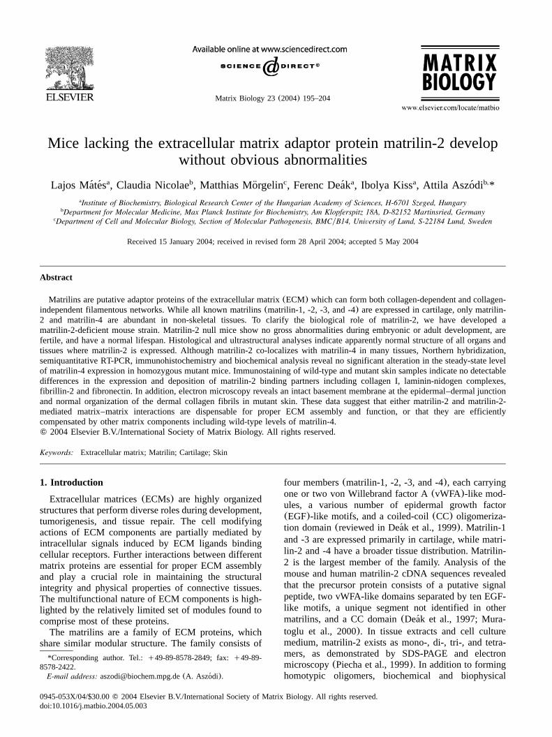
LacZ-neomycin cassette(Fig. 1a). Insertion of thetargeting construct into theMatn2 locus was designedto result in the deletion of the exon 2B containing thetranslation start site, the signal peptide and a positivelycharged region of the protein. Such a deletion waspredicted to abolish the synthesis of both matrilin-2mRNA and protein. Out of 360 ES cell clones survivingG418 selection, 18 correctly targeted ES cell cloneswere identified by Southern blot analysis ofBglII-digested genomic DNA(Fig. 1c) using an externalprobe to detect a 12-kb mutant fragment and a 9-kbwild-type fragment of 129ySv origin. Interestingly, ourprobe hybridized to a 6-kb wild-type band on C57BLy6 background indicating restriction enzyme polymor-phism between the 129ySv and C57BLy6 mouse strains(Fig. 1b). Chimeric males were generated using thestandard techniques and subsequently crossed withC57BLy6 and 129ySv females to produce outbred andinbred strains, respectively. Southern blot analysis of358 offspring from heterozygous intercrosses showedthe expected Mendelian ratio of genotypes(Fig. 1d).Northern blot analysis of total RNA from primaryfibroblasts isolated at embryonic day E14.5(E14.5)revealed a complete lack ofMatn2 mRNA in homozy-gous mutant samples(Fig. 1e).
2.2. Apparently normal embryonic development in theabsence of matrilin-2
Immunohistochemical analysis at E16.5 revealed thatall matrilins were expressed in the developing skeletalsystem in wild-type embryos(Fig. 2a). In mutantembryos, no matrilin-2 protein could be detected(Fig.2b). Analysis of control sections treated with collagenaseor protease XXIV before immunostaining showed theoverlapping distribution of matrilin-2 and matrilin-4 invarious non-skeletal dense and loose connective tissuesincluding those of tendons and ligaments(Fig. 2c,c’),dermis of the skin(Fig. 2d,d’), submucosa of theintestine (Fig. 2e,e’), valves of the heart(Fig. 2f,f’),basement membranes around the main bronchus andterminal bronchioli of the lung(Fig. 2g,g’), and in theperineurium of the spinal nerves(Fig. 2h,h’). Hematox-ylin-eosin (HE) staining of serial sagittal or crosssections at E14.5 and E16.5 showed an apparentlynormal tissue architecture in both outbred and inbredmutant embryos, which was indistinguishable from con-trol littermates (data not shown), indicating that theabsence of matrilin-2 plays do not interfere with normalembryonic development.
2.3. Endochondral bone formation and intervertebraldisc development in Matn2 null mice
Adult Matn2-deficient mice showed no obviousabnormalities, were fertile, and had a normal lifespan.Since matrilin-2 is expressed during endochondral bone
197L. Mates et al. / Matrix Biology 23 (2004) 195–204´ ´
Fig. 1. Targeted inactivation of the mouse gene encoding matrilin-2.(a) Partial map of wild-typeMatn2 allele, structure of targeting vector andrecombinant locus. E1 and E2 A, B indicate exons. The expected fragment sizes afterBglII digestion and hybridization with an external probeare 9 kb or 6 kb for wild-type allele, depending on the genetic background, and 12 kb for recombinant allele.(b) Southern blot analysis of wild-typeBglII-digested tail DNA with the external probe reveals restriction enzyme polymorphisms of theMatn2 gene between 129ySv and C57BLy6Jmouse strains.(c) Southern blot analysis of genomic DNA from ES cell clones shows the wild-type(9 kb) and the targeted(12 kb) alleles.(d)Southern blot analysis of mouse tail DNA derived from progeny of heterozygous mating of outbred(left panel) or inbred(right panel) mice.(e)Northern blot analysis of total RNA isolated from mouse embryonic fibroblasts of wild-type(qyq) and homozygous mutant(yyy) E14.5embryos. The blot was sequentially hybridized with probes specific for mouseMatn2 andGapdh.
formation (Fig. 2a; and in Segat et al., 2000; Klatt etal., 2002), we analyzed the development of axial andappendicular skeleton in more detail at various stages.Whole-skeleton staining with alcian blue(for cartilagi-nous tissues) and alizarin red(for bony tissues) atnewborn stage showed no significant size alteration orskeletal malformation in the mutant compared with thecontrol (Fig. 3a). X-ray analysis of 9-month-old micerevealed normal bone density and no abnormalities ofribs, vertebral bodies, or long bones in mutant mice(Fig. 3b). Using immunohistochemistry at E16.5, matri-lin-2 was detected in the perichondriumyperiosteum, atthe joint surface, and weekly in the proliferative andupper hypertrophic zones of the wild type growth plate(Fig. 3c). In contrast, matrilin-1, -3 and -4 were stronglydeposited throughout the cartilage. In addition, matrilin-4 expression was observed in the perichondriumyperi-osteum(Fig. 3c). The deposition of matrilin-1, -3, -4(Fig. 3c) and other cartilage-specific proteins, includingcollagen type II and aggrecan(not shown), were appar-ently unchanged in the matrilin-2-deficient cartilage.Northern blot analysis of newborn limb cartilage sam-ples revealed no obvious upregulation ofMatn2, -3 or -4 gene expression(data not shown). Histological
analysis of the epiphyseal and growth plate cartilagesduring long bone development at E16.5, newborn andvarious adult stages showed no detectable differencesbetween control and homozygous mutant mice(Fig. 3ddata not shown). Finally, safranin orange staining of thecartilage matrix for proteoglycans at 8 months of ageshowed no sign of degenerative changes at the articularsurfaces(Fig. 3e).Matrilin-2 displays strong expression during embry-
onic intervertebral disc(IVD) development(Piecha etal., 1999 and Fig. 2a) and in the annulus fibrosus andnucleus pulposus of adult IVDs(Deak et al., 1999).´Therefore, we examined IVD development on histolog-ical sections at various embryonic and adult stages. Noapparent differences between wild type and mutant IVDdevelopment were observed(Fig. 3f and data notshown).
2.4. Development of non-skeletal tissues in Matn2-deficient mice
Our previous studies revealed that matrilin-2 isexpressed in various adult non-skeletal mouse tissuesmainly associated with basement membrane-like

198 L. Mates et al. / Matrix Biology 23 (2004) 195–204´ ´
Fig. 2. Immunostaining of E16.5 mouse embryo.(a) All matrilins are expressed in the developing skeleton of a wild-type embryo.(b) Immu-nostaining shows the complete lack of matrilin-2 protein in a homozygous mutant embryo. The section was counterstained with hematoxylin.(c–h and c9–h9) Comparative immunostaining for matrilin-2 and matrilin-4 on collagenase-treated tissue sections of a E16.5 wild-type mouse embryo.Matrilin-2 and -4 are co-localized in tendons(tn) (c and c9), dermis(d) and epidermal–dermal junction of the skin(d and d9), submucosa(sm)of the intestine(e and e9), valves(v) of the heart(f and f9), basement membranes around the main bronchus(b) and terminal bronchioli(arrows)of the lung(g and g9), and spinal nerves(sn) (h and h9). Abbreviations: c, cartilage; e, epidermis; ep, epithelium.
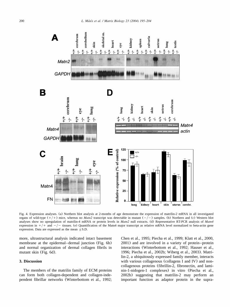
structures(Deak et al., 1997; Piecha et al., 1999). In´this study, the wide-ranging expression of theMatn2gene was further confirmed by Northern blot hybridi-zation indicating the presence of the matrilin-2 tran-scripts in all investigated organs(Fig. 4a). A carefulhistological analysis of different non-skeletal tissuessuch as brain, skin, kidney, heart, lung, intestine, uterus,spleen and eye, however, showed no obvious abnormal-ities in mutants compared with wild-type mice(data notshown). Similarly, electron microscopic analysis ofselected organs including kidney and lung revealed noultrastructural abnormalities in Matn2-deficient mice(data not shown).Since matrilin-4 shows a very similar expression
pattern to that of matrilin-2 in adult tissues, we investi-gated the possible upregulation of Matn4 in Matn2 nullmice. Northern and Western blot analyses of selectedtissues (brain, eye and lung) at 2 months of age
demonstrated that the expression levels of matrilin-4mRNA and protein in mutant mice were comparable tothose of control mice(Fig. 4b,c). To quantify moreprecisely the Matn4 mRNA level we employed semi-quantitative RT-PCR(Fig. 4d,e). No statistically signif-icant differences between wild-type and mutant micewere observed for Matn4 expression in any tissuesisolated from 2 weeks old animals.
2.5. The immunohistochemical distribution of the bind-ing partners of matrilin-2 is apparently normal in themutant skin
It has previously been shown that recombinantlyexpressed matrilin-2 binds various collagenous(colla-gens I and IV) and non-collagenous(fibronectin, lami-nin-1-nidogen-1 complex, fibrillin-2) components of theECM (Piecha et al., 2002b) and that matrilin-2, at least

199L. Mates et al. / Matrix Biology 23 (2004) 195–204´ ´
Fig. 3. Analysis of skeletal development in wild-type(qyq) and matrilin-2-deficient(yyy) mice. (a) No gross abnormalities are detectableby skeletal staining of newborn mice or by X-ray examination(b) of 9-month-old mice.(c) Hematoxilin-eosin(H&E) staining of E16.5 tibiasindicates no altered cartilage morphology. Immunostaining shows matrilin-2(Matn2) expression in the perichondriumyperiosteum, at the jointsurface and in the cartilage of wild-type, but not mutant tissues. The deposition of matrilin-1(Matn1), matrilin-3 (Matn3) and matrilin-4(Matn4)is comparable in wild-type and mutant cartilage.(d) H&E staining and Safranin orange staining(e) indicate no degeneration or altered proteoglycancontent of the articular cartilage(ac) at 8-months of age; gp, growth plate.(f). H&E staining shows normal intervertebral disc formation in 4-month-old mutant. Abbreviations: af, annulus fibrosus; np, nucleus pulposus.
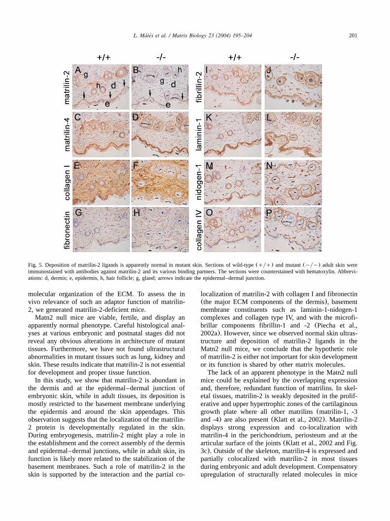
partially, co-localizes with collagens I and IV, fibronec-tin, and fibrillin-1 in the skin in vivo (Piecha et al.,2002a). These observations prompted us to compare thedistribution of these putative matrilin-2 binding ligandsin wild-type and mutant adult skin. In wild-type mice,matrilin-2 was strongly deposited at the epidermal–dermal junction, around hair follicles, vessels, and seba-ceous glands, whereas a very weak expression was ob-served in the upper part of the dermis(Fig. 5a). Matrilin-4 displayed similar tissue distribution with addition-
al strong expression in the whole epidermis and theupper dermis(Fig. 5c). Mutant skin lacked matrilin-2expression(Fig. 5b) and showed normal matrilin-4 imm-unostaining(Fig. 5d). The expression of the potentialligands for matrilin-2, like the basement membrane com-ponents laminin-1(Fig. 5k,l), nidogen-1(Fig. 5m,n),collagen IV (Fig. 5o,p), the microfibrillar constituentfibrillin-2 (Fig. 5i,j), and the major dermal ECM pro-teins collagen I and fibronectin, was also indistinguish-able between wild-type and mutant animals. Further-
200 L. Mates et al. / Matrix Biology 23 (2004) 195–204´ ´
Fig. 4. Expression analyses.(a) Northern blot analysis at 2-months of age demonstrate the expression of matrilin-2 mRNA in all investigatedorgans of wild-type(qyq) mice, whereas noMatn2 transcript was detectable in mutant(yyy) samples.(b) Northern and(c) Western blotanalyses show no upregulation of matrilin-4 mRNA or protein levels inMatn2 null extracts.(d) Representative RT-PCR analysis ofMatn4expression inqyq andyyy tissues.(e) Quantification of the Matn4 major transcript as relative mRNA level normalized to beta-actin geneexpression. Data are expressed as the mean"S.D.
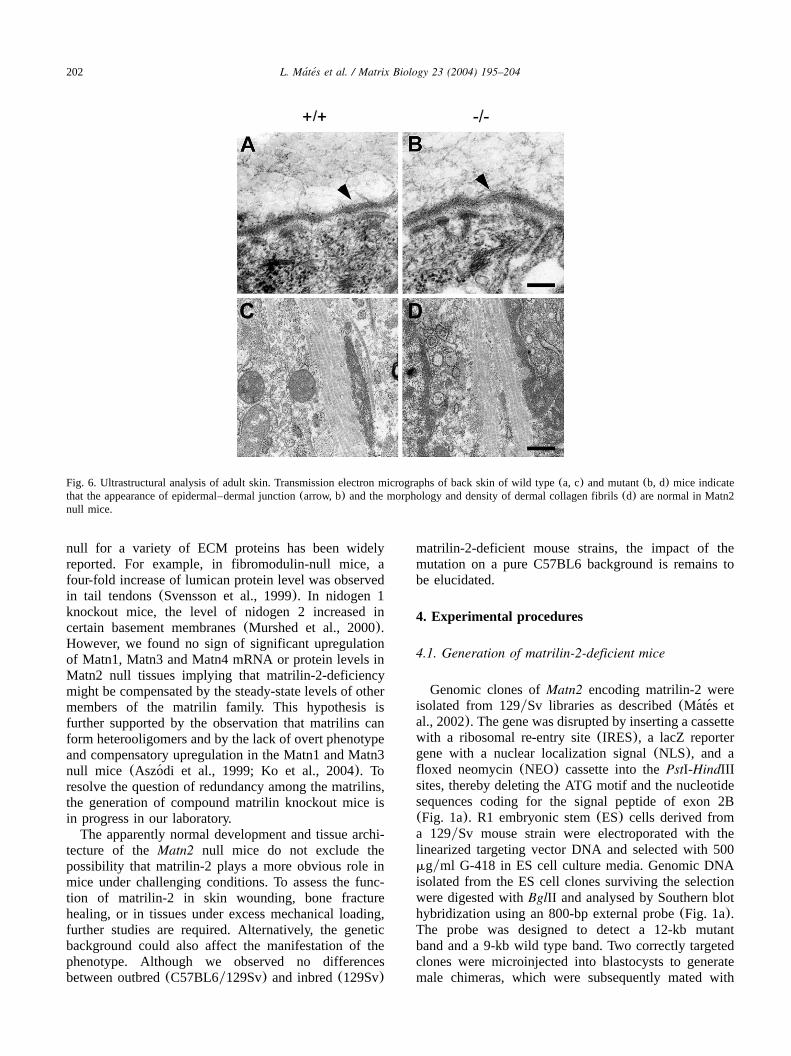
more, ultrastructural analysis indicated intact basementmembrane at the epidermal–dermal junction(Fig. 6b)and normal organization of dermal collagen fibrils inmutant skin(Fig. 6d).
3. Discussion
The members of the matrilin family of ECM proteinscan form both collagen-dependent and collagen-inde-pendent fibrillar networks(Winterbottom et al., 1992;
Chen et al., 1995; Piecha et al., 1999; Klatt et al., 2000,2001) and are involved in a variety of protein–proteininteractions(Winterbottom et al., 1992; Hauser et al.,1996; Piecha et al., 2002b; Wiberg et al., 2003). Matri-lin-2, a ubiquitously expressed family member, interactswith various collagenous(collagens I and IV) and non-collagenous proteins(fibrillin-2, fibronectin, and lami-nin-1-nidogen-1 complexes) in vitro (Piecha et al.,2002b) suggesting that matrilin-2 may perform animportant function as adaptor protein in the supra-
201L. Mates et al. / Matrix Biology 23 (2004) 195–204´ ´
Fig. 5. Deposition of matrilin-2 ligands is apparently normal in mutant skin. Sections of wild-type(qyq) and mutant(yyy) adult skin wereimmunostained with antibodies against matrilin-2 and its various binding partners. The sections were counterstained with hematoxylin. Abbrevi-ations: d, dermis; e, epidermis, h, hair follicle; g, gland; arrows indicate the epidermal–dermal junction.
molecular organization of the ECM. To assess the invivo relevance of such an adaptor function of matrilin-2, we generated matrilin-2-deficient mice.Matn2 null mice are viable, fertile, and display an
apparently normal phenotype. Careful histological anal-yses at various embryonic and postnatal stages did notreveal any obvious alterations in architecture of mutanttissues. Furthermore, we have not found ultrastructuralabnormalities in mutant tissues such as lung, kidney andskin. These results indicate that matrilin-2 is not essentialfor development and proper tissue function.In this study, we show that matrilin-2 is abundant in
the dermis and at the epidermal–dermal junction ofembryonic skin, while in adult tissues, its deposition ismostly restricted to the basement membrane underlyingthe epidermis and around the skin appendages. Thisobservation suggests that the localization of the matrilin-2 protein is developmentally regulated in the skin.During embryogenesis, matrilin-2 might play a role inthe establishment and the correct assembly of the dermisand epidermal–dermal junctions, while in adult skin, itsfunction is likely more related to the stabilization of thebasement membranes. Such a role of matrilin-2 in theskin is supported by the interaction and the partial co-
localization of matrilin-2 with collagen I and fibronectin(the major ECM components of the dermis), basementmembrane constituents such as laminin-1-nidogen-1complexes and collagen type IV, and with the microfi-brillar components fibrillin-1 and -2(Piecha et al.,2002a). However, since we observed normal skin ultras-tructure and deposition of matrilin-2 ligands in theMatn2 null mice, we conclude that the hypothetic roleof matrilin-2 is either not important for skin developmentor its function is shared by other matrix molecules.The lack of an apparent phenotype in the Matn2 null
mice could be explained by the overlapping expressionand, therefore, redundant function of matrilins. In skel-etal tissues, matrilin-2 is weakly deposited in the prolif-erative and upper hypertrophic zones of the cartilaginousgrowth plate where all other matrilins(matrilin-1, -3and -4) are also present(Klatt et al., 2002). Matrilin-2displays strong expression and co-localization withmatrilin-4 in the perichondrium, periosteum and at thearticular surface of the joints(Klatt et al., 2002 and Fig.3c). Outside of the skeleton, matrilin-4 is expressed andpartially colocalized with matrilin-2 in most tissuesduring embryonic and adult development. Compensatoryupregulation of structurally related molecules in mice
202 L. Mates et al. / Matrix Biology 23 (2004) 195–204´ ´
Fig. 6. Ultrastructural analysis of adult skin. Transmission electron micrographs of back skin of wild type(a, c) and mutant(b, d) mice indicatethat the appearance of epidermal–dermal junction(arrow, b) and the morphology and density of dermal collagen fibrils(d) are normal in Matn2null mice.
null for a variety of ECM proteins has been widelyreported. For example, in fibromodulin-null mice, afour-fold increase of lumican protein level was observedin tail tendons(Svensson et al., 1999). In nidogen 1knockout mice, the level of nidogen 2 increased incertain basement membranes(Murshed et al., 2000).However, we found no sign of significant upregulationof Matn1, Matn3 and Matn4 mRNA or protein levels inMatn2 null tissues implying that matrilin-2-deficiencymight be compensated by the steady-state levels of othermembers of the matrilin family. This hypothesis isfurther supported by the observation that matrilins canform heterooligomers and by the lack of overt phenotypeand compensatory upregulation in the Matn1 and Matn3null mice (Aszodi et al., 1999; Ko et al., 2004). To´resolve the question of redundancy among the matrilins,the generation of compound matrilin knockout mice isin progress in our laboratory.The apparently normal development and tissue archi-
tecture of theMatn2 null mice do not exclude thepossibility that matrilin-2 plays a more obvious role inmice under challenging conditions. To assess the func-tion of matrilin-2 in skin wounding, bone fracturehealing, or in tissues under excess mechanical loading,further studies are required. Alternatively, the geneticbackground could also affect the manifestation of thephenotype. Although we observed no differencesbetween outbred(C57BL6y129Sv) and inbred(129Sv)
matrilin-2-deficient mouse strains, the impact of themutation on a pure C57BL6 background is remains tobe elucidated.
4. Experimental procedures
4.1. Generation of matrilin-2-deficient mice
Genomic clones ofMatn2 encoding matrilin-2 wereisolated from 129ySv libraries as described(Mates et´ ´al., 2002). The gene was disrupted by inserting a cassettewith a ribosomal re-entry site(IRES), a lacZ reportergene with a nuclear localization signal(NLS), and afloxed neomycin(NEO) cassette into thePstI-HindIIIsites, thereby deleting the ATG motif and the nucleotidesequences coding for the signal peptide of exon 2B(Fig. 1a). R1 embryonic stem(ES) cells derived froma 129ySv mouse strain were electroporated with thelinearized targeting vector DNA and selected with 500mgyml G-418 in ES cell culture media. Genomic DNAisolated from the ES cell clones surviving the selectionwere digested withBglII and analysed by Southern blothybridization using an 800-bp external probe(Fig. 1a).The probe was designed to detect a 12-kb mutantband and a 9-kb wild type band. Two correctly targetedclones were microinjected into blastocysts to generatemale chimeras, which were subsequently mated with

203L. Mates et al. / Matrix Biology 23 (2004) 195–204´ ´
C57BLy6 and 129ySv females to establish outbred andinbredMatn2-null mice, respectively.
4.2. RNA analysis
Total RNA was isolated from various adult organsand embryonic primary fibroblasts as described previ-ously (Aszodi et al., 1996). For Northern analysis, 5–´20 mg of total RNA was size fractionated on a 1%agarose-2.2 M formaldehyde gel and transferred toHybond Nq membrane (Amersham). Filters werehybridized with P -labeled cDNA probes specific for32
matrilin-1, -2, -3(Aszodi et al., 1999), matrilin-4(Wag-´ener et al., 1998) and glyceraldehyde phosphodehydro-genase. For semiquantitative RT-PCR, two microgramsof total RNA were transcribed into cDNA using theSuperScript� III RNaseH reverse transcriptase(Invitro--
gen). For PCR reactions, 0.5 unit AmpliTaq DNApolymerase(Invitrogen) and 2ml cDNA were used in50 ml amplification buffer containing 0.2 mM dNTPs,1.5 mM MgCl and 200mM each of forward and2
reverse primers. The primer pairs specific for exon 0bof the mouseMatn4 gene were described previously(Wagener et al., 2001). The primer pairs specific forbeta-actin were: 59-TGACGGGGTCACCCACAC-39(forward) and 59-CTAGAAGCATTTGCGGTG-GAA-39(reverse). PCR was performed using the PTC-200 Pel-tier Thermal Cycler(MJ Research) under the followingconditions: 948C for 4 min, 948C for 1 min, 608C for1 min and 728C for 1 min, 35 cycles forMatn4 and20 cycles for beta-actin, followed by 728C for 10 min.PCR reactions were repeated three times and relativemRNA levels were determined by optical densitometryusing the Bio-Profile computer-assisted imaging system(Vilber Lourmat).
4.3. Histology and immunohistochemistry of embryonicand adult non-skeletal tissues
The histological and immunohistochemical analyseswere conducted on both C57BL6y129Sv and 129ySvgenetic backgrounds. For histology, 14.5-, 15.5-, and16.5-day-old embryos and various organs form 1-, 2-,and 4-month-old mice were fixed in 4% paraformalde-hyde(PFA) in phosphate-buffered saline(PBS, pH 7.2).After paraffin embedding, sections of 6mm were cutand stained with hematoxylin-eosin(H&E). For eachstages, at least three wild-type and mutant samples wereanalyzed using serial sections representing the embry-onic or adult tissues.For immunohistochemistry, samples were fixed in
95% ethanol-5% glacial acetic acid and embedded inparaffin. Immunostaining was performed by the avidin-biotin complex(ABC) procedure using a commerciallyavailable kit(Vectastain). To improve antibody penetra-tion to the tissue, sections were routinely digested before
immunostaining with protease XXIV(10mgyml in PBS,Sigma) for 20 min or with collagenase(5 mgyml,collagenase type II, Worthington) for 5 min at 378C.Rabbit polyclonal antibodies against the following anti-gens were used: matrilin-1, matrilin-2, matrilin-3, matri-lin-4 (see Ko et al., 2004, and references therein);collagen I, collagen IV, laminin-1, nidogen-1(all kindlyprovided by Dr Rupert Timpl, MPI, Martinsried, Ger-many) and fibrillin-2 (kind gift from Dr Dieter P.Reinhard, University of Lubeck, Lubeck, Germany).¨ ¨
4.4. Skeletal analysis
Skeletal staining of newborn mice with alcian blueyalizarin red and X-ray analysis of adult mice wereperformed as previously described(Aszodi et al., 1999).´For histology, adult bones were fixed in 4% PFA,decalcified in 10% EDTA and stained with H&E orSafranin Orange-Weigert hematoxylin. Immunostainingof skeletal tissues with antibodies against matrilin-1, -2,-3 and -4, collagen type II and aggrecan was performedas previously described(Aszodi et al., 1999).´
4.5. Ultrastructural analysis
For electron microscopy, tissue samples were fixed in0.15 M sodium cacodylate buffer, pH 7.4, containing2.5% glutaraldehyde for 1 day at room temperature.They were subsequently rinsed three times in isotonicsodium cacodylate buffer for 30 min and postfixed in0.15 M sodium cacodylate, pH 7.4, containing 1%(wtyvol) osmium tetroxide for 2 h. Samples were then rinsedin isotonic buffer solution, dehydrated in a graded seriesof ethanol, and embedded in Epon 812. Semithin(1mm) and thin (60 nm) sections were cut on a LeicaUltracut S(Deerfield, IL). Sections were stained in 5%uranyl acetate for 2 h and then in a saturated lead citratesolution for 7 min. All samples were viewed in aJeol1200 EX transmission electron microscope operatedat 60-kV accelerating voltage.
4.6. Biochemical analysis
Various organs from 1- and 3-month-old mice werehomogenised in buffer containing 0.25 M NaCl, 50 mMTris–HCl (pH 7.4) and 10 mM EDTA and extractedwith gentle agitation for 1.5 h at 378C after the additionof 2 mgyml collagenase type II. Unextracted materialwas removed by centrifugation at 8000=g for 10 minat 4 8C. Nonreducing SDS-polyacrylamide gel electro-phoresis sample buffer(5x) was added to the samplesand the proteins were separated on 7% SDS-polyacryl-amide gels then blotted onto Trans-Blot nitrocellulosemembranes(Bio-Rad). The blots were subsequentlyhybridized with polyclonal antibodies specific for matri-lin-2, -4 and tubulin or fibronectin as a control for
204 L. Mates et al. / Matrix Biology 23 (2004) 195–204´ ´
protein loading. Bound antibodies were hybridized tohorseradish peroxidase-conjugated swine anti-rabbitimmunoglobulin G (Sigma) and detected using theenhanced chemiluminescence(ECL) kit (Amersham).
Acknowledgments
We thank C. Cramnert, K. Sakai, A. Simon, I. Kravjar,´I. Fekete and Zs. Farkas for technical assistance, Dr Eva´
Kemeny, Dr. Kathryn Rodgers for reading the manu-´script, and Dr Reinhard Fassler(MPI, Dept. of Molec-¨ular Medicine) for supporting the project. This studywas supported by grants from OTKA T022224,T029142, T034399 and T034729 from the HungarianNational Scientific Research Foundation to I.K. andF.D.; by the National Research and Development Pro-gram NKFP-1Ay0023y2002 to I. K.; by the Max-PlanckSociety, the Deutsche Forschungsgemeinschaft(AS150y1-1; 436 UNG 113y154y2-1), the Swedish MedicalResearch Council, the Anna-Greta Crafoords Founda-tion, the Greta and Johan Kocks Foundation to A.A.;and by a short term fellowship from EMBO to L.M.
References
Aszodi, A., Hauser, N., Studer, D., Paulsson, M., Hiripi, L., Bosze,´ ¨Z., 1996. Cloning, sequencing and expression analysis of mousecartilage matrix protein cDNA. Eur. J. Biochem. 236, 970–977.
Aszodi, A., Bateman, J.F., Hirsch, E., Baranyi, M., Hunziker, E.B.,´Hauser, N., et al., 1999. Normal skeletal development of micelacking matrilin 1: redundant function of matrilins in cartilage?Mol. Cell Biol. 19, 7841–7845.
Chapman, K.L., Mortier, G.R., Chapman, K., Loughlin, J., Grant,M.E., Briggs, M.D., 2001. Mutations in the region encoding thevon Willebrand factor A domain of matrilin-3 are associated withmultiple epiphyseal dysplasia. Nat. Genet. 28, 393–396.
Chen, Q., Johnson, D.M., Haudenschild, D.R., Tondravi, M.M.,Goetinck, P.F., 1995. Cartilage matrix protein forms a type IIcollagen-independent filamentous network: analysis in primary cellcultures with a retrovirus expression system. Mol. Biol. Cell 6,1743–1753.
Deak, F., Piecha, D., Bachrati, C., Paulsson, M., Kiss, I., 1997.´Primary structure and expression of matrilin-2, the closest relativeof cartilage matrix protein within the von Willebrand factor typeA module superfamily. J. Biol. Chem. 272, 9268–9274.
Deak, F., Wagener, R., Kiss, I., Paulsson, M., 1999. The matrilins: a´novel family of oligomeric extracellular matrix proteins. MatrixBiol. 18, 55–66.
Frank, S., Schulthess, T., Landwehr, R., Lustig, A., Mini, T., Jeno,¨P., et al., 2002. Characterization of the matrilin coiled-coil domainsreceals seven novel isoforms. J. Biol. Chem. 277, 19071–19079.
Hauser, N., Paulsson, M., Heinegard, D., Morgelin, M., 1996. Inter-¨˚action of cartilage matrix protein with aggrecan: increased covalentcross-linking with tissue maturation. J. Biol. Chem. 271,32247–32252.
Huang, X., Birk, D.E., Goetinck, P.F., 1999. Mice lacking matrilin-1(cartilage matrix protein) have alterations in type II collagenfibrillogenesis and fibril organization. Dev. Dynam. 216, 434–441.
Klatt, A.R., Nitsche, P.D., Kobbe, B., Morgelin, M., Paulsson, M.,¨Wagener, R., 2000. Molecular structure and tissue distribution of
matrilin-3, a filament-forming extracellular matrix proteinexpressed during skeletal development. J. Biol. Chem. 275,3999–4006.
Klatt, A.R., Nitsche, D.P., Kobbe, B., Macht, M., Paulsson, M.,Wagener, R., 2001. Molecular structure, processing and tissuedistribution of matrilin-4. J. Biol. Chem. 276, 17267–17275.
Klatt, A.R., Paulsson, M., Wagener, R., 2002. Expression of matrilinsduring maturation of mouse skeletal tissues. Matrix Biol. 21,289–296.
Ko, Y., Kobbe, B., Nicolae, C., Miosge, N., Paulsson, M., Wagener,R., et al., 2004. Matrilin-3 is dispensable for mouse skeletal growthand development. Mol. Cell Biol. 24, 1691–1699.
Mates, L., Korpos, E., Deak, F., Liu, Z., Beier, D.R., Aszodi, A., et´´ ´ ´ ´al., 2002. Comparative analysis of the mouse and human genes(Matn2 and MATN2) for Matrilin-2, a filament-forming proteinwidely distributed in extracellular matrices. Matrix Biol. 21,163–174.
Mostert, A.K., Dijkstra, P.F., Jansen, B.R.H., van Horn, J.R., de Graaf,B., Heutink, P., et al., 2003. Familial multiple epiphyseal dysplasiadue to a matrilin-3 mutation: Further delineation of the phenotypeincluding 40 years follow-up. Am. J. Med. Genet. 120A, 490–497.
Muratoglu, S., Krysan, K., Balazs, M., Sheng, H., Zakany, R., Modis,´ ´ ´ ´L., et al., 2000. Primary structure of human matrilin-2, chromosomelocation of theMATN2 gene and conservation of an AT-AC intronin matrilin genes. Cytogenet. Cell Genet. 90, 323–327.
Murshed, M., Smyth, N., Miosge, N., Karolat, J., Krieg, T., Paulsson,M., et al., 2000. The absence of nidogen 1 does not affect murinebasement membrane formation. Mol. Cell Biol. 20, 7007–7012.
Piecha, D., Muratoglu, S., Morgelin, M., Hauser, N., Studer, D., Kiss,¨I., et al., 1999. Matrilin-2, a large, oligomeric matrix protein, isexpressed by a great variety of cells and forms fibrillar networks.J. Biol. Chem. 274, 13353–13361.
Piecha, D., Hartmannm, K., Kobbe, B., Haase, I., Mauch, C., Krieg,T., et al., 2002a. Expression of matrilin-2 in human skin. J. Invest.Dermatol. 119, 38–43.
Piecha, D., Wiberg, C., Morgelin, M., Reinhardt, D.P., Deak, F.,¨ ´Maurer, P., et al., 2002b. Matrilin-2 interacts with itself and withother extracellular matrix proteins. Biochem. J. 367, 715–721.
Segat, D., Nitsche, P.D., Klatt, A.R., Piecha, D., Korpos, E., Deak,´F., et al., 2000. Expression of matrilin-1, -2 and -3 in developingmouse limbs and heart. Matrix Biol. 19, 649–655.
Stefansson, S.E., Jonsson, H., Ingvarsson, T., Manolescu, I., Jonsson,H.H., Olafsdottir, G., et al., 2003. Genomewide scan for handosteoarthritis: a novel mutation in matrilin-3. Am. J. Hum. Genet.72, 1448–1459.
Svensson, L., Aszodi, A., Reinholt, F.P., Fassler, R., Heinegard, D.,´ ¨ ˚Oldberg, A., 1999. Fibromodulin-null mice have abnormal collagenfibrils, tissue organization, and altered lumican deposition intendon. J. Biol. Chem. 274, 9636–9647.
Wagener, R., Kobbe, B., Paulsson, M., 1998. Matrilin-4, a newmember of the matrilin family of extracellular matrix proteins.FEBS Lett. 436, 123–127.
Wagener, R., Kobbe, B., Aszodi, A., Aeschlimann, D., Paulsson, M.,´2001. Characterization of the marse Matrilin-4 gene: a 59antiparalleloverlap with the gene encoding the transcription factor RBP-L.Genomics 76, 89–98.
Wiberg, C., Klatt, A.R., Wagener, R., Paulsson, M., Bateman, J.F.,Heinegard, D., et al., 2003. Complexes of matrilin-1 and biglycan˚or decorin connect collagen VI microfibrils to both collagen II andaggrecan. J. Biol. Chem. 278, 37698–37704.
Winterbottom, N., Tondravi, M.M., Harrington, T.L., Klier, F.G.,Vertel, B.M., Goetinck, P.F., 1992. Cartilage matrix protein is acomponent of the collagen fibril of cartilage. Dev. Dynam. 193,266–276.
Related Documents