Mice Lacking D 5 Dopamine Receptors Have Increased Sympathetic Tone and Are Hypertensive Tom R. Hollon, 1 * Martin J. Bek, 3 * Jean E. Lachowicz, 4 Marjorie A. Ariano, 5 Eva Mezey, 2 Ramesh Ramachandran, 6 Scott R. Wersinger, 6 Patricio Soares-da-Silva, 7 Zhi Fang Liu, 1 Alexander Grinberg, 8 John Drago, 8 W. Scott Young III, 6 Heiner Westphal, 8 Pedro A. Jose, 3 and David R. Sibley 1 1 Molecular Neuropharmacology Section and 2 Basic Neurosciences Program, National Institute of Neurological Disorders and Stroke, National Institutes of Health, Bethesda, Maryland 20892-1406, 3 Department of Pediatrics, Georgetown University Medical Center, Washington, DC 20007, 4 CNS/Cardiovascular Research, Schering-Plough Research Institute, Kenilworth, New Jersey 07033, 5 Department of Neuroscience, The Chicago Medical School, North Chicago, Illinois 60064, 6 Section on Neural Gene Expression, National Institute of Mental Health, Bethesda, Maryland 20892, 7 Institute of Pharmacology and Therapeutics, Faculty of Medicine of Porto, Porto, Portugal, and 8 Laboratory of Mammalian Genes and Development, National Institute of Child Health and Human Development, National Institutes of Health, Bethesda, Maryland 20892 Dopamine is an important transmitter in the CNS and PNS, critically regulating numerous neuropsychiatric and physiolog- ical functions. These actions of dopamine are mediated by five distinct receptor subtypes. Of these receptors, probably the least understood in terms of physiological functions is the D 5 receptor subtype. To better understand the role of the D 5 dopamine receptor (DAR) in normal physiology and behavior, we have now used gene-targeting technology to create mice that lack this receptor subtype. We find that the D 5 receptor- deficient mice are viable and fertile and appear to develop normally. No compensatory alterations in other dopamine re- ceptor subtypes were observed. We find, however, that the mutant mice develop hypertension and exhibit significantly elevated blood pressure (BP) by 3 months of age. This hyper- tension appears to be caused by increased sympathetic tone, primarily attributable to a CNS defect. Our data further suggest that this defect involves an oxytocin-dependent sensitization of V 1 vasopressin and non-NMDA glutamatergic receptor- mediated pathways, potentially within the medulla, leading to increased sympathetic outflow. These results indicate that D 5 dopamine receptors modulate neuronal pathways regulating blood pressure responses and may provide new insights into mechanisms for some forms of essential hypertension in hu- mans, a disease that afflicts up to 25% of the aged adult population in industrialized societies. Key words: D 5 receptor; gene knock-out; hypertension; sym- pathetic tone; oxytocin; vasopressin Dopamine is an important neurotransmitter in the brain as well as the periphery and plays a critical role in regulating numerous locomotor, neuroendocrine, cognitive, and emotional f unctions. Dysregulation of dopaminergic systems has also been hypothe- sized to underlie several neuropsychiatric and endocrine disor- ders, including Parkinson’s disease, schizophrenia, Tourette’s syndrome, and hyperprolactinemia. The actions of dopamine are mediated by five distinct receptor subtypes that belong to the G-protein-coupled receptor super-family and are divided into two major subgroups, D 1 -like and D 2 -like, the basis of their structure and pharmacology (Neve and Neve, 1997). The D 1 -like subfamily consists of the D 1 and D 5 subtypes (also called D 1A and D 1B , respectively), both of which transduce their signals by in- creasing intracellular cAMP levels. The D 2 -like subfamily con- sists of the D 2 ,D 3 , and D 4 receptors, all of which can diminish cAMP production as well as regulate the activity of various ion channels. Although the D 1 -like and D 2 -like subfamilies can be differentiated pharmacologically, it is difficult to discriminate between receptors within each subfamily using selective ligands. This has led to uncertainties in ascribing specific physiological and behavioral functions to individual receptor subtypes. Inves- tigators have approached this issue, in part, by creating geneti- cally altered animals that lack individual receptor subtypes. Thus far, mice lacking D 1 ,D 2 ,D 3 , or D 4 receptors have been produced, all of which have exhibited informative phenotypes (Sibley, 1999; Glickstein and Schmauss, 2001). This has resulted in the elucida- tion of receptor functions that could not have been obtained through other means such as the predominant presynaptic and postsynaptic roles of the D 2S and D 2L receptor isoforms, respec- tively (Usiello et al., 2000). The D 5 dopamine receptor (DAR) has generated significant interest because of its relatively high affinity for dopamine, com- pared with other DARs, and its purported constitutive activity (Sunahara et al., 1991; Tiberi and Caron, 1994). This has sug- gested that the D 5 DAR may be activated in the absence or presence of low concentrations of endogenous agonist. Although the D 5 DAR is f unctionally coupled to the activation of adenylate Received May 31, 2002; revised Sept. 19, 2002; accepted Sept. 20, 2002. This work was partially supported by Department of Defense Grant 17-99-1-9542 to M.A.A. We thank Dr. Dong Jiang, Dr. Laureano D. Asico, David Cabrera, Sing Ping Huang, and Binu Tharakan for their assistance. *T.R.H. and M.J.B. contributed equally to this work. Correspondence should be addressed to Dr. David R. Sibley, Molecular Neuro- pharmacology Section, National Institute of Neurological Disorders and Stroke/ National Institutes of Health, Building 10, Room 5C108, 10 Center Drive, MSC 1406, Bethesda, MD 20892-1406. E-mail: [email protected]. J. Drago’s present address: Neurosciences Group, Monash University Department of Medicine, Monash Medical Centre, Clayton, Victoria, 3168, Australia. M. J. Bek’s present address: Department of Internal Medicine, Freiburg Univer- sity Medical School, Freiburg, Germany. Copyright © 2002 Society for Neuroscience 0270-6474/02/2210801-10$15.00/0 The Journal of Neuroscience, December 15, 2002, 22(24):10801–10810

Welcome message from author
This document is posted to help you gain knowledge. Please leave a comment to let me know what you think about it! Share it to your friends and learn new things together.
Transcript

Mice Lacking D5 Dopamine Receptors Have Increased SympatheticTone and Are Hypertensive
Tom R. Hollon,1* Martin J. Bek,3* Jean E. Lachowicz,4 Marjorie A. Ariano,5 Eva Mezey,2Ramesh Ramachandran,6 Scott R. Wersinger,6 Patricio Soares-da-Silva,7 Zhi Fang Liu,1 Alexander Grinberg,8John Drago,8 W. Scott Young III,6 Heiner Westphal,8 Pedro A. Jose,3 and David R. Sibley1
1Molecular Neuropharmacology Section and 2Basic Neurosciences Program, National Institute of Neurological Disordersand Stroke, National Institutes of Health, Bethesda, Maryland 20892-1406, 3Department of Pediatrics, GeorgetownUniversity Medical Center, Washington, DC 20007, 4CNS/Cardiovascular Research, Schering-Plough Research Institute,Kenilworth, New Jersey 07033, 5Department of Neuroscience, The Chicago Medical School, North Chicago, Illinois60064, 6Section on Neural Gene Expression, National Institute of Mental Health, Bethesda, Maryland 20892, 7Institute ofPharmacology and Therapeutics, Faculty of Medicine of Porto, Porto, Portugal, and 8Laboratory of Mammalian Genesand Development, National Institute of Child Health and Human Development, National Institutes of Health, Bethesda,Maryland 20892
Dopamine is an important transmitter in the CNS and PNS,critically regulating numerous neuropsychiatric and physiolog-ical functions. These actions of dopamine are mediated by fivedistinct receptor subtypes. Of these receptors, probably theleast understood in terms of physiological functions is the D5
receptor subtype. To better understand the role of the D5
dopamine receptor (DAR) in normal physiology and behavior,we have now used gene-targeting technology to create micethat lack this receptor subtype. We find that the D5 receptor-deficient mice are viable and fertile and appear to developnormally. No compensatory alterations in other dopamine re-ceptor subtypes were observed. We find, however, that themutant mice develop hypertension and exhibit significantlyelevated blood pressure (BP) by 3 months of age. This hyper-
tension appears to be caused by increased sympathetic tone,primarily attributable to a CNS defect. Our data further suggestthat this defect involves an oxytocin-dependent sensitization ofV1 vasopressin and non-NMDA glutamatergic receptor-mediated pathways, potentially within the medulla, leading toincreased sympathetic outflow. These results indicate that D5
dopamine receptors modulate neuronal pathways regulatingblood pressure responses and may provide new insights intomechanisms for some forms of essential hypertension in hu-mans, a disease that afflicts up to 25% of the aged adultpopulation in industrialized societies.
Key words: D5 receptor; gene knock-out; hypertension; sym-pathetic tone; oxytocin; vasopressin
Dopamine is an important neurotransmitter in the brain as well asthe periphery and plays a critical role in regulating numerouslocomotor, neuroendocrine, cognitive, and emotional functions.Dysregulation of dopaminergic systems has also been hypothe-sized to underlie several neuropsychiatric and endocrine disor-ders, including Parkinson’s disease, schizophrenia, Tourette’ssyndrome, and hyperprolactinemia. The actions of dopamine aremediated by five distinct receptor subtypes that belong to theG-protein-coupled receptor super-family and are divided intotwo major subgroups, D1-like and D2-like, the basis of theirstructure and pharmacology (Neve and Neve, 1997). The D1-likesubfamily consists of the D1 and D5 subtypes (also called D1A andD1B, respectively), both of which transduce their signals by in-
creasing intracellular cAMP levels. The D2-like subfamily con-sists of the D2, D3, and D4 receptors, all of which can diminishcAMP production as well as regulate the activity of various ionchannels. Although the D1-like and D2-like subfamilies can bedifferentiated pharmacologically, it is difficult to discriminatebetween receptors within each subfamily using selective ligands.This has led to uncertainties in ascribing specific physiologicaland behavioral functions to individual receptor subtypes. Inves-tigators have approached this issue, in part, by creating geneti-cally altered animals that lack individual receptor subtypes. Thusfar, mice lacking D1, D2, D3, or D4 receptors have been produced,all of which have exhibited informative phenotypes (Sibley, 1999;Glickstein and Schmauss, 2001). This has resulted in the elucida-tion of receptor functions that could not have been obtainedthrough other means such as the predominant presynaptic andpostsynaptic roles of the D2S and D2L receptor isoforms, respec-tively (Usiello et al., 2000).
The D5 dopamine receptor (DAR) has generated significantinterest because of its relatively high affinity for dopamine, com-pared with other DARs, and its purported constitutive activity(Sunahara et al., 1991; Tiberi and Caron, 1994). This has sug-gested that the D5 DAR may be activated in the absence orpresence of low concentrations of endogenous agonist. Althoughthe D5 DAR is functionally coupled to the activation of adenylate
Received May 31, 2002; revised Sept. 19, 2002; accepted Sept. 20, 2002.This work was partially supported by Department of Defense Grant 17-99-1-9542
to M.A.A. We thank Dr. Dong Jiang, Dr. Laureano D. Asico, David Cabrera, SingPing Huang, and Binu Tharakan for their assistance.
*T.R.H. and M.J.B. contributed equally to this work.Correspondence should be addressed to Dr. David R. Sibley, Molecular Neuro-
pharmacology Section, National Institute of Neurological Disorders and Stroke/National Institutes of Health, Building 10, Room 5C108, 10 Center Drive, MSC1406, Bethesda, MD 20892-1406. E-mail: [email protected].
J. Drago’s present address: Neurosciences Group, Monash University Departmentof Medicine, Monash Medical Centre, Clayton, Victoria, 3168, Australia.
M. J. Bek’s present address: Department of Internal Medicine, Freiburg Univer-sity Medical School, Freiburg, Germany.Copyright © 2002 Society for Neuroscience 0270-6474/02/2210801-10$15.00/0
The Journal of Neuroscience, December 15, 2002, 22(24):10801–10810

cyclase, recent studies suggest that the D5 DAR may also modu-late GABAA receptor-mediated activity through both secondmessenger cascades (Yan and Surmeier, 1997) as well as throughdirect receptor–receptor interactions (Liu et al., 2000). Localiza-tion of the D5 DAR in the brain has revealed a widespreaddistribution, with the highest expression in the cerebral cortex,hippocampus, and basal ganglia (Ariano et al., 1997; Ciliax et al.,2000). Interestingly, recent reports have suggested a possibleassociation of the D5 DAR gene with schizophrenia (Muir et al.,2001) or substance abuse (Vanyukov et al., 1998). D5 DARs arealso expressed in the hypothalamus, where they may regulatecircadian rhythms (Rivkees and Lachowicz, 1997) and femalesexual behaviors (Apostolakis et al., 1996a,b). Within the periph-ery, D5 DARs have been found in adrenal tissue (Dahmer andSenogles, 1996), kidney (Sanada et al., 2000), and also the gas-trointestinal tract, where they may exert a protective effect on theintestinal mucosa (Mezey et al., 1996). To further elucidate thephysiological roles of the D5 DAR, we have now used gene-targeting technology to generate mice lacking functional D5
DARs.
MATERIALS AND METHODSConstruction of targeting vector and gene targeting. The targeting construct“pD5KO” contained a 9.6 kb genomic fragment of the mouse D5 receptorgene isolated from a 129/Sv genomic library. This genomic sequence wassubcloned as an EcoRV–NheI fragment into the gene targeting vectorpPNT (Tybulewicz et al., 1991). Through a number of intermediatesubcloning steps, a neomycin resistance gene was ligated, in reverseorientation, at the unique SfiI site of the D5 receptor gene, thus disrupt-ing the reading frame within the coding region. The length of D5receptor genomic DNA flanking the neomycin gene in the targetingvector was 6.9 kb (3�) and 2.7 kb (5�). The targeting vector pD5KO wassubsequently electroporated into the J1 line (Li et al., 1992) of embryonicstem (ES) cells, and simultaneous G418 positive selection and gancyclo-vir negative selection were used to enrich for ES cell colonies withsuccessful gene targeting. Southern hybridization analysis was used toexamine DNA from ES colonies for gene-targeting events. Homologousrecombination at the 5� end of the targeting construct was detected bydigesting ES cell genomic DNA with NcoI and hybridizing the Southernblot with probe A (see Results). The normal allele was 6 kb in length, andthe recombinant allele was 3.4 kb. Probe B (see Results) was hybridizedto Southern blots of ES genomic DNA cut with KpnI to detect homolo-gous recombination at the 3� end. The normal allele was 28 kb, and thelength of the recombinant allele was 18 kb. A hybridization probe,derived from the Neo gene, detected a single 3.4 kb band on NcoISouthern blots for those ES colonies in which gene targeting was suc-cessful; bands different in length from 3.4 kb on these blots indicatedrandom integration of one of more copies of the targeting vector into theES cell genome.
Mutant mouse generation and genotyping. C57BL/6 blastocysts wereinjected with ES cells from five different recombinant ES colonies andimplanted into foster mothers. The 129/Sv–C57BL/6 chimeric offspringproduced by blastocyst injection were bred with C57BL/6 mice to passthe recombinant D5 receptor allele from the germline of these chimerasto an F1 generation. Southern blots of NcoI- or KpnI-digested genomicDNA from mouse tail biopsies, hybridized with probes A or B, respec-tively, were used to detect germline passage of the recombinant D5receptor allele in the F1 generation mice. Homozygous mutant (�/�)and wild-type (�/�) mice were generated from heterozygous mouseintermatings. In the later stages of this study, the mice were genotypedusing a PCR-based method involving amplification from mouse genomicDNA isolated from tail biopsies. Oligonucleotide primers were designedto flank the SfiI restriction site into which the neomycin cassette wasligated. Primer 1 (5�-ACTCTCTTAATCGTCTGGACCTTG-3�) andprimer 2 (5�-TCGCAGGCTGGGGTCAGGTTCGCA-3�) were used toamplify the wild-type allele, whereas primer 3 (5�-TGATCAACTAG-TGCCCGGGCGGTA-3�), which was unique to the neomycin cassette,was used with primer 1 to amplify the recombinant allele. The PCRreaction used 0.2 �g of genomic tail DNA in a 50 �l reaction (50 mM KCl,10 mM Tris-HCl, 1.5 mM MgCl2, 0.2 mM dNTP, 0.5 �M of each forwardand reverse primer with 2.5 U TaqDNA polymerase). The initial cycle of
amplification was as follows: denaturation at 94°C for 1 min, primerannealing at 55°C for 2 min, and extension at 72°C for 2 min. Thereaction then was carried through 30 cycles consisting of 94°C for 45 sec,55°C for 1 min, and 72°C for 1 min. The extension time at 72°C for thefinal cycle was 10 min.
Immunohistochemistry and in situ hybridization. The immunohisto-chemical analysis of the D5 receptor protein was performed as describedpreviously (Ariano et al., 1997). Adult male wild-type and homozygousmutant mice were killed, the brains were removed, and 10-�M-thickfresh-frozen brain sections were generated. Tissue sections from mutantsand wild-type brains were processed simultaneously. In situ hybridizationhistochemistry was performed as described (Malik et al., 1996).
Receptor autoradiography. Brains from wild-type and D5 mutant mice(n � 4 each) were removed after decapitation, frozen on dry ice, andstored at �80°C. Brain sections in the coronal plane were cut at 12 �mthickness and thaw-mounted on Superfrost plus slides (Fisher Scientific,Pittsburgh, PA). One set of slides from each animal that representedforebrain and midbrain regions was used for labeling the oxytocin recep-tor (OTR), whereas another set was used for labeling the vasopressin-1areceptor (VP). OTR autoradiography was performed using 125I–labeledornithine vasotocin analog [D(CH2)5-[Tyr(Me) 2, Thr 4, Tyr-NH2
9], 125I-OTA; specific activity, 2200 Ci/mmol; NEN Life Sciences Products,Boston, MA; NEX 254] as described previously (Insel and Shapiro,1992). VP autoradiography was performed using 125I-labeled linear VPligand [HO-phenylacetyl1-D-Tyr(Me) 2-Phe 3-Gln 4-Asn 5-Arg 6-Pro 7-Arg 8-NH2; specific activity, 2200 Ci/mmol; NEN Life Sciences Products;NEX 310] as described previously (Young et al., 2000).
Receptor binding assays. Homogenate radioligand binding assays usingstriatal or kidney membrane preparations were performed as describedpreviously for D1-like receptors (Jiang and Sibley, 1999) and D2-likereceptors (Schetz et al., 2000). [ 3H]-SCH-23390 (DuPont/NEN; 71.3Ci /mmol) was used to label D1-like receptors, whereas [ 3H]-methylspiperone (DuPont/NEN; 84 Ci/mmol) was used to label D2-likereceptors. Adult male or female wild-type and homozygous mutant micewere killed, the brains were removed, and the corpus striatum was rapidlydissected and immediately frozen before subsequent membrane prepa-rations and radioligand binding assays. Membrane protein concentra-tions were determined with the bicinchoninic acid protein reagent(Pierce, Rockford, IL) and a BSA standard curve.
Blood pressure studies. The mice were anesthetized with pentobarbital(50 mg/kg, i.p.), placed on heated board to maintain body temperature at37°C, and tracheotomized (PE 90). Catheters were inserted into thefemoral vessels and right jugular vein (PE 50 heat-stretched to 180 �mtip) for fluid administration, blood drawing, and blood pressure (BP)monitoring. After a 60 min stabilization period after the surgical proce-dures, the following agents were infused intravenously in random order:[1-(�-mercapto-�, �-cyclopentamethylene propionic acid), 2-(O-methyl)tyrosine]-Arg 8-vasopressin (Peninsula Laboratories, San Carlos,CA) at 10 �g/kg over 30 sec; BQ610 (Peninsula Laboratories) at 100�g � kg �1 � min �1 for 10 min; BQ788 (Peninsula Laboratories) at 6.6�g � kg �1 � min �1 for 15 min; phentolamine (RBI, Natick, MA) at 5ng � kg �1 � min �1 for 30 min; losartan (Merck, Philadelphia, PA) at 3mg/kg over 30 sec; GYKI 52466 (RBI) at 8 mg/kg over 30 sec; andCNQX (RBI) given at 1 mg/kg over 30 sec. The effects of these drugs, ifany, on blood pressure and heart rate were monitored for 20–45 min.The blood pressure was allowed to stabilize at pre-infusion values for30–60 min before the administration of subsequent drugs. In preliminarystudies, the nonglutamatergic antagonists were shown to completelyblock the vasopressor effects of their respective agonists: arginine-vasopressin, phenylephrine, endothelin-1, and angiotensin II given over30 sec at volumes of 40 �l (data not shown). In some mice, an oxytocinantagonist, D(CH2)5[Tyr(Me) 2, Thr 4, Thy-NH2]OVT (Bachem AG,Torrance, CA) was administered intraperitoneally (0.3 �g/kg) 12 and 24hr before blood pressure determination. The blood pressures were de-termined before oxytocin antagonist administration under a short-actinganesthetic agent, 2,2,2-tribromoethanol. The blood pressure effects of theV1 vasopressin receptor antagonist and GYKI 52466 were subsequentlytested under pentobarbital anesthesia as described above.
In some 2,2,2-tribromoethanol-anesthetized mice, blood pressureswere measured during the placement of a femoral artery catheter, coatedwith 5% heparin complex, that was threaded upward and out of a 5 mmincision at the nape of the neck. Analgesia (buprenorphine) was givenduring the recovery period and continued on the first day after surgery.One-third ml of a sterile solution (1/2 mg plasmin and 1000 U heparin/mlof sterile saline) was used to flush the catheter immediately and every 2 d
10802 J. Neurosci., December 15, 2002, 22(24):10801–10810 Hollon et al. • D5 Receptor-Deficient Mice Exhibit Hypertension

thereafter. Blood pressures were subsequently measured in freely mov-ing, unanesthetized mice, 1–3 d after catheter placement.
Determination of catecholamine levels. The adrenal glands were homog-enized with 0.1 M HClO4 and centrifuged at 6000 � g for 20 min at 4°C,and catecholamine concentrations were determined by HPLC and elec-trochemical detection (Caramona and Soares-da-Silva, 1985).
RESULTSGeneration of D5 DAR-deficient miceA PCR-generated cDNA encoding the rat D5 DAR was used toscreen a 129/Sv mouse genomic library to isolate the mouse D5
DAR gene. Several clones were isolated and characterizedthrough partial sequencing and restriction mapping to confirmthat they encoded the D5 DAR gene. A restriction map of themouse D5 DAR gene is shown at the top of Figure 1A. Toinactivate the D5 DAR gene, a neomycin resistance gene wasligated, in reverse orientation, into a unique SfiI site of the D5
receptor gene, thereby disrupting the reading frame within thecoding region (Fig. 1A). A stop codon was engineered into theproximal neomycin gene linker such that the recombinant D5
receptor would be prematurely truncated subsequent to Gly-190in the second extracellular loop of the receptor. A total of 216transfected ES cell lines were screened by Southern blotting (Fig.1B), resulting in the identification of six cell lines exhibitinghomologous recombination. ES cells amplified from five of theselines were used to generate chimeric male mice that were subse-quently bred with C57/BL6 females. Only one line (Fig. 1B, 112)produced chimeras capable of transmitting the mutant allele totheir offspring. Southern analysis of tail DNA from the progeny ofheterozygous matings revealed the predicted restriction patternsfor wild-type (�/�), heterozygote (�/�), and mutant (�/�)genotypes (Fig. 1C).
The D5 DAR null mutant mice develop normally with nonotable differences from wild-type littermates with respect toappearance, body weight, or home cage behaviors. The mutantallele appears to be inherited in a Mendelian manner, and the nullmutants were fertile and capable of reproduction. Histologicalexaminations revealed no abnormalities in major organ systems,and no obvious neurological or behavioral anomalies were noted.A more detailed behavioral characterization of these animals ispresented elsewhere (Holmes et al., 2001).
Confirmation of D5 DAR gene disruptionTo confirm the disruption of the D5 DAR gene, we took twodifferent approaches. Because the D1 and D5 DARs are pharma-cologically similar, there are no radioligands that selectively labelthe D5 subtype, and because as the D1 DAR is more prevalentthan the D5, binding assays with D1-like ligands will predomi-nantly label the D1 DAR with little signal contributed by the D5
subtype. Thus, we initially verified that the recombinant tran-script was expressed by the mutant animals by performing RT-PCR analysis using RNA extracted from mouse brains. Figure 2Ashows our strategy for the identification and detection of thewild-type and mutant transcripts by PCR. We designed threeprimers: 1 and 2 are unique to the coding sequence of the D5
receptor and flank the SfiI site into which the neomycin resistancegene was inserted, whereas primer 3 is unique to the inserted neogene sequence. Amplification with primers 1 and 2 should onlyresult in a 702 bp fragment from the wild-type allele, whereasamplification with primers 1 and 3 should only result in a 468 bpfragment from the mutant allele. Although theoretically primers1 and 2 could also give rise to a very large fragment from the
Figure 1. Generation of D5 receptor–deficient mice. A, The D5 receptorgene, targeting construct, and mutant loci of the D5 gene. The open boxrepresents the coding region of the D5 gene, the solid box depicts theneomycin phosphotransferase gene, and the stippled box represents theherpes simplex thymidine kinase gene. Probes used in Southern hybrid-ization analysis and predicted lengths of restriction fragments are alsoshown. A, AscI; E, EcoRV; K, KpnI; N, NcoI; Nh, NheI; S, SfiI, S/E,ligation junction of SrfI and EcoRV sites. B, Genomic analysis of targetedES cell DNA. Recombination was detected at the 5� end by digesting EScell DNA with NcoI and hybridizing the Southern blot with probe A. J1represents the wild-type ES cell DNA, and 112 represents a targeted EScell line exhibiting homologous recombination. C, Representative pedi-gree obtained from genotyping a litter of mice derived from crossingheterozygote parents. The genomic DNA was obtained from tail biopsiesand digested with NcoI. The blot was subsequently hybridized with ProbeA as described in B.
Hollon et al. • D5 Receptor-Deficient Mice Exhibit Hypertension J. Neurosci., December 15, 2002, 22(24):10801–10810 10803

mutant allele, the PCR conditions were not optimized for this tooccur. Figure 2B shows an RT-PCR experiment using RNAextracted from the brains of homozygous wild-type (�/�), het-erozygous (�/�), and homozygous mutant (�/�) mice that hadbeen genotyped via Southern blotting as shown in Figure 1C. Ascan be seen, all of the genotypes gave the predicted pattern ofPCR fragments. Similar RT-PCR data were generated usingRNA extracted from kidneys of wild-type and mutant genotypes(data not shown). These results indicate that the mutant mice areexpressing the recombinant transcript as expected.
Disruption of the D5 DAR gene was further confirmed bydirectly examining the expression of the D5 receptor proteinusing immunohistochemistry. We have previously described se-lective antisera for labeling the D5 receptor protein in rat brain(Ariano et al., 1997). Two antisera were generated using peptidesderived from the third extracellular and third intracellular loops
of the D5 DAR. Both of these epitopes are “downstream” of thetruncation site in the recombinant D5 DAR and should not beexpressed in the mutant animals. Figure 3 shows immunofluores-cence in fresh-frozen sections of frontal cortices from wild-typeand mutant mouse brains. Both anti-D5 DAR antisera detectedneurons in the frontal cortices of wild-type animals (Fig. 3A,D),analogous to results in rat brain tissue (Ariano et al., 1997). Incontrast, no staining was observed in brains from the D5 mutantanimals (Fig. 3B,E), and the fluorescence was at backgroundlevels (C, F). Taken together, the data in Figures 2 and 3 confirmthat the D5 DAR gene was disrupted in the mutant animals.
D1- and D2-like receptor binding is normal in the D5mutant miceAs part of our initial characterization of the D5 mutant mice, wewished to assess whether there were any alterations in the expres-sion of other dopamine receptor subtypes, perhaps arising as aconsequence of developmental compensation. We thus per-formed radioligand binding assays using striatal membrane ho-mogenates because the striatum is one of the regions of highestexpression for the D1 and D2 receptor subtypes and also containsD3 and D4 receptors (Ariano, 1996). Figure 4A shows a satura-tion radioligand binding experiment using the D1-like selectiveantagonist [ 3H]SCH-23390 in membranes prepared from wild-type and mutant mice. As can be seen, there were no differencesbetween the genotypes. Similar results were obtained via D1
receptor radioligand binding assays in kidney membranes derivedfrom both genotypes (data not shown). Because only the D1
receptor is being labeled in the D5 mutant mice, these resultsindicated that there were no compensatory alterations in thisreceptor subtype and further illustrated the predominance of theD1 DAR. In other studies, we also used D1-selective antisera and
Figure 2. Documentation that the D5 mutant allele is expressed in vivo.A, RT-PCR strategy for detection of the wild-type and mutant D5 recep-tor alleles in mouse tissues. Top, The coding region of the wild-type alleleis shown with the unique SfiI restriction site. Primers 1 and 2 are derivedfrom the coding region sequence. The predicted wild-type fragment is 702bp in length. Bottom, The recombinant gene is shown with the neomycinresistance gene insert. Primer 3 is derived from the neomycin resistancegene. The predicted mutant fragment is 468 bp in length. B, RT-PCRanalysis of RNA isolated from brain tissue. Wild-type (�/�), heterozy-gous (�/�), and mutant (�/�) animals, identified by genotyping, werekilled, brains were removed, and total RNA was isolated using theRNEasy Mini Kit (Qiagen, Hilden, Germany) and used for first-strandcDNA synthesis (Superscript II, Invitrogen, Gaithersburg, MD). An ali-quot of this cDNA was subjected to amplification by PCR using thegenotyping procedure described in Materials and Methods. The primersused in each amplification reaction are indicated at the top of each lane;the size of the fragments is indicated on the lef t.
Figure 3. Immunohistochemical detection of D5 receptor protein inwild-type and mutant mouse brains. Fresh-frozen mouse brains weremounted in the coronal plane and sectioned at 10 �m. Sections derivedfrom frontal cortex were subsequently processed as described in Materialsand Methods. A–C show results with antisera P14, which is directed to anextracellular epitope in the third external loop of the receptor. D–F showresults with antisera C14, which is directed to an intracellular epitope inthe third cytoplasmic loop of the receptor. C and F show wild-type tissueprocessed in the absence of primary antisera. Scale bar, 25 �m.
10804 J. Neurosci., December 15, 2002, 22(24):10801–10810 Hollon et al. • D5 Receptor-Deficient Mice Exhibit Hypertension

immunohistochemical techniques to verify that the cellular stain-ing of the D1 receptor was unaltered in various brain regions ofthe D5 DAR-deficient mice (data not shown).
Figure 4B shows a saturation radioligand binding assay usingthe D2-like selective antagonist [ 3H]methylspiperone. This ligandwill label the D2, D3, and D4 receptors with approximately equalaffinity. Within the striatum, however, �90–95% of the D2-likereceptors are composed of the D2 subtype, with the rest consist-ing of the lower abundant D3 and D4 receptors. As can be seen,there are no significant differences in [3H]methylspiperone bind-ing between the two mouse genotypes. Although the D3 and D4
receptors remain to be analyzed in greater detail, these resultssuggested that there were no alterations in D2 receptor expressionin the D5 mutant animals.
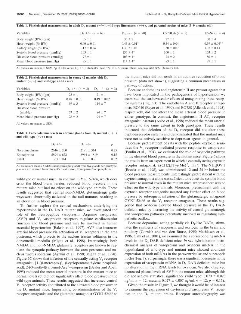
The D5 receptor mutant mice are hypertensive andhave increased sympathetic toneSeveral physiological parameters were examined in adult D5 mu-tant mice (�/�), and comparisons were made with wild-typelittermates (�/�) as well as with the parental C57BL/6 and129/Sv mouse strains (Table 1). Although there were no differ-ences in body or kidney weights between the D5 �/� and D5 �/�genotypes, the D5 mutant mice exhibited significantly elevatedheart weights as well as elevated systolic, diastolic, and meanblood pressures. Blood pressures were also significantly elevatedin the mutant animals even in the absence of anesthesia: wild-type (n � 4) systolic BP � 119 � 4, mean BP � 103 � 3, diastolicBP � 93 � 4; mutant (n � 5) systolic BP � 154 � 6, mean BP �127 � 8, diastolic BP � 115 � 8; p � 0.05 for all wild-type versusmutant values; Student’s t test. No gender differences were noted.
Because the 129 parental strain also showed significantly higherheart weights (Table 1), the elevated heart weights in the D5 �/�mice could be attributable to 129-linked genes. Alternatively, theincreased heart size in the mutants may represent a compensatoryresponse to the elevation in blood pressure. To address this issue,we evaluated the cardiovascular parameters in young mice notolder than 2 months of age. Table 2 shows these results. Nodifferences were observed in the genotypes for the parametersobtained using the young mice. The blood pressures were slightlybut not significantly elevated at this age, and there were nodifferences in the heart weights. These results indicate that thehypertension exhibited by the D5 �/� mice was age dependentand suggest that the cardiac hypertrophy observed in the adultmutants was a result of the elevated blood pressure.
Because D5 receptors inhibit catecholamine release from adre-nal chromaffin cells (Dahmer and Senogles, 1996), we testedwhether elevated adrenal catecholamines might contribute to theincreased blood pressure in the null mutants. Adrenal norepi-nephrine and epinephrine levels are presented in Table 3. Nosignificant differences in the absolute levels of norepinephrine orepinephrine were found between genotypes, but there was asignificant elevation in the epinephrine/norepinephrine ratio inthe mutant animals. Furthermore, acute adrenalectomy resultedin a greater reduction in mean arterial pressure in the D5 mutantanimals (110 � 8 to 62 � 6 mmHg; n � 7) compared withwild-type mice (88 � 0.4 to 56 � 5 mmHg; n � 6). Given theseresults, we wondered whether sympathetic blockade would nor-malize the blood pressure in the mutant mice relative to wild-typeanimals. Infusion of the �-adrenergic antagonist phentolamineresulted in a greater and more rapid reduction of blood pressurein the mutant mice compared with wild-type animals (Fig. 5A).Taken together, these experiments suggested that activation ofthe sympathetic nervous system occurred after disruption of theD5 receptor gene.
Because of the low-level expression of D5 receptors in theadrenal medulla, we thought that the increased sympathetic tonein the D5-deficient mice might be explained more readily by adefect within the CNS. Because central sympathetic nerve re-sponses originating in the pons and medulla are regulated bynon-NMDA glutamatergic pathways (Butcher and Cechetto,1998), we evaluated the effect of glutamatergic blockade on theblood pressure responses. Figure 5B shows the results of infusingthe AMPA/kainate glutamatergic receptor antagonists CNQXand GYKI 52466 (Yoshiyama et al., 1995) into wild-type andmutant mice. CNQX does not cross the blood–brain barrier andhad no effect on the mean arterial blood pressure in either
Figure 4. D1- and D2-like receptor binding assays in striatal membranehomogenates of D5 mutant mice. Striatal membranes were prepared fromwild-type (WT ) and mutant (KO) mice, and radioligand binding assayswere performed as described in Materials and Methods with the indicatedconcentrations of ligands. Only specific binding is shown. The data rep-resent the mean � SEM values from three separate experiments usingindividual mice. A, Saturation binding analyses in striatal membranesusing the D1-like selective antagonist [ 3H]SCH 23390. Computer analysisof the radioligand binding data resulted in the following parameters:wild-type, KD � 0.34 � 0.01 nM, Bmax � 1.01 � 0.18 pmol/mg protein;mutant, KD � 0.38 � 0.03 nM, Bmax � 1.17 � 0.28 pmol/mg protein. UsingStudent’s t test, there was no significant difference between the Bmaxvalues ( p � 0.51). B, Saturation binding analyses in striatal membranesusing the D2-like selective antagonist [ 3H]methylspiperone. Computeranalysis of the radioligand binding data resulted in the following param-eters: wild-type, KD � 0.30 � 0.04 nM, Bmax � 430 � 81 fmol/mg protein;mutant, KD � 0.28 � 0.02 nM, Bmax � 460 � 60 fmol/mg protein. UsingStudent’s t test, there was no significant difference between the Bmaxvalues ( p � 0.67).
Hollon et al. • D5 Receptor-Deficient Mice Exhibit Hypertension J. Neurosci., December 15, 2002, 22(24):10801–10810 10805
wild-type or mutant mice. In contrast, GYK1 52466, which doescross the blood–brain barrier, reduced blood pressure in themutant mice but had no effect on the wild-type animals. Theseresults suggested that central non-NMDA glutamatergic path-ways were abnormally activated in the null mutants, resulting inan elevation in blood pressure.
To further explore the central mechanisms underlying thehypertension in the D5 DAR mutant mice, we investigated therole of the neuropeptide vasopressin. Arginine vasopressin(AVP) and V1 vasopressin receptors regulate cardiovascularfunction and blood pressure and may be involved in humanessential hypertension (Bakris et al., 1997). AVP also increasesarterial blood pressure via activation of V1 receptors in the areapostrema, which projects to the nucleus tractus solitarius in thedorsomedial medulla (Migita et al., 1998). Interestingly, bothNMDA and non-NMDA glutamate receptors are known to reg-ulate the synaptic pathway between the area postrema and nu-cleus tractus solitarius (Aylwin et al., 1998; Migita et al., 1998).Figure 5C shows that infusion of the centrally acting V1 receptorantagonist, [1-(�-mercapto-�, �-cyclopentamethylene propionicacid), 2-(O-methyl)tyrosine]-Arg8-vasopressin (Bealer and Abell,1995) reduced the mean arterial pressure in the mutant mice tonormal levels yet did not significantly affect blood pressure in thewild-type animals. These results suggested that increased centralV1 receptor activity contributed to the elevated blood pressure inthe D5 mutant mice. Importantly, co-administration of the V1
receptor antagonist and the glutamate antagonist GYKI 52466 to
the mutant mice did not result in an additive reduction of bloodpressure (data not shown), suggesting a common mechanism orpathway of action.
Because endothelins and angiotensin II are pressor agents thathave been implicated in the pathogenesis of hypertension, weexamined the cardiovascular effects of antagonizing these recep-tor systems (Fig. 5D). The endothelin A and B receptor antago-nists, BQ610 (Beyer et al., 1999) and BQ788 (Allcock et al., 1995),respectively, did not affect the mean arterial blood pressure ineither genotype. In contrast, the angiotensin II AT1 receptorantagonist losartan (Asico et al., 1998) reduced the mean arterialpressure to the same extent in both genotypes. These resultsindicated that deletion of the D5 receptor did not alter thesepeptide/receptor systems and demonstrated that the mutant micewere not selectively sensitive to depressor agents in general.
Because pretreatment of rats with the peptide oxytocin sensi-tizes the V1 receptor-mediated pressor response to vasopressin(Poulin et al., 1994), we evaluated the role of oxytocin pathwaysin the elevated blood pressure in the mutant mice. Figure 6 showsthe results from an experiment in which a centrally acting oxytocinreceptor antagonist, D(CH2)5[Tyr(Me)2, Thr4, Thy-NH2]OVT(Boccia et al., 1998), was administered 12 and 24 hr before theblood pressure measurements. Interestingly, pretreatment with theoxytocin antagonist alone was sufficient to reduce the systolic bloodpressure to normal levels in the mutant mice, whereas there was noeffect on the wild-type animals. Moreover, pretreatment with theoxytocin receptor antagonist negated any further effect on bloodpressure by subsequent infusion of the glutamatergic antagonistGYKI 52466 or the V1 receptor antagonist. These results sug-gested that oxytocin elevated blood pressure in the D5 DAR-deficient mice by increasing the activity of central glutamatergicand vasopressin pathways potentially involved in regulating sym-pathetic outflow.
Because dopamine, acting partially via D1-like DARs, stimu-lates the synthesis of vasopressin and oxytocin in the brain andpituitary (Cornish and van den Buuse, 1995; Mathiasen et al.,1996; Galfi et al., 2001), we examined their corresponding mRNAlevels in the D5 DAR-deficient mice. In situ hybridization histo-chemical analysis of vasopressin and oxytocin mRNA in thehypothalami of wild-type and mutant mice showed abundantexpression of both mRNAs in the paraventricular and supraopticnuclei (Fig. 7). Surprisingly, there was a significant decrease in theexpression of vasopressin mRNA in D5 DAR-deficient mice butno alteration in the mRNA levels for oxytocin. We also observeddecreased plasma levels of AVP in the mutant mice, although thisdid not achieve statistical significance (wild type: 0.070 � 0.023ng/ml, n � 12; mutant: 0.027 � 0.007 ng/ml, n � 12; p � 0.15).
Given the results in Figure 7, we thought it would be of interestto examine the expression of oxytocin and vasopressin V1 recep-tors in the D5 mutant brains. Receptor autoradiography was
Table 1. Physiological measurements in adult D5 mutant (�/�), wild-type littermates (�/�), and parental strains of mice (3–9 months old)
Variables D5 �/� (n � 67) D5 �/� (n � 70) C57BL/6 (n � 5) 129/Sv (n � 4)
Body weight (BW) (gm) 35 � 1 35 � 2 27 � 1 30 � 4Heart weight (% BW) 0.40 � 0.01 0.45 � 0.01* 0.44 � 0.04 0.59 � 0.04**Kidney weight (% BW) 1.17 � 0.04 1.30 � 0.08 1.30 � 0.07 1.07 � 0.13Systolic blood pressure (mmHg) 103 � 1 136 � 4* 100 � 1 103 � 2Diastolic blood pressure (mmHg) 77 � 1 103 � 4* 74 � 2 80 � 1Mean blood pressure (mmHg) 85 � 1 114 � 4* 83 � 1 87 � 1
All values are means � SEM. *p � 0.05 versus D5 �/�; Student’s t test. **p � 0.05 versus others; one-way ANOVA; Duncan’s test.
Table 2. Physiological measurements in young (2 months old) D5mutant (�/�) and wild-type (�/�) mice
Variables D5 �/� (n � 3) D5 �/� (n � 3)
Body weight (BW) (gm) 23 � 1 21 � 1Heart weight (% BW) 0.48 � 0.01 0.49 � 0.02Systolic blood pressure (mmHg) 99 � 3 114 � 7Diastolic blood pressure
(mmHg) 67 � 2 84 � 7Mean blood pressure (mmHg) 78 � 2 94 � 7
All values are means � SEM.
Table 3. Catecholamine levels in adrenal glands from D5 mutant (�/�)and wild-type (�/�) mice
D5 �/� D5 �/� p
Norepinephrine 2646 � 200 2181 � 314 0.25Epinephrine 5894 � 672 8944 � 1819 0.15E/NE 2.3 � 0.4 4.1 � 0.5 0.02
All values are means � SEM (nanograms per gland) from five glands per genotype.p values are derived from Student’s t test. E/NE, Epinephrine/norepinephrine.
10806 J. Neurosci., December 15, 2002, 22(24):10801–10810 Hollon et al. • D5 Receptor-Deficient Mice Exhibit Hypertension

performed throughout multiple serial brain sections using radio-iodinated ligands for both V1 vasopressin and oxytocin receptors.Figure 8 shows representative coronal sections through the hypo-thalami of wild-type and mutant mice. No consistent differenceswere noted between the genotypes at any level examined. Dele-tion of the D5 receptor therefore did not appear to affect theexpression levels of these receptors.
DISCUSSIONSince its initial discovery and characterization, the exact physio-logical and behavioral roles of the D5 receptor have been difficultto clarify with certainty. This has been attributable, in large part,to the fact that the D1 and D5 DARs are pharmacologicallyindistinguishable. There are few, if any, ligands that exhibit 10-fold selectivity for either subtype (Neve and Neve, 1997). Inter-estingly, dopamine is one of the most selective agents demon-strating �10-fold higher affinity at the D5 DAR compared withthe D1. This lack of selective ligands has made it virtually impos-sible to selectively activate or block D1 or D5 receptors in vivo.Genetic approaches to this problem have been used by investiga-tors using antisense technologies to downregulate D1 or D5 DARexpression as well as the creation of D1 DAR-deficient mice(Sibley, 1999; Glickstein and Schmauss, 2001). These studies havedemonstrated a predominant role of the D1 receptor in regulatingvarious locomotor, cognitive, and other behaviors. Reports ofantisense “knock-down” of D5 receptor expression have beensparse but have suggested a role for the D5 DAR in regulatingfemale sexual behaviors (Apostolakis et al., 1996a,b) and loco-motor responses to dopaminergic agonists (Dziewczapolski et al.,1998).
In the present study, we have used gene targeting technology togenerate mice completely lacking functional D5 DARs. Docu-
menting the inactivation of the D5 DAR gene presented a chal-lenge because there are no radioligands that can be used toselectively label the D5 subtype without simultaneously labelingthe D1 DAR, and because as the D1 receptor is more abundantrelative to the D5, radioligand binding assays with D1-like ligandswill predominantly label the D1 DAR with very little signal beingcontributed by the D5 subtype. This has been well demonstratedin the recent publication of Montague et al. (2001), who per-formed radioligand binding assays in mice lacking the D1 receptorsubtype. Using [3H]SCH-23390, which labels both D1 and D5
receptors, almost all D1-like binding was found to be ablated inthe brains of these animals. There were, however, demonstrablelevels of [3H]SCH-23390 binding in the hippocampi of the D1
knock-out mice. This was presumed to represent binding to theD5 receptor because the hippocampus is a brain region of (rela-tively) high expression for this subtype. Although the use ofradioiodinated ligands could probably detect more D5 receptorbinding sites in the D1 DAR-deficient mice, it is clear that the D1
receptor is more abundant than the D5, and approaches otherthan radioligand binding are needed to demonstrate the absenceof a functional D5 receptor. We thus used two complementaryapproaches. First, we used RT-PCR analyses to show that the D5
mutant mice were expressing the recombinant transcript as ex-pected. Second, we used selective antisera to the D5 DAR andshowed that the D5-deficient mice lacked specific immunohisto-chemical staining that was observed in wild-type mice. Takentogether, these results indicate that the D5 DAR gene was inac-tivated as planned.
The D5 DAR-deficient mice were viable, appeared to developnormally, and were fertile and capable of reproduction. Thislatter observation was especially interesting given the antisense
Figure 5. Mean arterial blood pressureresponses in mice resulting from infu-sion of various pharmacological agents.Infusions were performed as describedin Materials and Methods. The data rep-resent the means � SEM. In some cases,the error bars are smaller than the datapoints. A, The �-adrenergic antagonistphentolamine was infused into wild-type (n � 7) and mutant (n � 14). B, Theperipherally restricted (CNQX ) and cen-trally acting (GYKI 52466) glutamatergicantagonists were infused into wild-typeand mutant mice: CNQX: wild-type(n � 6), mutant (n � 6); GYKI 52466:wild-type (n � 9), mutant (n � 9). C,The V1 vasopressin antagonist, [1-(�-mer-capto-�, �-cyclopentamethylene propi-onic acid), 2-(O-methyl)tyrosine]-Arg8-vasopressin was infused into wild-type(n � 8) and mutant (n � 14) mice. D,The angiotensin II AT1 receptor antag-onist, losartan, and the endothelin Aand B receptor antagonists, BQ610 andBQ788, respectively, were infused intowild-type and mutant mice (n � 6–14per group).
Hollon et al. • D5 Receptor-Deficient Mice Exhibit Hypertension J. Neurosci., December 15, 2002, 22(24):10801–10810 10807

studies (Apostolakis et al., 1996a,b) that described suppression oflordosis behavior in receptive females after D5 DAR knock-downin the ventromedial nucleus of the hypothalamus. Despite theirability to reproduce, it will be interesting to determine whetherthe sexual behaviors of these animals are abnormal in any way. Ingeneral, the home cage behaviors of the D5 DAR-deficient miceappeared normal. Casual observation could not distinguish be-tween mutant and wild-type animals. As is reported elsewhere,however, the D5 mutant mice did show some altered behavioralresponses to dopaminergic agonist stimulation (Holmes et al.,2001). Additional experimentation using the D5 DAR-deficientmice is currently underway to elucidate additional behavioralroles of this receptor subtype.
Because functional deletion of the D5 DAR might result incompensatory upregulation of other dopamine receptor subtypes,especially the D1 DAR, we examined the expression of D1-likeand D2-like receptors in the striatum, a brain region that ex-presses all DAR subtypes (Ariano, 1996). Using radioligands,which label either D1-like or D2-like receptors, we found nodifferences in the receptor binding activities when comparingmutant and wild-type animals. Similarly, no alterations in D1
receptor expression were noted using immunohistochemicalmethods. These results indicate that there are no compensatoryalterations in the expression of the D1 DAR and suggest that theD2-like receptors (D2, D3, and D4) are similarly unaffected.
As part of our initial characterization of the D5 mutant ani-mals, we discovered that they were hypertensive, exhibiting sig-nificantly elevated blood pressures. The elevation in the epineph-rine/norepinephrine ratio and the greater reduction in meanarterial pressure after adrenalectomy, or with �-adrenergic block-ade, in the mutant mice compared with wild-types, suggested thatthe hypertension was caused by increased sympathetic activity.However, because the percentage decrease in systolic blood pres-
sure after adrenalectomy was similar in both mutant and wild-type mice, we sought to determine whether there were CNSmechanisms that may have contributed to the increase in bloodpressure in the D5 mutant mice.
Dopamine receptors, including the D5 DAR, are present in theprefrontal cortex (Ariano et al., 1997; Ciliax et al., 2000), whichprojects to several brain areas involved with cardiovascular regu-lation (Verbene and Owens, 1998). Sympathetic responses fromthe prefrontal cortex are mediated within the lateral hypotha-lamic area (LHA) and ventrolateral medulla (VLM). Moreover,sympathetic responses originating in the prefrontal cortex andLHA are mediated by non-NMDA glutamate receptors in theVLM (Butcher and Cechetto, 1998). Indeed, CNS stimulation ofnon-NMDA glutamate receptors, specifically in the VLM, in-creases blood pressure (Chen et al., 1994; Araujo et al., 1999).Our studies suggest that the increased blood pressure in the D5
DAR-deficient mice may be caused by activation of asympathetic /non-NMDA glutamatergic axis because only a cen-trally acting non-NMDA glutamatergic antagonist decreasedblood pressure in D5 mutant mice.
The D5 receptor may also negatively interact with oxytocin andvasopressin pathways in the prefrontal cortex and other brainareas associated with autonomic control (Ariano et al., 1997;Hermes et al., 1998; Buijs and Van Eden, 2000; Ciliax et al.,2000). Thus, V1 vasopressin (Bealer and Abell, 1995) and oxyto-cin (Boccia et al., 1998) antagonists that cross the blood–brain
Figure 6. Blood pressure responses in mice pretreated with a centrallyacting oxytocin receptor antagonist. The blood pressures were initiallydetermined (BASAL) followed by two injections of the oxytocin receptorantagonist D(CH2)5[Tyr(Me)2, Thr 4, Thy-NH2]OVT, as described inMaterials and Methods. Systolic blood pressures were then evaluatedalone (OTR) or subsequent to the infusion of GYKI 52466 (OTR �GYKI ) or [1-(�-mercapto-�, �-cyclopentamethylene propionic acid),2-(O-methyl)tyrosine]-Arg 8-vasopressin (OTR � V1) as described in Fig-ure 5, B and C. The data represent the mean � SEM values from five tonine animals per group. In some cases, the error bars are smaller than thedata points. *p � 0.05 versus other groups; ANOVA; Newman–Keulstest. Similar results were observed for diastolic blood pressures (data notshown).
Figure 7. In situ hybridization histochemical (ISHH) analyses of vaso-pressin and oxytocin mRNA in the hypothalami of wild-type (n � 8) andmutant mice (n � 9). ISHH was performed as described in Materials andMethods, with representative images shown at the bottom of the figure.Phosphorimage analysis was performed by choosing a level that wentthrough the middle of the paraventricular and supraoptic nuclei in eachbrain. The y-axis represents arbitrary optical density (OD) values as-signed by the Image Gauge program of the Fuji phosphor imager. Thevasopressin mRNA levels were significantly decreased in the mutantbrains: OD mean � SEM values: wild-type, 9.0 � 0.73; mutant, 5.1 �0.73; p � 0.05; Student’s t test.
10808 J. Neurosci., December 15, 2002, 22(24):10801–10810 Hollon et al. • D5 Receptor-Deficient Mice Exhibit Hypertension

barrier were found to decrease the blood pressure in the D5
mutant but not wild-type mice. Interestingly, the hypotensiveeffect of the oxytocin antagonist occurred only 24 hr after itsadministration and negated any further reduction in blood pres-sure by vasopressin or glutamatergic blockade. These results areconsistent with the observation that oxytocin has been shown tosensitize V1 vasopressin receptors (Poulin et al., 1994) and fur-ther suggests that the decrease in blood pressure in the mutantmice engendered by these various antagonists occurs via a com-mon output pathway.
In summary, we have found that functional deletion of the D5
DAR gene produces hypertension in mice. The elevated bloodpressure appears to be attributable to increased sympathetic tonewith an involvement of adrenal catecholamines. The exact defectleading to the increase in sympathetic tone is unclear, although itappears to be primarily central in origin. Our current resultssuggest that D5 receptor deletion results in an oxytocin-dependent sensitization of V1 vasopressin and non-NMDA glu-tamatergic receptor-mediated pathways, potentially within themedulla, leading to increased sympathetic outflow in the mutantmice. This change is not associated with increased synthesis ofeither oxytocin or vasopressin, and in fact, vasopressin synthesisappears reduced in the D5 DAR-deficient mice. Furthermore,there is no increase in oxytocin or V1 vasopressin receptor num-bers. This suggests that the increased sensitivity must occur at thelevel of receptor signaling, possibly via enhanced G-protein in-teractions, or other regulatory mechanisms, and/or downstreamintracellular signaling pathways. The physiological events de-scribed here resulting from D5 DAR deletion may provide new
insights into mechanisms for some forms of essential hyperten-sion in humans and may lead to new therapeutic approaches forits treatment.
REFERENCESAllcock GH, Warner TD, Vane JR (1995) Roles of endothelin receptors
in the regional and systemic vascular responses to ET-1 in the anaes-thetized ganglion-blocked rat: use of selective antagonists. Br J Phar-macol 116:2482–2486.
Apostolakis EM, Garai J, Fox C, Smith CL, Watson SJ, Clark JH,O’Malley BW (1996a) Dopaminergic regulation of progesterone re-ceptors: brain D5 dopamine receptors mediate induction of lordosis byD1-like agonists in rats. J Neurosci 16:4823–4834.
Apostolakis EM, Garai J, Clark JH, O’Malley BW (1996b) In vivo reg-ulation of central nervous system progesterone receptors: cocaine in-duces steroid-dependent behavior through dopamine transporter mod-ulation of D5 receptors in rats. Mol Endocrinol 10:1595–1604.
Araujo GC, Lopes OU, Campos RR (1999) Importance of glycinergicand glutamatergic synapses within the rostral ventrolateral medulla forblood pressure regulation in conscious rats. Hypertension 34:752–755.
Ariano MA (1996) Dopamine receptor localization. The dopamine re-ceptors (Neve KA, Neve RL, eds), pp 77–104. Totowa, NJ: Humana.
Ariano MA, Wang J, Noblett KL, Larson ER, Sibley DR (1997) Cellu-lar distribution of the rat D1B receptor in central nervous system usinganti-receptor antisera. Brain Res 746:141–150.
Asico LD, Ladines C, Fuchs S, Accili D, Carey RM, Semeraro C,Pocchiari F, Felder RA, Eisner GM, Jose PA (1998) Disruption of thedopamine D3 receptor gene produces renin-dependent hypertension.J Clin Invest 102:493–498.
Aylwin ML, Horowitz JM, Bonham AC (1998) Non-NMDA andNMDA receptors in the synaptic pathway between area postrema andnucleus tractus solitarius. Am J Physiol 275:H1236–H1246.
Bakris G, Bursztyn M, Gavras I, Bresnahan M, Gavras H (1997) Role ofvasopressin in essential hypertension: racial differences. J Hypertens15:545–550.
Bealer SL, Abell SO (1995) Paraventricular nucleus histamine increasesblood pressure by adrenoceptor stimulation of vasopressin release.Am J Physiol 269:H80–H85.
Figure 8. Receptor autoradiography of V1 vasopressin and oxytocin receptors in brain sections of wild-type (n � 3) and mutant mice (n � 3).Representative autoradiograms of sections from wild-type (A, C) and mutant (B, D) mice through the hypothalamus show binding of the oxytocin (A,B) and vasopressin (C, D) receptor ligands. No consistent differences were noted between the wild-type and knock-out mice at any level examined. IV,Cortical layer 4; En, endopiriform nucleus; HF, hippocampal formation; LH, lateral hypothalamus; PL, posterolateral cortical amygdaloid nucleus; PV,thalamic paraventricular nucleus; VM, ventromedial hypothalamic nucleus. Three sets of wild-type and D5 mutant brains were examined with similarresults.
Hollon et al. • D5 Receptor-Deficient Mice Exhibit Hypertension J. Neurosci., December 15, 2002, 22(24):10801–10810 10809

Beyer ME, Slesak G, Hovelborn T, Kazmaier S, Nerz S, Hoffmeister HM(1999) Inotropic effects of endothelin-1: interaction with molsidomineand with BQ 610. Hypertension 33:145–152.
Boccia MM, Kopf SR, Baratti CM (1998) Effects of a single administra-tion of oxytocin or vasopressin and their interactions with two selectivereceptor antagonists on memory storage in mice. Neurobiol LearnMem 69:136–146.
Buijs RM, Van Eden CG (2000) The integration of stress by the hypo-thalamus, amygdala and prefrontal cortex: balance between the auto-nomic nervous system and the neuroendocrine system. Prog Brain Res126:117–132.
Butcher KS, Cechetto DF (1998) Neurotransmission in the medullamediating insular cortical and lateral hypothalamic sympathetic re-sponses. Can J Physiol Pharmacol 76:737–746.
Caramona MM, Soares-da-Silva P (1985) The effects of chemical sym-pathectomy on dopamine, noradrenaline and adrenaline content insome peripheral tissues. Br J Pharmacol 86:351–356.
Chen K, Hernandez YM, Dretchen KL, Gillis RA (1994) IntravenousNBQX inhibits spontaneously occurring sympathetic nerve activity andreduces blood pressure in cats. Eur J Pharmacol 252:155–160.
Ciliax BJ, Nash N, Heilman C, Sunahara R, Hartney A, Tiberi M, RyeDB, Caron MG, Niznik HB, Levey AI (2000) Dopamine D5 receptorimmunolocalization in rat and monkey brain. Synapse 37:125–145.
Cornish JL, van den Buuse M (1995) Stimulation of the rat mesolimbicdopaminergic system produces a pressor response which is mediated bydopamine D-1 and D-2 receptor activation and the release of vasopres-sin. Brain Res 701:28–38.
Dahmer MK, Senogles SE (1996) Dopaminergic inhibition of catechol-amine secretion from chromaffin cells: evidence that inhibition is me-diated by D4 and D5 dopamine receptors. J Neurochem 66:222–232.
Dziewczapolski G, Menalled LB, Garcia MC, Mora MA, Gershanik OS,Rubinstein M (1998) Opposite roles of D1 and D5 dopamine receptorsin locomotion revealed by selective antisense oligonucleotides. Neuro-Report 9:1–5.
Galfi M, Janaky T, Toth R, Prohaszka G, Juhasz A, Varga C, Laszlo FA(2001) Effects of dopamine and dopamine-active compounds on oxy-tocin and vasopressin production in rat neurohypophyseal tissue cul-tures. Regul Pept 98:49–54.
Glickstein SB, Schmauss C (2001) Dopamine receptor function: lessonsfrom knockout mice. Pharmacol Ther 91:63–83.
Hermes ML, Buijs RM, Masson-Pevet M, Pevet P (1998) Oxytocinergicinnervation of the brain of the garden dormouse (Eliomys quercinus L.).J Comp Neurol 273:252–262.
Holmes A, Hollon TR, Gleason TC, Liu Z, Dreiling J, Sibley DR,Crawley JN (2001) Behavioral characterization of dopamine D5 re-ceptor null mutant mice. Behav Neurosci 115:1129–1144.
Insel TR, Shapiro LE (1992) Oxytocin receptor distribution reflectssocial organization in monogamous and polygamous voles. Proc NatlAcad Sci USA 89:5981–5985.
Jiang D, Sibley DR (1999) Agonist-induced desensitization of D1 dopa-mine receptors with mutations of cyclic AMP-dependent protein ki-nase phosphorylation sites: attenuation of the rate of agonist-induceddesensitization. Mol Pharmacol 56:675–683.
Li E, Bestor TH, Jaenisch R (1992) Targeted mutation of the DNAmethyltransferase gene results in embryonic lethality. Cell 69:915–926.
Liu F, Wan Q, Pristupa ZB, Yu XM, Wang YT, Niznik HB (2000)Direct protein-protein coupling enables cross-talk between dopamineD5 and �-aminobutyric acid A receptors. Nature 403:274–280.
Malik KF, Kim J, Hartman AL, Kim P, Young III WS (1996) Bindingpreferences of the POU domain protein Brain-4: implications forautoregulation. Mol Brain Res 38:209–221.
Mathiasen JR, Larson ER, Ariano MA, Sladek CD (1996) Neurophysinexpression is stimulated by dopamine D1 agonist in dispersed hypotha-lamic cultures. Am J Physiol 39:R404–R412.
Mezey E, Eisenhofer G, Harta G, Hansson S, Gould L, Hunyady B,Hoffman BJ (1996) A novel nonneuronal catecholaminergic system:exocrine pancreas synthesizes and releases dopamine. Proc Natl AcadSci USA 93:10377–10382.
Migita K, Hori N, Manako J, Saito R, Takano Y, Kamiya H (1998)Effects of arginine-vasopressin on neuronal interaction from the areapostrema to the nucleus tractus solitari in rat brain slices. Neurosci Lett256:45–48.
Montague DM, Striplin CD, Overcash JS, Drago J, Lawler CP, MailmanRB (2001) Quantification of D1B (D5) receptors in dopamine D1Areceptor-deficient mice. Synapse 39:319–322.
Muir WJ, Thomson ML, McKeon P, Mynett-Johnson L, Whitton C,Evans KL, Porteous DJ, Blackwood DH (2001) Markers close to thedopamine D5 receptor gene (DRD5) show significant association withschizophrenia but not bipolar disorder. Am J Med Genet 105:152–158.
Neve KA, Neve RL (1997) The dopamine receptors. Totowa, NJ:Humana.
Poulin P, Komulainen A, Takahashi Y, Pittman (1994) QJ enhancedpressor responses to ICV vasopressin after pretreatment with oxytocin.Am J Physiol 266:R592–R598.
Rivkees S, Lachowicz JE (1997) Functional D1 and D5 dopamine recep-tors are expressed in the suprachiasmatic, supraoptic and paraventricu-lar nuclei of primates. Synapse 26:1–10.
Sanada H, Xu J, Watanabe H, Jose PA, Felder RA (2000) Differentialexpression and regulation of dopamine-1 (D1) and dopamine-5 (D5)receptor function in human kidney. Am J Hypertens 13:156A.
Schetz JA, Benjamin PS, Sibley DR (2000) Non-conserved residues inthe second transmembrane-spanning domain of the D4 dopamine re-ceptor are molecular determinants of D4-selective pharmacology. MolPharmacol 57:144–152.
Sibley DR (1999) New insights into dopaminergic receptor function us-ing antisense and genetically altered animals. Annu Rev PharmacolToxicol 39:313–341.
Sunahara RK, Guan HC, O’Dowd BF, Seeman P, Laurier LG, Ng G,George SR, Torchia J, Van Tol HH, Niznik HB (1991) Cloning of thegene for a human dopamine D5 receptor with higher affinity fordopamine than D1. Nature 350:614–619.
Tiberi M, Caron MG (1994) High agonist-independent activity is adistinguishing feature of the dopamine D1B receptor subtype. J BiolChem 269:27925–27931.
Tybulewicz VLJ, Crawford CE, Jackson PK, Bronson RT, Mulligan RC(1991) Neonatal lethality and lymphopenia in mice with a homozygousdisruption of the c-abl protooncogene. Cell 65:1153–1163.
Usiello A, Baik JH, Rouge-Pont F, Picetti R, Dierich A, LeMeur M,Piazza PV, Borrelli E (2000) Distinct functions of the two isoforms ofthe dopamine D2 receptors. Nature 408:199–203.
Vanyukov MM, Moss HB, Gioio AE, Hughes HB, Kaplan BB, Tarter RE(1998) An association between a microsatellite polymorphism at theDRD5 gene and the liability to substance abuse: pilot study. BehavGenet 28:75–82.
Verbene AJ, Owens NC (1998) Cortical modulation of the cardiovascu-lar system. Prog Neurobiol 54:149–168.
Yan Z, Surmeier DJ (1997) D5 dopamine receptors regulate Zn 2�-sensitive GABAA currents in striatal cholinergic interneurons througha PKA/PP1 cascade. Neuron 19:1115–1126.
Yoshiyama M, Roppolo JR, de Groat WC (1995) Effects of GYKI 52466and CNQX, AMPA/kainate receptor antagonists, on the micturitionreflex in the rat. Brain Res 691:185–194.
Young LJ, Wang Z, Cooper TT, Albers HE (2000) Vasopressin (V1a)receptor binding, mRNA expression and transcriptional regulation byandrogen in the Syrian hamster brain. J Neuroendocrinol 12:1179–1185.
10810 J. Neurosci., December 15, 2002, 22(24):10801–10810 Hollon et al. • D5 Receptor-Deficient Mice Exhibit Hypertension
Related Documents