MICE EXPRESSING CONSTITUTIVELY ACTIVE G s α EXHIBIT STIMULUS ENCODING DEFICITS SIMILAR TO THOSE OBSERVED IN SCHIZOPHRENIA PATIENTS C. R. MAXWELL a , Y. LIANG a , M. P. KELLY a,b , S. J. KANES a , T. ABEL b , and S. J. SIEGEL a,* a Division of Neuropsychiatry, Department of Psychiatry, University of Pennsylvania, Translational Research Laboratories, Room 2223, 125 South 31st Street, Philadelphia, PA 19104, USA b Department of Biology, University of Pennsylvania, Philadelphia, PA 19104, USA Abstract People with schizophrenia display sensory encoding deficits across a broad range of electrophysiological and behavioral measures, suggesting fundamental impairments in the ability to transduce the external environment into coherent neural representations. This inability to create basic components of complex stimuli interferes with a high fidelity representation of the world and likely contributes to cognitive deficits. The current study evaluates the effects of constitutive forebrain activation of the G s α G-protein subunit on auditory threshold and gain using acoustic brainstem responses and cortically generated N40 event-related potentials to assess the role of cyclic AMP signaling in sensory encoding. Additionally, we examine the ability of pharmacological treatments that mimic (amphetamine) or ameliorate (haloperidol) positive symptoms of schizophrenia to test the hypothesis that the encoding deficits observed in G s α transgenic mice can be normalized with treatment. We find that G s α transgenic mice have decreased amplitude of cortically generated N40 but normal acoustic brainstem response amplitude, consistent with forebrain transgene expression and a schizophrenia endophenotype. Transgenic mice also display decreased stimulus intensity response (gain) in both acoustic brainstem response and N40, indicating corticofugal influence on regions that lack transgene expression. N40 deficits in transgenic animals were ameliorated with low dose haloperidol and reversed with higher dose, suggesting dopamine D 2 receptor-linked G i activity contributes to the impairment. Consistent with this hypothesis, we recreated the G s α transgenic deficit in wild type animals using the indirect dopamine agonist amphetamine. This transgenic model of sensory encoding deficits provides a foundation for identifying biochemical contributions to sensory processing impairments associated with schizophrenia. Keywords G s α; N40; schizophrenia; cAMP; haloperidol; event-related potentials People with schizophrenia have difficulty interpreting events and stimuli in their environment. Such difficulties are dramatically manifest as delusions of reference and sensory illusions but may also result in confusion and inability to piece together signals required to interact in society. This integrated process of detecting and interpreting the constellation of multimodal stimuli is too complex to understand as a singular phenomenon or study with interpretable measures in the human or animal laboratory. However, evidence © 2006 IBRO. Published by Elsevier Ltd. All rights reserved * Corresponding author. Tel: +1-215-573-0278; fax: +1-215-573-2041. [email protected] (S. J. Siegel).. NIH Public Access Author Manuscript Neuroscience. Author manuscript; available in PMC 2012 March 24. Published in final edited form as: Neuroscience. 2006 September 1; 141(3): 1257–1264. doi:10.1016/j.neuroscience.2006.04.028. NIH-PA Author Manuscript NIH-PA Author Manuscript NIH-PA Author Manuscript

Welcome message from author
This document is posted to help you gain knowledge. Please leave a comment to let me know what you think about it! Share it to your friends and learn new things together.
Transcript

MICE EXPRESSING CONSTITUTIVELY ACTIVE Gsα EXHIBITSTIMULUS ENCODING DEFICITS SIMILAR TO THOSEOBSERVED IN SCHIZOPHRENIA PATIENTS
C. R. MAXWELLa, Y. LIANGa, M. P. KELLYa,b, S. J. KANESa, T. ABELb, and S. J. SIEGELa,*
aDivision of Neuropsychiatry, Department of Psychiatry, University of Pennsylvania, TranslationalResearch Laboratories, Room 2223, 125 South 31st Street, Philadelphia, PA 19104, USAbDepartment of Biology, University of Pennsylvania, Philadelphia, PA 19104, USA
AbstractPeople with schizophrenia display sensory encoding deficits across a broad range ofelectrophysiological and behavioral measures, suggesting fundamental impairments in the abilityto transduce the external environment into coherent neural representations. This inability to createbasic components of complex stimuli interferes with a high fidelity representation of the worldand likely contributes to cognitive deficits. The current study evaluates the effects of constitutiveforebrain activation of the Gsα G-protein subunit on auditory threshold and gain using acousticbrainstem responses and cortically generated N40 event-related potentials to assess the role ofcyclic AMP signaling in sensory encoding. Additionally, we examine the ability ofpharmacological treatments that mimic (amphetamine) or ameliorate (haloperidol) positivesymptoms of schizophrenia to test the hypothesis that the encoding deficits observed in Gsαtransgenic mice can be normalized with treatment. We find that Gsα transgenic mice havedecreased amplitude of cortically generated N40 but normal acoustic brainstem responseamplitude, consistent with forebrain transgene expression and a schizophrenia endophenotype.Transgenic mice also display decreased stimulus intensity response (gain) in both acousticbrainstem response and N40, indicating corticofugal influence on regions that lack transgeneexpression. N40 deficits in transgenic animals were ameliorated with low dose haloperidol andreversed with higher dose, suggesting dopamine D2 receptor-linked Gi activity contributes to theimpairment. Consistent with this hypothesis, we recreated the Gsα transgenic deficit in wild typeanimals using the indirect dopamine agonist amphetamine. This transgenic model of sensoryencoding deficits provides a foundation for identifying biochemical contributions to sensoryprocessing impairments associated with schizophrenia.
KeywordsGsα; N40; schizophrenia; cAMP; haloperidol; event-related potentials
People with schizophrenia have difficulty interpreting events and stimuli in theirenvironment. Such difficulties are dramatically manifest as delusions of reference andsensory illusions but may also result in confusion and inability to piece together signalsrequired to interact in society. This integrated process of detecting and interpreting theconstellation of multimodal stimuli is too complex to understand as a singular phenomenonor study with interpretable measures in the human or animal laboratory. However, evidence
© 2006 IBRO. Published by Elsevier Ltd. All rights reserved*Corresponding author. Tel: +1-215-573-0278; fax: +1-215-573-2041. [email protected] (S. J. Siegel)..
NIH Public AccessAuthor ManuscriptNeuroscience. Author manuscript; available in PMC 2012 March 24.
Published in final edited form as:Neuroscience. 2006 September 1; 141(3): 1257–1264. doi:10.1016/j.neuroscience.2006.04.028.
NIH
-PA Author Manuscript
NIH
-PA Author Manuscript
NIH
-PA Author Manuscript

from multiple investigators using complimentary approaches indicates that the inability tointerpret complex stimuli may begin with abnormalities in early detection and encoding ofstimulus characteristics. Disruption of early neural processes likely degrades the buildingblocks of interpretation and is consistent with abnormalities in cognitive domains thatrequire qualitative assessment of stimulus parameters on which to base decisions. If a signalis misallocated with respect to its relevance, its encoding in memory will be altered anddecisions based on distorted information.
Event-related potentials (ERPs) allow for evaluation of sensory processing in which thelatency of potentials is determined by generators of neural activity. In humans, early ERPs(1–8 ms) represent activation of brainstem structures such as the cochlear nucleus; mid-latency ERPs (8–40 ms) indicate forebrain activity including thalamus, hippocampus andprimary auditory cortex and long-latency ERPs (50–300 ms) represent higher processing,involving primary and association cortices (Picton et al., 1974). Patients with schizophreniadisplay abnormalities in auditory processing including reductions in amplitude of long-latency cortical ERP components. One report indicates that patients with schizophreniademonstrate impaired cortical ERPs with no differences in brainstem-generated potentials(Pfefferbaum et al., 1980; Boutros et al., 2004). Additional ERP components includingmismatch negativity have been proposed as predictive markers of cognitive function inpatients with schizophrenia (Boutros et al., 2004; Light and Braff, 2005a,b).
Early, middle and long-latency ERPs recorded from mice resemble corresponding humancomponents in polarity and response properties (Connolly et al., 2003; Maxwell et al.,2004a; Umbricht et al., 2004; Siegel et al., 2005) As such, ERPs in mice provide usefulmodels for genetic and mechanistic understanding of brain abnormalities and treatmentdevelopment in schizophrenia. Specifically, rodents share many similarities with humans forspecific portions of the ERP, including the mouse P1, N1, P2 and P3. These components arealso named the P20, N40, P80 and P120 for latency and share stimulus and pharmacologicresponse properties with the human P50, N100, P200 and P300 respectively (Siegel et al.,2003, 2005; Connolly et al., 2004; Maxwell et al., 2004b; Umbricht et al., 2005).
Previous reports indicate that auditory ERPs are modulated by compounds that influence thecyclic AMP (cAMP) signaling cascade. For example, antipsychotic compounds, whichregulate cAMP production through dopamine D2 receptor blockade, and phosphodiesteraseinhibitors, which prevent cAMP hydrolysis, increase auditory ERP amplitude (Maxwell etal., 2004a,b). Conversely, the indirect dopamine agonist amphetamine decreases ERPamplitude (Maxwell et al., 2004a). Additionally, G-protein-coupled receptors including Gsand Gi that influence intracellular cAMP concentrations modulate sensory-motor gating inrodents (Culm et al., 2003; Gould et al., 2004; Pineda et al., 2004). Therefore, the G-protein-coupled cAMP signaling cascade is a target for evaluating auditory ERPs to help understandsensory processing deficits similar to those in schizophrenia. The current study utilizes amouse model of schizophrenia that limits constitutively active Gsα transgene expression topostnatal forebrain structures through use of a CaMKIIα promoter, providing a degree ofanatomic localization for observed abnormalities.
EXPERIMENTAL PROCEDURESAnimals/surgery
Gsα transgenic mice were bred and group-housed at the University of Pennsylvania in ahemizygous state on a C57BL/6 background (N12–N14). As previously described, Gsα*transgenic mice express an isoform of the G-protein subunit Gsα that is constitutively activedue to a point mutation (Q227L) that prevents hydrolysis of bound GTP (Wand et al., 2001;Gould et al., 2004). Expression of the transgene is driven by the CaMKIIα-promoter, which
MAXWELL et al. Page 2
Neuroscience. Author manuscript; available in PMC 2012 March 24.
NIH
-PA Author Manuscript
NIH
-PA Author Manuscript
NIH
-PA Author Manuscript

restricts expression to postnatal forebrain neurons. Specifically, in situ hybridization studiesindicate that the transgene is expressed in striatum, hippocampus and cortex but notcerebellum, thalamus or brainstem (Wand et al., 2001). Animals were genotyped bySouthern blot using a transgene-specific probe as described (Abel et al., 1997). All protocolswere conducted in accordance with University Laboratory Animal Resources guidelines andwere approved by the National Institutes of Health. Animals were individually housedfollowing surgery in a light and temperature-controlled Association for Assessment andAccreditation of Laboratory Animal Care–accredited animal facility. Food and water wereavailable ad libitum. All efforts were made to minimize animal numbers and suffering.
Thirteen wild type (six male, seven female) and 11 transgenic Gsα* (five male, six female)mice aged 10–13 weeks were implanted with unipolar electrodes for non-anesthetizedrecordings of whole brain EEG activity two weeks later (Maxwell et al., 2004a). We havepreviously shown that this recording method closely resembles human scalp EEG recordings(Siegel et al., 2003). Animals were anesthetized with isoflurane. Recording electrodes wereplaced in the CA3 hippocampal region, (1.4 mm posterior, 2.65 mm lateral and 2.75 mmdeep relative to bregma) and referenced to the ipsilateral frontal sinus. The electrodepedestal was secured to the skull using dental cement and superglue. Electrode placementwas verified to be in the target region using the Perl's iron reaction (LaBossiere andGlickstein, 1976).
Experimental designThis study consisted of three experiments that had a within animal design. Experiment 1:Genotype differences were determined by evaluating the average across two saline trialsadministered into the i.p. cavity and conducted one week apart. Experiment 2: Vehicle, 0.1mg/kg and 1 mg/kg haloperidol (Sigma) were administered i.p. to examine the ability of aknown antipsychotic medication to reverse baseline differences in auditory evokedpotentials. Experiment 3: Vehicle, 0.1 mg/kg, 0.5 mg/kg and 1 mg/kg d-amphetamine(Sigma) were administered i.p. to evaluate the hypothesis that the transgene would recreateboth the effects of amphetamine seen in wild type mice as well as those seen in untreatedpatients with schizophrenia. Experiments 2 and 3 were performed in a counterbalancedfashion in which half of the animals received drug and half received vehicle on each testday. Age- and gender-matched wild type littermates were run in parallel to Gsα transgenicmice. Additionally, animals were given at least 48 h between drug exposures.
Auditory evoked potentialsThe generation of a positive deflection at 3 ms (P3) and a negative deflection at 5 ms (N5)poststimulus is thought to represent activation of hindbrain structures including the auditorynerve and brainstem based on the hypothesis that latency of the evoked potential reflectsrelative generation site (Henry, 1979; Connolly et al., 2003). Similarly, a negative deflectionoccurring at 40 ms (N40) poststimulus indicates activation of forebrain structures includinghippocampus, thalamus and auditory cortex (Maxwell et al., 2004b; Mamiya et al., 2005).Evaluations of gene effects on the P3N5 complex and the N40 allow for regionalassessments of brainstem responses and higher cortical processing of auditory stimuli.
Brainstem responseAuditory brainstem responses were evaluated prior to injection on each testing session at sixstimulus intensities to determine baseline differences in brainstem-evoked potentials.Consistent with previous publications, the protocol consists of 1000 white noise stimuli (3ms duration) presented at intensities of 0, 58, 66, 74, 82 and 90 dB (Connolly et al., 2003).These stimulus intensities were chosen to provide a thorough assessment of auditorythreshold. Raw EEG data were sampled at 6250 Hz using Micro 1401 hardware and spike 5
MAXWELL et al. Page 3
Neuroscience. Author manuscript; available in PMC 2012 March 24.
NIH
-PA Author Manuscript
NIH
-PA Author Manuscript
NIH
-PA Author Manuscript

software (Cambridge Electronic Design, Cambridge, UK). Waveforms were filteredbetween 1 and 300 Hz, baseline corrected at stimulus onset, and average waveforms weremade from 2 ms prior to stimulus presentation through 9 ms after stimulus onset. Aspreviously published, the amplitude of the P3 (2–4 ms window) to N5 (3–6 ms window)difference waveform was analyzed using a repeated measure ANOVA to examine maineffects of genotype and interactions between genotype and intensity (Connolly et al., 2003).
Cortical processingFifty white noise stimuli (10 ms duration) were presented with an 8 s inter-stimulus intervalat intensities of 75, 85 and 95 dB compared with a background of 70 dB. These stimulusintensities were chosen to provide a wide range of intensities within the time limitationsduring which the pharmacological treatments were effective. As such, auditory brainstemresponses were not evaluated following drug treatment in these animals. Raw EEG datawere sampled at 1667 Hz. Waveforms were baseline corrected at stimulus onset andindividual trials rejected for movement artifact based on twice the root mean squaredamplitude per mouse. Average waveforms were created from 50 ms pre-stimulus to 200 mspost stimulus (Fig. 1).
The amplitude of the N40 (25–60 ms w and drug (Statistica; Statsoft, Tulsa, OK, USA).Significant interactions were followed with Fisher LSD post hoc analyses. Initial analyses ofN40 amplitude included gender as a variable. No significant effects of gender or interactionsbetween gender and other variables were found on N40 amplitude at baseline or with eitherdrug condition. Therefore, the gender variable was removed from this analysis in order focuson the effects of genotype.
RESULTSWe previously reported that constitutive activation of Gsα causes impairments in pre-pulseinhibition (PPI) and proposed this mutation as a model of schizophrenia-like sensory-motorgating deficits (Gould et al., 2004). The current study demonstrates the effects of theforebrain-specific Gsα transgenic manipulation on auditory ERPs to determine regionalcontributions to specific phases of sensory encoding deficits relevant to schizophrenia.Additionally, we test the hypothesis that pharmacological treatments that ameliorate positivesymptoms of schizophrenia (hallucinations and delusions) will normalize sensory encodingdeficits caused by the Gsα mutation. These studies provide a novel transgenic mouse modelof sensory encoding deficits associated with schizophrenia and may facilitate understandingof neural and biochemical mechanisms that contribute to its pathophysiology.
Gsα transgenic mice display reduced acoustic brainstem response (ABR) intensityresponse but not threshold
The P3N5 complex was analyzed to determine baseline transgene effects at the brainstemprior to acute treatments (Table 1). The amplitude of the P3N5 complex increasesproportionally with stimulus intensity as evidenced by a main effect of stimulus intensity(Fig. 2). No main effect of genotype was found on this component. Although there were nosignificant differences between genotypes at any single intensity, a genotype by intensityinteraction suggests that there are differences in the slope of the stimulus intensity functionbetween genotypes (Table 1: gene×intensity, P=0.021; Fig. 2A). Fisher LSD post hocanalyses indicate that wild type mice show significant increases in the P3N5 response withsmaller increases in stimulus intensity (58 dB vs. 66 dB: P=0.063; 58 dB vs. 74 dB:P<0.001; 58 dB vs. 82 dB: P<0.001; 58 dB vs. 90 dB: P<0.001) than do transgenic mice (58dB vs. 66 dB: P=0.518; 58 dB vs. 74 dB: P=0.164; 58 dB vs. 82 dB: P=0.013; 58 dB vs. 90dB: P<0.001. ABR data were also analyzed by calculating the slope of the ABR intensity
MAXWELL et al. Page 4
Neuroscience. Author manuscript; available in PMC 2012 March 24.
NIH
-PA Author Manuscript
NIH
-PA Author Manuscript
NIH
-PA Author Manuscript

function for each mouse. We then used a two-tailed t-test to compare wild type to transgenicmice for this alternate method of assessment. The average slope was 0.064 for wild type and0.034 for transgenic with a P-value of 0.026. These data indicate that there are no significantdifferences in auditory threshold between wild type and transgenic animals but theseanimals differ in the stimulus intensity response (gain). Such data suggest that the there areno abnormalities in the mechanisms of detecting stimuli at the level of the cranial nerves andbrainstem in transgenic animals, but that the ability to encode stimulus intensity byrecruiting more neurons to fire in synchrony is impaired.
Gsα transgene expression impairs cortical N40 processingWe also measured the amplitude of the mouse N40 ERP, which shares homology with thehuman N100 primary cortical response (Connolly et al., 2003, 2004; Siegel et al., 2003,2005; Maxwell et al., 2004a,b). Transgenic animals had smaller N40 amplitude than wildtype mice as indicated by a main effect of genotype. This component also increased inamplitude in proportion to increasing stimulus intensity. Additionally, a genotype byintensity interaction demonstrated that transgenic mice had reduced amplitude at 85 and 95dB, suggesting an impaired stimulus intensity response (Fig. 2B).
Gsα transgenic deficit is reversed with haloperidolThe hypothesis that antipsychotic treatment would reverse the transgenic deficit, as shown inpharmacological models of schizophrenia, was tested by administration of haloperidol(Maxwell et al., 2004a,b; Siegel et al., 2005). Both doses of haloperidol increased the N40amplitude in transgenic mice (Table 1: gene×drug P=0.003). The 0.1 mg/kg dose ofhaloperidol increased the N40 amplitude in Gsα transgenic mice to match that of wild typelittermates. The 1.0 mg/kg dose of haloperidol enhanced N40 amplitude in Gsα transgenicmice above that of wild type littermates (transgenic vs. wild type: vehicle P=0.033; 0.1 mg/kg P=0.760; 1.0 mg/kg P=0.038, Fig. 3A), suggesting that the higher dose unmasked anunderlying augmentation from unopposed Gsα activity.
Gsα transgenic deficits are recreated in wild type mice using amphetamineAmphetamine was administered to assess the hypothesis that increased dopamine activity atboth D1 and D2-like DA receptors would recreate the Gsα transgenic deficit in wild typeanimals. A genotype by stimulus intensity interaction revealed the baseline stimulusintensity deficit in transgenic relative to wild type animals (Fig. 2D). Wild type micedisplayed a dose-dependent decrease in amplitude with amphetamine whereas the transgenicanimals were unchanged (Fig. 3B). A genotype by amphetamine by intensity interactionrevealed that all doses of amphetamine prevented wild type animals from displaying anormal stimulus intensity response which resembled the transgenic impairment (Table 2).
DISCUSSIONOverall, Gsα transgenic mice displayed decreased amplitude of cortically-generated N40with normal ABR ampli tude, consistent with forebrain transgene expression and theschizophrenia endophenotype. Gsα transgenic mice also displayed decreased stimulusintensity response (gain) for both ABR and N40 amplitude, indicating corticofugal influenceon brainstem regions that lack transgene expression. N40 deficits in Gsα transgenic animalswere ameliorated with haloperidol 0.1 mg/kg and reversed with the 1.0 dose, suggestingactivity of dopamine D2 receptor-linked Gi G-protein contributes to the observedimpairment. Consistent with this hypothesis, the indirect dopamine agonist amphetaminerecreated the Gsα transgenic deficit in wild type animals.
MAXWELL et al. Page 5
Neuroscience. Author manuscript; available in PMC 2012 March 24.
NIH
-PA Author Manuscript
NIH
-PA Author Manuscript
NIH
-PA Author Manuscript

Gsα transgenic deficit in forebrain processing consistent with transgene expressionThe patterns of normal ABR thresholds and impaired N1 (mouse N40, human N100)cortical ERP amplitude are consistent with findings in patients with schizophrenia(Pfefferbaum et al., 1980). Results using ABRs indicate that transgenic and wild typeanimals do not differ in amplitude for the lowest intensity stimuli at the level of cochlea orbrainstem, suggesting normal auditory threshold. However, the decreased N40 amplitude intransgenic animals provides evidence for impairments in auditory encoding of qualitativestimulus features such as intensity in cortical regions, indicating that this mutation ismanifest in higher order processing and/or the neuronal gain function. This hypothesis isconsistent with the pattern of targeted transgene expression in postnatal forebrain neurons(Wand et al., 2001; Gould et al., 2004).
Genotype influences stimulus intensity responseInterestingly, the slope of the stimulus intensity response is reduced in both the brainstemand cortical measures in Gsα transgenic mice. Specifically, the degree to which the stimulusintensity response increases differs with genotype such that the difference between softerand louder tones is less significant in transgenic mice. Because the transgene is notexpressed in brainstem, this reduction in ABR gain function may reflect descendingcorticofugal forebrain influences on hindbrain processing. This finding indicates thatalthough the transgene has limited expression, the Gsα mutation may alter multiple phases ofauditory processing and impact early, middle and late latency-evoked potentials. Based onthese data, we propose that forebrain structures mediate normal stimulus intensity responsesthroughout the auditory pathway. This finding has implications for auditory processingdeficits related to schizophrenia. Because most studies in patients with schizophrenia use asingle stimulus intensity, these data provide evidence for the need to examine the stimulusintensity responses in patients to more thoroughly characterize the auditory processingimpairments (Light and Braff, 1999; Boutros et al., 2004).
Antagonism of Gi-coupled D2 DA receptors normalizes Gsα transgenic deficitThe Gi-coupled dopamine D2-receptor antagonist haloperidol normalized the transgenicdeficit at the low dose (0.1 mg/kg) and resulted in augmentation of the N40 amplitude intransgenic animals beyond that of wild type mice at the higher dose (1 mg/kg). Importantly,this same dose of haloperidol also rescued the PPI deficits exhibited by these mice (Kelly etal., in press). We propose that this increased amplitude in transgenic mice following Gi-coupled D2-blockade likely results from the unopposed actions of constitutively active Gs onauditory processing. This suggests that the baseline ERP and PPI deficits in transgenic micemay result from compensatory changes that mimic or produce increased D2-mediateddopamine function. Thus, our model demonstrates that primary alterations in D1,5 receptor-mediated signal transduction can produce schizophrenia-like deficits in neural function thatare reversed with antipsychotic medication.
Dopamine facilitation recreates Gsα transgenic deficit in wild type miceThe indirect dopamine agonist amphetamine produces schizophrenia-like deficits in ERPs inboth humans and animals (Adler et al., 1986; Light et al., 1999). Although amphetaminedecreased the N40 amplitude in wild type mice, it did not produce further decrements in Gsαtransgenic littermates. This result suggests that transgenic mice may have increaseddopaminergic tone at baseline, obscuring the ability to produce further increases. Again,these data suggest that an initial increase in Gsα function produced Gi-coupled, D2-mediateddeficits similar to those seen in schizophrenia.
MAXWELL et al. Page 6
Neuroscience. Author manuscript; available in PMC 2012 March 24.
NIH
-PA Author Manuscript
NIH
-PA Author Manuscript
NIH
-PA Author Manuscript
CONCLUSIONIn summary, our data provide a transgenic mouse model of schizophrenia-like auditoryprocessing deficits that are ameliorated with haloperidol. Additionally, we have recreatedthis transgenic deficit using amphetamine in wild type littermates, suggesting a role forincreased catecholamine release or function in the abnormal auditory processing profile.This model can potentially be used to screen pharmacological compounds that may improvefundamental sensory processing abnormalities which underlie higher-order cognitiveimpairments associated with schizophrenia. This study provides support for the involvementof abnormal G-protein-mediated intracellular cAMP signal transduction in sensoryprocessing of schizophrenia.
AcknowledgmentsThis work was supported by the Stanley Medical Research Institute (S.J.S.) and the P50 MH 6404501 (S.J.S., T.A.and S.J.K., R.E. Gur, P.I.). The authors would like to thank Dr. Raquel E. Gur, Dr. Karen Stevens and Dr. RobertFreedman for helpful comments during the formulation of this manuscript.
Abbreviations
ABR acoustic brainstem response
cAMP cyclic AMP
ERP event-related potential
PPI pre-pulse inhibition
REFERENCESAbel T, Nguyen PV, Barad M, Deuel TA, Kandel ER, Bourtchouladze R. Genetic demonstration of a
role for PKA in the late phase of LTP and in hippocampus-based long-term memory. Cell. 1997;88:615–626. [PubMed: 9054501]
Adler LE, Rose G, Freedman R. Neurophysiological studies of sensory gating in rats: effects ofamphetamine, phencyclidine, and haloperidol. Biol Psychiatry. 1986; 21:787–798. [PubMed:3730461]
Boutros NN, Korzyukov O, Jansen B, Feingold A, Bell M. Sensory gating deficits during the mid-latency phase of information processing in medicated schizophrenia patients. Psychiatry Res. 2004;126:203–215. [PubMed: 15157747]
Connolly PM, Maxwell C, Liang Y, Kahn JB, Kanes SJ, Abel T, Gur RE, Turetsky BI, Siegel SJ. Theeffects of ketamine vary among inbred mouse strains and mimic schizophrenia for the P80, but notP20 or N40 auditory ERP components. Neurochem Res. 2004; 29:1179–1188. [PubMed: 15176475]
Connolly PM, Maxwell CR, Kanes SJ, Abel T, Liang Y, Tokarczyk J, Bilker WB, Turetsky BI, GurRE, Siegel SJ. Inhibition of auditory evoked potentials and prepulse inhibition of startle in DBA/2Jand DBA/2Hsd inbred mouse substrains. Brain Res. 2003; 992:85–95. [PubMed: 14604776]
Culm KE, Lim AM, Onton JA, Hammer RP Jr. Reduced G(i) and G(o) protein function in the ratnucleus accumbens attenuates sensorimotor gating deficits. Brain Res. 2003; 982:12–18. [PubMed:12915235]
Gould TJ, Bizily SP, Tokarczyk J, Kelly MP, Siegel SJ, Kanes SJ, Abel T. Sensorimotor gatingdeficits in transgenic mice expressing a constitutively active form of Gs alpha.Neuropsychopharmacology. 2004; 29:494–501. [PubMed: 14694347]
Henry KR. Auditory nerve and brain stem volume-conducted potentials evoked by pure-tone pips inthe CBA/J laboratory mouse. Audiology. 1979; 18:93–108. [PubMed: 435177]
LaBossiere, E.; Glickstein, M. Histological processing for the neural science. Charles C. Thomas;Springfield: 1976.
MAXWELL et al. Page 7
Neuroscience. Author manuscript; available in PMC 2012 March 24.
NIH
-PA Author Manuscript
NIH
-PA Author Manuscript
NIH
-PA Author Manuscript

Light GA, Braff DL. Human and animal studies of schizophrenia-related gating deficits. CurrPsychiatry Rep. 1999; 1:31–40. [PubMed: 11122903]
Light GA, Braff DL. Mismatch negativity deficits are associated with poor functioning inschizophrenia patients. Arch Gen Psychiatry. 2005a; 62:127–136. [PubMed: 15699289]
Light GA, Braff DL. Stability of mismatch negativity deficits and their relationship to functionalimpairments in chronic schizophrenia. Am J Psychiatry. 2005b; 162:1741–1743. [PubMed:16135637]
Light GA, Malaspina D, Geyer MA, Luber BM, Coleman EA, Sackeim HA, Braff DL. Amphetaminedisrupts P50 suppression in normal subjects. Biol Psychiatry. 1999; 46:990–996. [PubMed:10509182]
Mamiya N, Buchanan R, Wallace T, Skinner RD, Garcia-Rill E. Nicotine suppresses the P13 auditoryevoked potential by acting on the pedunculopontine nucleus in the rat. Exp Brain Res. 2005;164:109–119. [PubMed: 15754179]
Maxwell CR, Kanes SJ, Abel T, Siegel SJ. Phosphodiesterase inhibitors: a novel mechanism forreceptor-independent antipsychotic medications. Neuroscience. 2004a; 129:101–107. [PubMed:15489033]
Maxwell CR, Liang Y, Weightman BD, Kanes SJ, Abel T, Gur RE, Turetsky BI, Bilker WB, LenoxRH, Siegel SJ. Effects of chronic olanzapine and haloperidol differ on the mouse N1 auditoryevoked potential. Neuropsychopharmacology. 2004b; 29:739–746. [PubMed: 14735128]
Pfefferbaum A, Horvath TB, Roth WT, Tinklenberg JR, Kopell BS. Auditory brain stem and corticalevoked potentials in schizophrenia. Biol Psychiatry. 1980; 15:209–223. [PubMed: 7417612]
Picton TW, Hillyard SA, Krausz HI, Galambos R. Human auditory evoked potentials. I. Evaluation ofcomponents. Electroencephalogr Clin Neurophysiol. 1974; 36:179–190. [PubMed: 4129630]
Pineda VV, Athos JI, Wang H, Celver J, Ippolito D, Boulay G, Birnbaumer L, Storm DR. Removal ofG(ialpha1) constraints on adenylyl cyclase in the hippocampus enhances LTP and impairs memoryformation. Neuron. 2004; 41:153–163. [PubMed: 14715142]
Siegel SJ, Connolly P, Liang Y, Lenox RH, Gur RE, Bilker WB, Kanes SJ, Turetsky BI. Effects ofstrain, novelty, and NMDA blockade on auditory-evoked potentials in mice.Neuropsychopharmacology. 2003; 28:675–682. [PubMed: 12655312]
Siegel SJ, Maxwell CR, Majumdar S, Trief DF, Lerman C, Gur RE, Kanes SJ, Liang Y. Monoaminereuptake inhibition and nicotine receptor antagonism reduce amplitude and gating of auditoryevoked potentials. Neuroscience. 2005; 133:729–738. [PubMed: 15908134]
Umbricht D, Vyssotki D, Latanov A, Nitsch R, Lipp HP. Deviance-related electrophysiologicalactivity in mice: is there mismatch negativity in mice? Clin Neurophysiol. 2005; 116:353–363.[PubMed: 15661113]
Umbricht D, Vyssotky D, Latanov A, Nitsch R, Brambilla R, D'Adamo P, Lipp HP. Midlatencyauditory event-related potentials in mice: comparison to midlatency auditory ERPs in humans.Brain Res. 2004; 1019:189–200. [PubMed: 15306253]
Wand G, Levine M, Zweifel L, Schwindinger W, Abel T. The cAMP-protein kinase A signaltransduction pathway modulates ethanol consumption and sedative effects of ethanol. J Neurosci.2001; 21:5297–5303. [PubMed: 11438605]
MAXWELL et al. Page 8
Neuroscience. Author manuscript; available in PMC 2012 March 24.
NIH
-PA Author Manuscript
NIH
-PA Author Manuscript
NIH
-PA Author Manuscript

Fig. 1.Grand average waveforms for Gsα transgenic and wild type mice in each pharmacologicalcondition. Baseline ERP waveforms for wild type (black) and Gsα (gray) mice are shown inpanel A. Note that transgenic mice have significantly smaller N40 amplitude than wild typelittermates under basal conditions. Panel B demonstrates that haloperidol 0.1 mg/kgnormalizes N40 amplitude in Gsα mice relative to Wt mice. As shown in panel C,haloperidol 1.0 mg/kg causes a reversal of the baseline difference in N40 amplitude suchthat Gsα transgenic animals have significantly larger N40 than wild type littermates undermore complete D2 antagonism. Amphetamine causes a dose dependent decrease in N40amplitude among wild type mice as shown in panels D (0.1 mg/kg), E (0.5 mg/kg) and F(1.0 mg/kg). The N40 is marked with an arrow in panel A.
MAXWELL et al. Page 9
Neuroscience. Author manuscript; available in PMC 2012 March 24.
NIH
-PA Author Manuscript
NIH
-PA Author Manuscript
NIH
-PA Author Manuscript

Fig. 2.Gsα mice have normal auditory threshold with reduced ABR and N40 gain. ABR (mean+SEM) measured as the P3–N5 amplitude in wild type and transgenic mice is shown at 58–90 dB in panel A. Note that there is no overall difference in ABR amplitude betweentransgenic and wild type mice, indicating there is no deficit for auditory threshold or at anyindividual intensity. Comparison of ABR amplitude between wild type (black) vs. transgenic(gray) mice at each intensity using Fisher LSD post hoc analyses (Ms=0.56, df=84) are 58dB (P=0.456), 66 dB (P=0.846), 74 dB (P=0.733), 82 dB (P=0.441), 90 dB (P=0.281).However, there are differences in the amount of increased in ABR amplitude with increasingstimulus intensity (gain) within each genotype. Panel B shows baseline reduction in N40amplitude and intensity function (gain) in Gsα transgenic mice relative to wild typelittermates. Note that transgenic mice have significantly lower amplitude of N40 response atboth 85 and 95 dB, but not at 75 dB (MS=1732, df=61), 75 dB (P=0.849), 85 dB (P=0.006),and 95 dB (P<0.001). Panel C demonstrates that haloperidol (across doses) reverses the Gsαtransgenic deficit for N40 amplitude. Panel D demonstrates that amphetamine (across doses)causes a reduction in the overall amplitude and intensity function for N40 amplitude (gain)for wild type but not transgenic mice (MS=969, df=37) 75 dB (P=0.761), 85 dB (P=0.105),and 95 dB (P=0.033). Note the similarity between panels B and D for overall pattern of N40intensity function. Taken together, these data suggest that the Gsα transgene and increasedDA activity result in a schizophrenia-like pattern of reduced N1 amplitude. Wild type miceare shown in black and Gsα transgenic mice in gray in all panels. * Indicates P<0.05 for wildtype vs. transgenic mice.
MAXWELL et al. Page 10
Neuroscience. Author manuscript; available in PMC 2012 March 24.
NIH
-PA Author Manuscript
NIH
-PA Author Manuscript
NIH
-PA Author Manuscript

Fig. 3.Gsα transgenic endophenotype is reversed by haloperidol and mimicked by amphetamine:Panel A displays the interaction between haloperidol and genotype for N40 amplitude. Notethat transgenic mice have significantly reduced N40 amplitude at baseline, with nosignificant difference between genotypes after 0.1 mg/kg haloperidol, and the 1.0 mg/kgdose of haloperidol resulting in reversal of baseline deficits, with Gsα transgenic micedisplaying significantly larger N40 amplitude than wild type littermates (MS=2533, df=65).These data suggest that full D2 type DA receptor antagonism unmasks the selective effect ofthe Gsα transgene on auditory processing. Panel B shows the percent change relative tovehicle for both doses of haloperidol. Panel C displays the interaction between amphetamineand genotype for N40 amplitude. Gsα transgenic mice have significantly reduced amplitudeat baseline, as found in both experiments 1 and 2. However, the difference betweengenotypes is eliminated at all doses of amphetamine due to a dose dependent reduction inN40 amplitude among wild type but not transgenic mice (MS=1568, df=71). * IndicatesP<0.05 for wild type (black) vs. transgenic (gray). Panel D shows the percent changerelative to vehicle for each dose of amphetamine.
MAXWELL et al. Page 11
Neuroscience. Author manuscript; available in PMC 2012 March 24.
NIH
-PA Author Manuscript
NIH
-PA Author Manuscript
NIH
-PA Author Manuscript
NIH
-PA Author Manuscript
NIH
-PA Author Manuscript
NIH
-PA Author Manuscript
MAXWELL et al. Page 12
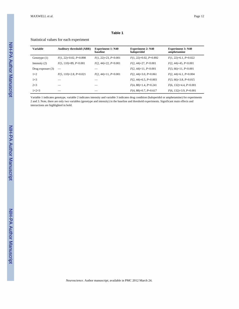
Table 1
Statistical values for each experiment
Variable Auditory threshold (ABR) Experiment 1: N40baseline
Experiment 2: N40haloperidol
Experiment 3: N40amphetamine
Genotype (1) F(1, 22)<0.02, P=0.898 F(1, 22)=23, P<0.001 F(1, 22)=0.02, P=0.892 F(1, 22)=6.1, P=0.022
Intensity (2) F(5, 110)=89, P<0.001 F(2, 44)=22, P<0.001 F(2, 44)=27, P<0.001 F(2, 44)=45, P<0.001
Drug exposure (3) — — F(2, 44)=11, P<0.001 F(3, 66)=11, P<0.001
1×2 F(5, 110)=2.8, P=0.021 F(2, 44)=11, P<0.001 F(2, 44)=3.0, P=0.061 F(2, 44)=6.1, P=0.004
1×3 — — F(2, 44)=6.5, P=0.003 F(3, 66)=3.8, P=0.015
2×3 — — F(4, 88)=1.4, P=0.241 F(6, 132)=4.4, P<0.001
1×2×3 — — F(4, 88)=0.7, P=0.617 F(6, 132)=3.9, P=0.001
Variable 1 indicates genotype, variable 2 indicates intensity and variable 3 indicates drug condition (haloperidol or amphetamine) for experiments2 and 3. Note, there are only two variables (genotype and intensity) in the baseline and threshold experiments. Significant main effects andinteractions are highlighted in bold.
Neuroscience. Author manuscript; available in PMC 2012 March 24.

NIH
-PA Author Manuscript
NIH
-PA Author Manuscript
NIH
-PA Author Manuscript
MAXWELL et al. Page 13
Table 2
Amphetamine by gene by intensity interaction for N40 amplitude
Amphetamine (mg/kg) Stimulus intensity (dB) Wild type Transgenic P-value
0
75 21±7 22±8 0.973
85 89±17 29±19 0.133
95 181±24 54±26 0.003
0.1
75 48±12 26±13 0.568
85 77±18 47±19 0.436
95 107±21 80±23 0.484
0.5
75 21±6 15±7 0.881
85 47±11 12±12 0.360
95 64±13 34±14 0.440
1
75 13±7 17±8 0.935
85 15±7 12±8 0.926
95 33±10 43±11 0.800
Note that amphetamine dose dependently decreases the normal intensity function (gain) for N40 amplitude in wild type but not transgenic animals.Fisher LSD post hoc analyses (MS=2531, df=77). P-values are listed for wild type vs. transgenic mice.
Neuroscience. Author manuscript; available in PMC 2012 March 24.
Related Documents