/mhj In Vivo Quantitative Assessment of Carotid Plaque component with multi-contrast MRI Jannie Wijnen 22 may 2003 using clustering algorithms, implemented in Mathematica

mhj In Vivo Quantitative Assessment of Carotid Plaque component with multi-contrast MRI Jannie Wijnen 22 may 2003 using clustering algorithms, implemented.
Dec 21, 2015
Welcome message from author
This document is posted to help you gain knowledge. Please leave a comment to let me know what you think about it! Share it to your friends and learn new things together.
Transcript
/mhj
In Vivo Quantitative Assessment of Carotid Plaque component with
multi-contrast MRI
Jannie Wijnen
22 may 2003
using clustering algorithms, implemented in Mathematica

/mhj
Question from the clinic
•If the highly thrombogenic plaque content is exposed to
the blood, thrombo-embolic incidents as myocardial
infarction, stroke, and peripheral vascular disease might
occur
•Design a program that automatically detects the
various components of the atherosclerotic plaque
• The program should be accurate and easy to use

/mhj
Solution to the question
•5 different MR images of the plaque -> notebook
•combining these images in a 5-dimensional
feature space
•search for clusters in feature space that represent
a tissue (hemorrhage, fibrous tissue, calcium and
lipid)
•show these clusters in the original image
/mhj
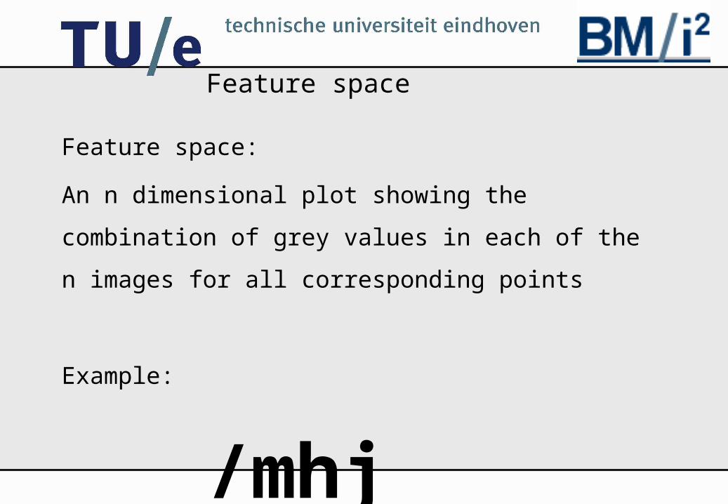
Feature space
Feature space:
An n dimensional plot showing the
combination of grey values in each of the n
images for all corresponding points
Example:
/mhj
ik10 3 74 6 71 8 0
y{0
2.5
5
7.5
10
2
4
6
8
2
4
6
8
10
0
2.5
5
7.5
10
2
4
6
8

/mhj
Clustering
Example of 3D feature space of the images
Kmeans cluster algorithm : Place K points into the
space represented by the objects that are being
clustered. These points represent initial group
centroids. Assign each object to the group that has the
closest centroid
Example of clusters in 3D space
/mhj
Problems
For the best classification the clusters should
be small and well- separated from each
other
•background intensity
•image miss-registration
•noise

/mhj
Background intensities
Example of clusters in images with a large intensity
gradient
Background correction by minimisation of
entropy of image with a known gradient.
Conclusion:
no background correction needed in these
images.
/mhj
Image Registration
•Pixels that are compared to each other must
represent exactly the same anatomical positions.
Example of non- registered images
•Minimisation of the mutual information (entropy)
leads to the displacement needed to register the
images.
Example of registered images

/mhj
Noise Reduction
The less noise the smaller the clusters
Euclideanshortening: preserves the
edges
example
Variance of the clusters after
euclideanshortening is smaller

/mhj
Starting Values
Difference between the clustering with randomly chosen starting values and starting values chosen by user

/mhj
Starting values
•The clustering is not reproducible when
starting values are chosen by hand
•It is better to extract starting values form
the information in the 5D space
•try to find starting values from the
maximum intensities in the 5D space
/mhj
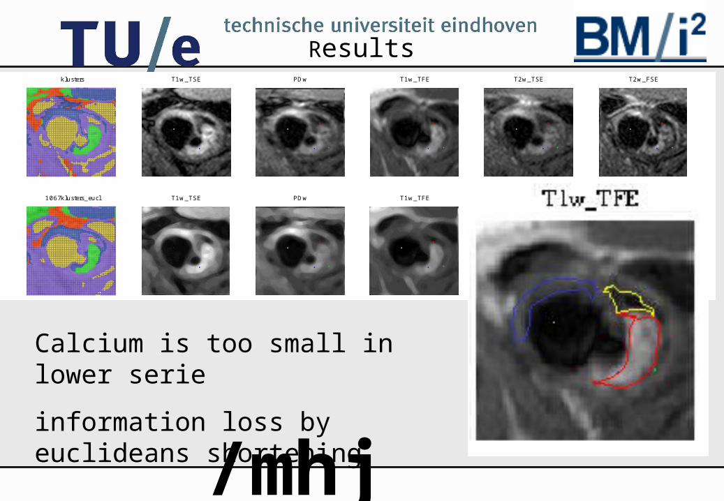
Resultsklusters T1w_TSE PDw T1w_TFE T2w_TSE T2w_FSE
1067klusters_eucl T1w_TSE PDw T1w_TFE T2w_TSE T2w_FSE
Calcium is too small in lower serie
information loss by euclideans shortening
/mhj
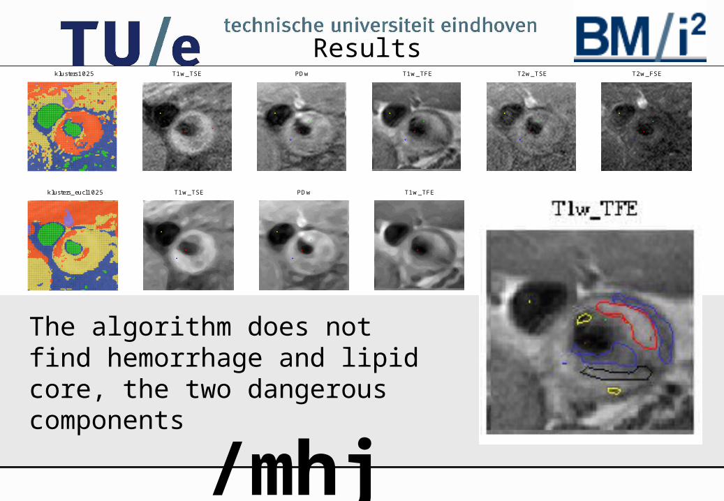
Resultsklusters1025 T1w_TSE PDw T1w_TFE T2w_TSE T2w_FSE
klusters_eucl1025 T1w_TSE PDw T1w_TFE T2w_TSE T2w_FSE
The algorithm does not find hemorrhage and lipid core, the two dangerous components
/mhj
Discussion /Conclusion
•Cluster techniques can be used for
tissue detection in atherosclerotic plaque
•A lot of progress can be made:
starting values, cluster technique, non-
linear registration, smoothing.
Related Documents