MFGE8 Does Not Influence Chorio-Retinal Homeostasis or Choroidal Neovascularization in vivo William Raoul 1,2,3 *, Lucie Poupel 5,6 , David-Alexandre Tregouet 9,15 , Sophie Lavalette 1,2,3 , Serge Camelo 8,9,10 , Nicole Keller 8,9,10 , Sophie Krumeich 11,12 , Bertrand Calippe 1,2,3 , Xavier Guillonneau 1,2,3 , Francine Behar-Cohen 4,8,9,10 , Salomon-Yves Cohen 14 , Holger Baatz 13 , Christophe Combadie `re 5,6,7 , Clotilde The ´ry 11,12 , Florian Sennlaub 1,2,3,4 * 1 INSERM, U968, Paris, France, 2 University Pierre and Marie Curie, Institut de la Vision, Paris, France, 3 Centre National de la Recherche Scientifique, Paris, France, 4 Assistance Publique-Ho ˆ pitaux de Paris, Ho ˆ tel Dieu, Service d’Ophtalmologie, Paris, France, 5 INSERM, UMR S945, Laboratory of Immunity and Infection, Paris, France, 6 University Pierre and Marie Curie, Laboratory of Immunity and Infection, Paris, France, 7 Assistance Publique-Ho ˆ pitaux de Paris, Groupe Hospitalier Pitie ´ -Salpe ´trie `re, Service d’Immunologie, Paris, France, 8 INSERM, UMR S 872, Centre de Recherche des Cordeliers, Paris, France, 9 University Pierre and Marie Curie, Paris, France, 10 Universite ´ Paris Descartes, Paris, France, 11 INSERM, U932, Paris, France, 12 Institut Curie, Centre de Recherche, Paris, France, 13 Augena ¨rztliche Gemeinschaftspraxis, Augenzentrum Recklinghausen und Zentrum der Augenheilkunde, J-W Goethe Universita ¨t Frankfurt am Main, Frankfurt, Germany, 14 Centre d’Angiographie et de Laser, Paris, France, 15 INSERM, UMR S 937, Faculte ´ de Me ´decine La Pitie ´ -Salpe ˆtrie `re, Paris, France Abstract Purpose: Milk fat globule-epidermal growth factor-factor VIII (MFGE8) is necessary for diurnal outer segment phagocytosis and promotes VEGF-dependent neovascularization. The prevalence of two single nucleotide polymorphisms (SNP) in MFGE8 was studied in two exsudative or ‘‘wet’’ Age-related Macular Degeneration (AMD) groups and two corresponding control groups. We studied the effect of MFGE8 deficiency on retinal homeostasis with age and on choroidal neovascularization (CNV) in mice. Methods: The distribution of the SNP (rs4945 and rs1878326) of MFGE8 was analyzed in two groups of patients with ‘‘wet’’ AMD and their age-matched controls from Germany and France. MFGE8-expressing cells were identified in Mfge8 +/2 mice expressing ß-galactosidase. Aged Mfge8 +/2 and Mfge8 2/2 mice were studied by funduscopy, histology, electron microscopy, scanning electron microscopy of vascular corrosion casts of the choroid, and after laser-induced CNV. Results: rs1878326 was associated with AMD in the French and German group. The Mfge8 promoter is highly active in photoreceptors but not in retinal pigment epithelium cells. Mfge8 2/2 mice did not differ from controls in terms of fundus appearance, photoreceptor cell layers, choroidal architecture or laser-induced CNV. In contrast, the Bruch’s membrane (BM) was slightly but significantly thicker in Mfge8 2/2 mice as compared to controls. Conclusions: Despite a reproducible minor increase of rs1878326 in AMD patients and a very modest increase in BM in Mfge8 2/2 mice, our data suggests that MFGE8 dysfunction does not play a critical role in the pathogenesis of AMD. Citation: Raoul W, Poupel L, Tregouet D-A, Lavalette S, Camelo S, et al. (2012) MFGE8 Does Not Influence Chorio-Retinal Homeostasis or Choroidal Neovascularization in vivo. PLoS ONE 7(3): e33244. doi:10.1371/journal.pone.0033244 Editor: Alfred Lewin, University of Florida, United States of America Received November 28, 2011; Accepted February 6, 2012; Published March 15, 2012 Copyright: ß 2012 Raoul et al. This is an open-access article distributed under the terms of the Creative Commons Attribution License, which permits unrestricted use, distribution, and reproduction in any medium, provided the original author and source are credited. Funding: This work was supported by grants from INSERM, Agence Nationale pour la Recherche (ANR) ‘‘blanc’’ (AO5120DD), ANR ‘‘Maladies Neurologiques et Maladies Psychiatriques’’ (R08098DS), ANR ‘‘Genopat’’ (R09099DS), European Grant ‘‘Innochem’’ (LSHB-CT-2005-518167), ERC starting Grant (ERC-2007 St.G. 210345), and a grant from Fondation de France to C.T. F.S. a is recipient of ERC starting grant. C.C. and F.S. are recipients of a contract ‘‘Interface’’ from Assistance Publique-Ho ˆ pitaux de Paris. The funders had no role in study design, data collection and analysis, decision to publish, or preparation of the manuscript. Competing Interests: The authors have declared that no competing interests exist. * E-mail: [email protected] (WR); [email protected] (FS) Introduction Milk fat globule-EGF-factor (MFGE8), also named lactadherin, PAS 6/7, SED1, BA46, p47, is a secreted glycoprotein first described in milk fat globules released in milk by mammary epithelial cells [1,2]. Secreted by different cell types, it promotes phagocytosis by linking phosphatidylserine at the surface of membrane vesicles [3] and apoptotic cells [4] to the avb3/b5 integrin on phagocytic cells. MFGE8 mediated phagocytosis induces a regulatory T cell response [5] and Mfge8 2/2 mice develop spontaneous late onset lupus-like disease and glomerulo- nephritis [6]. In humans, the association of two nucleotide polymorphisms in the coding region of MFGE8 predisposes subjects to systemic lupus erythematosus [7], suggesting that these single nucleotide polymorphisms (SNP) lead to a dysfunc- tional MFGE8. Furthermore, MFGE8 binds to avb3/b5 of vascular endothelial cells and promotes VEGF-driven neovascu- larization [8]. Phagocytosis of spent outer segments (OS) is critical for the long-term maintenance of the retina [9,10] and dependent upon a tyrosine kinase receptor (Mertk) [11,12] and the avb5 integrin [13]. The neural retina and the retinal pigment epithelium (RPE) PLoS ONE | www.plosone.org 1 March 2012 | Volume 7 | Issue 3 | e33244

Welcome message from author
This document is posted to help you gain knowledge. Please leave a comment to let me know what you think about it! Share it to your friends and learn new things together.
Transcript

MFGE8 Does Not Influence Chorio-Retinal Homeostasisor Choroidal Neovascularization in vivoWilliam Raoul1,2,3*, Lucie Poupel5,6, David-Alexandre Tregouet9,15, Sophie Lavalette1,2,3,
Serge Camelo8,9,10, Nicole Keller8,9,10, Sophie Krumeich11,12, Bertrand Calippe1,2,3,
Xavier Guillonneau1,2,3, Francine Behar-Cohen4,8,9,10, Salomon-Yves Cohen14, Holger Baatz13,
Christophe Combadiere5,6,7, Clotilde Thery11,12, Florian Sennlaub1,2,3,4*
1 INSERM, U968, Paris, France, 2 University Pierre and Marie Curie, Institut de la Vision, Paris, France, 3 Centre National de la Recherche Scientifique, Paris, France,
4 Assistance Publique-Hopitaux de Paris, Hotel Dieu, Service d’Ophtalmologie, Paris, France, 5 INSERM, UMR S945, Laboratory of Immunity and Infection, Paris, France,
6 University Pierre and Marie Curie, Laboratory of Immunity and Infection, Paris, France, 7 Assistance Publique-Hopitaux de Paris, Groupe Hospitalier Pitie-Salpetriere,
Service d’Immunologie, Paris, France, 8 INSERM, UMR S 872, Centre de Recherche des Cordeliers, Paris, France, 9 University Pierre and Marie Curie, Paris, France,
10 Universite Paris Descartes, Paris, France, 11 INSERM, U932, Paris, France, 12 Institut Curie, Centre de Recherche, Paris, France, 13 Augenarztliche Gemeinschaftspraxis,
Augenzentrum Recklinghausen und Zentrum der Augenheilkunde, J-W Goethe Universitat Frankfurt am Main, Frankfurt, Germany, 14 Centre d’Angiographie et de Laser,
Paris, France, 15 INSERM, UMR S 937, Faculte de Medecine La Pitie-Salpetriere, Paris, France
Abstract
Purpose: Milk fat globule-epidermal growth factor-factor VIII (MFGE8) is necessary for diurnal outer segment phagocytosisand promotes VEGF-dependent neovascularization. The prevalence of two single nucleotide polymorphisms (SNP) in MFGE8was studied in two exsudative or ‘‘wet’’ Age-related Macular Degeneration (AMD) groups and two corresponding controlgroups. We studied the effect of MFGE8 deficiency on retinal homeostasis with age and on choroidal neovascularization(CNV) in mice.
Methods: The distribution of the SNP (rs4945 and rs1878326) of MFGE8 was analyzed in two groups of patients with ‘‘wet’’AMD and their age-matched controls from Germany and France. MFGE8-expressing cells were identified in Mfge8+/2 miceexpressing ß-galactosidase. Aged Mfge8+/2 and Mfge82/2 mice were studied by funduscopy, histology, electronmicroscopy, scanning electron microscopy of vascular corrosion casts of the choroid, and after laser-induced CNV.
Results: rs1878326 was associated with AMD in the French and German group. The Mfge8 promoter is highly active inphotoreceptors but not in retinal pigment epithelium cells. Mfge82/2 mice did not differ from controls in terms of fundusappearance, photoreceptor cell layers, choroidal architecture or laser-induced CNV. In contrast, the Bruch’s membrane (BM)was slightly but significantly thicker in Mfge82/2 mice as compared to controls.
Conclusions: Despite a reproducible minor increase of rs1878326 in AMD patients and a very modest increase in BM inMfge82/2 mice, our data suggests that MFGE8 dysfunction does not play a critical role in the pathogenesis of AMD.
Citation: Raoul W, Poupel L, Tregouet D-A, Lavalette S, Camelo S, et al. (2012) MFGE8 Does Not Influence Chorio-Retinal Homeostasis or ChoroidalNeovascularization in vivo. PLoS ONE 7(3): e33244. doi:10.1371/journal.pone.0033244
Editor: Alfred Lewin, University of Florida, United States of America
Received November 28, 2011; Accepted February 6, 2012; Published March 15, 2012
Copyright: � 2012 Raoul et al. This is an open-access article distributed under the terms of the Creative Commons Attribution License, which permitsunrestricted use, distribution, and reproduction in any medium, provided the original author and source are credited.
Funding: This work was supported by grants from INSERM, Agence Nationale pour la Recherche (ANR) ‘‘blanc’’ (AO5120DD), ANR ‘‘Maladies Neurologiques etMaladies Psychiatriques’’ (R08098DS), ANR ‘‘Genopat’’ (R09099DS), European Grant ‘‘Innochem’’ (LSHB-CT-2005-518167), ERC starting Grant (ERC-2007 St.G.210345), and a grant from Fondation de France to C.T. F.S. a is recipient of ERC starting grant. C.C. and F.S. are recipients of a contract ‘‘Interface’’ from AssistancePublique-Hopitaux de Paris. The funders had no role in study design, data collection and analysis, decision to publish, or preparation of the manuscript.
Competing Interests: The authors have declared that no competing interests exist.
* E-mail: [email protected] (WR); [email protected] (FS)
Introduction
Milk fat globule-EGF-factor (MFGE8), also named lactadherin,
PAS 6/7, SED1, BA46, p47, is a secreted glycoprotein first
described in milk fat globules released in milk by mammary
epithelial cells [1,2]. Secreted by different cell types, it promotes
phagocytosis by linking phosphatidylserine at the surface of
membrane vesicles [3] and apoptotic cells [4] to the avb3/b5
integrin on phagocytic cells. MFGE8 mediated phagocytosis
induces a regulatory T cell response [5] and Mfge82/2 mice
develop spontaneous late onset lupus-like disease and glomerulo-
nephritis [6]. In humans, the association of two nucleotide
polymorphisms in the coding region of MFGE8 predisposes
subjects to systemic lupus erythematosus [7], suggesting that
these single nucleotide polymorphisms (SNP) lead to a dysfunc-
tional MFGE8. Furthermore, MFGE8 binds to avb3/b5 of
vascular endothelial cells and promotes VEGF-driven neovascu-
larization [8].
Phagocytosis of spent outer segments (OS) is critical for the
long-term maintenance of the retina [9,10] and dependent upon a
tyrosine kinase receptor (Mertk) [11,12] and the avb5 integrin
[13]. The neural retina and the retinal pigment epithelium (RPE)
PLoS ONE | www.plosone.org 1 March 2012 | Volume 7 | Issue 3 | e33244
express MFGE8 [14], whose interaction with avb5 integrin of
RPE cells is essential in the diurnal OS phagocytosis [15].
Furthermore, we have previously shown that OS phagocytosis by
the RPE is necessary for choroidal maintenance in vivo [16].
Age-related macular degeneration (AMD) is a major cause of
central vision loss in the elderly in Western countries [17]. AMD’s
most prominent pathological features are lesions involving the
RPE and Bruch’s membrane (BM), the degeneration of photore-
ceptors, [18] and VEGF-driven choroidal neovascularization
(CNV) [19], that occurs approximately in 10% of patients. The
causes of AMD are not well understood, but epidemiological
studies [20] and murine models [21] have identified key factors in
its pathogenesis such as age [17,22], family history [23] and
smoking [24,25].
In summary, MFGE8 has been shown to be essential in diurnal
OS phagocytosis and in angiogenesis. In the eye, its dysfunction
could therefore lead both to retinal degeneration and choroidal
involution as a consequence of impaired RPE phagocytosis and/or
to inhibition of neovascularization. We investigated whether a
SNP associated with increased prevalence of lupus in human
patients [7] is associated with AMD. Furthermore, we investigated
whether the deficiency of MFGE8 affects chorioretinal homeosta-
sis and CNV in vivo using Mfge82/2 mice.
Materials and Methods
Ethics statementIn accordance with the Declaration of Helsinki, patients and
volunteers provided written and informed consent for the studies,
which were approved by the Hotel Dieu ethics committee
(CCPPRB no. 0611303) and by the ethics committee of the
Arztekammer Westfalen-Lippe and the University of Munster
(no. 2007-246-f-S).
Animal experiments were approved by the Institutional Animal
Care and Use Committee, ‘‘Comite d’ethique pour l’experimen-
tation animale Charles Darwin’’ (ID Ce5/2010/002), and treated
in compliance with the ARVO Statement for the Use of Animals
in Ophthalmic and Vision Research.
Single Nucleotide Polymorphisms analysis in AMDStudies, including participants’ assessments and ethics details of
each cohort were described in Combadiere et al. [26] and Baatz et
al. [27]. White Caucasian control subjects and AMD patients were
recruited in Paris (France: 251 control subjects, 274 ‘‘wet’’ AMD
patients) and in Recklinghausen (Germany: 317 control subjects,
263 ‘‘wet’’ AMD patients). The mean age and gender distribution
are summarized in Tables 1 and 2.
Rs4945 is in open reading frame SNP and leads to Arginine
replacement of a Serine in the amino acid number 3. Rs1878326 is
also in open reading frame SNP of MFGE8 and leads to Leucine
replacement of Methionine in the amino acid number 76. Allelic
frequencies were calculated by gene counting.
We used the chi-square test (x2) to compare genotype
distributions and allele frequencies in participants. The Hardy-
Weinberg equilibrium was tested using a x2 test with 1 degree of
freedom. Association between SNP and ‘‘wet’’ AMD was assessed
by use of the Cochran-Armitage’s trend test [28]. Linkage
disequilibrium and haplotype association analyses were performed
by the use of THESIAS software [29].
AnimalsMfge82/2 and +/2 mouse strains were generated as described
before [8]. Mfge82/2 are functionally deficient in MFGE8 due to a
gene-trap insertion of ß-galactosidase in the Mfge8 gene (C57Bl/6
background, N8). The mice were maintained at the Institut Curie
animal facility or Centre de Recherche des Cordeliers animal
facility (Paris, France) under pathogen-free conditions. All animals
were housed in a 12/12 hour light/dark (100–500 lux) cycle with
food and water available ad libitum.
Fundus Photography and Laser-PhotocoagulationMice were anesthetized by intraperitoneal injection of pento-
barbital (40 mg/kg). Pupils were fully dilated with 1% tropic-
amide. Coverslips positioned on the mouse cornea were used as a
contact glass. Fundus photographs were taken with a digital CCD
camera (Nikon D3) coupled with an endoscope (Karl Storz,
Guyancourt, France) as previously described [30].
Laser-photocoagulations were performed 1 to 2 disc diameters
away from the papillae with an Argon laser (Viridis 532 nm,
Quantel Medical, Clermont-Ferrand, France) mounted on a slit
lamp (400 mW, 50 ms and 50 mm; Hagg-Streitt, BQ 900). For
CNV visualization at day 14, 3 months old mice were anesthetized
and perfused through their heart with phosphate buffered saline
(PBS) containing fluorescein (FITC)-dextran 50 mg/ml
(2.106 mW). Animals were sacrificed with 100% CO2 and their
eyes were removed and processed as described below. CNV was
quantified on photographs with Image J analysis software. CNV
was quantified as FITC positive surface.
Choroidal Flatmounts and ImmunohistochemistryThe eyes were enucleated, fixed in 4% paraformaldehyde (PFA)
for 20 minutes at room temperature, and sectioned at the limbus;
the cornea and lens were discarded. The retinas were peeled from
the RPE/choroid/sclera. Retinas and choroids were incubated
with the indicated primary and secondary antibodies. The
choroids and retinas were radially incised and flatmounted. The
primary antibodies used were rabbit anti-IBA1 (1:400; Wako,
Table 1. The mean age and gender distribution in the patientand control groups.
Patients FCTL, n (%) FAMD, n (%)
Men 91 (36,25) 81 (29,56)
Women 160 (63,75) 193 (70,44)
Total 251 274
Mean Age 71,8+/27,75 79,5+/26,72
P value (x2 test) 0.1
French groups (control group : FCTL; AMD group : FAMD).doi:10.1371/journal.pone.0033244.t001
Table 2. The mean age and gender distribution in the patientand control groups.
Patients GCTL, n (%) GAMD, n (%)
Men 115 (43,73) 126 (39,75)
Women 148 (56,27) 191 (60,25)
Total 263 317
Mean Age 77,8+/25,38 79,2+/25,47
P value (x2 test) 0.3
German groups (control group : GCTL; AMD group : GAMD).doi:10.1371/journal.pone.0033244.t002
MFGE8 in Aging Retina and CNV
PLoS ONE | www.plosone.org 2 March 2012 | Volume 7 | Issue 3 | e33244

Neuss, Germany) and rhodamine phalloidine (1:200; Invitrogen,
Cergy-Pontoise, France). Sections were counterstained with 4–6-
diamino-2-phenylindole (DAPI). Choroids, retinas and sections
were viewed with a fluorescence microscope (DM5500B, Leica,
Nanterre, France).
For ß-galactosidase activity detection, frozen sections from eyes
were fixed in 4% PFA before incubation. The samples were then
incubated in the beta-galactosidase detection reagent (5 mM
potassium ferricyanide, 5 mM potassium ferrocyanide, 2 mM
MgCl, 0.30 mg X-gal/ml in PBS) overnight at 37uC before
mounting in Immumount (Thermo Scientific, Courtaboeuf,
France).
Histology and Electron MicroscopyFor histology, eyes were fixed in 0.5% glutaraldehyde/4% PFA
for 2 h, dehydrated and mounted in historesin. 5-mm sections were
cut and stained with toluidin blue. For electron microscopy, eyes
were fixed in 2.5% glutaraldehyde of cacodylate buffer (0.1 M,
pH 7.4). After 1 hour, eyeballs were dissected, fixed for another
3 hours, post-fixed in 1% osmium tetroxide in cacodylate buffer,
and dehydrated in graduated ethanol solution. The samples were
included in epoxy resin and oriented. Semi-thin sections (1 mm),
obtained with an ultramicrotome Reichert Ultracut E (Leica),
were stained with toluidin blue, examined with a light microscope,
and photoreceptor layer thickness was measured. Ultra-thin
sections (80 nm) were contrasted by uranyl acetate and lead
citrate, then observed in an electron microscope JEOL 100 CX II
(JEOL, Tokyo, Japan) with 80 kV.
Vascular Corrosion CastsAnimals were sacrificed by CO2 inhalation. Vascular corrosion
cast was performed as previously described by Houssier et al. [16].
Briefly, a perfusion was done with Mercox resin+catalyst (Ladd
Research/Inland, Saint Loup sur Semouse, France) into the aorta
through left heart ventricle. After removal, eye tissues were
conserved overnight at 37uC in PBS to allow complete
polymerisation and digested by 5% KOH during 2 weeks at
37uC until only the vascular corrosion casts remained. The
specimens were prepared for electron microscopy, then scanned
and analyzed using Image J Software (NIH). The avascular area
was measured on frontal views and expressed as the percentage of
intercapillary surface of the whole area. The thickness of
choriocapillaries was measured on perpendicular views of the cast
from the retinal to scleral side of the choriocapillary cast.
Reverse Transcription and Real Time Polymerase ChainReaction
After tissues were separated (neural retina and choroid/RPE),
total RNA was isolated with NucleoSpin RNA II Kit (Macherey-
Nagel, Hoerdt, France). Single-stranded cDNA was synthesized
from total RNA (pre-treated with DNaseI amplification grade)
using oligodT as primer and superscript reverse transcriptase
(Invitrogen, Cergy Pontoise, France). Subsequent real-time
polymerase chain reaction (RT PCR) was performed using cDNA,
qPCR SuperMix-UDG Platinum SYBR Green (Invitrogen), and
the following primers (0.5 pmol/ml): mm actin sense: 59-AAGGC-
CAACCGTGAAAAGAT-39; mm actin antisense : 59-
GTGGTACGACCAGAGGCATAC-39; mm Mfge8 sense : 59-
GGATAATCAGGGCAAGATCA-39; mm Mfge8 antisense : 59-
TAGGACGCCACATACTGGAT-39
PCR reactions were performed in 40 cycles of 15 s at 95uC, 45 s
at 60uC. Product was not generated in control reactions in which
reverse transcriptase was omitted during cDNA synthesis.
Statistical AnalysisGraph Pad Prism 5 (GraphPad Software, San Diego, CA, USA)
was used for the data analysis and graphic representations. All
values are reported as means 6 SEM. Statistical analysis was
performed using the Mann-Whitney test for comparison among
means between two groups or ANOVA test between more than
two groups. Significance was set at P,0.05.
Results
MFGE8 single nucleotide polymorphism studies in twogroups of wet AMD patients and their controls
Two common SNP were previously identified in the open
reading frame of the MFGE8 gene called MFGE8 R3S (rs4945 for
nucleotide C.A) and MFGE8 M76L (rs1878326 for nucleotide
226 A.C), associated with an increased risk of systemic lupus
erythematosus [7]. The fact that Mfge82/2 mice develop a
spontaneous late onset lupus-like disease and glomerulonephritis
[6] suggests that rs4945 and rs1878326 lead to a decrease of
MFGE8 bioactivity, that would explain their association with the
human disease.
We thus investigated the described MFGE8 SNP in two DNA
collections of ‘‘wet’’ AMD patients recruited in France (FAMD,
274 subjects) and in Germany (GAMD, 317 subjects) and their age
matched controls (FCTL, 251 subjects; GCTL, 263 subjects). The
mean age and gender distribution are summarized in Tables 1 and
2. All of the subjects were of caucasian origin or decent. Risk
factors for ‘‘wet’’ AMD such as Factor H and smoking were more
frequent among cases than among controls as previously described
[31].
Genotype frequencies distribution of the rs4945 C.A (R3S)
and rs1878326 A.C (M76L) are shown in Tables 3 and 4,
respectively. All genotype distributions were compatible with
Hardy-Weinberg equilibrium (P.0.05) in cases and in controls,
from both French and German samples. In the French sample, the
rs4945 -A allele was more frequent in cases than in controls (0.36
vs 0.29, P = 0.017) but the inverse trend was observed in German
(0.30 vs 0.33, P = 0.229). Conversely, the pattern of association of
the rs1878326 with AMD was homogeneous across populations.
In both samples, the rs1878326-C allele tended to be more
frequent in cases than in controls, 0.36 vs 0.32, P = 0.184 and 0.41
vs 0.35, P = 0.053, in French and German, respectively. The
corresponding OR for AMD were then 1.19 [0.92–1.53] in
French and 1.27 [1.00–1.61] in German. The two ORs were not
significantly different (P = 0.92). Even though statistical signifi-
cance was not reached separately in each sample (likely due to
small sample size), the resulting combined OR for AMD
associated with the rs1878326-C allele was 1.23 [1.03–1.46] and
achieved significance (P = 0.019).
The pattern of linkage disequilibrium was homogeneous across
populations and was characterized by a moderate linkage
disequilibrium between the two studied SNP (r2 = 0.07,
D9 = 20.53 in French and r2 = 0.10, D9 = 20.60) in German).
As a consequence, the two SNP generated 4 common haplotypes
(Table 5) among which the two haplotypes carrying the
rs1878326-C allele were more frequent in cases than in controls
in the German and the French study, confirming the association of
the rs1878326-C allele with AMD observed in the univariate
analysis.
MFGE8 expressing cells in the retinaIn the litterature, immunohistochemistry using an anti-MFGE8
antibody localized MFGE8 protein mainly to the inner segments
of photoreceptors and to a lesser extent to RPE cells in mice using
MFGE8 in Aging Retina and CNV
PLoS ONE | www.plosone.org 3 March 2012 | Volume 7 | Issue 3 | e33244

two different antibodies [14]. We identified MFGE8 expressing
cells in Mfge8+/2 mice thanks to the gene-trap insertion of ß-
galactosidase in the Mfge8 gene. The resulting fusion protein is
retained in the cell and ß-galactosidase activity therefore allows to
identify MFGE8 expressing cells [8]. ß-galactosidase, detected by a
ß-galactosidase detection reagent, mainly localizes to the outer
nuclear layer (ONL) containing photoreceptor cell bodies (Fig. 1A).
Higher magnification reveals that staining is particularly strong in
the outer part of the ONL and that the RPE does express little ß-
galactosidase (Fig. 1B), confirming the previously published results
using MFGE8 antibodies [14]. There was no endogenous ß-
galactosidase detection in WT mice (data not shown). We
confirmed this staining pattern using RT PCR in separated tissues
(Fig. 1C). Mfge8 mRNA was highly expressed in young and aged
neural retina. In the RPE/choroid, basal mRNA expression was
significantly lower than in the neural retina. A slight but significant
increase in Mfge8 mRNA expression at 18 month old compared to
3-month-old C57Bl6 mice was observed. Taken together, our
results identify photoreceptors as the main source of MFGE8 in
the eye and suggest that MFGE8 localization in the RPE cells [14]
is due to uptake rather than RPE MFGE8 expression.
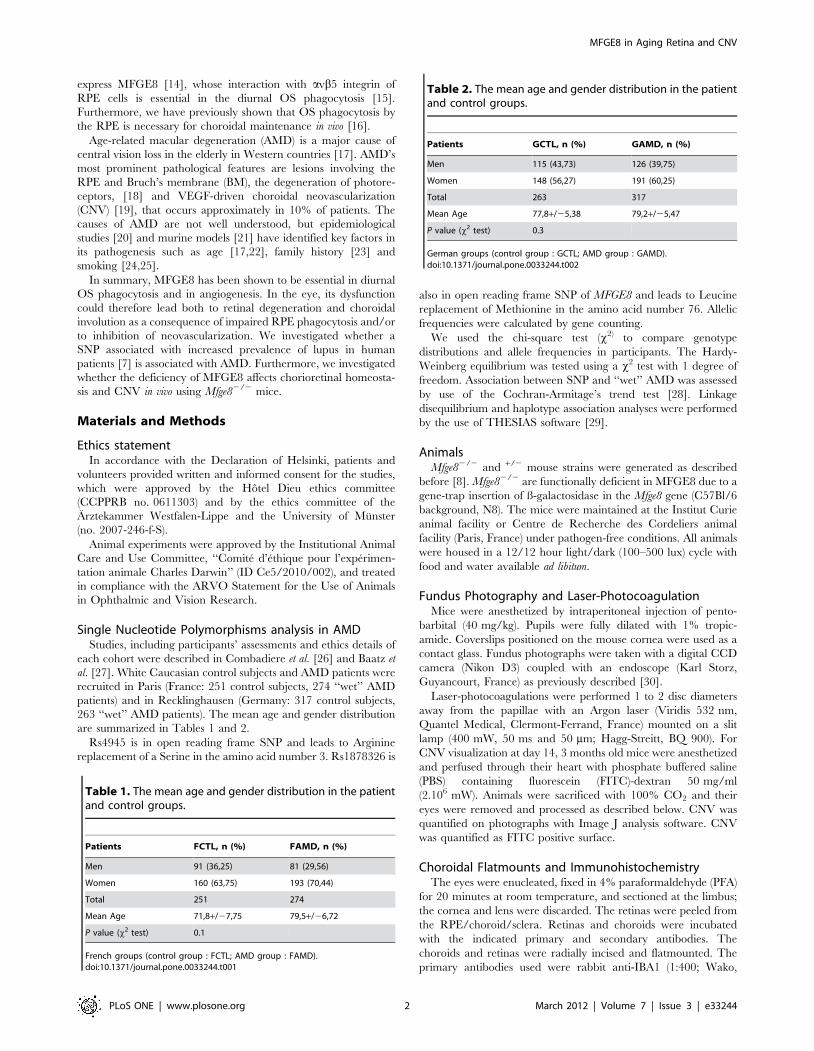
MFGE8 and retinal homeostasisMFGE8 has been shown to be essential in diurnal OS
phagocytosis [15] and disturbance of the phagocytosis of spent
OS by the RPE, as observed in the RCS rat, leads to
photoreceptor degeneration [10]. To evaluate if MFGE8 defi-
ciency alters long-term retinal homeostasis, we first studied the
fundoscopic appearance of 16–18 month old Mfge8+/2 (Fig. 2A)
and Mfge82/2 (Fig. 2B). Both fundi appeared smooth, devoid of
any remarkable lesions, and similar to each other. Furthermore,
histological sections of aged Mfge8+/2 (Fig. 2C) and Mfge82/2 mice
(Fig. 2D) showed a regular photoreceptor layer and quantification
of the number of photoreceptor cell nuclei layers in the ONL
revealed no signs of degeneration from the inferior to the superior
pole in Mfge82/2 (Fig. 2E grey line) compared to Mfge8+/2 mice
(Fig. 2E black line). Labelling for RPE (phalloidine, red) and
subretinal macrophages/microglial cells (IBA1, green) in aged
Mfge8+/2 (Fig. 2F) and Mfge82/2 mice (Fig. 2G) showed no
morphological abnormalities of RPE cells and no pathological
accumulation of phagocytes under the retina. Instead, we found a
tendency of decreased subretinal phagocytes in 18-month-old
Mfge82/2 compared to Mfge8+/2 (Fig. 2H, quantification of
subretinal IBA1 positive cells). Laminar deposits in the BM and
Drusen in the RPE/BM complex are early signs of AMD that
ultimately impact RPE and photoreceptor health [18,32]. To
evaluate if MFGE8 is implicated in homeostasis of BM, we
evaluated BM morphology of 16–18 month old Mfge8+/2 (Fig. 2I
and K) and Mfge82/2 (Fig. 2J and L) by electron microscopy.
Although retina and RPE were ultrastructurally similar, we
observed a very modest, but significant thickening of BM in
Mfge82/2 mice by 22% (Fig. 2M). To evaluate possible differences
in genes that are potentially involved in lipid clearance from the
BM, we analyzed Abca1, Abca4, Ldlr, cd36 and Caveolin1 mRNA in
aged Mfge8+/2 and Mfge82/2. However, we were not able to
detect an influence of MFGE8 on the expression of these genes
(Figure S1). Since BM is rich in collagen, and MFGE8 has been
shown to promote collagen phagocytosis to prevent reactive
fibrosis [33], BM thickening observed in aged Mfge82/2 mice
could be a result of a reactive fibrosis.
Table 3. Genotype distribution of MFGE8 rs4945 (R3S) polymorphism in AMD patients and controls.
MFGE8 rs4945 FCTL, n = 241 FAMD, n = 271 GCTL, n = 260 GAMD, n = 308
CC 122 (51%) 110 (41%) 118 (45%) 154 (50%)
CA 99 (41%) 128 (47%) 112 (43%) 125 (41%)
AA 20 (8%) 33 (12%) 30 (12%) 29 (9%)
MAF 0.29 0.36 0.33 0.30
OR [95%CI] 1.376 [1.056–1.791] 0.855 [0.665–1.100]
P 0.017 0.229
MAF: Minor Allele Frequency.OR: Allelic Odds Ratio with its 95% Confidence Interval.P: Cochran-Armitage trend test’s p-value.doi:10.1371/journal.pone.0033244.t003
Table 4. Genotype distribution of MFGE8 rs1878326 (M76L) polymorphism in AMD patients and controls.
MFGE8 rs1878326 FCTL, n = 251 FAMD, n = 274 GCTL, n = 263 GAMD, n = 317
AA 108 (43%) 114 (42%) 112 (43%) 113 (35%)
AC 123 (49%) 121 (44%) 116 (44%) 148 (47%)
CC 20 (8%) 39 (14%) 35 (13%) 56 (18%)
MAF 0.32 0.36 0.35 0.41
OR [95%CI] 1.186 [0.919–1.531] 1.271 [1.001–1.613]
P 0.184 0.053
MAF: Minor Allele Frequency.OR: Allelic Odds Ratio with its 95% Confidence Interval.P: Cochran-Armitage trend test’s p-value.doi:10.1371/journal.pone.0033244.t004
MFGE8 in Aging Retina and CNV
PLoS ONE | www.plosone.org 4 March 2012 | Volume 7 | Issue 3 | e33244

MFGE8 and choroidal homeostasis andneovascularization
Although MFGE8 deficiency did not lead to retinal degener-
ation, disturbances in RPE biology and diminished expression of
trophic factors can lead to choroidal involution [16]. However,
vascular corrosion casts of of 16–18 month old Mfge8+/2 (Fig. 3 I
and D) and Mfge82/2 (Fig. 3 B and E) showed no MFGE8-related
vascular drop-out, measured as a percentage of intercapillary
area of the total area, (Fig. 3C) or thinning, measured on
pericentral, perpendicular sections through the vascular casts
(Fig. 3F).
MFGE8 interacts with avb3 and avb5 on vascular endothelium
and specifically promotes VEGF-driven ischemic neovasculariza-
tion in vivo, but not bFGF induced neovascularization [8]. Since
laser-induced CNV has been shown to be VEGF and integrin
dependent [34,35], we quantified CNV in the MFGE8-deficient
mice. Interestingly, FITC-dextran perfused choroidal flatmounts
at 14 d after laser-induced neovascularization of Mfge8+/2 (Fig. 3G)
and Mfge82/2 (Fig. 3H), showed no difference of CNV in the two
groups (Fig. 3I). These results are consistent with our recent
observation that the promoting function of MFGE8 on develop-
ment of bladder tumors is not linked with alterations in intra-
tumor angiogenesis [36]. Thus, the interactions of MFGE8,
integrin dimers and VEGF are possibly tissue dependent.
Discussion
MFGE8s essential role in diurnal OS phagocytosis [15] and
critical involvement in VEGF-driven neovascularization [8] make
it a candidate gene for an involvement in the pathogenesis of
AMD, where photoreceptor degeneration, choroidal involution, or
CNV can occur. We here show a suggestive statistical evidence for
association of the rs1878326-C allele with increased risk of AMD
in two European populations.
To evaluate the influence of MFGE8 dysfunction in vivo on
retinal homeostasis and CNV we analyzed Mfge8+/2 and Mfge82/2
mice with ageing and in a model of CNV. We first confirmed that
MFGE8 is constitutively expressed by photoreceptors in vivo.
Interestingly, the suppression of the diurnal peak of OS phagocy-
tosis observed in Mfge82/2 mice [15] has very little negative effect
on long term chorioretinal homeostasis in 18-month-old knockout
mice, apart from a slight thickening of the BM. These findings
confirm and extend the lack of retinal degeneration in 12-month-old
Mfge82/2 mice [37]. Furthermore, even though MFGE8 interacts
with avb3 and avb5 on the vascular endothelium and strongly
promotes VEGF-driven neovascularization [8], it has no effect on
laser-induced CNV which is VEGF and avb3 integrin dependent
[34,35].
Taken together, our data shows that MFGE8 is expressed in
the retina but not critically involved in retinal homeostasis or
CNV. It seems unlikely that it is predominantly involved in the
pathogenesis of AMD. Although we present mostly negative
results, we feel that this extensive in vivo study is of interest to
researchers in the field of AMD pathogenesis, due to its exclusion
of a high potential candidate gene. We hope that the publication of
these negative results are helpful to the community to exclude a
valid hypothesis and avoid repetetion of expensive research in
other laboratories.
Table 5. Haplotype frequency distributions derived from MFGE8 rs1878326 (M76L) and rs4945 (R3S) polymorphisms in AMDpatients and controls.
Polymorphisms French German
rs1878326 rs4945 FCTL (n = 238) FAMD (n = 269) GCTL (n = 253) GAMD (n = 298)
A C 0.426 0.342 0.355 0.344
A A 0.252 0.293 0.289 0.245
C C 0.284 0.299 0.310 0.359
C A 0.038 0.066 0.045 0.052
doi:10.1371/journal.pone.0033244.t005
Figure 1. MFGE8 expressing cells in mouse retina. Representativemicrographs of ß-galactosidase localization in MFGE8+/GAL micedetected by a ß-galactosidase detection reagent (blue staining). Phasecontrast (A) image and light transmitted (B) image. CH: choroid; GCL:ganglion cell layer; INL: inner nuclear layer; ONL: outer nuclear layer;RPE: retinal pigment epithelium. Experiments were reproduced at least3 times. Scale bars: 100 mm (A); 25 mm (B). Mfge8 RT PCR in neural retina(RET) and choroid (CHO) at different time points in C57Bl6 mice (3month old vs 18 month old, n = 6/group; Dunnett test with 3 m oldCHO as control, **, P#0,01 and ***, P#0,001).doi:10.1371/journal.pone.0033244.g001
MFGE8 in Aging Retina and CNV
PLoS ONE | www.plosone.org 5 March 2012 | Volume 7 | Issue 3 | e33244
Supporting InformationFigure S1 Abca1, Abca4, Ldlr, Cd36, Cav1 RT PCR inchoroid/RPE in 16–18 month old Mfge82/2 (2/2)
compared to age-matched Mfge8+/2 (+/2). 8 eyes/group;
no statistical difference in all groups.
(TIF)
Figure 2. MFGE8 and retinal homeostasis. Fundus photographs of 18 month-old Mfge8+/2 mice (A) and Mfge82/2 mice (B). Histological sectionsof historesin-embedded eyes from 18 months old Mfge8+/2 mice (C) and Mfge82/2 mice (D). Quantification of the number of photoreceptor cellnuclei layers in the ONL from the inferior to the superior pole in 18 month old Mfge82/2 (E grey line) compared to Mfge8+/2(E black line, n = 4 mice/group). Rhodamine phalloidine (red) IBA1 (green) stained RPE flatmounts of 18 month-old Mfge8+/2 mice (F) and Mfge82/2 mice (G). Quantification ofsubretinal macrophages/microglia (MQ/MC) IBA1 positive cells, (H, n = 6–8 mice/group). Representative transmission electron micrograph of Bruchsmembrane (black arrows) of 18 month old Mfge8+/2 (I and K) and Mfge82/2 (J and L) mice. Bruchs membrane thickness measurements in 18 monthold Mfge8+/2 and Mfge82/2 mice (M, P#0,05, n = 4 mice/group) INL: inner nuclear layer; ONL: outer nuclear layer; RPE: retinal pigment epithelium; BM:Bruch membrane; CH: Choroid. Scale bars: 50 mm (C, D), 150 mm (F, G); 2 mm (I, J); 0,5 mm (K,L).doi:10.1371/journal.pone.0033244.g002
MFGE8 in Aging Retina and CNV
PLoS ONE | www.plosone.org 6 March 2012 | Volume 7 | Issue 3 | e33244

Acknowledgments
The authors wish to thank Christopher Murray for critical review, Soazig
Le Lay and Isabelle Dugail for data analysis and Isabel Le Disquet for
scanning electron microscopy.
Author Contributions
Conceived and designed the experiments: WR FS. Performed the
experiments: WR LP SL SC NK SK BC XG FS FBC SYC HB. Analyzed
the data: WR DAT CC CT FS. Wrote the paper: WR FS.
References
1. Ceriani RL, Peterson JA, Lee JY, Moncada R, Blank EW (1983) Character-
ization of cell surface antigens of human mammary epithelial cells with
monoclonal antibodies prepared against human milk fat globule. Somatic Cell
Genet 9: 415–427.
2. Stubbs JD, Lekutis C, Singer KL, Bui A, Yuzuki D, et al. (1990) cDNA cloning
of a mouse mammary epithelial cell surface protein reveals the existence of
epidermal growth factor-like domains linked to factor VIII-like sequences. Proc
Natl Acad Sci U S A 87: 8417–8421.
3. Oshima K, Aoki N, Kato T, Kitajima K, Matsuda T (2002) Secretion of a
peripheral membrane protein, MFG-E8, as a complex with membrane vesicles.
Eur J Biochem 269: 1209–1218.
4. Hanayama R, Tanaka M, Miwa K, Shinohara A, Iwamatsu A, et al. (2002)
Identification of a factor that links apoptotic cells to phagocytes. Nature 417:
182–187.
5. Jinushi M, Nakazaki Y, Dougan M, Carrasco DR, Mihm M, et al. (2007) MFG-
E8-mediated uptake of apoptotic cells by APCs links the pro- and
antiinflammatory activities of GM-CSF. J Clin Invest 117: 1902–1913.
6. Hanayama R, Tanaka M, Miyasaka K, Aozasa K, Koike M, et al. (2004)
Autoimmune disease and impaired uptake of apoptotic cells in MFG-E8-
deficient mice. Science 304: 1147–1150.
7. Hu CY, Wu CS, Tsai HF, Chang SK, Tsai WI, et al. (2009) Genetic
polymorphism in milk fat globule-EGF factor 8 (MFG-E8) is associated with
systemic lupus erythematosus in human. Lupus 18: 676–681.
8. Silvestre JS, Thery C, Hamard G, Boddaert J, Aguilar B, et al. (2005) Lactadherin
promotes VEGF-dependent neovascularization. Nat Med 11: 499–506.
9. Young RW, Bok D (1969) Participation of the retinal pigment epithelium in the
rod outer segment renewal process. J Cell Biol 42: 392–403.
10. Edwards RB, Szamier RB (1977) Defective phagocytosis of isolated rod outer
segments by RCS rat retinal pigment epithelium in culture. Science 197:
1001–1003.
11. Nandrot E, Dufour EM, Provost AC, Pequignot MO, Bonnel S, et al. (2000)
Homozygous deletion in the coding sequence of the c-mer gene in RCS rats
unravels general mechanisms of physiological cell adhesion and apoptosis.
Neurobiol Dis 7: 586–599.
12. D’Cruz PM, Yasumura D, Weir J, Matthes MT, Abderrahim H, et al. (2000)
Mutation of the receptor tyrosine kinase gene Mertk in the retinal dystrophic
RCS rat. Hum Mol Genet 9: 645–651.
13. Finnemann SC, Bonilha VL, Marmorstein AD, Rodriguez-Boulan E (1997)
Phagocytosis of rod outer segments by retinal pigment epithelial cells requires
alpha(v)beta5 integrin for binding but not for internalization. Proc Natl Acad
Sci U S A 94: 12932–12937.
14. Burgess BL, Abrams TA, Nagata S, Hall MO (2006) MFG-E8 in the retina
and retinal pigment epithelium of rat and mouse. Mol Vis 12: 1437–
1447.
15. Nandrot EF, Anand M, Almeida D, Atabai K, Sheppard D, et al. (2007)
Essential role for MFG-E8 as ligand for alphavbeta5 integrin in diurnal retinal
phagocytosis. Proc Natl Acad Sci U S A 104: 12005–12010.
16. Houssier M, Raoul W, Lavalette S, Keller N, Guillonneau X, et al. (2008) CD36
deficiency leads to choroidal involution via COX2 down-regulation in rodents.
PLoS Med 5: e39.
17. Friedman DS, O’Colmain BJ, Munoz B, Tomany SC, McCarty C, et al. (2004)
Prevalence of age-related macular degeneration in the United States. Arch
Ophthalmol 122: 564–572.
18. Sarks SH (1976) Ageing and degeneration in the macular region: a clinico-
pathological study. Br J Ophthalmol 60: 324–341.
19. Rosenfeld PJ, Brown DM, Heier JS, Boyer DS, Kaiser PK, et al. (2006)
Ranibizumab for neovascular age-related macular degeneration. N Engl J Med
355: 1419–1431.
20. Klein R, Peto T, Bird A, Vannewkirk MR (2004) The epidemiology of age-
related macular degeneration. Am J Ophthalmol 137: 486–495.
Figure 3. MFGE8 and choroidal homeostasis and neovascularization. Vascular corrosion casts of the retinal aspect of choriocapillaries of 16–18 month old Mfge8+/2 (A) and Mfge82/2 mice (B). Quantification of the avascular intracapillary area in Mfge8+/2 and Mfge82/2 mice (C, 5 mice/group). Vascular corrosion casts of perpendicularly cut choroid of 16–18 month old Mfge8+/2 (D) and Mfge82/2 (E). Quantification of the thickness ofthe choriocapillaries in Mfge8+/2 and Mfge82/2 mice (F, 3 mice/group). FITC-dextran perfused choroidal flatmounts at 14 d after laser-inducedneovascularization of Mfge8+/2 (G) and Mfge82/2 (H). Quantification of FITC positive choroidal neovascularizations in Mfge8+/2 and Mfge82/2 mice (I,n = 8 mice/group). Scale bars: 200 mm (A, B); 100 mm (D, E); 50 mm (G, H).doi:10.1371/journal.pone.0033244.g003
MFGE8 in Aging Retina and CNV
PLoS ONE | www.plosone.org 7 March 2012 | Volume 7 | Issue 3 | e33244

21. Ding X, Patel M, Chan CC (2009) Molecular pathology of age-related macular
degeneration. Prog Retin Eye Res 28: 1–18.22. Augood CA, Vingerling JR, de Jong PT, Chakravarthy U, Seland J, et al. (2006)
Prevalence of age-related maculopathy in older Europeans: the European Eye
Study (EUREYE). Arch Ophthalmol 124: 529–535.23. Bird AC, Bressler NM, Bressler SB, Chisholm IH, Coscas G, et al. (1995) An
international classification and grading system for age-related maculopathy andage-related macular degeneration. The International ARM Epidemiological
Study Group. Surv Ophthalmol 39: 367–374.
24. Vinding T, Appleyard M, Nyboe J, Jensen G (1992) Risk factor analysis foratrophic and exudative age-related macular degeneration. An epidemiological
study of 1000 aged individuals. Acta Ophthalmol (Copenh) 70: 66–72.25. Klein R, Klein BE, Linton KL, DeMets DL (1993) The Beaver Dam Eye Study:
the relation of age-related maculopathy to smoking. Am J Epidemiol 137:190–200.
26. Combadiere C, Feumi C, Raoul W, Keller N, Rodero M, et al. (2007)
CX3CR1-dependent subretinal microglia cell accumulation is associated withcardinal features of age-related macular degeneration. J Clin Invest 117:
2920–2928.27. Baatz H, Poupel L, Coudert M, Sennlaub F, Combadiere C (2009)
[Polymorphisms of complement factor genes and age-related macular
degeneration in a German population]. Klin Monbl Augenheilkd 226: 654–658.28. Sasieni PD (1997) From genotypes to genes: doubling the sample size. Biometrics
53: 1253–1261.29. Tregouet DA, Garelle V (2007) A new JAVA interface implementation of
THESIAS: testing haplotype effects in association studies. Bioinformatics 23:1038–1039.
30. Paques M, Guyomard JL, Simonutti M, Roux MJ, Picaud S, et al. (2007)
Panretinal, high-resolution color photography of the mouse fundus. Invest
Ophthalmol Vis Sci 48: 2769–2774.
31. Swaroop A, Branham KE, Chen W, Abecasis G (2007) Genetic susceptibility to
age-related macular degeneration: a paradigm for dissecting complex disease
traits. Hum Mol Genet 16 Spec No. 2: R174–182.
32. Green WR, Enger C (1993) Age-related macular degeneration histopathologic
studies. The 1992 Lorenz E. Zimmerman Lecture. Ophthalmology 100:
1519–1535.
33. Atabai K, Jame S, Azhar N, Kuo A, Lam M, et al. (2009) Mfge8 diminishes the
severity of tissue fibrosis in mice by binding and targeting collagen for uptake by
macrophages. J Clin Invest 119: 3713–3722.
34. Kwak N, Okamoto N, Wood JM, Campochiaro PA (2000) VEGF is major
stimulator in model of choroidal neovascularization. Invest Ophthalmol Vis Sci
41: 3158–3164.
35. Kamizuru H, Kimura H, Yasukawa T, Tabata Y, Honda Y, et al. (2001)
Monoclonal antibody-mediated drug targeting to choroidal neovascularization
in the rat. Invest Ophthalmol Vis Sci 42: 2664–2672.
36. Sugano G, Bernard-Pierrot I, Lae M, Battail C, Allory Y, et al. (2010) Milk fat
globule–epidermal growth factor–factor VIII (MFGE8)/lactadherin promotes
bladder tumor development. Oncogene 30: 642–653.
37. Nandrot EF, Finnemann SC (2008) Lack of alphavbeta5 integrin receptor or its
ligand MFG-E8: distinct effects on retinal function. Ophthalmic Res 40:
120–123.
MFGE8 in Aging Retina and CNV
PLoS ONE | www.plosone.org 8 March 2012 | Volume 7 | Issue 3 | e33244
Related Documents