METHODOLOGY Open Access An efficient procedure for protein extraction from formalin-fixed, paraffin-embedded tissues for reverse phase protein arrays Huifang Guo 1 , Wenbin Liu 2 , Zhenlin Ju 2 , Pheroze Tamboli 3 , Eric Jonasch 4 , Gordon B Mills 1 , Yiling Lu 1† , Bryan T Hennessy 5† and Dimitra Tsavachidou 4*† Abstract Introduction: Protein extraction from formalin-fixed paraffin-embedded (FFPE) tissues is challenging due to extensive molecular crosslinking that occurs upon formalin fixation. Reverse-phase protein array (RPPA) is a high-throughput technology, which can detect changes in protein levels and protein functionality in numerous tissue and cell sources. It has been used to evaluate protein expression mainly in frozen preparations or FFPE-based studies of limited scope. Reproducibility and reliability of the technique in FFPE samples has not yet been demonstrated extensively. We developed and optimized an efficient and reproducible procedure for extraction of proteins from FFPE cells and xenografts, and then applied the method to FFPE patient tissues and evaluated its performance on RPPA. Results: Fresh frozen and FFPE preparations from cell lines, xenografts and breast cancer and renal tissues were included in the study. Serial FFPE cell or xenograft sections were deparaffinized and extracted by six different protein extraction protocols. The yield and level of protein degradation were evaluated by SDS-PAGE and Western Blots. The most efficient protocol was used to prepare protein lysates from breast cancer and renal tissues, which were subsequently subjected to RPPA. Reproducibility was evaluated and Spearman correlation was calculated between matching fresh frozen and FFPE samples. The most effective approach from six protein extraction protocols tested enabled efficient extraction of immunoreactive protein from cell line, breast cancer and renal tissue sample sets. 85% of the total of 169 markers tested on RPPA demonstrated significant correlation between FFPE and frozen preparations (p < 0.05) in at least one cell or tissue type, with only 23 markers common in all three sample sets. In addition, FFPE preparations yielded biologically meaningful observations related to pathway signaling status in cell lines, and classification of renal tissues. Conclusions: With optimized protein extraction methods, FFPE tissues can be a valuable source in generating reproducible and biologically relevant proteomic profiles using RPPA, with specific marker performance varying according to tissue type. Keywords: Formalin-fixed, Paraffin-embedded tissue, Protein extraction, Reverse phase protein array, Breast cancer, Renal cancer * Correspondence: [email protected] † Equal contributors 4 Department of Genitourinary Medical Oncology, University of Texas MD Anderson Cancer Center, Houston, TX, USA Full list of author information is available at the end of the article © 2012 Guo et al.; licensee BioMed Central Ltd. This is an Open Access article distributed under the terms of the Creative Commons Attribution License (http://creativecommons.org/licenses/by/2.0), which permits unrestricted use, distribution, and reproduction in any medium, provided the original work is properly cited. Guo et al. Proteome Science 2012, 10:56 http://www.proteomesci.com/content/10/1/56

Welcome message from author
This document is posted to help you gain knowledge. Please leave a comment to let me know what you think about it! Share it to your friends and learn new things together.
Transcript
Guo et al. Proteome Science 2012, 10:56http://www.proteomesci.com/content/10/1/56
METHODOLOGY Open Access
An efficient procedure for protein extraction fromformalin-fixed, paraffin-embedded tissues forreverse phase protein arraysHuifang Guo1, Wenbin Liu2, Zhenlin Ju2, Pheroze Tamboli3, Eric Jonasch4, Gordon B Mills1, Yiling Lu1†,Bryan T Hennessy5† and Dimitra Tsavachidou4*†
Abstract
Introduction: Protein extraction from formalin-fixed paraffin-embedded (FFPE) tissues is challenging due toextensive molecular crosslinking that occurs upon formalin fixation. Reverse-phase protein array (RPPA) is ahigh-throughput technology, which can detect changes in protein levels and protein functionality in numeroustissue and cell sources. It has been used to evaluate protein expression mainly in frozen preparations or FFPE-basedstudies of limited scope. Reproducibility and reliability of the technique in FFPE samples has not yet beendemonstrated extensively. We developed and optimized an efficient and reproducible procedure for extraction ofproteins from FFPE cells and xenografts, and then applied the method to FFPE patient tissues and evaluated itsperformance on RPPA.
Results: Fresh frozen and FFPE preparations from cell lines, xenografts and breast cancer and renal tissues wereincluded in the study. Serial FFPE cell or xenograft sections were deparaffinized and extracted by six differentprotein extraction protocols. The yield and level of protein degradation were evaluated by SDS-PAGE and WesternBlots. The most efficient protocol was used to prepare protein lysates from breast cancer and renal tissues, whichwere subsequently subjected to RPPA. Reproducibility was evaluated and Spearman correlation was calculatedbetween matching fresh frozen and FFPE samples.The most effective approach from six protein extraction protocols tested enabled efficient extraction ofimmunoreactive protein from cell line, breast cancer and renal tissue sample sets. 85% of the total of 169 markerstested on RPPA demonstrated significant correlation between FFPE and frozen preparations (p < 0.05) in at leastone cell or tissue type, with only 23 markers common in all three sample sets. In addition, FFPE preparationsyielded biologically meaningful observations related to pathway signaling status in cell lines, and classification ofrenal tissues.
Conclusions: With optimized protein extraction methods, FFPE tissues can be a valuable source in generatingreproducible and biologically relevant proteomic profiles using RPPA, with specific marker performance varyingaccording to tissue type.
Keywords: Formalin-fixed, Paraffin-embedded tissue, Protein extraction, Reverse phase protein array, Breast cancer,Renal cancer
* Correspondence: [email protected]†Equal contributors4Department of Genitourinary Medical Oncology, University of Texas MDAnderson Cancer Center, Houston, TX, USAFull list of author information is available at the end of the article
© 2012 Guo et al.; licensee BioMed Central Ltd. This is an Open Access article distributed under the terms of the CreativeCommons Attribution License (http://creativecommons.org/licenses/by/2.0), which permits unrestricted use, distribution, andreproduction in any medium, provided the original work is properly cited.
Guo et al. Proteome Science 2012, 10:56 Page 2 of 12http://www.proteomesci.com/content/10/1/56
BackgroundFormalin fixation coupled with paraffin embedding(FFPE) is the standard tissue fixation and storage methodadopted by most health institutions. FFPE tissues arehighly stable, and can be stored at room temperature.Proteins are stabilized through cross-linking and cell ortissue structure is mainly preserved. Multiple reportsshow that proteins and protein modifications such asphosphorylation are maintained and can be determinedyears later by immunohistochemistry [1].However, releasing proteins from FFPE tissues for
proteomic analysis has proven to be a daunting task.The extensive molecular crosslinking that occurs uponformalin fixation reduces protein extraction efficiencyand may interfere with immunoreactivity [2], warrant-ing the development of more efficient extractionmethods [3]. Several proteomic studies using archivalFFPE tissues have been reported in recent years. Themajority of these studies employ protein extractionmethods that are derived from heat-induced antigenretrieval techniques originally developed for immuno-histochemistry [4-8].In the era of personalized medicine, new therapeutic
and diagnostic options are rapidly becoming available tocancer patients, based on novel high-throughput tech-nologies. Reverse-phase protein array (RPPA) can effi-ciently quantify changes in protein levels and proteinfunctionality in numerous tissue and cell sources [9-12].This methodology provides a suitable approach in gener-ating high-throughput protein profiles in a robust quan-titative manner, providing an advantage over traditionalmethods such as immunohistochemistry [13,14]. RPPAhas been used to evaluate protein expression mainly infrozen preparations. Reproducibility and reliability of thetechnique in FFPE samples have not been previouslyassessed extensively.In this study, we developed and optimized an efficient
and reproducible procedure for extraction of proteinsfrom FFPE tissues. We demonstrate that this approachproduces reliable results and biologically meaningfulproteomic profiles generated by RPPA.
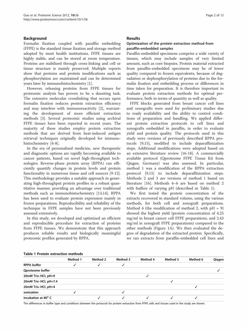
Table 1 Protein extraction methods
Method 1 Method 2 Metho
RPPA buffer ✓ ✓ ✓
Qproteome buffer
20mM Tris–HCl, pH=9
20mM Tris–HCl, pH=7.4
20mM Tris–HCl, pH=4
sonication ✓ ✓
incubation at 80o C ✓ ✓
The differences in buffer type and conditions between the protocols for protein ext
ResultsOptimization of the protein extraction method fromparaffin-embedded samplesParaffin-embedded specimens comprise a wide variety oftissues, which may include samples of very limitedamount, such as core biopsies. Protein material extractedfrom paraffin-embedded specimens may be of lowerquality compared to frozen equivalents, because of deg-radation or dephosphorylation of proteins due to the for-malin fixation and embedding process or differences intime taken for preparation. It is therefore important toevaluate protein extraction methods for optimal per-formance, both in terms of quantity as well as quality.FFPE blocks generated from breast cancer cell lines
and xenografts were used for preliminary studies dueto ready availability and the ability to control condi-tions of preparation and handling. We applied differ-ent protein extraction protocols to cell lines andxenografts embedded in paraffin, in order to evaluateyield and protein quality. The protocols used in thisstudy were versions of previously described RPPA pro-tocols [9,15], modified to include deparaffinizationsteps. Additional modifications were adopted based onan extensive literature review [16-18]. A commerciallyavailable protocol (Qproteome FFPE Tissue Kit fromQiagen, Germany) was also assessed. In particular,method 1 was a modification of the RPPA extractionprotocol [9,15] to include deparaffinization steps.Methods 2 and 3 are versions of method 1 based onliterature [16]. Methods 4–6 are based on method 2with buffers of varying pH (described in Table 1).We first tested the protein concentration of the
extracts recovered in standard volume, using the variousmethods, for both cell and xenograft preparations.Method 4 (the modification of method 2, with pH = 9)showed the highest yield (protein concentration of 4.25mg/ml in breast cancer cell FFPE preparations, and 2.43mg/ml in xenograft FFPE preparations) compared to theother methods (Figure 1A). We then evaluated the de-gree of degradation of the extracted protein. Specifically,we ran extracts from paraffin-embedded cell lines and
d 3 Method 4 Method 5 Method 6 Qiagen
✓
✓
✓
✓
✓ ✓ ✓
raction from FFPE cells and tissues used in the study are shown.
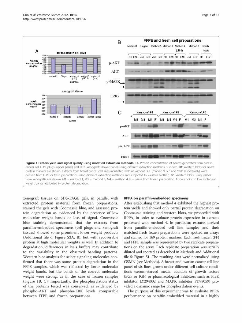
Figure 1 Protein yield and signal quality using modified extraction methods. 1A: Protein concentration of lysates generated from breastcancer cell FFPE plugs (upper panel) and FFPE xenografts (lower panel) using different extraction methods is shown. 1B: Western blots for selectprotein markers are shown. Extracts from breast cancer cell lines incubated with or without EGF (marked “EGF” and “ctrl” respectively) werederived from FFPE or fresh preparations using different extraction methods and subjected to western blotting. 1C: Western blots using lysatesfrom xenografts are shown. M1 = method 1; M3 = method 3; M4 = method 4; F = lysate from frozen preparation. Arrows point to low molecularweight bands attributed to protein degradation.
Guo et al. Proteome Science 2012, 10:56 Page 3 of 12http://www.proteomesci.com/content/10/1/56
xenograft tissues on SDS-PAGE gels, in parallel withextracted protein material from frozen preparations,stained the gels with Coomassie blue, and assessed pro-tein degradation as evidenced by the presence of lowmolecular weight bands or loss of signal. Coomassieblue staining demonstrated that the extracts fromparaffin-embedded specimens (cell plugs and xenografttissues) showed some prominent lower weight products(Additional file 6: Figure S2A, B), but with recoverableprotein at high molecular weights as well. In addition todegradation, differences in lysis buffers may contributeto the variability in the observed banding patterns.Western blot analysis for select signaling molecules con-firmed that there was some protein degradation in theFFPE samples, which was reflected by lower molecularweight bands, but the bands of the correct molecularweight were strong, as in the case of frozen samples(Figure 1B, C). Importantly, the phosphorylation statusof the proteins tested was conserved, as evidenced byphospho-AKT and phospho-ERK levels comparablebetween FFPE and frozen preparations.
RPPA on paraffin-embedded specimensAfter establishing that method 4 exhibited the highest pro-tein yields and showed only partial protein degradation onCoomassie staining and western blots, we proceeded withRPPA, in order to evaluate protein expression in extractsprocessed with method 4. In particular, extracts derivedfrom paraffin-embedded cell line samples and theirmatched fresh frozen preparations were spotted on arraysand stained for 169 protein markers. Each fresh frozen (FF)and FFPE sample was represented by two replicate prepara-tions on the array. Each replicate preparation was seriallydiluted and spotted as described in Methods and Additionalfile 5: Figure S1. The resulting data were normalized usingGSMN (see Methods). A breast and ovarian cancer cell linepanel of six lines grown under different cell culture condi-tions (serum-starved media, addition of growth factors(EGF or IGF) or pharmacological inhibitors such as PI3Kinhibitor LY294002 and MAPK inhibitor PD98059) pro-vided a dynamic range for phosphorylation events.The purpose of this experiment was to evaluate RPPA
performance on paraffin-embedded material in a highly
Guo et al. Proteome Science 2012, 10:56 Page 4 of 12http://www.proteomesci.com/content/10/1/56
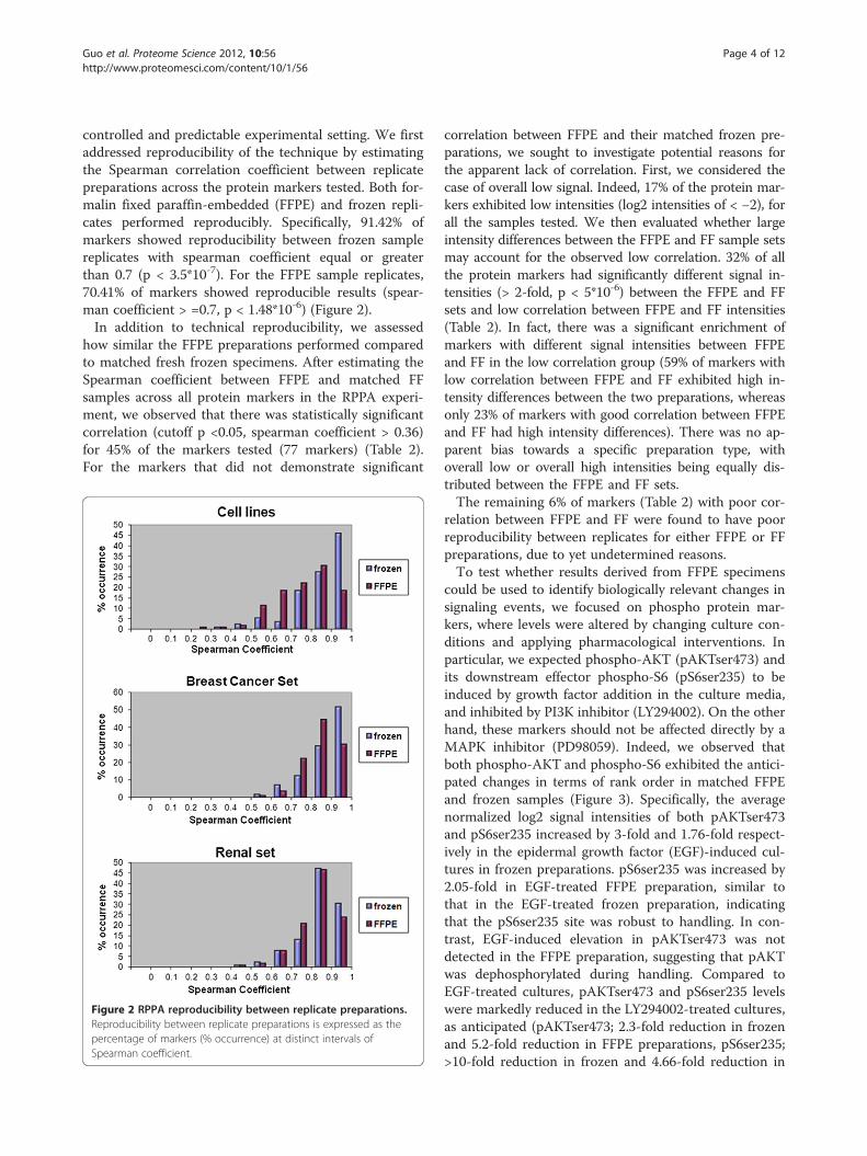
controlled and predictable experimental setting. We firstaddressed reproducibility of the technique by estimatingthe Spearman correlation coefficient between replicatepreparations across the protein markers tested. Both for-malin fixed paraffin-embedded (FFPE) and frozen repli-cates performed reproducibly. Specifically, 91.42% ofmarkers showed reproducibility between frozen samplereplicates with spearman coefficient equal or greaterthan 0.7 (p < 3.5*10-7). For the FFPE sample replicates,70.41% of markers showed reproducible results (spear-man coefficient > =0.7, p < 1.48*10-6) (Figure 2).In addition to technical reproducibility, we assessed
how similar the FFPE preparations performed comparedto matched fresh frozen specimens. After estimating theSpearman coefficient between FFPE and matched FFsamples across all protein markers in the RPPA experi-ment, we observed that there was statistically significantcorrelation (cutoff p <0.05, spearman coefficient > 0.36)for 45% of the markers tested (77 markers) (Table 2).For the markers that did not demonstrate significant
Figure 2 RPPA reproducibility between replicate preparations.Reproducibility between replicate preparations is expressed as thepercentage of markers (% occurrence) at distinct intervals ofSpearman coefficient.
correlation between FFPE and their matched frozen pre-parations, we sought to investigate potential reasons forthe apparent lack of correlation. First, we considered thecase of overall low signal. Indeed, 17% of the protein mar-kers exhibited low intensities (log2 intensities of < −2), forall the samples tested. We then evaluated whether largeintensity differences between the FFPE and FF sample setsmay account for the observed low correlation. 32% of allthe protein markers had significantly different signal in-tensities (> 2-fold, p < 5*10-6) between the FFPE and FFsets and low correlation between FFPE and FF intensities(Table 2). In fact, there was a significant enrichment ofmarkers with different signal intensities between FFPEand FF in the low correlation group (59% of markers withlow correlation between FFPE and FF exhibited high in-tensity differences between the two preparations, whereasonly 23% of markers with good correlation between FFPEand FF had high intensity differences). There was no ap-parent bias towards a specific preparation type, withoverall low or overall high intensities being equally dis-tributed between the FFPE and FF sets.The remaining 6% of markers (Table 2) with poor cor-
relation between FFPE and FF were found to have poorreproducibility between replicates for either FFPE or FFpreparations, due to yet undetermined reasons.To test whether results derived from FFPE specimens
could be used to identify biologically relevant changes insignaling events, we focused on phospho protein mar-kers, where levels were altered by changing culture con-ditions and applying pharmacological interventions. Inparticular, we expected phospho-AKT (pAKTser473) andits downstream effector phospho-S6 (pS6ser235) to beinduced by growth factor addition in the culture media,and inhibited by PI3K inhibitor (LY294002). On the otherhand, these markers should not be affected directly by aMAPK inhibitor (PD98059). Indeed, we observed thatboth phospho-AKT and phospho-S6 exhibited the antici-pated changes in terms of rank order in matched FFPEand frozen samples (Figure 3). Specifically, the averagenormalized log2 signal intensities of both pAKTser473and pS6ser235 increased by 3-fold and 1.76-fold respect-ively in the epidermal growth factor (EGF)-induced cul-tures in frozen preparations. pS6ser235 was increased by2.05-fold in EGF-treated FFPE preparation, similar tothat in the EGF-treated frozen preparation, indicatingthat the pS6ser235 site was robust to handling. In con-trast, EGF-induced elevation in pAKTser473 was notdetected in the FFPE preparation, suggesting that pAKTwas dephosphorylated during handling. Compared toEGF-treated cultures, pAKTser473 and pS6ser235 levelswere markedly reduced in the LY294002-treated cultures,as anticipated (pAKTser473; 2.3-fold reduction in frozenand 5.2-fold reduction in FFPE preparations, pS6ser235;>10-fold reduction in frozen and 4.66-fold reduction in
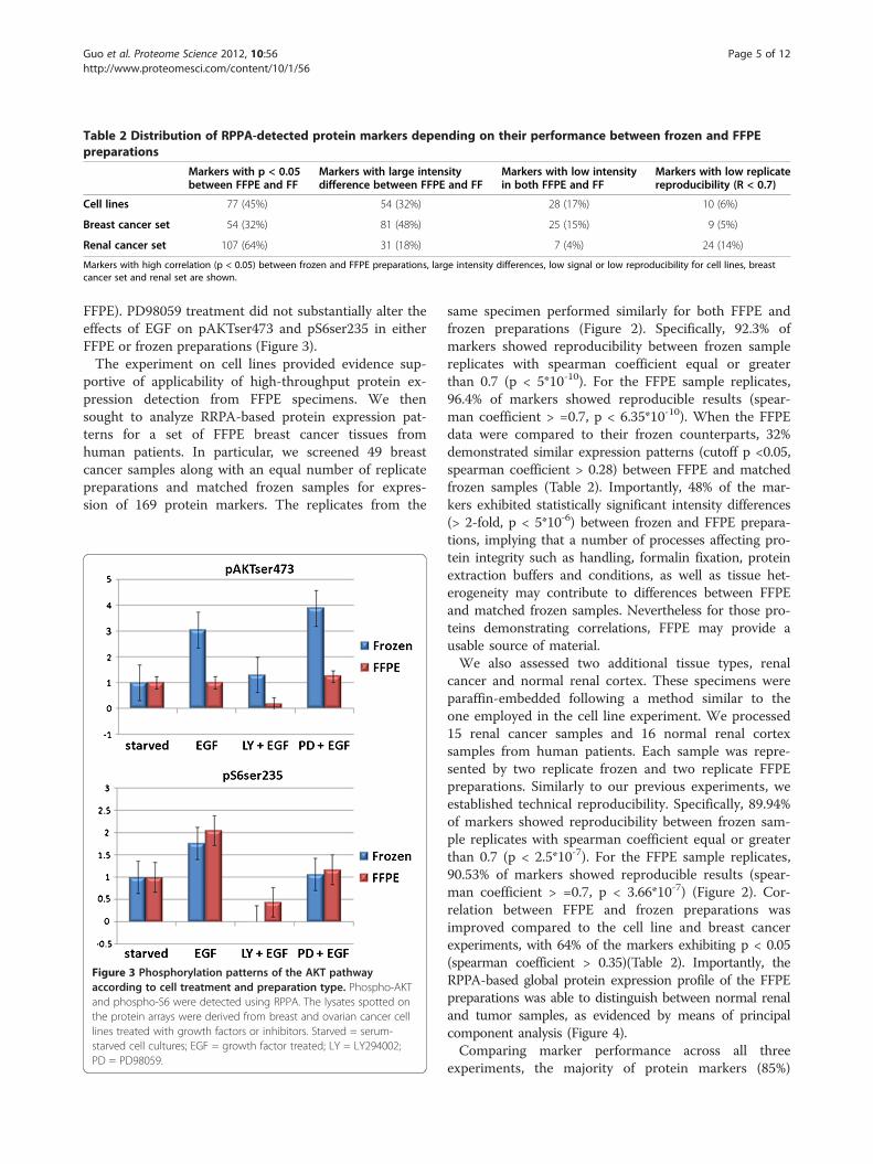
Table 2 Distribution of RPPA-detected protein markers depending on their performance between frozen and FFPEpreparations
Markers with p < 0.05between FFPE and FF
Markers with large intensitydifference between FFPE and FF
Markers with low intensityin both FFPE and FF
Markers with low replicatereproducibility (R < 0.7)
Cell lines 77 (45%) 54 (32%) 28 (17%) 10 (6%)
Breast cancer set 54 (32%) 81 (48%) 25 (15%) 9 (5%)
Renal cancer set 107 (64%) 31 (18%) 7 (4%) 24 (14%)
Markers with high correlation (p < 0.05) between frozen and FFPE preparations, large intensity differences, low signal or low reproducibility for cell lines, breastcancer set and renal set are shown.
Guo et al. Proteome Science 2012, 10:56 Page 5 of 12http://www.proteomesci.com/content/10/1/56
FFPE). PD98059 treatment did not substantially alter theeffects of EGF on pAKTser473 and pS6ser235 in eitherFFPE or frozen preparations (Figure 3).The experiment on cell lines provided evidence sup-
portive of applicability of high-throughput protein ex-pression detection from FFPE specimens. We thensought to analyze RRPA-based protein expression pat-terns for a set of FFPE breast cancer tissues fromhuman patients. In particular, we screened 49 breastcancer samples along with an equal number of replicatepreparations and matched frozen samples for expres-sion of 169 protein markers. The replicates from the
Figure 3 Phosphorylation patterns of the AKT pathwayaccording to cell treatment and preparation type. Phospho-AKTand phospho-S6 were detected using RPPA. The lysates spotted onthe protein arrays were derived from breast and ovarian cancer celllines treated with growth factors or inhibitors. Starved = serum-starved cell cultures; EGF = growth factor treated; LY = LY294002;PD = PD98059.
same specimen performed similarly for both FFPE andfrozen preparations (Figure 2). Specifically, 92.3% ofmarkers showed reproducibility between frozen samplereplicates with spearman coefficient equal or greaterthan 0.7 (p < 5*10-10). For the FFPE sample replicates,96.4% of markers showed reproducible results (spear-man coefficient > =0.7, p < 6.35*10-10). When the FFPEdata were compared to their frozen counterparts, 32%demonstrated similar expression patterns (cutoff p <0.05,spearman coefficient > 0.28) between FFPE and matchedfrozen samples (Table 2). Importantly, 48% of the mar-kers exhibited statistically significant intensity differences(> 2-fold, p < 5*10-6) between frozen and FFPE prepara-tions, implying that a number of processes affecting pro-tein integrity such as handling, formalin fixation, proteinextraction buffers and conditions, as well as tissue het-erogeneity may contribute to differences between FFPEand matched frozen samples. Nevertheless for those pro-teins demonstrating correlations, FFPE may provide ausable source of material.We also assessed two additional tissue types, renal
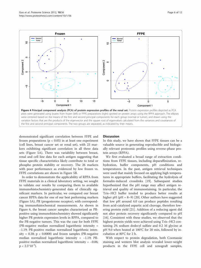
cancer and normal renal cortex. These specimens wereparaffin-embedded following a method similar to theone employed in the cell line experiment. We processed15 renal cancer samples and 16 normal renal cortexsamples from human patients. Each sample was repre-sented by two replicate frozen and two replicate FFPEpreparations. Similarly to our previous experiments, weestablished technical reproducibility. Specifically, 89.94%of markers showed reproducibility between frozen sam-ple replicates with spearman coefficient equal or greaterthan 0.7 (p < 2.5*10-7). For the FFPE sample replicates,90.53% of markers showed reproducible results (spear-man coefficient > =0.7, p < 3.66*10-7) (Figure 2). Cor-relation between FFPE and frozen preparations wasimproved compared to the cell line and breast cancerexperiments, with 64% of the markers exhibiting p < 0.05(spearman coefficient > 0.35)(Table 2). Importantly, theRPPA-based global protein expression profile of the FFPEpreparations was able to distinguish between normal renaland tumor samples, as evidenced by means of principalcomponent analysis (Figure 4).Comparing marker performance across all three
experiments, the majority of protein markers (85%)
Frozen FFPE
Figure 4 Principal component analysis (PCA) of protein expression profiles of the renal set. Protein expression profiles depicted as PCAplots were generated using lysates from frozen (left) or FFPE preparations (right) spotted on protein arrays using the RPPA approach. The ellipseswere centered based on the means of the first and second principal components for each group (normal or tumor), and drawn using thevariation factors that are the products of the eigenvector and the square root of eigenvalues calculated from the variances and covariances ofthe first and second principal components. The two groups are separated, as indicated by their means.
Guo et al. Proteome Science 2012, 10:56 Page 6 of 12http://www.proteomesci.com/content/10/1/56
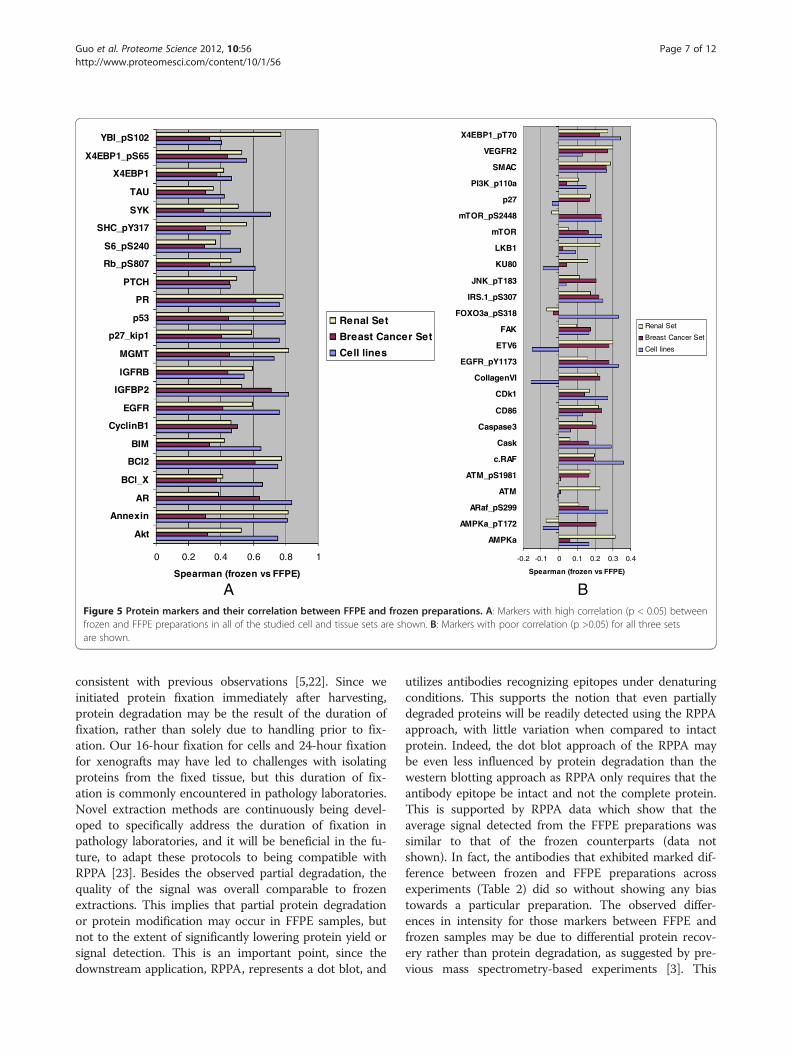
demonstrated significant correlation between FFPE andfrozen preparations (p < 0.05) in at least one experiment(cell lines, breast cancer set or renal set), with 23 mar-kers exhibiting significant correlation in all three datasets (Figure 5A). There was variability between breast,renal and cell line data for each antigen suggesting thattissue specific characteristics likely contribute to total orphospho protein stability or recovery. The 26 markerswith poor performance as evidenced by low frozen vs.FFPE correlations are shown in Figure 5B.In order to demonstrate the applicability of RPPA from
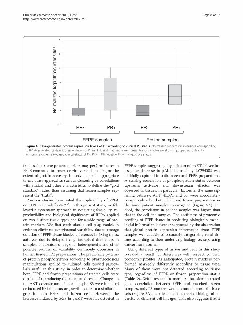
FFPE materials in a clinical laboratory setting, we soughtto validate our results by comparing them to availableimmunohistochemistry-generated data of clinically sig-nificant markers. In particular, we compared our breastcancer RPPA data for one of our best performing markers(Figure 5A), PR (progesterone receptor), with correspond-ing immunohistochemical measurements. As shown inFigure 6, the breast cancer samples characterized as PR-positive using immunohistochemistry showed significantlyhigher PR protein expression levels in RPPA, compared tothe PR-negative tumors. This was the case for both FFPE(PR-negative median normalized logarithmic intensity =−1.19; PR-positive median normalized logarithmic inten-sity = 0.38; p < 0.0008) and frozen samples (PR-negativemedian normalized logarithmic intensity = −1.19; PR-positive median normalized logarithmic intensity = −0.06;p < 2.5*10-5).
DiscussionIn this study, we have shown that FFPE tissues can be avaluable source in generating reproducible and biologic-ally relevant proteomic profiles using reverse-phase pro-tein arrays (RPPA).We first evaluated a broad range of extraction condi-
tions from FFPE tissues, including deparaffinization, re-hydration, buffer components, pH conditions andtemperatures. In the past, antigen retrieval techniqueswere used that mainly focused on applying high tempera-tures in appropriate buffers, facilitating the hydrolysis offormalin-induced crosslinks [19]. Subsequent studieshypothesized that the pH range may affect antigen re-trieval and quality of immunostaining. In particular, theTris–HCl buffer tended to produce better results athigher pH (pH = 8–9) [20]. Other authors have suggestedthat low pH around 4.0 can produce peptides resultingfrom acid-catalyzed aspartic acid cleavage, therefore low-ering protein yield [21]. Addition of a reducing agent didnot alter protein recovery significantly compared to pH[16]. Consistent with these studies, we observed that thehighest protein yields were achieved using Tris–HCl con-taining 2% sodium dodecyl sulfate and 0.2 M glycine atpH 9.0 when heated at 100°C for 20 min, followed by in-cubation at 80°C for 2 h.With respect to protein degradation, both Coomassie
staining and western blot analysis revealed lower-weightproducts in the FFPE cell and xenograft samples,
0 0.2 0.4 0.6 0.8 1
Akt
Annexin
AR
BCl_X
BCl2
BIM
CyclinB1
EGFR
IGFBP2
IGFRB
MGMT
p27_kip1
p53
PR
PTCH
Rb_pS807
S6_pS240
SHC_pY317
SYK
TAU
X4EBP1
X4EBP1_pS65
YBI_pS102
Spearman (frozen vs FFPE)
Renal Set
Breast Cancer Set
Cell lines
-0.2 -0.1 0 0.1 0.2 0.3 0.4
AMPKa
AMPKa_pT172
ARaf_pS299
ATM
ATM_pS1981
c.RAF
Cask
Caspase3
CD86
CDk1
CollagenVI
EGFR_pY1173
ETV6
FAK
FOXO3a_pS318
IRS.1_pS307
JNK_pT183
KU80
LKB1
mTOR
mTOR_pS2448
p27
PI3K_p110a
SMAC
VEGFR2
X4EBP1_pT70
Spearman (frozen vs FFPE)
Renal Set
Breast Cancer Set
Cell lines
A BFigure 5 Protein markers and their correlation between FFPE and frozen preparations. A: Markers with high correlation (p < 0.05) betweenfrozen and FFPE preparations in all of the studied cell and tissue sets are shown. B: Markers with poor correlation (p >0.05) for all three setsare shown.
Guo et al. Proteome Science 2012, 10:56 Page 7 of 12http://www.proteomesci.com/content/10/1/56
consistent with previous observations [5,22]. Since weinitiated protein fixation immediately after harvesting,protein degradation may be the result of the duration offixation, rather than solely due to handling prior to fix-ation. Our 16-hour fixation for cells and 24-hour fixationfor xenografts may have led to challenges with isolatingproteins from the fixed tissue, but this duration of fix-ation is commonly encountered in pathology laboratories.Novel extraction methods are continuously being devel-oped to specifically address the duration of fixation inpathology laboratories, and it will be beneficial in the fu-ture, to adapt these protocols to being compatible withRPPA [23]. Besides the observed partial degradation, thequality of the signal was overall comparable to frozenextractions. This implies that partial protein degradationor protein modification may occur in FFPE samples, butnot to the extent of significantly lowering protein yield orsignal detection. This is an important point, since thedownstream application, RPPA, represents a dot blot, and
utilizes antibodies recognizing epitopes under denaturingconditions. This supports the notion that even partiallydegraded proteins will be readily detected using the RPPAapproach, with little variation when compared to intactprotein. Indeed, the dot blot approach of the RPPA maybe even less influenced by protein degradation than thewestern blotting approach as RPPA only requires that theantibody epitope be intact and not the complete protein.This is supported by RPPA data which show that theaverage signal detected from the FFPE preparations wassimilar to that of the frozen counterparts (data notshown). In fact, the antibodies that exhibited marked dif-ference between frozen and FFPE preparations acrossexperiments (Table 2) did so without showing any biastowards a particular preparation. The observed differ-ences in intensity for those markers between FFPE andfrozen samples may be due to differential protein recov-ery rather than protein degradation, as suggested by pre-vious mass spectrometry-based experiments [3]. This
Nor
mal
ized
loga
rithm
ic in
tens
ities
PR PR+ PR PR+
FFPE samples Frozen samplesFigure 6 RPPA-generated protein expression levels of PR according to clinical PR status. Normalized logarithmic intensities correspondingto RPPA-generated protein expression levels of PR in FFPE and matched frozen breast tumor samples are shown, grouped according toimmunohistochemistry-based clinical status of PR (PR - = PR-negative; PR + = PR-positive status).
Guo et al. Proteome Science 2012, 10:56 Page 8 of 12http://www.proteomesci.com/content/10/1/56
implies that some protein markers may perform better inFFPE compared to frozen or vice versa depending on theextent of protein recovery. Indeed, it may be appropriateto use other approaches such as clustering or correlationswith clinical and other characteristics to define the “goldstandard” rather than assuming that frozen samples rep-resent the “truth”.Previous studies have tested the applicability of RPPA
on FFPE materials [3,24-27]. In this present study, we fol-lowed a systematic approach in evaluating feasibility, re-producibility and biological significance of RPPA appliedon two distinct tissue types and for a wide range of pro-tein markers. We first established a cell plug model, inorder to eliminate experimental variability due to storageduration of FFPE tissue blocks, differences in fixing times,autolysis due to delayed fixing, individual differences insamples, anatomical or regional heterogeneity, and otherpossible sources of variability commonly occurring inhuman tissue FFPE preparations. The predictable patternsof protein phosphorylation according to pharmacologicalmanipulations applied to cultured cells proved particu-larly useful in this study, in order to determine whetherboth FFPE and frozen preparations of treated cells werecapable of reproducing the anticipated results. Changes inthe AKT downstream effector phospho-S6 were inhibitedor induced by inhibitors or growth factors to a similar de-gree in both FFPE and frozen cells. However, theincreases induced by EGF in pAKT were not detected in
FFPE samples suggesting degradation of pAKT. Neverthe-less, the decrease in pAKT induced by LY294002 wasfaithfully captured in both frozen and FFPE preparations.A striking correlation of phosphorylation status betweenupstream activator and downstream effector wasobserved in tissues. In particular, factors in the same sig-naling pathway, AKT, 4EBP1 and S6, were coordinatelyphosphorylated in both FFPE and frozen preparations inthe same patient samples interrogated (Figure 5A). In-deed, the correlation in patient samples was higher thanthat in the cell line samples. The usefulness of proteomicprofiling of FFPE tissues in producing biologically mean-ingful information is further supported by the observationthat global protein expression information from FFPEsamples was capable of accurately categorizing renal tis-sues according to their underlying biology i.e. separatingcancer from normal.Using different types of tissues and cells in this study
revealed a wealth of differences with respect to theirproteomic profiles. As anticipated, protein markers per-formed markedly differently according to tissue type.Many of them were not detected according to tissuetype, regardless of FFPE or frozen preparation status(Table 2). With respect to markers that demonstratedgood correlation between FFPE and matched frozensamples, only 23 markers were common across all tissuesets (Figure 5A), as a testament to marked biological di-versity of different cell lineages. This also suggests that it
Guo et al. Proteome Science 2012, 10:56 Page 9 of 12http://www.proteomesci.com/content/10/1/56
may be necessary to select different antibodies to inter-rogate the diverse array of tissue lineages as protein andparticular phosphoprotein stability may be tissuespecific.With respect to the reproducibility of the approach, it
is important to note that separate preparations, eitherFFPE or frozen, from the same tissue sample (replicates)performed similarly on RPPA (Figure 2). This questionsthe notion that tissue heterogeneity between replicatepreparations may introduce critical variations in proteinprofiling preventing useful interpretation if laser-capturemicrodissection is not applied [28]. It is noteworthy thatour replicate preparations were derived from adjacentserial sections or pieces, therefore limiting tumor hetero-geneity. But unlike our replicate preparations fromwithin the same sample, FFPE and matched frozen sam-ples came from tissue pieces that were located a signifi-cant distance from one another, especially in the case ofbreast cancer samples. Thus, tissue heterogeneity mayhave contributed to discrepancies between FFPE andmatched frozen RPPA results. This is consistent with re-cently published studies of spatial heterogeneity of DNAmutations [29].With respect to the applicability of RPPA in the clin-
ical routine setting, it is encouraging that the RPPA-based measurements of PR correlated significantly withthe immunohistochemical results of this clinically sig-nificant marker in breast tumors (Figure 6).FFPE tissues represent a valuable resource to conduct
retrospective studies aimed to biomarker discovery andvalidation, in cancer as well as other diseases. The mosteffective approach may be to both discover and validatemolecular markers on FFPE preparations to decreasevariability potentially induced by tissue handling whencomparing FFPE and frozen samples. In order to im-prove the ability to characterize proteins from tissuesused for pathological evaluation, a number of stepscould be considered: using a consistent and short periodof fixation in formalin, limiting the duration of time be-tween tissue collection and addition of formalin, usingsmall tissue pieces where formalin will permeate morefrequently, obtaining tissue from multiple parts of thetissue and the consideration of new fixation approachesthat are under development. Clearly, FFPE tissue studieshold the promise of producing highly reproducibleand meaningful data when linked to powerful high-throughput methodologies such as reverse-phase proteinarrays.
Materials and methodsPreparation of FFPE cell blocksMDA-MB-231 and MDA-MB-468 cells were plated in15 cm Petri dishes, and allowed to grow to 80% conflu-ence. They were subsequently starved with serum-free
medium overnight and then incubated with EGF or IGFfor 10 minutes for cell signaling stimulation. The starvedand growth factor stimulated cell monolayers wererinsed twice with TBS (50 mM Tris–HCl pH 7.5 and150 mM NaCl) and harvested with rubber cell scrapers.Then cell pellets were processed as fresh frozen or FFPEcell blocks. Cell pellets were fixed in 10% formalin(Fisher Scientific, Pittsburgh, PA, USA) for 16 hours,and embedded in paraffin as a piece of tissue accordingto standard histological procedures. Paraffin cell blockswere stored at room temperature until analysis.
Xenografts and human tissuesThe mammary pads of four-week-old female nude micewere inoculated with 5 × 10⁶ MDA-MB-231 cells. Micewith mean tumor diameters of 0.5 cm were sacrificedand the grafted tumors were cut in halves. Half tumorwas fixed in 10% formalin for 24 hours and paraffin em-bedded using standard procedures; the other half wasimmediately snap-frozen and then stored at −80°C untiluse. All experiments with mice were performed accord-ing to the IACUC guidelines for the care and use of liv-ing animals in scientific research.Renal tumor, renal cortex and breast tumor tissue
FFPE blocks and their matched frozen pieces from thesame patients were obtained from the University ofTexas MD Anderson Cancer Center tissue bank, follow-ing Institutional Review Board (IRB) approval and guide-lines. In the case of renal tissues, the FFPE and matchedfrozen preparations were derived from adjacent tissuepieces, whereas in the case of breast cancer tissues, theywere derived from distant tumor pieces. Renal tissuesamples were confirmed to have at least 70% cellularcontent, whereas the breast cancer tissue samples had atleast 50% cellular content. All samples used in the studywere histologically confirmed using H&E.
Deparaffinization and protein extractionTo define technical reproducibility of methodology,replicates of four serial FFPE cell or tissue sections,10 μm thick, were placed in Eppendorf tubes and depar-affinized by incubation at room temperature in xylene(Fisher Scientific, Pittsburgh, PA, USA) for 10 min. Aftereach incubation, tissue was pelleted at 14000 × g for3 min, and incubation/centrifugation steps wererepeated two times. The deparaffinized tissue pelletswere then rehydrated with a graded series of ethanol(Pharmaco products Inc, Brookfield, IL, USA). The rehy-drated tumor tissue and cell sections were resuspendedin a panel of extraction buffers as described (see Table 1).The extraction buffers evaluated included QproteomeFFPE tissue kit (Qiagen); 20 mM Tris HCl buffers atvarious pH values (4, 6 and 9), with 2%(w/v)SDS; thelysis buffer routinely used for RPPA assay (1% Triton X-

Guo et al. Proteome Science 2012, 10:56 Page 10 of 12http://www.proteomesci.com/content/10/1/56
100, 50 mM Hepes, pH 7.4, 150 mM NaCl, 1.5 mMMgCl2, 1 mM EGTA, 10 mM NaF, 100 mM Na Pyru-vate, 1 mM Na3VO4, 10% glycerol, containing proteaseinhibitors and phosphatase inhibitors from Roche Ap-plied Science) [9] plus 4 × SDS buffer (3:1, v/v). For tis-sue samples, the amount of extraction buffer added tothe pellets from paraffin sections was estimated accord-ing to the size of the tissue surface area. In particular,tissue blocks were first scanned with an Image Scanner(CanoScan 8400F, Canon, Lake Success, NY, USA) andthen the surface area of the paraffin-embedded tissuewas calculated using ImageJ (NIH). For frozen samples,adjustment of buffer volume according to weight of eachfrozen piece was performed.The samples were first incubated on ice for 5 min, and
mixed by vortexing, then boiled at 100°C for 20 min fol-lowed by an optional sonication step and an optional incu-bation at 80° C in a water bath for 2 hours. The extractionmethods followed are described in detail in Table 1. Afterprotein extraction, any remaining unsolubilized materialwas pelleted at 14000 × g for 20 min, and protein concen-tration of total protein extracted was determined by theBCA Protein Assay (Pierce Chemicals Co., Rockford, IL,USA). The Pierce BCA Protein Assay is a detergent-compatible formulation and the protein standards wereprepared using the same lysis buffer as the samples.
SDS-PAGE and western blotProtein extracts obtained from fresh-frozen and FFPE tis-sues were subjected to SDS-PAGE in a Bio-Rad MiniProtean II system. Proteins were stained with Brilliant R250 according to Westermeier et al. [30]. Parallel gelswere transferred onto PVDF (Polyvinylidene Fluoride,BioRad, Richmond, USA) membrane. Membranes wereblocked and probed with antibodies against differentsized molecules and visualized using ECL (Amersham/GE health care).
RPPA assays and analysisProtein extracts from cell and tissue paraffin blocks wereprobed for expression of validated antibodies by RPPA[9,11,31,32]. Specifically, five serial 2-fold dilutions of theprotein extracts were performed using RPPA lysis buffercontaining 1% SDS. The diluted lysates were spotted onnitrocellulose-coated FAST slides (Whatman, Schleicher& Schuell BioScience, Inc., Keene, NH) by an Aushon2470 arrayer (Aushon Biosystems, Burlington, MA) permanufacturer’s protocol. The construction of the proteinarray is shown in Additional file 5: Figure S1. Eachspotted slide was incubated with a primary antibody(Additional file 1: Table S1) in the appropriate dilution. Atotal of 169 slides were stained for 169 antibodies. Thespecific protein-antibody interaction was recognized bybiotin-conjugated secondary antibody and amplified by
tyramide deposition. The analyte was detected by avidin-conjugated peroxidase reactive to its substrate chromogendiaminobenzidine (DAKO catalyzed signal amplification(CSA) system, DAKO, Carpinteria, CA). The stainedRPPA slides were scanned by the Hewlett-Packard (HP)scanner and its companying scanning software, and theslide images were quantified for raw signal intensities bythe software MicroVigene (VigeneTech Inc., Carlisle, MA)per manufacturer’s protocol. The raw signal intensities(which are provided in detail, in Additional files 2, 3 and4: Tables S2, S3, and S4) were then processed by the Rpackage SuperCurve [33] developed by the Department ofBioinformatics and Computational Biology at Universityof Texas MD Anderson Cancer Center (http://bioinfor-matics.mdanderson.org/Software/OOMPA/). The log2-scaled protein concentrations were normalized by globalsample median normalization (GSMN) (the median of allprotein marker intensities for a single sample is subtractedfrom each of the data points of this specific sample[22,34]). Student’s t test was used to evaluate statisticalsignificance of differences in protein expression levelsbetween FFPE and frozen sample groups, or betweenPR-negative and PR-positive breast cancer groups, inorder to determine markers with intensity differences be-tween FFPE and frozen preparations, or between PR-positive and PR-negative tumors. The reproducibility ofsampling methods was determined by Spearman correl-ation coefficient. Principal component analysis was per-formed to show the relatedness of the samples and of theproteins. All statistical analysis was performed using thestatistics software package R [35].
Additional files
Additional file 1: Table S1. List of the primary antibodies used in theRPPA experiments.
Additional file 2: Table S2. RPPA raw signal intensities of the cell linesexperiment (FFPE and frozen).
Additional file 3: Table S3. RPPA raw signal intensities of the breastcancer tissue experiment (FFPE and frozen).
Additional file 4: Table S4. RPPA raw signal intensities of the renaltissue experiment (FFPE and frozen).
Additional file 5: Figure S1. Protein Microarray Construction. The size,design and dilution arrangement of the protein microarray are shown.
Additional file 6: Figure S2. SDS-PAGE analysis of protein lysatesextracted from fresh frozen and FFPE cell blocks or xenografts by usingdifferent extraction protocols. (A) Extracts from breast cancer cell linesincubated with or without EGF were derived from FFPE or freshpreparations using different extraction methods and subjected to SDS-PAGE analysis. (B) SDS-PAGE image of protein extracts from fresh frozenand FFPE xenograft tissues are shown. Lane M: molecular weight marker.Arrows point to low molecular weight bands attributed to proteindegradation.
AbbreviationsBCA: Bicinchoninic Acid; ECL: Enhanced Chemiluminescence; FF: FreshFrozen; FFPE: Formalin-fixed Paraffin-embedded; GSMN: Global SampleMedian Normalization; PVDF: Polyvinylidene Fluoride; RPPA: Reverse-phase
Guo et al. Proteome Science 2012, 10:56 Page 11 of 12http://www.proteomesci.com/content/10/1/56
Protein Array; SDS-PAGE: Sodium Dodecyl Sulfate Polyacrylamide GelElectrophoresis.
Competing interestsThe authors declare that they have no competing interests.
Authors’ contributionsHG: experiments and manuscript preparation, WL: statistics, ZJ: statistics, PT:tissue evaluation/pathology, EJ: tissue evaluation, GBM: manuscriptpreparation, YL: RPPA experiments and manuscript preparation, BTH:manuscript preparation, DT: RPPA experiments and manuscript preparation.All authors read and approved the final manuscript.
Authors’ informationHG: Research scientist, Systems Biology, MD Anderson Cancer Center, WL:Research Statistical Analyst, MD Anderson Cancer Center, ZJ: Sr. StatisticalAnalyst, MD Anderson Cancer Center, PT: Associate Professor, Pathology, MDAnderson Cancer Center, EJ: Associate Professor, Genitourinary MedicalOncology, MD Anderson Cancer Center, GBM: Professor, Chair, SystemsBiology, MD Anderson Cancer Center, YL: Associate professor, SystemsBiology, director of RPPA Core, BTH: Senior Lecturer, RCSI, Consultant MedicalOncologist, Beaumont Hospital, Royal, College of Surgeons of Ireland, Dublin,Ireland, DT: Instructor, Genitourinary Medical Oncology, MD Anderson CancerCenter.
AcknowledgementsThis work was supported by grants from:BTH: Career Development Award (CDA) from the Conquer CancerFoundation of the American Society of Clinical Oncology (ASCO),Translational research award TRA/2010/8 from the Health Research Board(HRB) of Ireland the Science Foundation Ireland (SFI).DT: University of Texas MD Anderson Cancer Center Kidney Pilot Program,Mary K. Chapman Foundation Charitable Fund.MD Anderson Cancer Center Support Grant (CCSG) CA016672 from NationalInstitutes of Health.Komen RPPA Grant.
Author details1Department of Systems Biology, University of Texas MD Anderson CancerCenter, Houston, TX, USA. 2Department of Bioinformatics and ComputationalBiology, University of Texas MD Anderson Cancer Center, Houston, TX, USA.3Department of Pathology, University of Texas MD Anderson Cancer Center,Houston, TX, USA. 4Department of Genitourinary Medical Oncology,University of Texas MD Anderson Cancer Center, Houston, TX, USA.5Department of Medical Oncology, Beaumont Hospital, Royal College ofSurgeons of Ireland, Dublin, Ireland.
Received: 17 February 2012 Accepted: 10 September 2012Published: 24 September 2012
References1. Lim MS, Elenitoba-Johnson KS: Proteomics in pathology research. Lab
Invest 2004, 84(10):1227–1244.2. Crockett DK, Lin Z, Vaughn CP, Lim MS, Elenitoba-Johnson KS: Identification
of proteins from formalin-fixed paraffin-embedded cells by LC-MS/MS.Lab Invest 2005, 85(11):1405–1415.
3. Addis MF, Tanca A, Pagnozzi D, Crobu S, Fanciulli G, Cossu-Rocca P, Uzzau S:Generation of high-quality protein extracts from formalin-fixed,paraffin-embedded tissues. Proteomics 2009, 9(15):3815–3823.
4. Hood BL, Darfler MM, Guiel TG, Furusato B, Lucas DA, Ringeisen BR,Sesterhenn IA, Conrads TP, Veenstra TD, Krizman DB: Proteomic analysis offormalin-fixed prostate cancer tissue. Mol Cell Proteomics 2005,4(11):1741–1753.
5. Shi SR, Liu C, Balgley BM, Lee C, Taylor CR: Protein extraction fromformalin-fixed, paraffin-embedded tissue sections: quality evaluation bymass spectrometry. J Histochem Cytochem 2006, 54(6):739–743.
6. Palmer-Toy DE, Krastins B, Sarracino DA, Nadol JB Jr, Merchant SN: Efficientmethod for the proteomic analysis of fixed and embedded tissues.J Proteome Res 2005, 4(6):2404–2411.
7. Guo T, Wang W, Rudnick PA, Song T, Li J, Zhuang Z, Weil RJ, DeVoe DL, LeeCS, Balgley BM: Proteome analysis of microdissected formalin-fixed and
paraffin-embedded tissue specimens. J Histochem Cytochem 2007,55(7):763–772.
8. Hwang SI, Thumar J, Lundgren DH, Rezaul K, Mayya V, Wu L, Eng J, WrightME, Han DK: Direct cancer tissue proteomics: a method to identifycandidate cancer biomarkers from formalin-fixed paraffin-embeddedarchival tissues. Oncogene 2007, 26(1):65–76.
9. Tibes R, Qiu Y, Lu Y, Hennessy B, Andreeff M, Mills GB, Kornblau SM: Reversephase protein array: validation of a novel proteomic technology andutility for analysis of primary leukemia specimens and hematopoieticstem cells. Mol Cancer Ther 2006, 5(10):2512–2521.
10. Park ES, Rabinovsky R, Carey M, Hennessy BT, Agarwal R, Liu W, Ju Z, DengW, Lu Y, Woo HG, et al: Integrative analysis of proteomic signatures,mutations, and drug responsiveness in the NCI 60 cancer cell line set.Mol Cancer Ther 2010, 9(2):257–267.
11. Carey MS, Agarwal R, Gilks B, Swenerton K, Kalloger S, Santos J, Ju Z, Lu Y,Zhang F, Coombes KR, et al: Functional proteomic analysis of advancedserous ovarian cancer using reverse phase protein array: TGF-betapathway signaling indicates response to primary chemotherapy. ClinCancer Res 2010, 16(10):2852–2860.
12. Sheehan KM, Calvert VS, Kay EW, Lu Y, Fishman D, Espina V, Aquino J, SpeerR, Araujo R, Mills GB, et al: Use of reverse phase protein microarrays andreference standard development for molecular network analysis ofmetastatic ovarian carcinoma. Mol Cell Proteomics 2005, 4(4):346–355.
13. Spurrier B, Honkanen P, Holway A, Kumamoto K, Terashima M, TakenoshitaS, Wakabayashi G, Austin J, Nishizuka S: Protein and lysate arraytechnologies in cancer research. Biotechnol Adv 2008, 26(4):361–369.
14. Speer R, Wulfkuhle J, Espina V, Aurajo R, Edmiston KH, Liotta LA, Petricoin EF3rd: Development of reverse phase protein microarrays for clinicalapplications and patient-tailored therapy. Cancer Genomics Proteomics2007, 4(3):157–164.
15. Hennessy BT, Lu Y, Gonzalez-Angulo AM, Carey MS, Myhre S, Ju Z, DaviesMA, Liu W, Coombes K, Meric-Bernstam F, et al: A Technical Assessment ofthe Utility of Reverse Phase Protein Arrays for the Study of theFunctional Proteome in Non-microdissected Human Breast Cancers. ClinProteomics 2010, 6(4):129–151.
16. Fowler CB, Cunningham RE, O'Leary TJ, Mason JT: 'Tissue surrogates' as amodel for archival formalin-fixed paraffin-embedded tissues. Lab Invest2007, 87(8):836–846.
17. Shi SR, Cote RJ, Taylor CR: Antigen retrieval immunohistochemistry andmolecular morphology in the year 2001. Appl Immunohistochem MolMorphol 2001, 9(2):107–116.
18. Chung JY, Lee SJ, Kris Y, Braunschweig T, Traicoff JL, Hewitt SM: Awell-based reverse-phase protein array applicable to extracts fromformalin-fixed paraffin-embedded tissue. Proteomics Clin Appl 2008,2:1539–1547.
19. Shi SR, Key ME, Kalra KL: Antigen retrieval in formalin-fixed, paraffin-embedded tissues: an enhancement method for immunohistochemicalstaining based on microwave oven heating of tissue sections.J Histochem Cytochem 1991, 39(6):741–748.
20. Shi SR, Imam SA, Young L, Cote RJ, Taylor CR: Antigen retrievalimmunohistochemistry under the influence of pH using monoclonalantibodies. J Histochem Cytochem 1995, 43(2):193–201.
21. Fowler CB, Cunningham RE, Waybright TJ, Blonder J, Veenstra TD, O'LearyTJ, Mason JT: Elevated hydrostatic pressure promotes protein recoveryfrom formalin-fixed, paraffin-embedded tissue surrogates. Lab Invest2008, 88(2):185–195.
22. Zien A, Aigner T, Zimmer R, Lengauer T: Centralization: a new methodfor the normalization of gene expression data. Bioinformatics 2001,17(Suppl 1):S323–S331.
23. Wolff C, Schott C, Porschewski P, Reischauer B, Becker KF: Successfulprotein extraction from over-fixed and long-term stored formalin-fixedtissues. PLoS One 2011, 6(1):e16353.
24. Becker KF, Schott C, Hipp S, Metzger V, Porschewski P, Beck R, Nahrig J,Becker I, Hofler H: Quantitative protein analysis from formalin-fixedtissues: implications for translational clinical research and nanoscalemolecular diagnosis. J Pathol 2007, 211(3):370–378.
25. Berg D, Langer R, Tran K, Walch A, Schuster T, Bronger H, Becker KF: ProteinMicroarray-based Comparison of HER2, Estrogen Receptor, andProgesterone Receptor Status in Core Biopsies and Surgical SpecimensFrom FFPE Breast Cancer Tissues. Appl Immunohistochem Mol Morphol2011, 19(4):300–305.
Guo et al. Proteome Science 2012, 10:56 Page 12 of 12http://www.proteomesci.com/content/10/1/56
26. Berg D, Wolff C, Malinowsky K, Tran K, Walch A, Bronger H, Schuster T,Hofler H, Becker KF: Profiling signalling pathways in formalin-fixed andparaffin-embedded breast cancer tissues reveals cross-talk betweenEGFR, HER2, HER3 and uPAR. J Cell Physiol 2012, 227(1):204–212.
27. Wolff C, Malinowsky K, Berg D, Schragner K, Schuster T, Walch A, Bronger H,Hofler H, Becker KF: Signalling networks associated with urokinase-typeplasminogen activator (uPA) and its inhibitor PAI-1 in breast cancertissues: new insights from protein microarray analysis. J Pathol 2011,223(1):54–63.
28. Silvestri A, Colombatti A, Calvert VS, Deng J, Mammano E, Belluco C, DeMarchi F, Nitti D, Liotta LA, Petricoin EF, et al: Protein pathway biomarkeranalysis of human cancer reveals requirement for upfront cellular-enrichment processing. Lab Invest 2010, 90(5):787–796.
29. Gerlinger M, Rowan AJ, Horswell S, Larkin J, Endesfelder D, Gronroos E,Martinez P, Matthews N, Stewart A, Tarpey P, et al: Intratumorheterogeneity and branched evolution revealed by multiregionsequencing. N Engl J Med 2012, 366(10):883–892.
30. Westermeier R: Sensitive, quantitative, and fast modifications forCoomassie Blue staining of polyacrylamide gels. Proteomics 2006,6(Suppl 2):61–64.
31. Hennessy BT, Lu Y, Poradosu E, Yu Q, Yu S, Hall H, Carey MS, Ravoori M,Gonzalez-Angulo AM, Birch R, et al: Pharmacodynamic markers ofperifosine efficacy. Clin Cancer Res 2007, 13(24):7421–7431.
32. Stemke-Hale K, Gonzalez-Angulo AM, Lluch A, Neve RM, Kuo WL, Davies M,Carey M, Hu Z, Guan Y, Sahin A, et al: An integrative genomic andproteomic analysis of PIK3CA, PTEN, and AKT mutations in breast cancer.Cancer Res 2008, 68(15):6084–6091.
33. Hu J, He X, Baggerly KA, Coombes KR, Hennessy BT, Mills GB: Non-parametric quantification of protein lysate arrays. Bioinformatics 2007,23(15):1986–1994.
34. Quackenbush J: Microarray data normalization and transformation. NatGenet 2002, 32(Suppl):496–501.
35. Team RDC: R: A language and environment for statistical computing. Vienna,Austria: R Foundation for Statistical Computing; 2010:409.
doi:10.1186/1477-5956-10-56Cite this article as: Guo et al.: An efficient procedure for proteinextraction from formalin-fixed, paraffin-embedded tissues for reversephase protein arrays. Proteome Science 2012 10:56.
Submit your next manuscript to BioMed Centraland take full advantage of:
• Convenient online submission
• Thorough peer review
• No space constraints or color figure charges
• Immediate publication on acceptance
• Inclusion in PubMed, CAS, Scopus and Google Scholar
• Research which is freely available for redistribution
Submit your manuscript at www.biomedcentral.com/submit
Related Documents