METATARSAL OSTEOTOMY USING DOUBLE-THREADED SCREWS: BIOMECHANICAL ANALYSIS Anna ZIĘBOWICZ, Anita KAJZER, Wojciech KAJZER, and Jan MARCINIAK Silesian University of Technology, Institute of Engineering Materials and Biomaterials, ul. Konarskiego 18a, 44-100 Gliwice, Poland email: [email protected] Abstract: The fundamental purpose of this research was to determine the biomechanical characteristics of the first metatarsal bone – double-threaded screws system made of stainless steel (Cr-Ni-Mo) and an assessment of its stability. To define the biomechanical characteristics of the system, the finite element method and experimental method were applied. Geometric models of metatarsal bone and double-threaded screws, were discretized by means of SOLID 95 element. Appropriate boundary conditions imitating phenomena in the real system with appropriate accuracy were established. The aim of biomechanical analysis was calculation of displacements and stresses in the bone and the stabilizers in a function of the applied loading. The experimental method was carried out to calculate displacements of the analyzed system. The obtained results can be applied to determine the construction features of the stabilizer and to select mechanical properties of metallic biomaterial (selection of degree of strain hardening). 1. INTRODUCTION The human foot combines mechanical complexity and structural strength. The ankle serves as a foundation, shock absorber and propulsion engine. The foot can sustain enormous pressure (several tons over the course of a one-mile run) and provides flexibility and resilience. Structurally, the foot has three main parts: the forefoot, the midfoot, and the hindfoot. The foot and ankle contain: 26 bones that can be divided into the tarsal bones, the metatarsal bones and the phalanges – Fig.1. The tarsal bones are the larger bones that form the back section of foot, with the calcaneum being the largest. There are five metatarsal bones and these are given names from the first to the fifth. The first metatarsal bone is the largest and is the bone that joins to the big toe. It also has a lack of interconnecting ligaments between itself and the second metatarsal. This allows for independent motion. Fig.1. Top view of foot bones [1]

Welcome message from author
This document is posted to help you gain knowledge. Please leave a comment to let me know what you think about it! Share it to your friends and learn new things together.
Transcript
METATARSAL OSTEOTOMY USING DOUBLE-THREADED SCREWS:
BIOMECHANICAL ANALYSIS
Anna ZIĘBOWICZ, Anita KAJZER, Wojciech KAJZER, and Jan MARCINIAK
Silesian University of Technology, Institute of Engineering Materials and Biomaterials, ul. Konarskiego 18a,
44-100 Gliwice, Poland
email: [email protected]
Abstract: The fundamental purpose of this research was to determine the biomechanical characteristics of the
first metatarsal bone – double-threaded screws system made of stainless steel (Cr-Ni-Mo) and an assessment of
its stability. To define the biomechanical characteristics of the system, the finite element method and
experimental method were applied. Geometric models of metatarsal bone and double-threaded screws, were
discretized by means of SOLID 95 element. Appropriate boundary conditions imitating phenomena in the real
system with appropriate accuracy were established. The aim of biomechanical analysis was calculation of
displacements and stresses in the bone and the stabilizers in a function of the applied loading. The experimental
method was carried out to calculate displacements of the analyzed system. The obtained results can be applied to
determine the construction features of the stabilizer and to select mechanical properties of metallic biomaterial
(selection of degree of strain hardening).
1. INTRODUCTION
The human foot combines mechanical complexity and structural strength. The ankle serves
as a foundation, shock absorber and propulsion engine. The foot can sustain enormous
pressure (several tons over the course of a one-mile run) and provides flexibility and
resilience. Structurally, the foot has three main parts: the forefoot, the midfoot, and the
hindfoot.
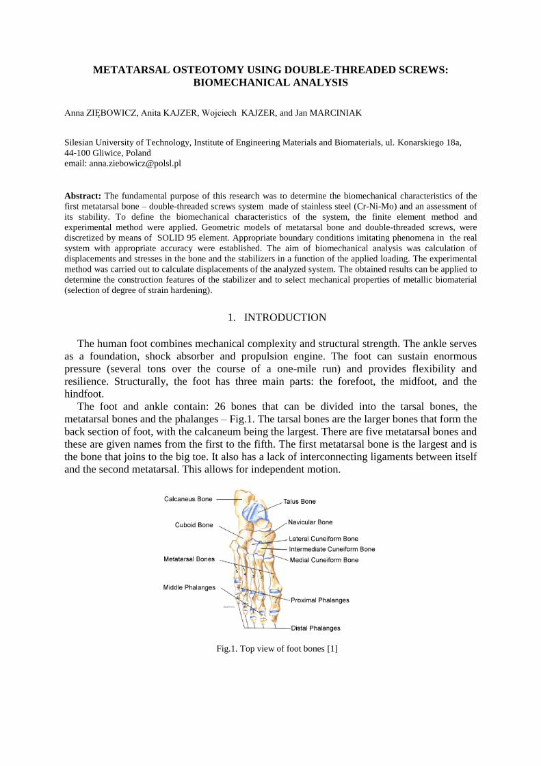
The foot and ankle contain: 26 bones that can be divided into the tarsal bones, the
metatarsal bones and the phalanges – Fig.1. The tarsal bones are the larger bones that form the
back section of foot, with the calcaneum being the largest. There are five metatarsal bones and
these are given names from the first to the fifth. The first metatarsal bone is the largest and is
the bone that joins to the big toe. It also has a lack of interconnecting ligaments between itself
and the second metatarsal. This allows for independent motion.
Fig.1. Top view of foot bones [1]
The foot also contains: 33 joints; more than 100 muscles, tendons (fibrous tissues that
connect muscles to bones), ligaments (fibrous tissues that connect bones to other bones) and
a network of blood vessels, nerves, skin and soft tissue.
These components work together to provide the body with support, balance and mobility.
A structural flaw or malfunction in any one part can result in the development of problems
elsewhere in the body. Abnormalities in other parts of the body can lead to problems in the
feet [2,3].
The metatarsal bones are some of the most commonly fractured (broken) bones in the foot.
There are two main types of metatarsal fractures:
acute fractures – due to a sudden injury to the foot (commonly dropping a heavy
object onto the foot, a fall or a sporting injury) [4]
stress fractures – due to overuse, or repetitive, injury to a normal metatarsal bone [5].
The kind of fracture should be characterized and treatment initiated. Metatarsal fractures
are divided into three sections – 1st, 5
th and 2
nd – 4
th [6,7]. Due to the head of the first
metatarsal being thought to bear one third of our body weight, any evidence of instability
requires operative fixation [8]. The present-day alternative of small-bones reconstructions in
orthopaedics is the double-threaded screws, that indicate many favorable features especially
with reference to minimization of tissue traumas. The matter of these solutions is application
of two threads of diverse diameter, that assure stabilization of bone fragments with the use of
physiological effects [9].
2. MATERIALS AND METHODS
The main aim of the work was the determination of the biomechanical characteristics of the
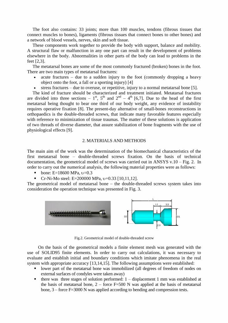
first metatarsal bone – double-threaded screws fixation. On the basis of technical
documentation, the geometrical model of screws was carried out in ANSYS v.10 – Fig. 2. In
order to carry out the numerical analysis, the following material properties were as follows:
bone: E=18600 MPa, =0.3
Cr-Ni-Mo steel: E=200000 MPa, =0.33 [10,11,12].
The geometrical model of metatarsal bone – the double-threaded screws system takes into
consideration the operation technique was presented in Fig. 3.
Fig.2. Geometrical model of double-threaded screw
On the basis of the geometrical models a finite element mesh was generated with the
use of SOLID95 finite elements. In order to carry out calculations, it was necessary to
evaluate and establish initial and boundary conditions which imitate phenomena in the real
system with appropriate accuracy [13,14,15]. The following assumptions were established:
lower part of the metatarsal bone was immobilized (all degrees of freedom of nodes on
external surfaces of condyles were taken away)
there was three stages of solution performed: 1 – displacement 1 mm was established at
the basis of metatarsal bone, 2 – force F=500 N was applied at the basis of metatarsal
bone, 3 – force F=3000 N was applied according to bending and compression tests.
L
L1 3,5
D2
D1
2,0
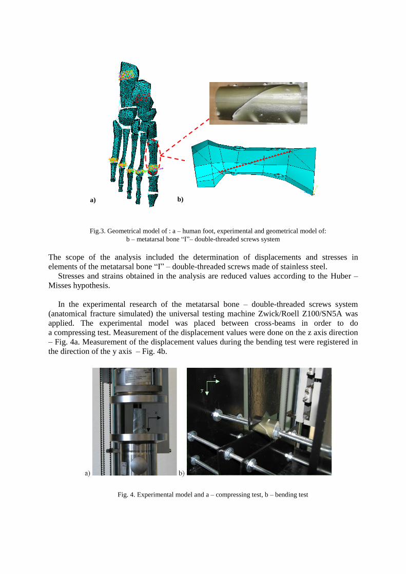
Fig.3. Geometrical model of : a – human foot, experimental and geometrical model of:
b – metatarsal bone “I”– double-threaded screws system
The scope of the analysis included the determination of displacements and stresses in
elements of the metatarsal bone “I” – double-threaded screws made of stainless steel.
Stresses and strains obtained in the analysis are reduced values according to the Huber –
Misses hypothesis.
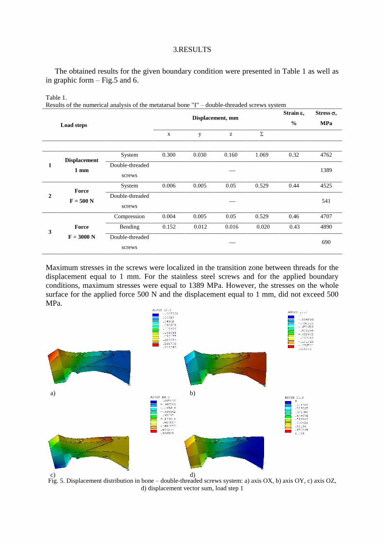
In the experimental research of the metatarsal bone – double-threaded screws system
(anatomical fracture simulated) the universal testing machine Zwick/Roell Z100/SN5A was
applied. The experimental model was placed between cross-beams in order to do
a compressing test. Measurement of the displacement values were done on the z axis direction
– Fig. 4a. Measurement of the displacement values during the bending test were registered in
the direction of the y axis – Fig. 4b.
Fig. 4. Experimental model and a – compressing test, b – bending test
a) b)
3.RESULTS
The obtained results for the given boundary condition were presented in Table 1 as well as
in graphic form – Fig.5 and 6.
Table 1.
Results of the numerical analysis of the metatarsal bone "I" – double-threaded screws system
Load steps
Displacement, mm
Strain ,
%
Stress ,
MPa
x y z
1 Displacement
1 mm
System 0.300 0.030 0.160 1.069 0.32 4762
Double-threaded
screws 1389
2 Force
F = 500 N
System 0.006 0.005 0.05 0.529 0.44 4525
Double-threaded
screws 541
3 Force
F = 3000 N
Compression 0.004 0.005 0.05 0.529 0.46 4707
Bending 0.152 0.012 0.016 0.020 0.43 4890
Double-threaded
screws 690
Maximum stresses in the screws were localized in the transition zone between threads for the
displacement equal to 1 mm. For the stainless steel screws and for the applied boundary
conditions, maximum stresses were equal to 1389 MPa. However, the stresses on the whole
surface for the applied force 500 N and the displacement equal to 1 mm, did not exceed 500
MPa.
Fig. 5. Displacement distribution in bone – double-threaded screws system: a) axis OX, b) axis OY, c) axis OZ,
d) displacement vector sum, load step 1
a) b)
c) d)

Fig. 6. Displacement distribution in bone – double-threaded screws system: a) axis OX, b) axis OY, c) axis OZ,
d) displacement vector sum, load step 2
On the basis of the performed analyses, it can be stated that the displacement
characteristics of the first metatarsal bone – double-threaded screws system in the
experimental and numerical conditions were similar – Fig. 7.
Fig. 7. Comparison of displacements for experimental and numerical analysis
Observations of the experimental model after the compression test revealed that one of the
compression screws was broken.
Damage localization corresponded with the maximum values of stresses obtained from the
numerical analysis – Fig. 8.
a) b)
c) d)
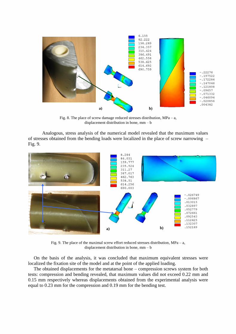
Fig. 8. The place of screw damage reduced stresses distribution, MPa – a,
displacement distribution in bone, mm – b
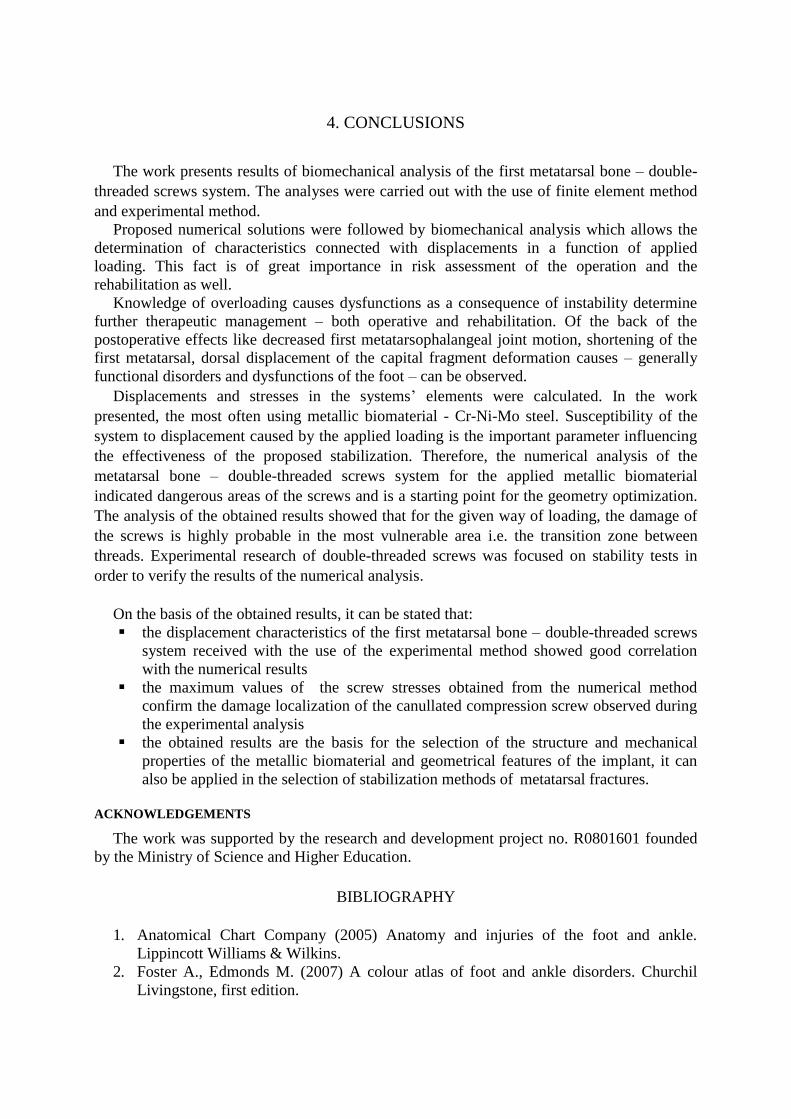
Analogous, stress analysis of the numerical model revealed that the maximum values
of stresses obtained from the bending loads were localized in the place of screw narrowing –
Fig. 9.
Fig. 9. The place of the maximal screw effort reduced stresses distribution, MPa – a,
displacement distribution in bone, mm – b
On the basis of the analysis, it was concluded that maximum equivalent stresses were
localized the fixation site of the model and at the point of the applied loading.
The obtained displacements for the metatarsal bone – compression screws system for both
tests: compression and bending revealed, that maximum values did not exceed 0.22 mm and
0.15 mm respectively whereas displacements obtained from the experimental analysis were
equal to 0.23 mm for the compression and 0.19 mm for the bending test.
b) a)
a) b)
4. CONCLUSIONS
The work presents results of biomechanical analysis of the first metatarsal bone – double-
threaded screws system. The analyses were carried out with the use of finite element method
and experimental method.
Proposed numerical solutions were followed by biomechanical analysis which allows the
determination of characteristics connected with displacements in a function of applied
loading. This fact is of great importance in risk assessment of the operation and the
rehabilitation as well.
Knowledge of overloading causes dysfunctions as a consequence of instability determine
further therapeutic management – both operative and rehabilitation. Of the back of the
postoperative effects like decreased first metatarsophalangeal joint motion, shortening of the
first metatarsal, dorsal displacement of the capital fragment deformation causes – generally
functional disorders and dysfunctions of the foot – can be observed.
Displacements and stresses in the systems’ elements were calculated. In the work
presented, the most often using metallic biomaterial - Cr-Ni-Mo steel. Susceptibility of the
system to displacement caused by the applied loading is the important parameter influencing
the effectiveness of the proposed stabilization. Therefore, the numerical analysis of the
metatarsal bone – double-threaded screws system for the applied metallic biomaterial
indicated dangerous areas of the screws and is a starting point for the geometry optimization.
The analysis of the obtained results showed that for the given way of loading, the damage of
the screws is highly probable in the most vulnerable area i.e. the transition zone between
threads. Experimental research of double-threaded screws was focused on stability tests in
order to verify the results of the numerical analysis.
On the basis of the obtained results, it can be stated that:
the displacement characteristics of the first metatarsal bone – double-threaded screws
system received with the use of the experimental method showed good correlation
with the numerical results
the maximum values of the screw stresses obtained from the numerical method
confirm the damage localization of the canullated compression screw observed during
the experimental analysis
the obtained results are the basis for the selection of the structure and mechanical
properties of the metallic biomaterial and geometrical features of the implant, it can
also be applied in the selection of stabilization methods of metatarsal fractures.
ACKNOWLEDGEMENTS
The work was supported by the research and development project no. R0801601 founded
by the Ministry of Science and Higher Education.
BIBLIOGRAPHY
1. Anatomical Chart Company (2005) Anatomy and injuries of the foot and ankle.
Lippincott Williams & Wilkins.
2. Foster A., Edmonds M. (2007) A colour atlas of foot and ankle disorders. Churchil
Livingstone, first edition.

3. Arndt A., Ekenman I., Westblad P., Lundberg A. (2002) Effects of fatigue and load
variation of metatarsal deformation measured in vivo during barefoot walking. Journal
of Biomechanics 35: 621-628.
4. Rosenberg G.A., Sferra J.J. (2000) Treatment strategies for acute fractures and
nonunions of the proximal fifth metatarsal. Journal of the American Academy of
Orthopaedic Surgeons 8(5): 332-338.
5. Buckwalter J.A., Brander E.A. (1997): Stress and insufficiency fractures. American
Academy of Family Physician 56: 175-182.
6. Beck M., Mittlmeier T. (2008) Metatarsal fractures. Der Unfallchirurg.111(10): 829-
839 (in German).
7. Rammelt S., Heineck J., Zwipp H. (2004) Metatarsal fractures. Injury 35 Suppl 2:
SB77-86.
8. Sorensen M.D., Hyer C.F. (2009) Metatarsus primus varus correction: the osteotomies.
Clinics in Podiatric Medicine and Surgery 26(3): 409-425.
9. Stryker Leibinger GmbH & Co. KG (2004) TwinFix cannulated compression screw,
Leibinger solutions for hand surgery. Procedural Guide.
10. Marciniak J. (2000) Austenitic steel – the basic implantation material used in
orthopaedic surgery. Orthopaedy, Traumatology, Rehabilitation 3: 52-58. (in Polish)
11. Marciniak J. (2002) Biomaterials, Edit by Silesian University of Technology, Gliwice:
238-252 (in Polish).
12. ISO 5832-1 (2007) Implants for surgery metallic materials -- Part 1 -- Wrought
stainless steel.
13. Walke W., Marciniak J., Paszenda Z., Kaczmarek M., Cieplak J. (2008) Biomechanical
behaviour of double threaded screw in tibia fixation. Information Technologies in
Biomedicine. Advances in Soft Computing Springer-Verlag 47, Berlin Heidelberg:
521-528.
14. Ziębowicz A., Kajzer A., Kajzer W., Marciniak J. (2009) Biomechanical analysis of
the 1st metatarsal - compression screws system. Engineering of Biomaterials.
15. Kajzer W., Kajzer A., Marciniak J. (2009) FEM analysis of compression screws used
for small bone treatment. Journal of Achievements in Materials and Manufacturing
Engineering 33(2): 189-196.
Related Documents


