Metastatic lymph node ratio as a prognostic factor after laparoscopic total mesorectal excision for extraperitoneal rectal cancer Marco Ettore Allaix • Alberto Arezzo • Paola Cassoni • Massimiliano Mistrangelo • Giuseppe Giraudo • Mario Morino Received: 7 June 2012 / Accepted: 23 October 2012 Ó Springer Science+Business Media New York 2012 Abstract Background The lymph node ratio (LNR; number of positive nodes divided by total nodes harvested) has been demonstrated to be a prognostic factor in colon cancer, but its role in extraperitoneal rectal cancer is still debated; furthermore, no data are available on laparoscopic rectal resection. The aim of this study was to evaluate the prog- nostic impact of LNR on long-term outcomes after laparoscopic total mesorectal excision (LTME) for extra- peritoneal cancer in consecutive patients with a 5-year minimum follow-up. Methods This study is a prospective analysis of consec- utive patients who underwent LTME for adenocarcinoma of the extraperitoneal rectum. Results LTME was performed in 158 patients. The median number of LN harvested was 12 (range = 3–25). The proportion of specimens with fewer than 12 examined LN was significantly higher in patients who had neoadju- vant chemoradiotherapy (p \ 0.001). During a median follow-up period of 122 months, the local recurrence rate was 8 %. At univariate analysis, disease-free survival and overall survival significantly decreased with increasing LNR (p \ 0.001). Multivariate analysis showed that the distal margin B1 cm was the only independent predictor of local recurrence (p = 0.028). LNR (cutoff value = 0.25) and lymphovascular invasion were significant prognostic factors for both disease-free (p = 0.015 and p = 0.046, respectively) and overall survival (p = 0.031 and p = 0.040, respectively). Even in the subgroup of patients in whom fewer than 12 LN were examined, LNR confirmed its prognostic role, with a statistical trend toward worse disease-free survival and overall survival. Conclusion Metastatic LNR is an independent prognostic factor for disease-free survival and overall survival after LTME for extraperitoneal rectal cancer. Keywords Lymph node ratio Á Survival Á Laparoscopy Á Total mesorectal excision Á Rectal cancer Excellence of surgical technique is of particular relevance in the treatment of extraperitoneal rectal cancer. Routine excision of the intact mesorectum during resection of cancer of the middle and lower rectum has resulted in a significant decrease in local recurrence rates [1]. Devel- oped and popularized by Heald and co worker [1], total mesorectal excision (TME) is presently the surgical gold standard, with a 4 % local recurrence rate and a 78 % tumor-free survival rate in curative cases at 5 years [2]. A recent meta-analysis by Huang et al. [3] of random- ized controlled trials that included small numbers of patients with upper or mid-to-low rectal cancer did not show differences between laparoscopic and open surgery in terms of the number of lymph nodes (LN) harvested, local recurrence, 3-year disease-free survival, and overall sur- vival. Although a minimum of 12 LN in the tumor speci- men is recommended for an adequate assessment of tumor M. E. Allaix Á A. Arezzo Á M. Mistrangelo Á G. Giraudo Á M. Morino (&) Digestive, Colorectal, Oncologic and Minimally Invasive Surgery, Department of Surgical Sciences, University of Torino, Corso A. M. Dogliotti 14, 10126 Turin, Italy e-mail: [email protected] M. E. Allaix e-mail: [email protected] P. Cassoni Department of Medical Sciences, University of Torino, Turin, Italy 123 Surg Endosc DOI 10.1007/s00464-012-2694-5 and Other Interventional Techniques

Welcome message from author
This document is posted to help you gain knowledge. Please leave a comment to let me know what you think about it! Share it to your friends and learn new things together.
Transcript
Metastatic lymph node ratio as a prognostic factorafter laparoscopic total mesorectal excision for extraperitonealrectal cancer
Marco Ettore Allaix • Alberto Arezzo •
Paola Cassoni • Massimiliano Mistrangelo •
Giuseppe Giraudo • Mario Morino
Received: 7 June 2012 / Accepted: 23 October 2012
� Springer Science+Business Media New York 2012
Abstract
Background The lymph node ratio (LNR; number of
positive nodes divided by total nodes harvested) has been
demonstrated to be a prognostic factor in colon cancer, but
its role in extraperitoneal rectal cancer is still debated;
furthermore, no data are available on laparoscopic rectal
resection. The aim of this study was to evaluate the prog-
nostic impact of LNR on long-term outcomes after
laparoscopic total mesorectal excision (LTME) for extra-
peritoneal cancer in consecutive patients with a 5-year
minimum follow-up.
Methods This study is a prospective analysis of consec-
utive patients who underwent LTME for adenocarcinoma
of the extraperitoneal rectum.
Results LTME was performed in 158 patients. The
median number of LN harvested was 12 (range = 3–25).
The proportion of specimens with fewer than 12 examined
LN was significantly higher in patients who had neoadju-
vant chemoradiotherapy (p \ 0.001). During a median
follow-up period of 122 months, the local recurrence rate
was 8 %. At univariate analysis, disease-free survival and
overall survival significantly decreased with increasing
LNR (p \ 0.001). Multivariate analysis showed that the
distal margin B1 cm was the only independent predictor of
local recurrence (p = 0.028). LNR (cutoff value = 0.25)
and lymphovascular invasion were significant prognostic
factors for both disease-free (p = 0.015 and p = 0.046,
respectively) and overall survival (p = 0.031 and p =
0.040, respectively). Even in the subgroup of patients in
whom fewer than 12 LN were examined, LNR confirmed
its prognostic role, with a statistical trend toward worse
disease-free survival and overall survival.
Conclusion Metastatic LNR is an independent prognostic
factor for disease-free survival and overall survival after
LTME for extraperitoneal rectal cancer.
Keywords Lymph node ratio � Survival � Laparoscopy �Total mesorectal excision � Rectal cancer
Excellence of surgical technique is of particular relevance
in the treatment of extraperitoneal rectal cancer. Routine
excision of the intact mesorectum during resection of
cancer of the middle and lower rectum has resulted in a
significant decrease in local recurrence rates [1]. Devel-
oped and popularized by Heald and co worker [1], total
mesorectal excision (TME) is presently the surgical gold
standard, with a 4 % local recurrence rate and a 78 %
tumor-free survival rate in curative cases at 5 years [2].
A recent meta-analysis by Huang et al. [3] of random-
ized controlled trials that included small numbers of
patients with upper or mid-to-low rectal cancer did not
show differences between laparoscopic and open surgery in
terms of the number of lymph nodes (LN) harvested, local
recurrence, 3-year disease-free survival, and overall sur-
vival. Although a minimum of 12 LN in the tumor speci-
men is recommended for an adequate assessment of tumor
M. E. Allaix � A. Arezzo � M. Mistrangelo � G. Giraudo �M. Morino (&)
Digestive, Colorectal, Oncologic and Minimally Invasive
Surgery, Department of Surgical Sciences, University of Torino,
Corso A. M. Dogliotti 14, 10126 Turin, Italy
e-mail: [email protected]
M. E. Allaix
e-mail: [email protected]
P. Cassoni
Department of Medical Sciences, University of Torino,
Turin, Italy
123
Surg Endosc
DOI 10.1007/s00464-012-2694-5
and Other Interventional Techniques
staging, the number of resected LN after TME is highly
variable.
While the prognostic role of the lymph node ratio (LNR)
in colon cancer patients has been demonstrated, its role in
extraperitoneal rectal cancer is still under debate. Further-
more, no cutoff values have been clearly identified, and no
prospective data are available in patients who underwent
laparoscopic TME.
The aim of this study was to prospectively evaluate the
prognostic value of the LNR in consecutive patients who
underwent laparoscopic TME for extraperitoneal rectal
cancer with a 5-year minimum follow-up.
Materials and methods
The data of all patients admitted to our institution with
histologically proven adenocarcinoma of extraperitoneal
(mid and low) rectum were entered into a prospective
database. In the absence of specific contraindications to
laparoscopy (e.g., severe cardiopulmonary disease and
glaucoma), patients with tumors in the extraperitoneal
rectum were selected for laparoscopic TME based on the
following criteria: elective surgery, absence of acute
intestinal occlusion or perforation, and American Society
of Anesthesiologists (ASA) status of I–III. Neither morbid
obesity nor prior pelvic surgery was considered a contra-
indication to laparoscopic TME.
The preoperative workup included clinical evaluation,
total colonoscopy, chest and upper abdominal computed
tomography (CT) scan, endoscopic ultrasound and pelvic
CT scan until 2003, then pelvic magnetic resonance
imaging (MRI), and tumor marker assay for carcinoem-
bryonic antigen (CEA) and cancer antigen 19-9.
Neoadjuvant chemoradiotherapy (CRT) was discussed
in a multidisciplinary setting. Patients preoperatively
staged as T3-4 N0-1 without distant metastases received
preoperative CRT (45 Gy over 4 weeks, together with
systemic 5-fluorouracil intravenous infusion) and were
reevaluated by clinical examination, rigid rectoscopy,
endoscopic ultrasound, and CT or MRI 4 weeks after the
completion of CRT. Definitive inclusion in the study was
decided at this point, but patients with T4 tumors that did
not show clinical downstaging or downsizing were exclu-
ded as they were considered a contraindication to the lap-
aroscopic approach.
All surgical procedures were performed by surgeons
experienced in colorectal and laparoscopic advanced sur-
gery. They followed the same oncologic principles as
described by Heald and co worker [1]: adequate resection
margins; en bloc high ligation of the inferior mesenteric
artery (IMA) and lymphadenectomy; and minimal intra-
operative manipulation of the tumor mass. Our technique
of laparoscopic anterior resection with TME has been
previously described [4]. When digital examination
revealed that the neoplasm reached the anatomic anal canal
or was fixed to the pelvic floor, a laparoscopic abdomino-
perineal resection was performed.
Only patients with a minimum follow-up of 60 months
were included in the study. For this prospective study, a
database was created to contain the patient’s characteristics
(age, gender, and ASA status), preoperative assessment,
operative variables, pathological examination, and short-
term and long-term outcomes. Operative variables included
duration of the operation (from skin incision to the appli-
cation of dressings), intraoperative morbidity and mortal-
ity, and conversion rate to abdominal surgery. Conversion
to laparotomy was defined as an unplanned incision or an
incision made longer or earlier than planned. Pathological
examination included stage of disease (TNM), length of the
surgical specimen, number of LN harvested, LNR (defined
as the number of positive nodes divided by total nodes
harvested), and longitudinal and radial margins of excision.
Lymph nodes in the mesorectal fatty tissue were identified
after formalin fixation of the specimen. Long-term out-
comes included the local recurrence rate, incidence of
abdominal wall and distant metastases, disease-free sur-
vival, and overall survival for rectal cancer.
Patients were classified in four groups according to the
LN metastases distribution (LND): (1) LND0, no LN
metastasis; (2) LND1, metastases in the perirectal nodes;
(3) LND2, metastases in the intermediate nodes; and (4)
LND3, metastases in nodes at the origin of the IMA. Stage
III patients were divided into four categories according to
quartiles for the LNR: 0.01–0.10, 0.11–0.25, 0.26–0.43,
and C0.44.
All patients who received neoadjuvant CRT and stage
II–III–IV patients were offered an adjuvant treatment after
a clinical oncologic evaluation within 8 weeks after
surgery:
Follow-up assessment consisted of a digital examina-
tion, rectoscopy, and tumor marker assay every 3 months
for the first 2 years, then every 6 months thereafter. A full
colonoscopy was performed at 12 months and then every
3 years, and chest and abdominopelvic CT scans were
performed at 6 and 12 months and every year thereafter.
The data were collected prospectively from the time of
diagnosis.
Statistical analysis
Quantitative data are given as median and range and
qualitative data as frequency and percentage. Patients with
a minimum follow-up of 60 months were included in the
analysis. Univariate analyses of 5-year overall survival and
disease-free survival rates were performed using the
Surg Endosc
123
Kaplan–Meier method, and the differences between the
groups were analyzed using the log-rank test. Patients’
observations were censored on the date of last examination
or death.
A multivariable Cox regression analysis was performed
to identify predictive factors of local recurrence, disease-
free survival, and overall survival using both forward and
backward stepwise selection. Explanatory variables with
univariable P B 0.200 were included in the multivariable
analysis. This significance level was chosen to incorporate
all potentially important predictor variables in the final
modeling process. All sets of variables were analyzed: age,
gender, type of surgery, conversion to open surgery, pT
stage, tumor grade, number of LN harvested, LNR, LND,
peritumoral lymphocytic infiltrate, lymphovascular inva-
sion, distal resection margins, postoperative anastomotic
leakage, neoadjuvant treatment, and postoperative treat-
ment. A level of 5 % was set as the criterion for statistical
significance. The data were collected in an Excel spread-
sheet. The statistical analysis was performed using
SYSTAT ver. 10 (Systat Software, Inc., Chicago, IL, USA).
Results
Between July 1996 and July 2006, 158 patients with
extraperitoneal rectal adenocarcinoma underwent laparo-
scopic TME (Table 1). One hundred twenty-six (79.7 %)
patients underwent a ‘‘sphincter-saving’’ procedure and 32
(20.3 %) underwent abdominoperineal resection. There
were 21 (13.3 %) conversions to laparotomy. The 30-day
postoperative morbidity rate was 22.2 % (35/158). The
reoperation rate was 7.6 % (12/158). The 30-day mortality
rate was 0.6 % (1/158).
Anatomopathological results
The clearance of the distal margin was B1 cm in 30
(18.9 %) cases, with no distal margin tumor infiltration. All
circumferential margins were clear. The rectal cancer
stages, according to the 7th AJCC TNM staging system, for
the 158 patients were stage I in 48 patients, stage II in 38,
stage III in 50, and stage IV in 22. The median number of
LN harvested was 12 (range = 3–25). The proportion of
specimens with fewer than 12 examined LN was signifi-
cantly higher in the group of 35 patients who underwent
neoadjuvant CRT (77.1 vs. 40.7 %; p \ 0.001). Further-
more, the median number of LN harvested was lower in
stage I–II patients (n = 10.5) than in stage III patients
(n = 11) (p = 0.079). Among the stage III patients, there
was a higher percentage of pN2 in the group with more
than 12 LN in the surgical specimen (40 vs. 20 %;
p = 0.100). LN metastases were distributed among the
stage I–III patients as follows: 86 patients were in the
LND0 group, 35 in LND1, 13 in LND2, and 2 in LND3.
Long-term results
The median follow-up period was 122 months
(range = 60–180). Seven (4.4 %) patients were lost to
follow-up (4 stage I and 3 stage II). A total of 72 (48 %)
patients received adjuvant chemotherapy and 16 (10.7 %)
adjuvant CRT. The local recurrence rate was 8 % (12/150)
at a median time of 24.5 months (range = 10–56).
The distribution of stages was similar between the group
of patients with local recurrence (LR group) and the group
of patients who did not experience a local recurrence (non-
LR group): stage I: 25 % (n = 3) versus 29.7 % (n = 41),
p = 0.989; stage II: 33.3 % (n = 4) versus 22.5 %
(n = 31), p = 0.618; stage III: 33.3 % (n = 4) versus
33.3 % (n = 46), p = 0.750; stage IV: 8.4 % (n = 1)
versus 14.5 % (n = 20), p = 0.876. A significantly higher
rate of patients with fewer than 12 LN was found in the LR
group than in the non-LR group (83.3 vs. 42.2 %,
p = 0.014). Both groups did not differ in terms of use of
neoadjuvant CRT (33.3 vs. 21 %, p = 0.532).
Distant metastases developed in 23 (17.8 %) stage I–III
patients. The port-site metastases rate was 1.3 % (2/150),
involving a stage IV patient 17 months after surgery and a
stage III patient 28 months after surgery.
The 5-year overall survival rate was 69.8 % and the
disease-free survival rate was 60.5 %. The 5-year overall
survival rate was 92.3 % for stage I patients, 85.6 % for
stage II, and 63.1 % for stage III; no patient with stage IV
disease was alive at 41 months after surgery (p \ 0.001).
The 5-year disease-free survival rate was 86.5 % for stage I
patients, 75.6 % for stage II, and 48.4 % for stage III; no
patient with stage IV was disease-free at 41 months after
surgery (p \ 0.001).
Excluding the stage IV patients, univariate analysis
showed that for the risk of local recurrence (Table 2),
tumor grade (p = 0.006), lymphovascular invasion (p =
0.010), distal surgical margins B1 cm (p = 0.018), and
number of LN harvested (p = 0.050) were all statistically
significant, while pT stage and neoadjuvant CRT showed a
statistical trend (p = 0.111 and p = 0.085, respectively).
Multivariate analysis indicated distal surgical margins
B1 cm as an independent predictor of local recurrence
(p = 0.028), while the number of LN harvested (p =
0.087), tumor grade (p = 0.052), and pT stage (p = 0.100)
had a statistical trend.
At univariate analysis, the factors associated with a
poorer disease-free survival and overall survival (Table 3, 4)
were age, pT stage, tumor grade, number of LN harvested,
LNR, lymphovascular invasion, peritumoral lymphocytic
infiltrate, and postoperative treatment. Both 5-year disease-
Surg Endosc
123
free survival and overall survival significantly decreased
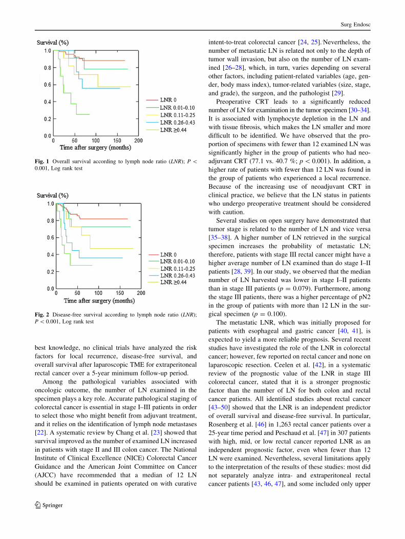
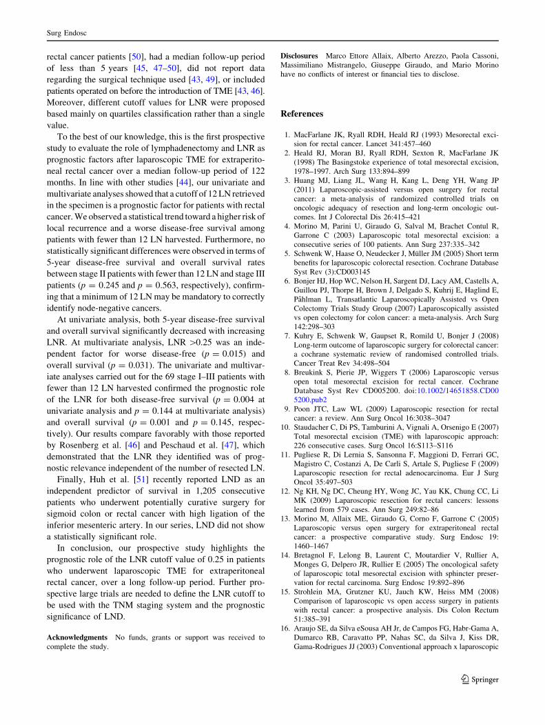
with increasing LNR (p \ 0.001) (Figs. 1, 2). At multi-
variate analysis, tumor grade (p = 0.007), LNR [ 0.25
(p = 0.015), and lymphovascular invasion (p = 0.046)
were significant predictors of poorer disease-free survival
(Table 3), while pT stage (p = 0.088), number of LN har-
vested (p = 0.174), and peritumoral lymphocytic infiltrate
(p = 0.168) showed a statistical trend. For overall survival,
the only independent factors were LNR[0.25 (p = 0.031)
and lymphovascular invasion (p = 0.040), while tumor
grade showed a statistical trend (p = 0.091) (Table 4).
Univariate and multivariate analyses were also carried
out for the 69 stage I–III patients in whom fewer than 12
LN were examined. Even in the analysis of this subgroup,
the LNR [0.25 confirmed its prognostic role for both
disease-free survival (p = 0.004 and p = 0.144 at univar-
iate and multivariate analyses, respectively) and overall
survival (p = 0.001 and p = 0.155, respectively).
No statistically significant differences were observed for
5-year disease-free survival and overall survival rates
between the 19 stage II patients with fewer than 12 LN and
the 50 stage III patients (p = 0.245 and p = 0.563,
respectively).
Discussion
Evidence-based data support the use of laparoscopic sur-
gery for colon cancer [5–7], whereas data on laparoscopic
TME with or without a sphincter-saving procedure are
limited [8, 9]. Evidence comes mainly from several case
series [4, 10–12], comparative nonrandomized studies [13–
15], or randomized clinical trials (RCTs) [16–21] with a
limited number of patients or a relatively short follow-up
period.
A recent meta-analysis by Huang et al. [3] to assess the
oncologic adequacy of resection and the oncologic out-
comes after laparoscopic versus open surgery for rectal
cancer showed that laparoscopic surgery is comparable to
open surgery in terms of anatomopathological findings and
the local recurrence rate, although no data about the
prognostic role of lymphadenectomy were given. To our
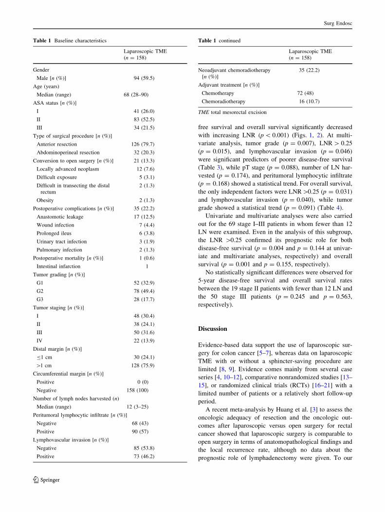
Table 1 Baseline characteristics
Laparoscopic TME
(n = 158)
Gender
Male [n (%)] 94 (59.5)
Age (years)
Median (range) 68 (28–90)
ASA status [n (%)]
I 41 (26.0)
II 83 (52.5)
III 34 (21.5)
Type of surgical procedure [n (%)]
Anterior resection 126 (79.7)
Abdominoperineal resection 32 (20.3)
Conversion to open surgery [n (%)] 21 (13.3)
Locally advanced neoplasm 12 (7.6)
Difficult exposure 5 (3.1)
Difficult in transecting the distal
rectum
2 (1.3)
Obesity 2 (1.3)
Postoperative complications [n (%)] 35 (22.2)
Anastomotic leakage 17 (12.5)
Wound infection 7 (4.4)
Prolonged ileus 6 (3.8)
Urinary tract infection 3 (1.9)
Pulmonary infection 2 (1.3)
Postoperative mortality [n (%)] 1 (0.6)
Intestinal infarction 1
Tumor grading [n (%)]
G1 52 (32.9)
G2 78 (49.4)
G3 28 (17.7)
Tumor staging [n (%)]
I 48 (30.4)
II 38 (24.1)
III 50 (31.6)
IV 22 (13.9)
Distal margin [n (%)]
B1 cm 30 (24.1)
[1 cm 128 (75.9)
Circumferential margin [n (%)]
Positive 0 (0)
Negative 158 (100)
Number of lymph nodes harvested (n)
Median (range) 12 (3–25)
Peritumoral lymphocytic infiltrate [n (%)]
Negative 68 (43)
Positive 90 (57)
Lymphovascular invasion [n (%)]
Negative 85 (53.8)
Positive 73 (46.2)
Table 1 continued
Laparoscopic TME
(n = 158)
Neoadjuvant chemoradiotherapy
[n (%)]
35 (22.2)
Adjuvant treatment [n (%)]
Chemotherapy 72 (48)
Chemoradiotherapy 16 (10.7)
TME total mesorectal excision
Surg Endosc
123
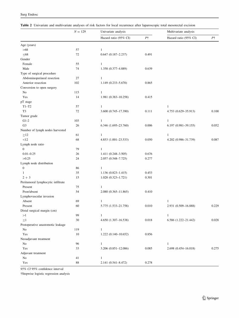
Table 2 Univariate and multivariate analyses of risk factors for local recurrence after laparoscopic total mesorectal excision
N = 129 Univariate analysis Multivariate analysis
Hazard ratio (95% CI) P� Hazard ratio (95% CI) P�
Age (years)
[68 57 1
B68 72 0.647 (0.187–2.237) 0.491
Gender
Female 55 1
Male 74 1.358 (0.377–4.889) 0.639
Type of surgical procedure
Abdominoperineal resection 27 1
Anterior resection 102 1.149 (0.233–5.670) 0.865
Conversion to open surgery
No 115 1
Yes 14 1.981 (0.383–10.258) 0.415
pT stage
T1–T2 57 1 1
T3 72 3.600 (0.745–17.390) 0.111 4.753 (0.629–35.913) 0.100
Tumor grade
G1-2 103 1 1
G3 26 6.346 (1.695–23.760) 0.006 6.197 (0.981–39.155) 0.052
Number of lymph nodes harvested
C12 61 1 1
\12 68 4.853 (1.001–23.533) 0.050 4.202 (0.986–31.739) 0.087
Lymph node ratio
0 79 1
0.01–0.25 26 1.411 (0.248–3.505) 0.676
[0.25 24 2.057 (0.548–7.725) 0.277
Lymph node distribution
0 86 1
1 35 1.136 (0.823–1.415) 0.453
2 ? 3 15 1.028 (0.323–1.721) 0.301
Peritumoral lymphocytic infiltrate
Present 75 1
Poor/absent 54 2.080 (0.365–11.865) 0.410
Lymphovascular invasion
Absent 69 1 1
Present 60 5.775 (1.533–21.758) 0.010 2.931 (0.509–16.888) 0.229
Distal surgical margin (cm)
[1 99 1 1
B1 30 4.650 (1.307–16.538) 0.018 6.586 (1.222–21.442) 0.028
Postoperative anastomotic leakage
No 119 1
Yes 10 1.222 (0.140–10.652) 0.856
Neoadjuvant treatment
No 96 1 1
Yes 33 3.206 (0.851–12.086) 0.085 2.698 (0.454–16.018) 0.275
Adjuvant treatment
No 41 1
Yes 88 2.141 (0.541–8.472) 0.278
95% CI 95% confidence interval
�Stepwise logistic regression analysis
Surg Endosc
123
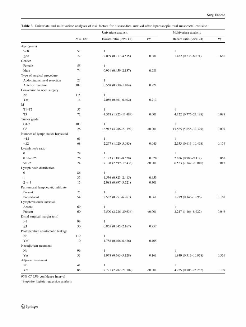
Table 3 Univariate and multivariate analyses of risk factors for disease-free survival after laparoscopic total mesorectal excision
Univariate analysis Multivariate analysis
N = 129 Hazard ratio (95% CI) P� Hazard ratio (95% CI) P�
Age (years)
[68 57 1 1
B68 72 2.039 (0.917–4.535) 0.081 1.452 (0.238–8.871) 0.686
Gender
Female 55 1
Male 74 0.991 (0.459–2.137) 0.981
Type of surgical procedure
Abdominoperineal resection 27 1
Anterior resection 102 0.568 (0.230–1.404) 0.221
Conversion to open surgery
No 115 1
Yes 14 2.056 (0.661–6.402) 0.213
M
T1–T2 57 1 1
T3 72 4.578 (1.825–11.484) 0.001 4.122 (0.775–23.198) 0.088
Tumor grade
G1-2 103 1 1
G3 26 16.917 (4.986–27.392) \0.001 15.565 (5.655–32.329) 0.007
Number of lymph nodes harvested
C12 61 1 1
\12 68 2.277 (1.020–5.083) 0.045 2.533 (0.613–10.468) 0.174
Lymph node ratio
0 79 1 1
0.01–0.25 26 3.173 (1.181–8.528) 0.0280 2.856 (0.988–9.112) 0.063
[0.25 24 7.108 (2.599–19.436) \0.001 6.523 (2.347–20.010) 0.015
Lymph node distribution
0 86 1
1 35 1.536 (0.823–2.415) 0.453
2 ? 3 15 2.088 (0.897–3.721) 0.301
Peritumoral lymphocytic infiltrate
Present 75 1 1
Poor/absent 54 2.582 (0.957–6.967) 0.061 1.279 (0.146–1.696) 0.168
Lymphovascular invasion
Absent 69 1 1
Present 60 7.500 (2.726–20.636) \0.001 2.247 (1.166–8.922) 0.046
Distal surgical margin (cm)
[1 99 1
B1 30 0.865 (0.345–2.167) 0.757
Postoperative anastomotic leakage
No 119 1
Yes 10 1.758 (0.466–6.626) 0.405
Neoadjuvant treatment
No 96 1 1
Yes 33 1.978 (0.763–5.128) 0.161 1.849 (0.313–10.928) 0.556
Adjuvant treatment
No 41 1 1
Yes 88 7.771 (2.782–21.707) \0.001 4.225 (0.706–25.282) 0.109
95% CI 95% confidence interval
�Stepwise logistic regression analysis
Surg Endosc
123
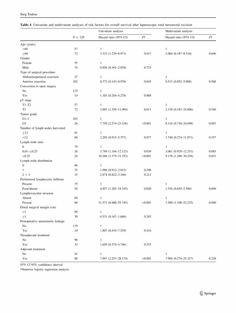
Table 4 Univariate and multivariate analyses of risk factors for overall survival after laparoscopic total mesorectal excision
Univariate analysis Multivariate analysis
N = 129 Hazard ratio (95% CI) P� Hazard ratio (95% CI) P�
Age (years)
[68 57 1 1
B68 72 3.333 (1.239–8.971) 0.017 2.004 (0.187–8.510) 0.696
Gender
Female 55 1
Male 74 0.856 (0.361–2.029) 0.723
Type of surgical procedure
Abdominoperineal resection 27 1 1
Anterior resection 102 0.373 (0.143–0.976) 0.045 0.513 (0.052–5.088) 0.568
Conversion to open surgery
No 115 1
Yes 14 1.103 (0.284–4.278) 0.888
pT stage
T1–T2 57 1 1
T3 72 3.883 (1.359–11.094) 0.011 2.129 (0.183–24.806) 0.546
Tumor grade
G1-2 103 1 1
G3 26 7.750 (2.574–23.336) \0.001 8.116 (0.718–34.698) 0.091
Number of lymph nodes harvested
C12 61 1 1
\12 68 2.295 (0.915–5.757) 0.077 1.740 (0.274–11.071) 0.557
Lymph node ratio
0 79 1 1
0.01–B0.25 26 3.789 (1.184–12.123) 0.039 3.061 (0.929–12.251) 0.085
[0.25 24 10.286 (3.375–31.353) \0.001 9.178 (1.288–30.258). 0.031
Lymph node distribution
0 86 1
1 35 1.986 (0.912–2.915) 0.298
2 ? 3 15 2.874 (0.822–3.166) 0.211
Peritumoral lymphocytic infiltrate
Present 75 1 1
Poor/absent 54 4.857 (1.285–18.355) 0.020 1.536 (0.659–5.580) 0.696
Lymphovascular invasion
Absent 69 1 1
Present 60 31.571 (6.400–55.745) \0.001 7.580 (1.100–52.235) 0.040
Distal surgical margin (cm)
[1 99 1
B1 30 0.531 (0.167–1.686) 0.283
Postoperative anastomotic leakage
No 119 1
Yes 10 1.807 (0.434–7.529) 0.416
Neoadjuvant treatment
No 96 1
Yes 33 1.650 (0.574–4.746) 0.353
Adjuvant treatment
No 41 1 1
Yes 88 7.967 (2.253–28.174) \0.001 7.904 (0.274–25.327) 0.228
95% CI 95% confidence interval
�Stepwise logistic regression analysis
Surg Endosc
123
best knowledge, no clinical trials have analyzed the risk
factors for local recurrence, disease-free survival, and
overall survival after laparoscopic TME for extraperitoneal
rectal cancer over a 5-year minimum follow-up period.
Among the pathological variables associated with
oncologic outcome, the number of LN examined in the
specimen plays a key role. Accurate pathological staging of
colorectal cancer is essential in stage I–III patients in order
to select those who might benefit from adjuvant treatment,
and it relies on the identification of lymph node metastases
[22]. A systematic review by Chang et al. [23] showed that
survival improved as the number of examined LN increased
in patients with stage II and III colon cancer. The National
Institute of Clinical Excellence (NICE) Colorectal Cancer
Guidance and the American Joint Committee on Cancer
(AJCC) have recommended that a median of 12 LN
should be examined in patients operated on with curative
intent-to-treat colorectal cancer [24, 25]..Nevertheless, the
number of metastatic LN is related not only to the depth of
tumor wall invasion, but also on the number of LN exam-
ined [26–28], which, in turn, varies depending on several
other factors, including patient-related variables (age, gen-
der, body mass index), tumor-related variables (size, stage,
and grade), the surgeon, and the pathologist [29].
Preoperative CRT leads to a significantly reduced
number of LN for examination in the tumor specimen [30–34].
It is associated with lymphocyte depletion in the LN and
with tissue fibrosis, which makes the LN smaller and more
difficult to be identified. We have observed that the pro-
portion of specimens with fewer than 12 examined LN was
significantly higher in the group of patients who had neo-
adjuvant CRT (77.1 vs. 40.7 %; p \ 0.001). In addition, a
higher rate of patients with fewer than 12 LN was found in
the group of patients who experienced a local recurrence.
Because of the increasing use of neoadjuvant CRT in
clinical practice, we believe that the LN status in patients
who undergo preoperative treatment should be considered
with caution.
Several studies on open surgery have demonstrated that
tumor stage is related to the number of LN and vice versa
[35–38]. A higher number of LN retrieved in the surgical
specimen increases the probability of metastatic LN;
therefore, patients with stage III rectal cancer might have a
higher average number of LN examined than do stage I–II
patients [28, 39]. In our study, we observed that the median
number of LN harvested was lower in stage I–II patients
than in stage III patients (p = 0.079). Furthermore, among
the stage III patients, there was a higher percentage of pN2
in the group of patients with more than 12 LN in the sur-
gical specimen (p = 0.100).
The metastatic LNR, which was initially proposed for
patients with esophageal and gastric cancer [40, 41], is
expected to yield a more reliable prognosis. Several recent
studies have investigated the role of the LNR in colorectal
cancer; however, few reported on rectal cancer and none on
laparoscopic resection. Ceelen et al. [42], in a systematic
review of the prognostic value of the LNR in stage III
colorectal cancer, stated that it is a stronger prognostic
factor than the number of LN for both colon and rectal
cancer patients. All identified studies about rectal cancer
[43–50] showed that the LNR is an independent predictor
of overall survival and disease-free survival. In particular,
Rosenberg et al. [46] in 1,263 rectal cancer patients over a
25-year time period and Peschaud et al. [47] in 307 patients
with high, mid, or low rectal cancer reported LNR as an
independent prognostic factor, even when fewer than 12
LN were examined. Nevertheless, several limitations apply
to the interpretation of the results of these studies: most did
not separately analyze intra- and extraperitoneal rectal
cancer patients [43, 46, 47], and some included only upper
Fig. 1 Overall survival according to lymph node ratio (LNR); P \0.001, Log rank test
Fig. 2 Disease-free survival according to lymph node ratio (LNR);
P \ 0.001, Log rank test
Surg Endosc
123
rectal cancer patients [50], had a median follow-up period
of less than 5 years [45, 47–50], did not report data
regarding the surgical technique used [43, 49], or included
patients operated on before the introduction of TME [43, 46].
Moreover, different cutoff values for LNR were proposed
based mainly on quartiles classification rather than a single
value.
To the best of our knowledge, this is the first prospective
study to evaluate the role of lymphadenectomy and LNR as
prognostic factors after laparoscopic TME for extraperito-
neal rectal cancer over a median follow-up period of 122
months. In line with other studies [44], our univariate and
multivariate analyses showed that a cutoff of 12 LN retrieved
in the specimen is a prognostic factor for patients with rectal
cancer. We observed a statistical trend toward a higher risk of
local recurrence and a worse disease-free survival among
patients with fewer than 12 LN harvested. Furthermore, no
statistically significant differences were observed in terms of
5-year disease-free survival and overall survival rates
between stage II patients with fewer than 12 LN and stage III
patients (p = 0.245 and p = 0.563, respectively), confirm-
ing that a minimum of 12 LN may be mandatory to correctly
identify node-negative cancers.
At univariate analysis, both 5-year disease-free survival
and overall survival significantly decreased with increasing
LNR. At multivariate analysis, LNR [0.25 was an inde-
pendent factor for worse disease-free (p = 0.015) and
overall survival (p = 0.031). The univariate and multivar-
iate analyses carried out for the 69 stage I–III patients with
fewer than 12 LN harvested confirmed the prognostic role
of the LNR for both disease-free survival (p = 0.004 at
univariate analysis and p = 0.144 at multivariate analysis)
and overall survival (p = 0.001 and p = 0.145, respec-
tively). Our results compare favorably with those reported
by Rosenberg et al. [46] and Peschaud et al. [47], which
demonstrated that the LNR they identified was of prog-
nostic relevance independent of the number of resected LN.
Finally, Huh et al. [51] recently reported LND as an
independent predictor of survival in 1,205 consecutive
patients who underwent potentially curative surgery for
sigmoid colon or rectal cancer with high ligation of the
inferior mesenteric artery. In our series, LND did not show
a statistically significant role.
In conclusion, our prospective study highlights the
prognostic role of the LNR cutoff value of 0.25 in patients
who underwent laparoscopic TME for extraperitoneal
rectal cancer, over a long follow-up period. Further pro-
spective large trials are needed to define the LNR cutoff to
be used with the TNM staging system and the prognostic
significance of LND.
Acknowledgments No funds, grants or support was received to
complete the study.
Disclosures Marco Ettore Allaix, Alberto Arezzo, Paola Cassoni,
Massimiliano Mistrangelo, Giuseppe Giraudo, and Mario Morino
have no conflicts of interest or financial ties to disclose.
References
1. MacFarlane JK, Ryall RDH, Heald RJ (1993) Mesorectal exci-
sion for rectal cancer. Lancet 341:457–460
2. Heald RJ, Moran BJ, Ryall RDH, Sexton R, MacFarlane JK
(1998) The Basingstoke experience of total mesorectal excision,
1978–1997. Arch Surg 133:894–899
3. Huang MJ, Liang JL, Wang H, Kang L, Deng YH, Wang JP
(2011) Laparoscopic-assisted versus open surgery for rectal
cancer: a meta-analysis of randomized controlled trials on
oncologic adequacy of resection and long-term oncologic out-
comes. Int J Colorectal Dis 26:415–421
4. Morino M, Parini U, Giraudo G, Salval M, Brachet Contul R,
Garrone C (2003) Laparoscopic total mesorectal excision: a
consecutive series of 100 patients. Ann Surg 237:335–342
5. Schwenk W, Haase O, Neudecker J, Muller JM (2005) Short term
benefits for laparoscopic colorectal resection. Cochrane Database
Syst Rev (3):CD003145
6. Bonjer HJ, Hop WC, Nelson H, Sargent DJ, Lacy AM, Castells A,
Guillou PJ, Thorpe H, Brown J, Delgado S, Kuhrij E, Haglind E,
Pahlman L, Transatlantic Laparoscopically Assisted vs Open
Colectomy Trials Study Group (2007) Laparoscopically assisted
vs open colectomy for colon cancer: a meta-analysis. Arch Surg
142:298–303
7. Kuhry E, Schwenk W, Gaupset R, Romild U, Bonjer J (2008)
Long-term outcome of laparoscopic surgery for colorectal cancer:
a cochrane systematic review of randomised controlled trials.
Cancer Treat Rev 34:498–504
8. Breukink S, Pierie JP, Wiggers T (2006) Laparoscopic versus
open total mesorectal excision for rectal cancer. Cochrane
Database Syst Rev CD005200. doi:10.1002/14651858.CD00
5200.pub2
9. Poon JTC, Law WL (2009) Laparoscopic resection for rectal
cancer: a review. Ann Surg Oncol 16:3038–3047
10. Staudacher C, Di PS, Tamburini A, Vignali A, Orsenigo E (2007)
Total mesorectal excision (TME) with laparoscopic approach:
226 consecutive cases. Surg Oncol 16:S113–S116
11. Pugliese R, Di Lernia S, Sansonna F, Maggioni D, Ferrari GC,
Magistro C, Costanzi A, De Carli S, Artale S, Pugliese F (2009)
Laparoscopic resection for rectal adenocarcinoma. Eur J Surg
Oncol 35:497–503
12. Ng KH, Ng DC, Cheung HY, Wong JC, Yau KK, Chung CC, Li
MK (2009) Laparoscopic resection for rectal cancers: lessons
learned from 579 cases. Ann Surg 249:82–86
13. Morino M, Allaix ME, Giraudo G, Corno F, Garrone C (2005)
Laparoscopic versus open surgery for extraperitoneal rectal
cancer: a prospective comparative study. Surg Endosc 19:
1460–1467
14. Bretagnol F, Lelong B, Laurent C, Moutardier V, Rullier A,
Monges G, Delpero JR, Rullier E (2005) The oncological safety
of laparoscopic total mesorectal excision with sphincter preser-
vation for rectal carcinoma. Surg Endosc 19:892–896
15. Strohlein MA, Grutzner KU, Jauch KW, Heiss MM (2008)
Comparison of laparoscopic vs open access surgery in patients
with rectal cancer: a prospective analysis. Dis Colon Rectum
51:385–391
16. Araujo SE, da Silva eSousa AH Jr, de Campos FG, Habr-Gama A,
Dumarco RB, Caravatto PP, Nahas SC, da Silva J, Kiss DR,
Gama-Rodrigues JJ (2003) Conventional approach x laparoscopic
Surg Endosc
123
abdominoperineal resection for rectal cancer treatment after
neoadjuvant chemoradiation: results of a prospective randomized
trial. Rev Hosp Clin Fac Med Sao Paulo 58:133–140
17. Jayne DG, Guillou PJ, Thorpe H, Quirke P, Copeland J, Smith
AM, Heath RM, Brown JM, UK MRC CLASICC Trial Group
(2007) Randomized trial of laparoscopic-assisted resection of
colorectal carcinoma: 3-year results of the UK MRC CLASICC
Trial Group. J Clin Oncol 25:3061–3068
18. Braga M, Frasson M, Vignali A, Zuliani W, Capretti G, Di Carlo V
(2007) Laparoscopic resection in rectal cancer patients: outcome
and cost-benefit analysis. Dis Colon Rectum 50:464–471
19. Ng SS, Leung KL, Lee JF, Yiu RY, Li JC, Teoh AY (2008)
Laparoscopic-assisted versus open abdominoperineal resection
for low rectal cancer: a prospective randomized trial. Ann Surg
Oncol 15:2418–2425
20. Lujan J, Valero G, Hernandez Q, Sanchez A, Frutos MD, Parrilla P
(2009) Randomized clinical trial comparing laparoscopic and open
surgery in patients with rectal cancer. Br J Surg 96:982–989
21. Ng SS, Leung KL, Lee JF, Yiu RY, Li JC, Hon SS (2009) Long-
term morbidity and oncologic outcomes of laparoscopic-assisted
anterior resection for upper rectal cancer: ten-year results of a
prospective, randomized trial. Dis Colon Rectum 52:558–566
22. Andre T, Boni C, Navarro M, Tabernero J, Hickish T, Topham C,
Bonetti A, Clingan P, Bridgewater J, Rivera F, de Gramont A
(2009) Improved overall survival with oxaliplatin, fluorouracil,
and leucovorin as adjuvant treatment in stage II or III colon
cancer in the MOSAIC trial. J Clin Oncol 27:3109–3116
23. Chang GJ, Rodriguez-Bigas MA, Skibber JM, Moyer VA (2007)
Lymph node evaluation and survival after curative resection of
colon cancer: systematic review. J Natl Cancer Inst 99:433–441
24. NICE Improving Outcomes in Colorectal Cancer, Manual
Update, London: National Institute for Clinical Excellence, May
2004
25. Nelson H, Petrelli N, Carlin A, Couture J, Fleshman J, Guillem J,
Miedema B, Ota D, Sargent D, National Cancer Institute Expert
Panel (2001) Guidelines 2000 for colon and rectal cancer surgery.
J Natl Cancer Inst 93:583–596
26. Goldstein NS, Sanford W, Coffey M, Layfield LJ (1996) Lymph
node recovery from colorectal resection specimens removed for
adenocarcinoma. Trends over time and a recommendation for a
minimum number of lymph nodes to be recovered. Am J Clin
Pathol 106:209–216
27. Hernanz F, Revuelta S, Redondo C, Madrazo C, Castillo J,
Gomez-Fleitas M (1994) Colorectal adenocarcinoma: quality of the
assessment of lymph node metastases. Dis Colon Rectum 37:
373–376
28. Wong JH, Severino R, Honnebier MB, Tom P, Namiki TS (1999)
Number of nodes examined and staging accuracy in colorectal
carcinoma. J Clin Oncol 17:2896–2900
29. Evans MD, Barton K, Rees A, Stamatakis JD, Karandikar SS
(2008) The impact of surgeon and pathologist on lymph node
retrieval in colorectal cancer and its impact on survival for
patients with Dukes’ stage B disease. Colorectal Dis 10:157–164
30. Sermier A, Gervaz P, Egger JF, Dao M, Allal AS, Bonet M, Morel P
(2006) Lymph node retrieval in abdominoperineal surgical speci-
men is radiation time-dependent. World J Surg Oncol 4:29
31. Wichmann MW, Muller C, Meyer G, Strauss T, Hornung HM,
Lau-Werner U, Angele MK, Schildberg FW (2002) Effect of
preoperative radiochemotherapy on lymph node retrieval after
resection of rectal cancer. Arch Surg 137:206–210
32. Baxter NN, Morris AM, Rothenberger DA, Tepper JE (2005)
Impact of preoperative radiation for rectal cancer on subsequent
lymph node evaluation: a population-based analysis. Int J Radiat
Oncol Biol Phys 61:426–431
33. Nagtegaal ID, van de Velde CJ, van der Worp E, Kapiteijn E,
Quirke P, van Krieken JH (2002) Macroscopic evaluation of
rectal cancer resection specimen: clinical significance of the
pathologist in quality control. J Clin Oncol 20:1729–1734
34. Rullier A, Laurent C, Capdepont M, Vendrely V, Belleannee G,
Bioulac-Sage P, Rullier E (2008) Lymph nodes after preoperative
chemoradiotherapy for rectal carcinoma: number, status, and
impact on survival. Am J Surg Pathol 32:45–50
35. Joseph NE, Sigurdson ER, Hanlon AL, Wang H, Mayer RJ,
MacDonald JS, Catalano PJ, Haller DG (2003) Accuracy of
determining nodal negativity in colorectal cancer on the basis of
the number of nodes retrieved on resection. Ann Surg Oncol
10:213–218
36. Jakub JW, Russell G, Tillman CL, Lariscy C (2009) Colon cancer
and low lymph node count. Who is to blame? Arch Surg 144:
1115–1120
37. Baxter NN, Virnig DJ, Rothenberger DA, Morris AM, Jessurun J,
Virnig BA (2005) Lymph node evaluation in colorectal cancer
patients: a population-based study. J Natl Cancer Inst 97:219–225
38. Gelos M, Gelhaus J, Mehnert P, Bonhag G, Sand M, Philippou S,
Mann B (2008) Factors influencing lymph node harvest in colo-
rectal surgery. Int J Colorectal Dis 23:53–59
39. Tepper JE, O’Connell MJ, Niedzwiecki D, Hollis D, Compton C,
Benson AB 3rd, Cummings B, Gunderson L, Macdonald JS,
Mayer RJ (2001) Impact of number of nodes retrieved on out-
come in patients with rectal cancer. J Clin Oncol 19:157–163
40. Marchet A, Mocellin S, Ambrosi A, Morgagni P, Garcea D,
Marrelli D, Roviello F, de Manzoni G, Minicozzi A, Natalini G,
De Santis F, Baiocchi L, Coniglio A, Nitti D, Italian Research
Group for Gastric Cancer (IRGGC) (2007) The ratio between
metastatic and examined lymph nodes (N Ratio) is an indepen-
dent prognostic factor in gastric cancer regardless of the type of
lymphadenectomy: results of an Italian multicentric study in 1853
patients. Ann Surg 245:543–552
41. Mariette C, Piessen G, Briez N, Triboulet JP (2008) The number
of metastatic lymph nodes and the ratio between metastatic and
examined lymph nodes are independent prognostic factors in
oesophageal cancer regardless of neoadjuvant chemoradiation or
lympadenectomy extent. Ann Surg 247:365–371
42. Ceelen W, Van Nieuwenhove Y, Pattyn P (2010) Prognostic
value of the lymph node ratio in stage III colorectal cancer: a
systematic review. Ann Surg Oncol 17:2847–2855
43. Stocchi L, Nelson H, Sargent DJ, O’Connell MJ, Tepper JE,
Krook JE, Beart R Jr, North Central Cancer Treatment Group
(2001) Impact of surgical and pathologic variables in rectal
cancer: a United States Community and Cooperative Group
report. J Clin Oncol 19:3895–3902
44. Edler D, Ohrling K, Hallstrom M, Karlberg M, Ragnhammar P
(2007) The number of analyzed lymph nodes - a prognostic factor
in colorectal cancer. Acta Oncol 46:975–981
45. Peng JJ, Xu Y, Guan ZQ, Zhu J, Wang M, Cai G, Sheng W, Cai S
(2008) Prognostic significance of the metastatic lymph node ratio
in node-positive rectal cancer. Ann Surg Oncol 15:3118–3123
46. Rosenberg R, Friederichs J, Schuster T, Gertler R, Maak M,
Becker K, Grebner A, Ulm K, Hofler H, Nekarda H, Siewert JR
(2008) Prognosis of patients with colorectal cancer is associated
with lymph node ratio. A single-center analysis of 3026 patients
over a 25-year time period. Ann Surg 248:968–978
47. Peschaud F, Benoist S, Julie C, Beauchet A, Penna C, Rougier P,
Nordlinger B (2008) Prognosis of patients with colorectal cancer
is associated with lymph node ratio: a single-center analysis
of 3026 patients over a 25-year time period. Ann Surg 248:
1067–1073
48. Kim YS, Kim JH, Yoon SM, Choi EK, Ahn SD, Lee SW, Kim
JC, Yu CS, Kim HC, Kim TW, Chang HM (2009) Lymph node
ratio as a prognostic factor in patients with stage III rectal cancer
treated with total mesorectal excision followed by chemoradio-
therapy. Int J Radiat Oncol Biol Phys 74:796–802
Surg Endosc
123

49. Moug SJ, Saldanha JD, McGregor JR, Balsitis M, Diament RH
(2009) Positive lymph node retrieval ratio optimises patient
staging in colorectal cancer. Br J Cancer 100:1530–1533
50. Priolli DG, Cardinalli IA, Pereira JA, Alfredo CH, Margarido NF,
Martinez CA (2009) Metastatic lymph node ratio as an
independent prognostic variable in colorectal cancer: study of
113 patients. Tech Coloproctol 13:113–121
51. Huh JW, Kim YJ, Kim HR (2012) Distribution of lymph mode
metastases is an independent predictor of survival for sigmoid
colon and rectal cancer. Ann Surg 255:70–78
Surg Endosc
123
Related Documents