Iran J Radiol. 2015 April; 12(2): e11197. DOI: 10.5812/iranjradiol.11197 Published online 2015 April 20. Case Report Metastatic Adenocarcinoma Presenting as Extensive Cavoatrial Tumor Thrombus Bushra Johari 1,2,* ; Yang Faridah Abdul Aziz 1 ; Sivakumar Krishnasamy 3 ; Lai Meng Looi 4 ; Shahrul Amry Hashim 3 ; Raja Amin Raja Mokhtar 3 1 Department of Biomedical Imaging, University Malaya of Research Imaging Center, Faculty of Medicine, University Malaya, Kuala Lumpur, Malaysia 2 Faculty of Medicine, University Teknologi MARA, Jalan Hospital, Sungai Buloh Selangor, Malaysia 3 Department of Surgery, University of Malaya Medical Center, Kuala Lumpur, Malaysia 4 Department of Pathology, University of Malaya Medical Center, Kuala Lumpur, Malaysia *Corresponding author: Bushra Johari, Faculty of Medicine, MARA University of Technology, Jalan Hospital, Sungai Buloh Selangor, Malaysia. Tel: +6012-3729495, E-mail: bushra0271@ yahoo.com Received: March 14, 2013; Revised: May 24, 2013; Accepted: June 1, 2013 The presence of tumor thrombus in the right atrium is frequently the result of direct intraluminal extension of infra-diaphragmatic malignancy into the inferior vena cava (IVC) or supradiaphragmatic carcinoma into the superior vena cava (SVC). Right atrial tumor thrombus with extension into both SVC and IVC has not been reported in the literature. We present a patient who presented with symptoms of right atrial and SVC obstruction. Imaging revealed presence of a thrombus in the right atrium, extending to the SVC and IVC, with the additional findings of a left adrenal mass and multiple liver lesions. The histopathological examination of the right atrial mass revealed metastatic adenocarcinoma cells. The patient was given a presumptive diagnosis of metastatic adenocarcinoma, most likely adrenal in origin, with multiple hepatic lesions suspicious for metastasis. The clinical outcome of the patient was not favorable; the patient succumbed before the adrenal mass could be confirmed to be the primary tumor. This case highlights that in patients manifesting with extensive cavoatrial thrombus as, the existence of primary carcinoma should be considered especially in the adrenal cortex or in the lung. Keywords:Tumor; Thrombosis; Heart Atria; Cardiac Imaging Techniques Copyright © 2015, Tehran University of Medical Sciences and Iranian Society of Radiology. This is an open-access article distributed under the terms of the Creative Commons Attribution-NonCommercial 4.0 International License (http://creativecommons.org/licenses/by-nc/4.0/) which permits copy and redistribute the mate- rial just in noncommercial usages, provided the original work is properly cited. 1. Introduction The presence of tumor thrombus in the right atrium is frequently the result of direct intraluminal thrombotic extension of infradiaphragmatic malignancy such as re- nal or hepatocellular carcinoma into the inferior vena cava (IVC) or thyroid carcinoma into the superior vena cava (SVC). Right atrial tumor thrombus with extension into both SVC and IVC has not been reported in the lit- erature. Primary adrenal tumors presenting as cardiac masses are rare. 2. Case Presentation A 42-year-old man, with previous history of treated pulmonary tuberculosis, presented with progressive shortness of breath, reduced effort tolerance, facial puff- iness and upper limb swelling over a period of several months. On examination, the patient was hypotensive, tachycardic and hypoxic. Initial transthoracic echo- cardiogram revealed a large mass in the right atrium, with pericardial effusion (Figure 1). The left sided struc- tures were normal in size and function. The provision- al diagnosis at this time was right atrial myxoma. The electrocardiography (ECG)-gated cardiac computed to- mography (CT) using a 64-slice duals Source CT system (Siemens Medical Solutions, Malvern, PA, USA) revealed a minimally enhancing thrombus in the right atrium, extending to SVC the superior vena cava (SVC), subcla- vian veins and inferior vena cava (IVC), with mass effect on the left main and IVC, with mass pulmonary artery and the right ventricle (Figure 2). Doppler ultrasonog- raphy (US) of the abdomen and lower limbs showed extensive thrombus in the IVC and left femoral vein, as well as a left suprarenal mass (Figure 3). A decision was made to perform sternotomy for right atriotomy and SVC venotomy, as there was a high risk of pulmo- nary thromboembolic phenomenon. Intraoperatively, the right atrium was found to be filled with a densely adherent organized white thrombus, extending to IVC, SVC and innominate veins. Evacuation of clots was per- formed (Figure 4). Although the surgical procedure was complicated by massive blood loss, the patient slowly recovered with intensive care. The histopathological ex- amination of the right atrial mass revealed metastatic poorly-differentiated adenocarcinoma cells, exhibiting mucin-positive cytoplasmic vacuoles, which were im- munopositive for cytokeratin 7 and thyroid transcrip- CARDIAC IMAGING

Welcome message from author
This document is posted to help you gain knowledge. Please leave a comment to let me know what you think about it! Share it to your friends and learn new things together.
Transcript

Iran J Radiol. 2015 April; 12(2): e11197. DOI: 10.5812/iranjradiol.11197
Published online 2015 April 20. Case Report
Metastatic Adenocarcinoma Presenting as Extensive Cavoatrial Tumor Thrombus
Bushra Johari 1,2,*; Yang Faridah Abdul Aziz 1; Sivakumar Krishnasamy 3; Lai Meng Looi 4; Shahrul Amry Hashim 3; Raja Amin Raja Mokhtar 3
1Department of Biomedical Imaging, University Malaya of Research Imaging Center, Faculty of Medicine, University Malaya, Kuala Lumpur, Malaysia2Faculty of Medicine, University Teknologi MARA, Jalan Hospital, Sungai Buloh Selangor, Malaysia3Department of Surgery, University of Malaya Medical Center, Kuala Lumpur, Malaysia4Department of Pathology, University of Malaya Medical Center, Kuala Lumpur, Malaysia*Corresponding author: Bushra Johari, Faculty of Medicine, MARA University of Technology, Jalan Hospital, Sungai Buloh Selangor, Malaysia. Tel: +6012-3729495, E-mail: [email protected]
Received: March 14, 2013; Revised: May 24, 2013; Accepted: June 1, 2013
The presence of tumor thrombus in the right atrium is frequently the result of direct intraluminal extension of infra-diaphragmatic malignancy into the inferior vena cava (IVC) or supradiaphragmatic carcinoma into the superior vena cava (SVC). Right atrial tumor thrombus with extension into both SVC and IVC has not been reported in the literature. We present a patient who presented with symptoms of right atrial and SVC obstruction. Imaging revealed presence of a thrombus in the right atrium, extending to the SVC and IVC, with the additional findings of a left adrenal mass and multiple liver lesions. The histopathological examination of the right atrial mass revealed metastatic adenocarcinoma cells. The patient was given a presumptive diagnosis of metastatic adenocarcinoma, most likely adrenal in origin, with multiple hepatic lesions suspicious for metastasis. The clinical outcome of the patient was not favorable; the patient succumbed before the adrenal mass could be confirmed to be the primary tumor. This case highlights that in patients manifesting with extensive cavoatrial thrombus as, the existence of primary carcinoma should be considered especially in the adrenal cortex or in the lung.
Keywords:Tumor; Thrombosis; Heart Atria; Cardiac Imaging Techniques
Copyright © 2015, Tehran University of Medical Sciences and Iranian Society of Radiology. This is an open-access article distributed under the terms of the Creative Commons Attribution-NonCommercial 4.0 International License (http://creativecommons.org/licenses/by-nc/4.0/) which permits copy and redistribute the mate-rial just in noncommercial usages, provided the original work is properly cited.
1. IntroductionThe presence of tumor thrombus in the right atrium is
frequently the result of direct intraluminal thrombotic extension of infradiaphragmatic malignancy such as re-nal or hepatocellular carcinoma into the inferior vena cava (IVC) or thyroid carcinoma into the superior vena cava (SVC). Right atrial tumor thrombus with extension into both SVC and IVC has not been reported in the lit-erature. Primary adrenal tumors presenting as cardiac masses are rare.
2. Case PresentationA 42-year-old man, with previous history of treated
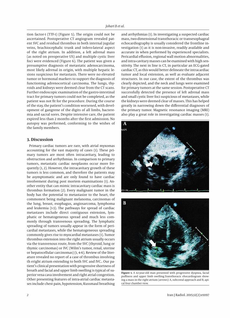
pulmonary tuberculosis, presented with progressive shortness of breath, reduced effort tolerance, facial puff-iness and upper limb swelling over a period of several months. On examination, the patient was hypotensive, tachycardic and hypoxic. Initial transthoracic echo-cardiogram revealed a large mass in the right atrium, with pericardial effusion (Figure 1). The left sided struc-tures were normal in size and function. The provision-al diagnosis at this time was right atrial myxoma. The electrocardiography (ECG)-gated cardiac computed to-
mography (CT) using a 64-slice duals Source CT system (Siemens Medical Solutions, Malvern, PA, USA) revealed a minimally enhancing thrombus in the right atrium, extending to SVC the superior vena cava (SVC), subcla-vian veins and inferior vena cava (IVC), with mass effect on the left main and IVC, with mass pulmonary artery and the right ventricle (Figure 2). Doppler ultrasonog-raphy (US) of the abdomen and lower limbs showed extensive thrombus in the IVC and left femoral vein, as well as a left suprarenal mass (Figure 3). A decision was made to perform sternotomy for right atriotomy and SVC venotomy, as there was a high risk of pulmo-nary thromboembolic phenomenon. Intraoperatively, the right atrium was found to be filled with a densely adherent organized white thrombus, extending to IVC, SVC and innominate veins. Evacuation of clots was per-formed (Figure 4). Although the surgical procedure was complicated by massive blood loss, the patient slowly recovered with intensive care. The histopathological ex-amination of the right atrial mass revealed metastatic poorly-differentiated adenocarcinoma cells, exhibiting mucin-positive cytoplasmic vacuoles, which were im-munopositive for cytokeratin 7 and thyroid transcrip-
CARDIAC IMAGING
Johari B et al.
Iran J Radiol. 2015;12(2):e111972
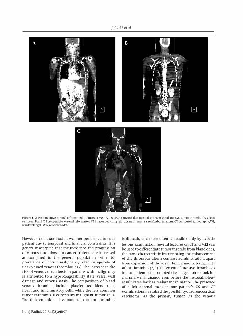
tion factor-1 (TTF-1) (Figure 5). The origin could not be ascertained. Postoperative CT angiogram revealed pat-ent SVC and residual thrombus in both internal jugular veins, brachiocephalic trunk and infero-lateral aspect of the right atrium. In addition, a left adrenal mass (as noted on preoperative US) and multiple cystic liver foci were evidenced (Figure 6). The patient was given a presumptive diagnosis of metastatic adenocarcinoma, most likely adrenal in origin, with multiple hepatic le-sions suspicious for metastasis. There were no elevated tumor or hormonal markers to support the diagnosis of functioning adrenocortical carcinoma. The lungs, thy-roids and kidneys were deemed clear from the CT scans. Further endoscopic examination of the gastro-intestinal tract for primary tumors could not be completed, as the patient was not fit for the procedure. During the course of the stay, the patient’s condition worsened, with devel-opment of gangrene of the digits of all limbs, bactere-mia and sacral sores. Despite intensive care, the patient expired less than 2 months after the first admission. No autopsy was performed, conforming to the wishes of the family members.
3. DiscussionPrimary cardiac tumors are rare, with atrial myxomas
accounting for the vast majority of cases (1). These pri-mary tumors are most often intracavitary, leading to obstruction and arrhythmias. In comparison to primary tumors, metastatic cardiac neoplasms occur more fre-quently (1, 2). However, the intracavitary growth of these tumors is less common, and therefore the patients may be asymptomatic and are only found to have cardiac involvement during post mortem examinations (1). An-other entity that can mimic intracavitary cardiac mass is thrombus formation (2). Every malignant tumor in the body has the potential to metastasize to the heart, the commonest being malignant melanoma, carcinomas of the lung, breast, esophagus, angiosarcoma, lymphoma and leukemia (1-3). The pathways for spread of cardiac metastases include direct contiguous extension, lym-phatic or hematogeneous spread and much less com-monly through transvenous spreading. The lymphatic spreading of tumors usually appear in the form of peri-cardial metastases, while the hematogeneous spreading commonly gives rise to myocardial metastases (1). Tumor thrombus extension into the right atrium usually occurs via the transvenous route, from the SVC (thyroid, lung or thymic carcinomas) or IVC (Wilm’s tumor, renal, uterine or hepatocellular carcinomas) (1, 4-6). Review of the liter-ature revealed no report of a case of thrombus involving th eright atrium extending to both SVC and IVC.. Our pa-tient’s clinical presentation with progressive shortness of breath and facial and upper limb swelling is typical of su-perior vena cava involvement and right atrial congestion. Other presenting features of intra-atrial cardiac metasta-ses include chest pain, hypotension, Kussmaul breathing
and arrhythmias (1). In investigating a suspected cardiac mass, two-dimensional transthoracic or transesophageal echocardiography is usually considered the frontline in-vestigation (1) as it is non-invasive, readily available and accurate in when performed by experienced specialists. Pericardial effusion, regional wall motion abnormalities, and intra-cavitary masses can be examined with high sen-sitivity. The next in line is CT, in particular an ECG-gated cardiac CT, as this would better delineate the intracardiac tumor and local extension, as well as evaluate adjacent structures. In our case, the extent of the thrombus was clearly depicted, and the neck and lungs were examined for primary tumors at the same session. Postoperative CT successfully detected the presence of left adrenal mass and small cystic liver foci suspicious of metastases, while the kidneys were deemed clear of masses. This has helped greatly in narrowing down the differential diagnoses of the primary tumor. Magnetic resonance imaging (MRI) also play a great role in investigating cardiac masses (1).
Figure 1. A 42-year-old man presented with progressive dyspnea, facial puffiness and upper limb swelling.Transthoracic ehocardiogram show-ing a mass in the right atrium (arrows): A, subcostal approach and B, api-cal four chamber view.

Johari B et al.
3Iran J Radiol. 2015;12(2):e11197
Figure 2. A, Computed tomography (WW: 350, WL: 50). coronal reconstruction showing right atrial tumor thrombus extending into the SVC (arrow) and right brachiocephalic vein; B, Computed Tomography (WW: 350, WL: 50) in axial section showing thrombus within the right atrium (arrow); C, Computed Tomography (WW: 600, WL: 200) in axial section showing thrombus within the intrahepatic IVC (arrow), extending to the hepatic veins; D, Computed Tomography (WW: 600, WL: 200) in axial section showing thrombus within the intrahepatic IVC (arrow); E, Computed Tomography (WW: 600, WL: 200). In coronal reconstruction Coronal reconstruction showing patent main pulmonary arteries (arrow).Abbreviations: IVC, inferior vena cava; SVC, superior vena cava; WL, window length; WW, window width.

Johari B et al.
Iran J Radiol. 2015;12(2):e111974
Figure 3. A, Doppler US showing thrombus in the proximal IVC (arrow); B, Doppler US showing thrombus in the distal IVC (arrow); C, Doppler US showing thrombus in right superficial femoral vein (arrow); D, US showing ill-defined left suprarenal mass (arrow). Abbreviations: IVC, inferior vena cava; SVC, superior vena cava; US, ultrasound.
Figure 4. A, Intraoperative finding of intra-atrial tumor thrombus extending to the SVC and IVC; B, Excised mass in macroscopic section. Abbreviations: IVC, inferior vena cava; SVC, superior vena cava.
Figure 5. A, Histopathological examination of the right atrial mass revealed malignant epithelial cells with moderate nuclear pleomorphism and high mitotic activity; these show poor gland formation and occasional cytoplasmic vacuoles containing mucin (H and E, 200 ×); B, Tumor cells from the atrial mass exhibiting nuclear immunopositivity for TTF-1. (Immunohistochemistry for TTF-1, 200 ×). Abbreviations: HE, hematoxylin and eosin; TTF-1, thyroid transcription factor-1.
Johari B et al.
5Iran J Radiol. 2015;12(2):e11197
Figure 6. A, Postoperative coronal reformatted CT images (WW: 350, WL: 50) showing that most of the right atrial and SVC tumor thrombus has been removed; B and C, Postoperative coronal reformatted CT images depicting left suprarenal mass (arrow). Abbreviations: CT, computed tomography; WL, window length; WW, window width.
However, this examination was not performed for our patient due to temporal and financial constraints. It is generally accepted that the incidence and progression of venous thrombosis in cancer patients are increased as compared to the general population, with 10% prevalence of occult malignancy after an episode of unexplained venous thrombosis (7). The increase in the risk of venous thrombosis in patients with malignancy is attributed to a hypercoagulability state, vessel wall damage and venous stasis. The composition of bland venous thrombus include platelet, red blood cells, fibrin and inflammatory cells, while the less common tumor thrombus also contains malignant tumor cells. The differentiation of venous from tumor thrombus
is difficult, and more often is possible only by hepatic lesions examination. Several features on CT and MRI can be used to differentiate tumor thrombi from bland ones, the most characteristic feature being the enhancement of the thrombus aftern contrast administration, apart from expansion of the vessel lumen and heterogeneity of the thrombus (7, 8). The extent of massive thrombosis in our patient has prompted the suggestion to look for a primary malignancy, even before the histopathology result came back as malignant in nature. The presence of a left adrenal mass in our patient’s US and CT examinations has raised the possibility of adrenocortical carcinoma, as the primary tumor. As the venous
Johari B et al.
Iran J Radiol. 2015;12(2):e111976
drainage of the adrenals follows the kidneys’ pathway, adrenal tumors have the propensity to extend into the IVC. However, right atrial involvement in malignant adrenal tumors is very rare (5, 9, 10). It is worthwhile to note that while the histopathological examination may not ascertain the primary origin of the tumor thrombus, the cells were TTF-1 positive, increasing the possibility of occult lung adenocarcinoma. The CT scans on both occasions did not reveal the presence of mass lesions in the lungs. However, occult carcinomas could arise within the tuberculosis scars within the parenchyma. Lung carcinomas are also known to spread to the adrenal glands, which could account for the left adrenal mass in this patient. In conclusion, we present a case of a patient with right atrial tumor thrombus extending to both IVC and SVC, should be considered especially in the adrenal cortex or in the lung and liver deposits, suspicious of metastases. The histopathological examination of the thrombus revealed metastatic adenocarcinoma. This case highlighted that in patients presenting with extensive cavoatrial thrombus as presented, the existence of primary carcinoma should be considered especially in the adrenal cortex or in the lung.
Authors’ ContributionsBushra Johari and Yang Faridah Abdul Aziz were in-
volved in radiological imaging of this patient and writing of the case report. Sivakumar Krishnasamy, Shahrul Amry Hashim and Raja Amin Raja Mokhtar were the cardiotho-racic surgeons managing the patient, and Lai Meng Looi prepared the histopathology report.
Funding/SupportWe thank the University Malaya Medical Center for their
valuable support.
References1. Chiles C, Woodard PK, Gutierrez FR, Link KM. Metastatic involve-
ment of the heart and pericardium: CT and MR imaging. Radio-graphics. 2001;21(2):439–49.
2. Kaul P, George R, Paniagua R, Balaji S, Sivananthan M, Sapsford R, et al. Massive inferior venacavo-atrial thrombus following neo-adjuvant chemotherapy. Br J Cardiol. 2012;19(4):184.
3. Chiappini B, Savini C, Marinelli G, Suarez SM, Di Eusanio M, Fio-rani V, et al. Cavoatrial tumor thrombus: single-stage surgical approach with profound hypothermia and circulatory arrest, including a review of the literature. J Thorac Cardiovasc Surg. 2002;124(4):684–8.
4. Abdou SM, Hajar R. Right atrial thrombus mimicking right atrial tumor. Heart Views. 2004;5(2):66.
5. Kim KH, Park JC, Lim SY, Sohn IS, Yun KH, Cho SH, et al. A case of non-functioning huge adrenocortical carcinoma extend-ing into inferior vena cava and right atrium. J Korean Med Sci. 2006;21(3):572–6.
6. Reynen K, Kockeritz U, Strasser RH. Metastases to the heart. Ann Oncol. 2004;15(3):375–81.
7. Khosa F, Otero HJ, Prevedello LM, Rybicki FJ, Di Salvo DN. Imaging presentation of venous thrombosis in patients with cancer. AJR Am J Roentgenol. 2010;194(4):1099–108.
8. Akin O, Dixit D, Schwartz L. Bland and tumor thrombi in ab-dominal malignancies: magnetic resonance imaging assess-ment in a large oncologic patient population. Abdom Imaging. 2011;36(1):62–8.
9. Rosen B, Rozenman Y, Harpaz D. Extension of adrenocortical car-cinoma into the right atrium--echocardiographic diagnosis: a case report. Cardiovasc Ultrasound. 2003;1:5.
10. Senthil R, Mittal BR, Kashyap R, Bhattacharya A, Radotra BD, Bhansali A. (1)(8)F FDG PET/CT demonstration of IVC and right atrial involvement in adrenocortical carcinoma. Jpn J Radiol. 2012;30(3):281–3.
Related Documents