Metal ion and antioxidant alterations in leaves between different sexes of Ginkgo biloba L. E ´ va Stefanovits-Ba ´nyai a , Kla ´ra Szentmiha ´lyi b , Attila Hegedu ˝s a , Noe ´mi Koczka c , La ´szlo ´ Va ´li d , Gabriella Taba b,d , Anna Bla ´zovics d, * a Department of Applied Chemistry, Faculty of Food Science, Corvinus University of Budapest, P.O. Box 53, Budapest, Hungary, H-1518 b Chemical Research Centre, Hungarian Academy, P.O. Box 17, Budapest, Hungary, H-1525 c Department of Horticultural Technology, Faculty of Agricultural and Environmental Sciences, Szent Istva ´n University, P.O. Box 53, Go ¨do ¨llo ˝, Hungary, H-2103 d Department of Medicine, Semmelweis University of Medicine 2nd P.O. Box 277, Budapest, Hungary, H-1444 Received 10 January 2005; accepted 10 June 2005 Abstract A comparative study was carried out to determine some valuable phytochemical components, macro- and microelement and redox parameters in leaves of male and female Ginkgo biloba trees and in extracts made from them. G. biloba extracts have become more popular as a therapeutic agent in the modern pharmacology in neurodegenerative diseases, in which increased brain metal levels can be observed and free radical reactions are involved. Macro- and microelement components, total phenol content, H-donating activity and reducing power as well as total scavenger capacity were determined in the samples. Well detectable differences were obtained for micro- and macroelement contents between male and female samples, but no toxic elements could be detected in the extracts. Male extracts contained more hazardous metals (e.g. Fe) compared to the female ones, while extracts from female leaves had higher levels of ions, which are known to have beneficial effects in neurodegenerative diseases. The ethanolic extracts of male leaves showed the highest H-donating activity, reducing power and total phenol content, as well as the best total scavenger activity. Ginkgo extracts due to the antioxidant properties may have favourable effects as dietary supplements in several neurodegenerative diseases, but this study draws the attention that critical evaluation is required in view of the potential hazard induced by their metal ion constitution. Our results lead us to the conclusion that although the aqueous extracts of female leaves are characterized by relatively lower antioxidant properties, they may be more eligible for these purposes due to their favourable metal ion constitution. D 2005 Elsevier Inc. All rights reserved. Keywords: Antioxidants; Ginkgo biloba; Metal elements; Neurodegenerative diseases; Phenol Introduction Ginkgo biloba L. is the most ancient living gymnosperm in the world. As being the only representative of the Ginkgoaceae family, it has often been called a ‘‘living fossil’’ by Charles Darwin (Michel, 1986). In the past few years G. biloba was one of the most extensively studied species. Scientific attention is needed because leaf-extract seems to have various curative effects and excellent antioxidant capacity (Schilcher, 1988; Lugasi et al., 1999; Ellnain-Wojtaszek et al., 2002). Many data support the efficacy of G. biloba extracts in biological systems, including in vitro and in vivo experiments and its therapeutic efficacy was also observed in clinical trials of elderly patients and patients with neurodegenerative diseases (Droy-Lefaix, 1997; Akiba et al., 1998; Lugasi et al., 1999; Wei et al., 2000; Kim, 2001; Bush, 2002; Maynard et al., 2002). In ageing processes, G. biloba may ameliorate the mitochondria respiratory chain function by quenching the superoxide anion, and the hydroxyl and peroxyl radicals. It protects the brain by facilitating the uptake of neurotransmitters and by reducing ischemia – reperfusion episodes and level of apoptosis (Droy-Lefaix, 1997). Some clinical data exist, that G. biloba extracts might be used as an effective drug for the treatment of neuronal diseases associated with the production of peroxynitrite (Wei et al., 2000). G. biloba has a nonsteroidal 0024-3205/$ - see front matter D 2005 Elsevier Inc. All rights reserved. doi:10.1016/j.lfs.2005.06.012 * Corresponding author. Tel.: +36 1 266 0926; fax: +36 1 266 0926. E-mail address: [email protected] (A. Bla ´zovics). Life Sciences 78 (2006) 1049 – 1056 www.elsevier.com/locate/lifescie

Welcome message from author
This document is posted to help you gain knowledge. Please leave a comment to let me know what you think about it! Share it to your friends and learn new things together.
Transcript
lsevier.com/locate/lifescie
Life Sciences 78 (200
Metal ion and antioxidant alterations in leaves between different sexes of
Ginkgo biloba L.
Eva Stefanovits-Banyai a, Klara Szentmihalyi b, Attila Hegedus a, Noemi Koczka c, Laszlo Vali d,
Gabriella Taba b,d, Anna Blazovics d,*
a Department of Applied Chemistry, Faculty of Food Science, Corvinus University of Budapest, P.O. Box 53, Budapest, Hungary, H-1518b Chemical Research Centre, Hungarian Academy, P.O. Box 17, Budapest, Hungary, H-1525
c Department of Horticultural Technology, Faculty of Agricultural and Environmental Sciences, Szent Istvan University, P.O. Box 53, Godollo, Hungary, H-2103d Department of Medicine, Semmelweis University of Medicine 2nd P.O. Box 277, Budapest, Hungary, H-1444
Received 10 January 2005; accepted 10 June 2005
Abstract
A comparative study was carried out to determine some valuable phytochemical components, macro- and microelement and redox parameters
in leaves of male and female Ginkgo biloba trees and in extracts made from them. G. biloba extracts have become more popular as a therapeutic
agent in the modern pharmacology in neurodegenerative diseases, in which increased brain metal levels can be observed and free radical reactions
are involved. Macro- and microelement components, total phenol content, H-donating activity and reducing power as well as total scavenger
capacity were determined in the samples. Well detectable differences were obtained for micro- and macroelement contents between male and
female samples, but no toxic elements could be detected in the extracts. Male extracts contained more hazardous metals (e.g. Fe) compared to the
female ones, while extracts from female leaves had higher levels of ions, which are known to have beneficial effects in neurodegenerative
diseases. The ethanolic extracts of male leaves showed the highest H-donating activity, reducing power and total phenol content, as well as the best
total scavenger activity. Ginkgo extracts due to the antioxidant properties may have favourable effects as dietary supplements in several
neurodegenerative diseases, but this study draws the attention that critical evaluation is required in view of the potential hazard induced by their
metal ion constitution. Our results lead us to the conclusion that although the aqueous extracts of female leaves are characterized by relatively
lower antioxidant properties, they may be more eligible for these purposes due to their favourable metal ion constitution.
D 2005 Elsevier Inc. All rights reserved.
Keywords: Antioxidants; Ginkgo biloba; Metal elements; Neurodegenerative diseases; Phenol
Introduction
Ginkgo biloba L. is the most ancient living gymnosperm in
the world. As being the only representative of the Ginkgoaceae
family, it has often been called a ‘‘living fossil’’ by Charles
Darwin (Michel, 1986).
In the past few years G. biloba was one of the most
extensively studied species. Scientific attention is needed
because leaf-extract seems to have various curative effects
and excellent antioxidant capacity (Schilcher, 1988; Lugasi et
al., 1999; Ellnain-Wojtaszek et al., 2002).
0024-3205/$ - see front matter D 2005 Elsevier Inc. All rights reserved.
doi:10.1016/j.lfs.2005.06.012
* Corresponding author. Tel.: +36 1 266 0926; fax: +36 1 266 0926.
E-mail address: [email protected] (A. Blazovics).
Many data support the efficacy of G. biloba extracts in
biological systems, including in vitro and in vivo experiments
and its therapeutic efficacy was also observed in clinical trials
of elderly patients and patients with neurodegenerative diseases
(Droy-Lefaix, 1997; Akiba et al., 1998; Lugasi et al., 1999;
Wei et al., 2000; Kim, 2001; Bush, 2002; Maynard et al.,
2002). In ageing processes, G. biloba may ameliorate the
mitochondria respiratory chain function by quenching the
superoxide anion, and the hydroxyl and peroxyl radicals. It
protects the brain by facilitating the uptake of neurotransmitters
and by reducing ischemia–reperfusion episodes and level of
apoptosis (Droy-Lefaix, 1997). Some clinical data exist, that G.
biloba extracts might be used as an effective drug for the
treatment of neuronal diseases associated with the production
of peroxynitrite (Wei et al., 2000). G. biloba has a nonsteroidal
6) 1049 – 1056
www.e
Table 1
Ginkgo biloba (L.) samples and extracts
Marks of samples Sex Extract
0911 FW Female Aqueous
0911 MW Male Aqueous
0911 FA Female Ethanolic
0911 MA Male Ethanolic
E. Stefanovits-Banyai et al. / Life Sciences 78 (2006) 1049–10561050
anti-inflammatory property on Alzheimer’s disease (Dorais-
wamy, 2002).
As a result of wide ranging research, flavonol-glycosides,
terpene lactones, ginkgolides A, B, C, bilobalide, amino acids
in G. biloba leaves were identified (Hodisan et al., 1998; Lolla
et al., 1998; Yuping and Fengchang, 2000). The G. biloba
extract showed copper-binding property (Lugasi et al., 1999).
Ginkgolic acids, the toxic phenolic compounds are also present
in the fruits and leaves of G. biloba L. Polyphenol and
flavonoid contents are responsible for radical scavenger
activity (Lugasi et al., 1999). Antioxidant properties of
polyphenols and flavonoids were identified on the apoptotic
processes in hippocampal cell cultures (Barkats et al., 1995;
Bastianetto et al., 2000). Ginkgolide B and bilobalide but not
its terpenoid constituents may play a beneficial role in
oxidative stress, to protect against free radicals through actions
on heme oxygenase gene expression and activity, and also
significantly increased GPx gene expression and GPx enzyme
activity (Chen et al., 2001).
However, meagre number of data is known about the metal
ion composition of G. biloba. Supraoptimal concentration of
several metals e.g. Fe, Zn, Cu, Al or Mn have toxic actions on
nerve cells and neurobehavioral functioning, which can be
expressed either as developmental effects or as an increased
risk of neurodegenerative diseases in old age. Tissue injury,
e.g. by ischemia or trauma, can cause increased metal ion
availability and accelerate free radical reactions, highly reactive
hydroxyl radical and other oxidants in the presence of
Fcatalytic_ Fe or Cu ions. Free radical reactions are involved
in the neurotoxicity of Al and in damage to the substantia nigra
in patients with Parkinson’s disease and beta amyloid
accumulation in the plaques are reviewed (Halliwell, 1992;
Bush, 2002). Mn neurotoxicity could be related to the
capability of this metal to increase catechol autooxidation in
catecholaminergic neurons, therefore, increasing the formation
of toxic compounds such as peroxides, superoxides, free
radicals, and semi-orthoquinones (Vescovi et al., 1989). Beta
amyloid aggregation is not spontaneous. The process is age
dependent and belongs to the metal elements, which induces
the protein to precipitate into metal-enriched masses. In this
precipitating process hydrogen peroxide, superoxide anion and
hydroxyl radicals take part (Bush, 2002). Beta amyloid is a
result of misprocessing of amyloid precursor protein, and
gamma-secetase is involved in the process (Torp et al., 2003).
Amyloid beta /Fe2+ can induce apoptotic death in neuronal
cells. Metal ion chelation and inhibitors of pro-apoptotic kinase
cascades may be beneficial in therapy of Alzheimer’s disease
(Kuperstein and Yavin, 2003). The ability of Al to alter the
degradation of beta amyloid suggests a way in which Al and
amyloid neurotoxic substances might interact (Banks et al.,
1996). The hippocampal Zn is decreased in Alzheimer disease,
and amyloid is formed within the walls of capillaries, disturb
the blood brain barrier and toxic metals may enter the cerebral
cortex, where they displace the zinc in Zn enzymes (Deloncle
and Guillard, 1990).
There is some epidemiological evidence for elevated risk of
Alzheimer’s disease in areas where there is high concentration
of aluminium in drinking water. The impacts of Al on humans
and its impact on major physiological systems, such as
neurological-system, musculoskeletal-, respiratory-, cardiovas-
cular-, hepatobiliary-, endocrine-, urinary-, and reproductive
system are well known (Nayak, 2002).
Other metals, especially Pb, Hg, Mn and Cu have been
implicated in amyotrophic lateral sclerosis and Parkinson’s
disease, as well (Carpenter, 2001). The activated transcellular
transport of Cu complexed with plant flavonoids and phenolic
carboxylic acids is ensured by a carrier protein, divalent cation
transporter (DCT1). Menkes protein transports it from the
endothelial cells of the brain-barrier to the brain. Free Cu(I) and
Cu(II) occur in the body in a small amount only; their
concentration is 10–18 mol/L in the plasma. These ions are
very reactive and catalyze the formation of oxygen free radicals
(Ferruzza et al., 1999). Ceruloplasmin can also be found in the
brain and has an oxidase function. Cu occurs in beta amyloid
protein and in prion caused Creutzfeld–Jacob disease (Siegel
and Siegel, 1981).
The metabolism of Zn is regulated by DCT1 carrier protein,
Zn transporters and metallothioneins. Zn is a key element in
superoxide dismutase. Zn-metallothionein has hydroxyl scav-
enging ability (Brando-Neto and Bell, 1994). Zn plays role in
the hippocampal neurons for memory, intellect, behaviour and
neuropsychological function (Sandstead et al., 2000).
Our purpose was to determine the metal ion content and
antioxidant/scavenger bioactive components in extracts of
female and male G. biloba leaves growing in Hungary to
prove their efficacy in vitro.
Materials and methods
Plant material
About 50 leaves from a similar position within the canopy
were collected in September (code number: 0911) 2003
severally from 3 male and 3 female G. biloba (L.) trees growing
at the Botanical Garden of Eotvos Lorand University, Budapest.
Preparation of G. biloba extracts
Extracts of G. biloba (L.) were prepared from young fresh
leaves. Leaves were cut into small pieces, and 5 g of fresh
leaves were infused with 100 ml boiling water or hot aqueous
ethanol (25 -C; water / ethanol 80 /20, v/v). The ethanolic and
aqueous extracts were stored at room temperature for 24 h.
After centrifugation (13,000 rpm, 10 min) the supernatant were
stored in a refrigerator until the analyses. Table 1 summarizes
the leaf samples and the extracts made.
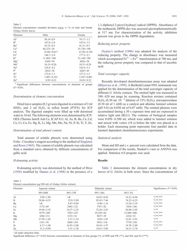
Table 2
Element concentrationT standard deviation (Ag/g, n =3) of male and female
Ginkgo biloba leaves
Elements Male Female
Ala 50.18T0.9 74.17T1.5
Ba 167.8T2.0 147.6T1.5Baa 43.53T0.5 41.31T0.3
Caa 44,223T28 35,720T190
Cd 0.168T0.017 0.150T0.150
Fea 144.7T2.0 177.5T2.1Ka 11, 062T100 21,144T180
Mga 6369T50 6026T58
Mna 16.33T0.20 18.51T0.29
Naa 124.4T4.2 142.8T5.3Pa 2822T30 6265T45
Sra 173.6T1.3 127.3T1.5
Tia 1.807T0.101 2.547T0.001Zna 8.93T0.21 18.43T0.32a Significant differences between concentrations of elements of groups
(P <0.05).
E. Stefanovits-Banyai et al. / Life Sciences 78 (2006) 1049–1056 1051
Determination of element concentration
Dried leave samples (0.2 g) were digested in amixture of 2 ml
HNO3 and 2 ml H2O2 in teflon bomb (PTFE) for ICP
analysis. The digested samples were filled up with deionised
water to 10ml. The following elements were determined by ICP-
OES (Thermo Jarrell Ash Co, ICAP 61): Al, As, B, Ba, Ca, Cd,
Co, Cr, Cu, Fe, Hg, K, Li, Mg, Mn, Mo, Na, Ni, P, Sr, Ti, V, Zn.
Determination of total phenol content
Total amount of soluble phenols were determined using
Folin–Ciocalteu’s reagent according to the method of Singleton
and Rossi (1965). The content of soluble phenols was calculated
from a standard curve obtained by different concentrations of
gallic acid.
H-donating activity
H-donating activity was determined by the method of Blois
(1958) modified by Hatano et al. (1988) in the presence of a
Table 3
Element concentration (Ag/100 ml) of Ginkgo biloba extracts
Elements Aqueous extract Eth
0911MW 0911 FW 09
Al 5.69T1.86 <dl 1
B 28.86T4.35 52.9T5.09 9
Ba <dl 8.87T0.89Ca 1172T69 2492T121 1
Fe 19.98T2.32 14.27T2.25 3
K 9279T289 7833T247 19
Mg 1696T211 3133T32 5
Mn 37.50T1.02 5.13T1.14
P 1161T38 2668T103 3
Sr 6.17T1.08 16.93T2.08 1
Zn 51.2T0.99 3.31T1.20 1
<dl under detection limit.
Significant difference ( P <0.05) between concentration of elements of four groups
1,1-diphenyl-2-picryl-hydrasyl radical (DPPH). Absorbance of
the methanolic DPPH-dye was assessed spectrophotometrically
at 517 nm. For characterization of the activity, inhibition
percent was given to the DPPH degradation.
Reducing power property
Oyaizu’s method (1986) was adopted for analysis of the
reducing property. The change in absorbance was measured
which accompanied Fe3+YFe2+ transformation at 700 nm, and
the reducing power property was compared to that of ascorbic
acid.
Total scavenger capacity
Recently developed chemiluminescense assay was adopted
(Blazovics et al., 1999). A Berthold Lumat 9501 instrument was
applied for the determination of the total scavenger capacity of
different G. biloba extracts. The emitted light was measured in
390–620 nm range by scanning. Reaction mixture contained
H2O2 (0.30 ml, 10�4 dilution of 33% H2O2), microperoxidase
(0.30 ml of 1 mM) as a catalyst and alkaline luminol solution
(pH 9.8) (in 0.050 ml of 0.07 mM). The emitted photons were
accumulated during a 30 s exposure time and are expressed in
relative light unit (RLU). The volumes of biological samples
were 0.050–0.200 ml, which were added to luminol solution
and mixed with vortex (10 s) before the tube was placed in a
holder. Each measuring point represents four parallel data in
luminol dependent chemiluminescence experiments.
Statistical analysis
Mean and SD and c.v. percent were calculated from the data.
For comparison of the results, Student’s t-test or ANOVA was
applied. Statistica 6.0 program was used.
Results
Table 2 demonstrates the element concentrations in dry
leaves of G. biloba in both sexes. Since the concentrations of
anolic extract Significance ( P <0.05)
11 MA 0911 FA
0.63T0.42 16.21T0.44 *,**,***
2.43T7.46 76.22T6.25 *,**,***
8.48T1.16 13.26T2.19 *,**,***
767T82 3410T166 *,**,***
2.26T2.83 26.80T2.05 *,**
,520T64 15,860T486 *,**,***
037T39 6324T52 *,**,***
8.69T1.85 12.01T3.01 *,**
032T118 5739T238 *,**,***
0.43T1.97 18.78T2.88 *,**,***
4.61T3.04 16.21T3.78 *,**
(*), of MW and FW (**), and MA and FA (***).
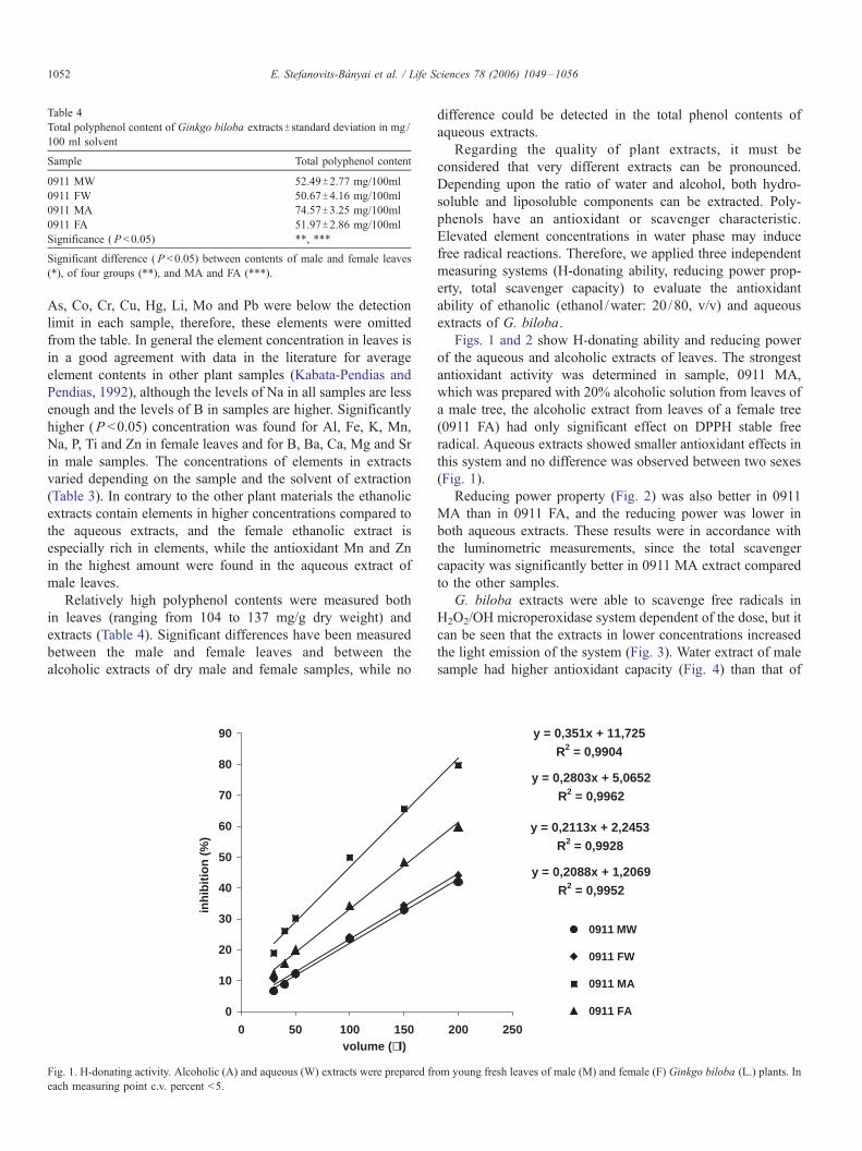
Table 4
Total polyphenol content of Ginkgo biloba extractsT standard deviation in mg/
100 ml solvent
Sample Total polyphenol content
0911 MW 52.49T2.77 mg/100ml
0911 FW 50.67T4.16 mg/100ml
0911 MA 74.57T3.25 mg/100ml
0911 FA 51.97T2.86 mg/100ml
Significance ( P <0.05) **, ***
Significant difference ( P <0.05) between contents of male and female leaves
(*), of four groups (**), and MA and FA (***).
E. Stefanovits-Banyai et al. / Life Sciences 78 (2006) 1049–10561052
As, Co, Cr, Cu, Hg, Li, Mo and Pb were below the detection
limit in each sample, therefore, these elements were omitted
from the table. In general the element concentration in leaves is
in a good agreement with data in the literature for average
element contents in other plant samples (Kabata-Pendias and
Pendias, 1992), although the levels of Na in all samples are less
enough and the levels of B in samples are higher. Significantly
higher (P <0.05) concentration was found for Al, Fe, K, Mn,
Na, P, Ti and Zn in female leaves and for B, Ba, Ca, Mg and Sr
in male samples. The concentrations of elements in extracts
varied depending on the sample and the solvent of extraction
(Table 3). In contrary to the other plant materials the ethanolic
extracts contain elements in higher concentrations compared to
the aqueous extracts, and the female ethanolic extract is
especially rich in elements, while the antioxidant Mn and Zn
in the highest amount were found in the aqueous extract of
male leaves.
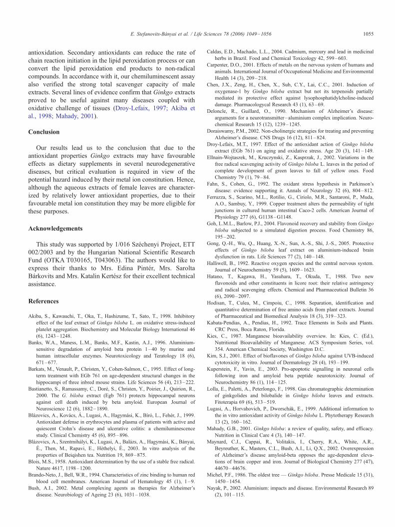
Relatively high polyphenol contents were measured both
in leaves (ranging from 104 to 137 mg/g dry weight) and
extracts (Table 4). Significant differences have been measured
between the male and female leaves and between the
alcoholic extracts of dry male and female samples, while no
0
10
20
30
40
50
60
70
80
90
0 50 100 150
inh
ibit
ion
(%
)
volume (µl)
Fig. 1. H-donating activity. Alcoholic (A) and aqueous (W) extracts were prepared fr
each measuring point c.v. percent <5.
difference could be detected in the total phenol contents of
aqueous extracts.
Regarding the quality of plant extracts, it must be
considered that very different extracts can be pronounced.
Depending upon the ratio of water and alcohol, both hydro-
soluble and liposoluble components can be extracted. Poly-
phenols have an antioxidant or scavenger characteristic.
Elevated element concentrations in water phase may induce
free radical reactions. Therefore, we applied three independent
measuring systems (H-donating ability, reducing power prop-
erty, total scavenger capacity) to evaluate the antioxidant
ability of ethanolic (ethanol /water: 20 /80, v/v) and aqueous
extracts of G. biloba.
Figs. 1 and 2 show H-donating ability and reducing power
of the aqueous and alcoholic extracts of leaves. The strongest
antioxidant activity was determined in sample, 0911 MA,
which was prepared with 20% alcoholic solution from leaves of
a male tree, the alcoholic extract from leaves of a female tree
(0911 FA) had only significant effect on DPPH stable free
radical. Aqueous extracts showed smaller antioxidant effects in
this system and no difference was observed between two sexes
(Fig. 1).
Reducing power property (Fig. 2) was also better in 0911
MA than in 0911 FA, and the reducing power was lower in
both aqueous extracts. These results were in accordance with
the luminometric measurements, since the total scavenger
capacity was significantly better in 0911 MA extract compared
to the other samples.
G. biloba extracts were able to scavenge free radicals in
H2O2/OH microperoxidase system dependent of the dose, but it
can be seen that the extracts in lower concentrations increased
the light emission of the system (Fig. 3). Water extract of male
sample had higher antioxidant capacity (Fig. 4) than that of
y = 0,2803x + 5,0652R2 = 0,9962
y = 0,2113x + 2,2453R2 = 0,9928
y = 0,2088x + 1,2069R2 = 0,9952
y = 0,351x + 11,725R2 = 0,9904
200 250
0911 MW
0911 FW
0911 MA
0911 FA
om young fresh leaves of male (M) and female (F) Ginkgo biloba (L.) plants. In

y = 0,0055x + 0,0231R2 = 0,9943
y = 0,0036x + 0,0244R2 = 0,9986
y = 0,0021x + 0,0118R2 = 0,9914
y = 0,0025x + 0,0042R2 = 0,9956
0
0,2
0,4
0,6
0,8
1
1,2
0 50 100 150 200 250
(µm
ol a
sco
rbic
aci
d/m
l)
0911 MW
0911 FW
0911 MA
0911 FA
volume (µl)
Fig. 2. Reducing power of alcoholic (A) and aqueous (W) extractions prepared from young fresh leaves of male (M) and female (F) Ginkgo biloba (L.) plants. In
each measuring point c.v. percent <5.
E. Stefanovits-Banyai et al. / Life Sciences 78 (2006) 1049–1056 1053
female. This measuring system can be induced with trace of
metal ions; therefore, probably this catalytic effect can be seen
in the figures.
Discussion
Active compounds of G. biloba were evidenced to produce
physiological effects against the pathogenesis of some neuro-
degenerative diseases (Wei et al., 2000; Bush, 2002; Maynard
et al., 2002; Gong et al., 2005). Since differences between the
inner contents of leaves from trees of different sexes may occur
as well as extraction methods may differ in the dissolution
efficiency of significant compounds, we compared the aqueous
and ethanolic extracts of leaves from male and female trees in
this respect.
Increasingly, the etiology of some neurodegenerative dis-
eases has been linked to exposures to environmental toxicants.
Transition metals (e.g. Fe, Cu) as well as Zn or Al were shown to
be involved in the pathogenesis of Parkinson’s disease, tardive
0,00E+00
5,00E+06
1,00E+07
1,50E+07
2,00E+07
2,50E+07
3,00E+07
0 50 100 1
chem
ilum
ines
cen
ce (
RL
U)
1,
1,
1,
1,
1,
(RL
U)
volume (µ
Fig. 3. Total chemiluminescent intensity of alcoholic (A) extractions made from you
measuring point c.v. percent <5.
dyskinesia, metal intoxication syndromes, Down’s syndrome,
and possibly also in schizophrenia, Huntington’s disease, where
free radicals have been implicated, as well. The abnormal
combination of trace elements with beta amyloid takes part in
the free radical formation (Bush, 2002; Maynard et al., 2002).
Well detectable differences were obtained between the
element compositions of the two sexes. Element content of
plant tissues on the one hand may vary according to weather,
soil or environmental conditions, which highlights the fact that
extracts for curative purposes must be carefully checked and
only those possessing favourable element constitution and free
from harmful substances must be allowed for use. In a recent
comprehensive study it was mentioned that in most countries
the standard quality control of medicinal plants is not always
enforced (Caldas and Machado, 2004). On the other hand, the
extraction methods used in this study highly influenced the
element content of extracts.
Aluminium concentration was significantly higher in
ethanolic extracts, especially in case of female leaves. Fe
50 200 250
0911 MA
0911 FA
00E+00
00E+02
00E+04
00E+06
00E+08
0 100 200 (µl)
l)
ng fresh leaves of male (M) and female (F) Ginkgo biloba (L.) plants. In each

0,00E+00
5,00E+06
1,00E+07
1,50E+07
2,00E+07
2,50E+07
3,00E+07
3,50E+07
0 50 100 150 200 250
chem
ilum
ines
cen
ce (
RL
U)
0911 MW
0911 FW
1,00E+00
1,00E+02
1,00E+04
1,00E+06
1,00E+08
0 100 200 (µl)
(RL
U)
volume (µl)
Fig. 4. Total chemiluminescent intensity of aqueous (W) extractions made from young fresh leaves of male (M) and female (F) Ginkgo biloba (L.) plants. In each
measuring point c.v. percent <5.
E. Stefanovits-Banyai et al. / Life Sciences 78 (2006) 1049–10561054
content of male leaf-extracts was slightly higher than that of
female leaf-extracts. Fe and Al are two metal ions, which
concentration is increased in the substantia nigra destruction of
neurons, in the substantia nigra pars compacta of the basal
ganglia of patients with Parkinson’s disease, and both metal
elements are suspected to be involved in the pathophysiology
of Alzheimer’s disease. Al induced brain dysfunction was
improved by Ginkgo extracts (Gong et al., 2005). Total Fe is
increased and ferritin is reduced in the zona compacta in
patients with Parkinson’s disease. Transition metals, as Fe and
Cu, may reduce dioxygen molecule to form superoxide radical
and catalyze Fenton reaction under aerobic circumstances and
lead to a membrane and cell damage (Siegel and Siegel,
1999a).
Mn is essential in the antioxidant defence system, since
superoxide dismutase enzyme, which contains Mn, scavenges
superoxide anions. Mn and Zn contents were 4.3- and 3.5-
times higher in aqueous extracts of male leaves compared to
those in the ethanolic extracts. Mn(II) is an antioxidant, since in
fast reaction it exterminates the alkyl peroxyl radicals formed
by peroxidation of fatty acids, while Fe(II) ions generate
alkoxy and hydroxyl radicals and continue the chain reaction
(Kies, 1987; Siegel and Siegel, 1999a). Mn(II) ions, similarly
to Zn ions, are able to decrease the formation of superoxide
radicals (Schramm and Wedler, 1986).
In the blood, Mn(II) ions are in the form of free aqua-
complexes or bounded to glycoproteins, but when oxidized,
Mn(III) ions change apotransferrine to transmanganine. Mn can
be transported through the blood–brain barrier (Siegel and
Siegel, 1999b), therefore in a supraoptimal concentration it
primarily damages the globus pallidus and substantia nigra pars
reticularis and relatively spares the nigrostriatal dopaminergic
system (Olanow et al., 1996).
It must be highlighted that our results revealed clearly
higher Mg content in extracts from female leaves compared to
male leaf-extracts in case of both extraction methods. Mg-rich
Ginkgo extract may contribute to the regeneration of brain or
brain barrier and change the generally low concentrations of
Mg and other elements (such as Ca, K, Na, P, S and Zn)
favourable in neurological diseases (Yasui et al., 1992).
Calcium, magnesium, sodium, and potassium are involved in
maintaining balance of sympathetic and parasympathetic
systems of the autonomic nervous systems. Mg takes place in
several enzyme functions and it is one of the most important
factors in the homeostasis of elements.
Antioxidant defence mechanisms appear to be reduced in
the parkinsonian substantia nigra with the findings of decreased
activities of glutathione peroxidase and catalase (Fahn and
Cohen, 1992). Since free radical induced oxidative reactions
are also involved in the pathogenesis of several neurodegen-
erative diseases (Halliwell, 1992), antioxidant properties of
curative agents may be of great consideration. Chen et al.
(2001) described that the extracts of G. biloba indeed induces
several antioxidant responses in human cells. We demonstrated
that total content of the antioxidant phenols involving poly-
phenols, flavonoids etc. is outstanding in case of ethanolic
extracts from male leaves. Significantly higher total phenol
content occurred in leaves of male trees and their ethanolic
extracts, while in aqueous extracts no difference could be
detected between the two sexes, which indicate that the
phenolic compounds particularly accumulating in male leaves
are characterized by a preferably liposoluble nature. Similar
deviations in the dissolution efficiency of several phenolic
compounds in ethanolic and aqueous extracts were reported by
Goh and Barlow (2004).
All the measured antioxidant parameters were in a good
accordance with the phenol content of samples. Ethanolic
extracts of male leaves were shown to possess the highest H-
donating ability, which represents the chain-breaking property
of extracts, a feature of how efficiently can an antioxidant
molecule serve H to free radicals and neutralize them
(Blazovics et al., 2003).
Ethanolic male extracts also produced the highest values in
case of the reducing power, which is an index of secondary
E. Stefanovits-Banyai et al. / Life Sciences 78 (2006) 1049–1056 1055
antioxidation. Secondary antioxidants can reduce the rate of
chain reaction initiation in the lipid peroxidation process or can
convert the lipid peroxidation end products to non-radical
compounds. In accordance with it, our chemiluminescent assay
also verified the strong total scavenger capacity of male
extracts. Several lines of evidence confirm that Ginkgo extracts
proved to be useful against many diseases coupled with
oxidative challenge of tissues (Droy-Lefaix, 1997; Akiba et
al., 1998; Mahady, 2001).
Conclusion
Our results lead us to the conclusion that due to the
antioxidant properties Ginkgo extracts may have favourable
effects as dietary supplements in several neurodegenerative
diseases, but critical evaluation is required in view of the
potential hazard induced by their metal ion constitution. Hence,
although the aqueous extracts of female leaves are character-
ized by relatively lower antioxidant properties, due to their
favourable metal ion constitution they may be more eligible for
these purposes.
Acknowledgements
This study was supported by 1/016 Szechenyi Project, ETT
002/2003 and by the Hungarian National Scientific Research
Fund (OTKA T030165, T043063). The authors would like to
express their thanks to Mrs. Edina Pinter, Mrs. Sarolta
Barkovits and Mrs. Katalin Kertesz for their excellent technical
assistance.
References
Akiba, S., Kawauchi, T., Oka, T., Hashizume, T., Sato, T., 1998. Inhibitory
effect of the leaf extract of Ginkgo biloba L. on oxidative stress-induced
platelet aggregation. Biochemistry and Molecular Biology International 46
(6), 1243–1248.
Banks, W.A., Maness, L.M., Banks, M.F., Kastin, A.J., 1996. Aluminium-
sensitive degradation of amyloid beta protein 1–40 by murine and
human intracellular enzymes. Neurotoxicology and Teratology 18 (6),
671–677.
Barkats, M., Venault, P., Christen, Y., Cohen-Salmon, C., 1995. Effect of long-
term treatment with EGb 761 on age-dependent structural changes in the
hippocampi of three inbred mouse strains. Life Sciences 56 (4), 213–222.
Bastianetto, S., Ramassamy, C., Dore, S., Christen, Y., Poirier, J., Quirion, R.,
2000. The G. biloba extract (Egb 761) protects hippocampal neurons
against cell death induced by beta amyloid. European Journal of
Neuroscience 12 (6), 1882–1890.
Blazovics, A., Kovacs, A., Lugasi, A., Hagymasi, K., Bıro, L., Feher, J., 1999.
Antioxidant defense in erythrocytes and plasma of patients with active and
quiescent Crohn’s disease and ulcerative colitis: a chemiluminescence
study. Clinical Chemistry 45 (6), 895–896.
Blazovics, A., Szentmihalyi, K., Lugasi, A., Balazs, A., Hagymasi, K., Banyai,
E., Then, M., Rapavi, E., Hethelyi, E., 2003. In vitro analysis of the
properties of Beiqishen tea. Nutrition 19, 869–875.
Blois, M.S., 1958. Antioxidant determination by the use of a stable free radical.
Nature 4617, 1198–1200.
Brando-Neto, J., Bell, W.R., 1994. Characteristics of zinc binding to human red
blood cell membranes. American Journal of Hematology 45 (1), 1–9.
Bush, A.I., 2002. Metal complexing agents as therapies for Alzheimer’s
disease. Neurobiology of Ageing 23 (6), 1031–1038.
Caldas, E.D., Machado, L.L., 2004. Cadmium, mercury and lead in medicinal
herbs in Brazil. Food and Chemical Toxicology 42, 599–603.
Carpenter, D.O., 2001. Effects of metals on the nervous system of humans and
animals. International Journal of Occupational Medicine and Environmental
Health 14 (3), 209–218.
Chen, J.X., Zeng, H., Chen, X., Sub, C.Y., Lai, C.C., 2001. Induction of
oxygenase-1 by Ginkgo biloba extract but not its terpenoids partially
mediated its protective effect against lysophosphatidylcholine-induced
damage. Pharmacologycal Research 43 (1), 63–69.
Deloncle, R., Guillard, O., 1990. Mechanism of Alzheimer’s disease:
arguments for a neurotransmitter–aluminium complex implication. Neuro-
chemical Research 15 (12), 1239–1245.
Doraiswamy, P.M., 2002. Non-cholinergic strategies for treating and preventing
Alzheimer’s disease. CNS Drugs 16 (12), 811–824.
Droy-Lefaix, M.T., 1997. Effect of the antioxidant action of Ginkgo biloba
extract (EGb 761) on aging and oxidative stress. Age 20 (3), 141–149.
Ellnain-Wojtaszek, M., Kruczynski, Z., Kasprzak, J., 2002. Variations in the
free radical scavenging activity of Ginkgo biloba L. leaves in the period of
complete development of green leaves to fall of yellow ones. Food
Chemistry 79 (1), 79–84.
Fahn, S., Cohen, G., 1992. The oxidant stress hypothesis in Parkinson’s
disease: evidence supporting it. Annals of Neurology 32 (6), 804–812.
Ferruzza, S., Scarino, M.L., Rotilio, G., Ciriolo, M.R., Santaroni, P., Muda,
A.O., Sambuy, Y., 1999. Copper treatment alters the permeability of tight
junctions in cultured human intestinal Caco-2 cells. American Journal of
Physiology 277 (6), G1138–G1148.
Goh, L.M.L., Barlow, P.J., 2004. Flavonoid recovery and stability from Ginkgo
biloba subjected to a simulated digestion process. Food Chemistry 86,
195–202.
Gong, Q.-H., Wu, Q., Huang, X.-N., Sun, A.-S., Shi, J.-S., 2005. Protective
effects of Ginkgo biloba leaf extract on aluminium-induced brain
dysfunction in rats. Life Sciences 77 (2), 140–148.
Halliwell, B., 1992. Reactive oxygen species and the central nervous system.
Journal of Neurochemistry 59 (5), 1609–1623.
Hatano, T., Kagawa, H., Yasuhara, T., Okuda, T., 1988. Two new
flavonoids and other constituents in licore root: their relative astringency
and radical scavenging effects. Chemical and Pharmaceutical Bulletin 36
(6), 2090–2097.
Hodisan, T., Culea, M., Cimpoiu, C., 1998. Separation, identification and
quantitative determination of free amino acids from plant extracts. Journal
of Pharmaceutical and Biomedical Analysis 18 (3), 319–323.
Kabata-Pendias, A., Pendias, H., 1992. Trace Elements in Soils and Plants.
CRC Press, Boca Raton, Florida.
Kies, C., 1987. Manganese bioavailability overview. In: Kies, C. (Ed.),
Nutritional Bioavailability of Manganese. ACS Symposium Series, vol.
354. American Chemical Society, Washington D.C.
Kim, S.J., 2001. Effect of bioflavones of Ginkgo biloba against UVB-induced
cytotoxicity in vitro. Journal of Dermatology 28 (4), 193–199.
Kuperstein, F., Yavin, E., 2003. Pro-apoptotic signalling in neuronal cells
following iron and amyloid beta peptide neurotoxicity. Journal of
Neurochemistry 86 (1), 114–125.
Lolla, E., Paletti, A., Peterlongo, F., 1998. Gas chromatographic determination
of ginkgolides and bilobalide in Ginkgo biloba leaves and extracts.
Fitoterapia 69 (6), 513–519.
Lugasi, A., Horvahovich, P., Dworschak, E., 1999. Additional information to
the in vitro antioxidant activity of Ginkgo biloba L. Phytotherapy Research
13 (2), 160–162.
Mahady, G.B., 2001. Ginkgo biloba: a review of quality, safety, and efficacy.
Nutrition in Clinical Care 4 (3), 140–147.
Maynard, C.J., Cappai, R., Volitakis, I., Cherry, R.A., White, A.R.,
Beyreuther, K., Masters, C.L., Bush, A.I., Li, Q.X., 2002. Overexpression
of Alzheimer’s disease amyloid-beta opposes the age-dependent eleva-
tions of brain copper and iron. Journal of Biological Chemistry 277 (47),
44670–44676.
Michel, P.F., 1986. The oldest tree — Ginkgo biloba. Presse Medicale 15 (31),
1450–1454.
Nayak, P., 2002. Aluminium: impacts and disease. Environmental Research 89
(2), 101–115.

E. Stefanovits-Banyai et al. / Life Sciences 78 (2006) 1049–10561056
Olanow, C.W., Good, P.F., Shinotoh, H., Hewitt, K.A., Vingerhoets, F., Snow,
B.J., Beal, M.F., Calne, D.B., Perl, D.P., 1996. Manganese intoxication in
rhesus monkey: a clinical, imaging, pathologic, and biochemical study.
Neurology 46, 492–498.
Oyaizu, M., 1986. Studies on products of browning reaction prepared from
glucosamine. Japanese Journal of Nutrition 44, 307–315.
Sandstead, H.H., Frederickson, C.P., Penland, J.G., 2000. History of zinc as
related to brain function. Journal of Nutrition 130 (2S Suppl.), 496S–502S.
Schilcher, H., 1988. Ginkgo biloba L. Untersuchungen zur Qualitat,
Wirkung, Wirksamkeit und Unbedenklichkeit. Zeitschrift fur Phytotherapie
9, 119–127.
Schramm, V.L., Wedler, F.C., 1986. Manganese in Metabolism and Enzyme
Function. Acad Press, New York, pp. 1–437.
Siegel, A., Siegel, H., 1981. Copper proteins. Metal Ions in Biological Systems
13, 1–394.
Siegel, A., Siegel, H., 1999a. Interactions between free radicals and metal ions
in life processes. Metal Ions in Biological Systems 36, 1–797.
Siegel, A., Siegel, H., 1999b. Manganese and its role in biological processes.
Metal Ions in Biological Systems 37, 1–788.
Singleton, V.L., Rossi, J.A., 1965. Colorimetry of total phenolics with
phosphomolibdic–phosphotungstic acid reagents. American Journal of
Enology and Viticulture 16, 144–158.
Torp, R., Ottersen, O.P., Cotman, C.W., Head, E., 2003. Identification of
neuronal plasma membrane microdomains that colocalize beta-amyloid and
presenilin: implication for beta-amyloid precursor protein processing.
Neuroscience 120 (2), 291–300.
Vescovi, A., Gebbia, M., Cappelletti, G., Parati, E.A., Santagostino, A., 1989.
Interactions of manganese with human brain glutathione-S-transferase.
Toxicology 57 (2), 183–191.
Wei, T., Hou, J., Chen, C., Zhao, B., Xin, W., 2000. The antioxidant EGb761
prevents peroxynitrite-induced apoptosis in rat cortical neurons. Medical
Science Research 28 (2), 93–95.
Yasui, M., Kihira, T., Ota, K., 1992. Calcium, magnesium and aluminium
concentrations in Parkinson’s disease. Neurotoxicology 13 (3), 593–600.
Yuping, T., Fengchang, L., 2000. Separation and isolation of terpene lactones
from Ginkgo biloba L. by direct high performance liquid chromatography.
Journal of Liquid Chromatography and Related Technologies 23 (18),
2897–2900.
Related Documents