This is a repository copy of Metal Fluorides: Tools for Structural and Computational Analysis of Phosphoryl Transfer Enzymes. White Rose Research Online URL for this paper: https://eprints.whiterose.ac.uk/113916/ Version: Published Version Article: Jin, Yi orcid.org/0000-0002-6927-4371, Molt. Jr., Robert W. and Blackburn, Michael (2017) Metal Fluorides: Tools for Structural and Computational Analysis of Phosphoryl Transfer Enzymes. Topics in Current Chemistry. https://doi.org/10.1007/s41061-017-0130-y [email protected] https://eprints.whiterose.ac.uk/ Reuse This article is distributed under the terms of the Creative Commons Attribution (CC BY) licence. This licence allows you to distribute, remix, tweak, and build upon the work, even commercially, as long as you credit the authors for the original work. More information and the full terms of the licence here: https://creativecommons.org/licenses/ Takedown If you consider content in White Rose Research Online to be in breach of UK law, please notify us by emailing [email protected] including the URL of the record and the reason for the withdrawal request.

Welcome message from author
This document is posted to help you gain knowledge. Please leave a comment to let me know what you think about it! Share it to your friends and learn new things together.
Transcript
This is a repository copy of Metal Fluorides: Tools for Structural and Computational Analysis of Phosphoryl Transfer Enzymes.
White Rose Research Online URL for this paper:https://eprints.whiterose.ac.uk/113916/
Version: Published Version
Article:
Jin, Yi orcid.org/0000-0002-6927-4371, Molt. Jr., Robert W. and Blackburn, Michael (2017)Metal Fluorides: Tools for Structural and Computational Analysis of Phosphoryl Transfer Enzymes. Topics in Current Chemistry.
https://doi.org/10.1007/s41061-017-0130-y
[email protected]://eprints.whiterose.ac.uk/
Reuse
This article is distributed under the terms of the Creative Commons Attribution (CC BY) licence. This licence allows you to distribute, remix, tweak, and build upon the work, even commercially, as long as you credit the authors for the original work. More information and the full terms of the licence here: https://creativecommons.org/licenses/
Takedown
If you consider content in White Rose Research Online to be in breach of UK law, please notify us by emailing [email protected] including the URL of the record and the reason for the withdrawal request.

REVIEW
Metal Fluorides: Tools for Structural
and Computational Analysis of Phosphoryl Transfer
Enzymes
Yi Jin1,2 • Robert W. Molt Jr.3,4,5 •
G. Michael Blackburn2
Received: 2 December 2016 / Accepted: 1 March 2017
� The Author(s) 2017. This article is published with open access at Springerlink.com
Abstract The phosphoryl group, PO3–, is the dynamic structural unit in the bio-
logical chemistry of phosphorus. Its transfer from a donor to an acceptor atom, with
oxygen much more prevalent than nitrogen, carbon, or sulfur, is at the core of a
great majority of enzyme-catalyzed reactions involving phosphate esters, anhy-
drides, amidates, and phosphorothioates. The serendipitous discovery that the
phosphoryl group could be labeled by ‘‘nuclear mutation,’’ by substitution of PO3–
by MgF3– or AlF4
–, has underpinned the application of metal fluoride (MFx)
complexes to mimic transition states for enzymatic phosphoryl transfer reactions,
with sufficient stability for experimental analysis. Protein crystallography in the
solid state and 19F NMR in solution have enabled direct observation of ternary and
quaternary protein complexes embracing MFx transition state models with precision.
These studies have underpinned a radically new mechanistic approach to enzyme
catalysis for a huge range of phosphoryl transfer processes, as varied as kinases,
phosphatases, phosphomutases, and phosphohydrolases. The results, without
This article is part of the Topical Collection ‘‘Phosphate Labeling in Chemical Biology’’; edited by
Henning Jessen.
& G. Michael Blackburn
1 Structural Biology Laboratory, Department of Chemistry, University of York, York YO31 7YD,
UK
2 Department of Molecular Biology and Biotechnology, Krebs Institute, University of Sheffield,
Sheffield S10 2TN, UK
3 ENSCO, Inc., 4849 North Wickham Road, Melbourne, FD 32940, USA
4 Department of Chemistry and Chemical Biology, Indiana University-Purdue University,
Indianapolis, IN 46202, USA
5 Department of Biochemistry and Molecular Biology, School of Medicine, Indiana University,
Indianapolis, IN 46202, USA
123
Top Curr Chem (Z) (2017) 375:36
DOI 10.1007/s41061-017-0130-y

exception, have endorsed trigonal bipyramidal geometry (tbp) for concerted, ‘‘in-
line’’ stereochemistry of phosphoryl transfer. QM computations have established the
validity of tbp MFx complexes as reliable models for true transition states, deliv-
ering similar bond lengths, coordination to essential metal ions, and virtually
identical hydrogen bond networks. The emergence of protein control of reactant
orbital overlap between bond-forming species within enzyme transition states is a
new challenging theme for wider exploration.
Keywords MFx � Phosphoryl group surrogates � Enzyme mechanisms � Transition
state analogs � QM/MM computation � KS-DFT analysis
1 Background
Alexander Todd1 and Frank Westheimer2 held complementary, and sometime
overlapping, views on the centrality of phosphates for life. Todd’s pronouncement:
‘‘Where there’s Life, there’s Phosphorus’’, encapsulated his conviction that enzymes
that manipulate phosphates have been at the heart of biology from the dawn of life
anywhere in the universe [1]. Westheimer identified the evolutionary centrality of
phosphate [2]. The cellular behavior of phosphate esters and anhydrides provides
one of the most remarkable chemical paradoxes: phosphate monoesters hydrolyze
spontaneously under physiological conditions with t1/2 1012 years, yet simple
phosphatase enzymes have kcat ca. 30 s-1. The enormous difference corresponds to
a remarkable catalytic rate enhancement of 1021 [3]. How do enzymes achieve this?
This article focuses on the use of aluminum and magnesium fluoride complexes to
mimic structures of transition states of enzymatic reactions that involve the
phosphoryl group, PO3-, and to provide a structural base for quantum chemical
computations to describe them in detail.
1.1 Basics of Phosphoryl Transfer
Studies on phosphoryl transfer reactions were greatly advanced by the use of
oxygen isotopes to show that they generally involved P–O cleavage, with transfer of
the phosphoryl group (PO3-) between a donor oxygen (OD) and an acceptor oxygen
(OA) or, less commonly, nitrogen or sulfur atoms [4]. Polyphosphates, such as ATP,
react by attack of water (or an alcohol) on the terminal c-phosphorus, breaking the
P–O bond to the O3B atom (usually oxygen and infrequently nitrogen) (Scheme 1a).
More advanced isotope work, deploying 16O, 17O, and 18O, established that the
near-universal stereochemistry for such processes, for both chemical and enzymatic
reactions, involves inversion of stereochemistry at the transferring phosphorus
(Scheme 1) [5–7].
An accurate description of the technical aspects of the varieties and uses of MFxmodels calls for a brief explanation of the terminology of phosphoryl transfer. The
1 Lord Todd of Trumpington, Nobel Laureate 1957, 1702 Professor of Chemistry 1944-71, Cambridge,
UK.2 Professor of Chemistry, 1953-2007, Harvard, Cambridge, USA.
36 Page 2 of 31 Top Curr Chem (Z) (2017) 375:36
123

phosphoryl group, PO3– is identified throughout organic chemistry and biology as
the anionic, trigonal planar assembly of a phosphorus and three oxygen atoms. It is
usually drawn without P=O double bonds, is highly electrophilic, and has not been
identified in any condensed phase (Scheme 2). It is helpful to perceive its
combination with an alcohol, such as adenosyl-50-OH, to generate a phosphate
monoester, illustrated for adenosine 50-phosphate, AMP. The addition of a second
phosphoryl group to a terminal oxygen generates a pyrophosphate monoester,
illustrated for adenosine 50-diphosphate, ADP; and capture of a third phosphoryl
group gives adenosine 50-triphosphate, ATP (Scheme 2). Strings of phosphorus
atoms in such chains have conventionally been labeled Pa, Pb, Pc, etc. but, with
new IUPAC nomenclature, are now better identified as PA, PB, PG, PD, etc [8].
1.2 Historic Development of Mechanisms
Todd [9] and Westheimer [2] thought that phosphoryl transfer reactions should be
stepwise, involving a monomeric metaphosphate intermediate species (Scheme 1a).
That concept, unproven after extended but fruitless effort, has now been discarded
in favor of concerted phosphoryl transfer reactions for phosphate monoesters and
anhydrides. These have ‘‘in-line’’ geometry for OD–P–OA in the transition state
(TS), with variable associative or dissociative character (Scheme 1b) [10–12].
Isotope labeling studies have contributed historically to studies on phosphoryl
transfer in biological systems [13] and 31P NMR has been applied effectively for
investigations on ATP [14] and phosphoarginine [15], but protein crystallography
before the mid-1990s was restricted to binary complexes with stable substrate and
bisubstrate analogs that gave limited information about reaction mechanisms
[16, 17]. In 1994, that situation changed dramatically with the crystallization of
ternary complexes of guanosine diphosphate (GDP) coordinated to tetrafluoroalu-
minate, AlF4–, and the small G protein, Gia1 (PDB: 1gfi) [18] and with transducin a
(PDB: 1tad) [19]. Although these complexes had octahedral geometry for the
Scheme 1 a The atomic identities in ATP employ the new IUPAC nomenclature [8]. b Threemechanisms for transfer of a phosphoryl group (PO3
–, top center) between a donor (left) and an acceptor(right) species. Sequential process (a, blue arrows) involves formation of a trigonal planar metaphosphateanion as an intermediate. Concerted process (b, green arrows) shows a trigonal bipyramidal (tbp)transition state with phosphorus fully bonded to three equatorial oxygens and partially bonded to the axialdonor (OD) and acceptor (OA) oxygens. An alternative, sequential process (c, magenta arrows), largelydiscarded, shows formation of a stable pentacoordinate phosphorane intermediate having full bonds to allfive oxygens
Top Curr Chem (Z) (2017) 375:36 Page 3 of 31 36
123
aluminum tetrafluoride moiety, they were immediately described as transition state
analogs (TSAs) for phosphoryl transfer. They were soon followed by further MFxspecies, notably BeF3
–, AlF30, and MgF3
–. The number of such complexes has grown
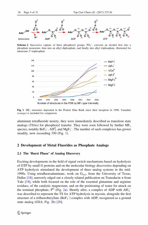
steadily, now exceeding 350 (Fig. 1).
2 Development of Metal Fluorides as Phosphate Analogs
2.1 The ‘Burst Phase’ of Analog Discovery
Exciting developments in the field of signal switch mechanisms based on hydrolysis
of GTP by small G proteins and on the molecular biology discoveries depending on
ATP hydrolysis stimulated the development of three analog systems in the mid-
1990s. Using tetrafluoroaluminate, work on Gia1, from the University of Texas,
Dallas [18], narrowly edged out a closely related publication on Transducin a from
Yale [19], while both focused on the role of the essential glutamine and arginine
residues, of the catalytic magnesium, and on the positioning of water for attack on
the terminal phosphate, PG (Fig. 2a). Shortly after, a complex of ADP with AlF4–
was described to represent the TS for ATP hydrolysis in myosin, alongside the first
structure of a trifluoroberyllate (BeF3–) complex with ADP, recognized as a ground
state analog (GSA; Fig. 2b) [20].
Scheme 2 Successive capture of three phosphoryl groups, PO3–, converts an alcohol first into a
phosphate monoester, then into an alkyl diphosphate, and finally into alkyl triphosphate, illustrated foradenosine 50-triphosphate
Fig. 1 MFx structures deposited in the Protein Data Bank since their inception in 1996. Vanadate(orange) is included for comparison
36 Page 4 of 31 Top Curr Chem (Z) (2017) 375:36
123
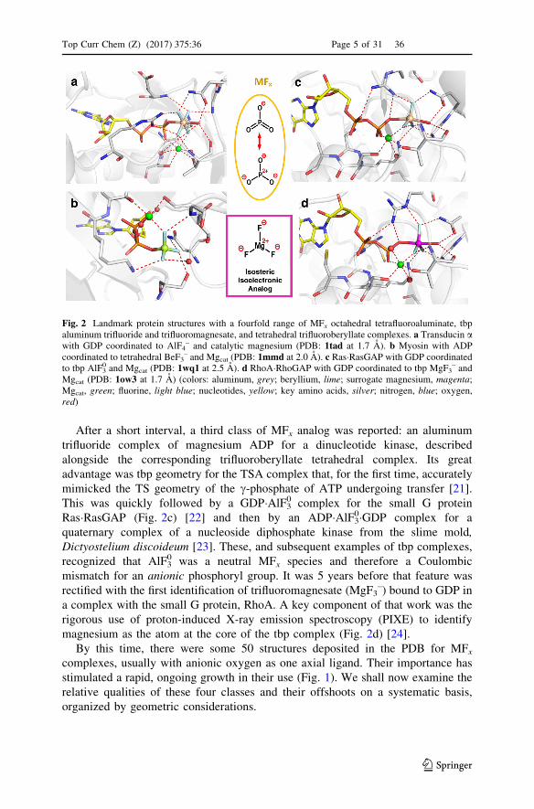
After a short interval, a third class of MFx analog was reported: an aluminum
trifluoride complex of magnesium ADP for a dinucleotide kinase, described
alongside the corresponding trifluoroberyllate tetrahedral complex. Its great
advantage was tbp geometry for the TSA complex that, for the first time, accurately
mimicked the TS geometry of the c-phosphate of ATP undergoing transfer [21].
This was quickly followed by a GDP�AlF30 complex for the small G protein
Ras�RasGAP (Fig. 2c) [22] and then by an ADP�AlF30�GDP complex for a
quaternary complex of a nucleoside diphosphate kinase from the slime mold,
Dictyostelium discoideum [23]. These, and subsequent examples of tbp complexes,
recognized that AlF30 was a neutral MFx species and therefore a Coulombic
mismatch for an anionic phosphoryl group. It was 5 years before that feature was
rectified with the first identification of trifluoromagnesate (MgF3–) bound to GDP in
a complex with the small G protein, RhoA. A key component of that work was the
rigorous use of proton-induced X-ray emission spectroscopy (PIXE) to identify
magnesium as the atom at the core of the tbp complex (Fig. 2d) [24].
By this time, there were some 50 structures deposited in the PDB for MFxcomplexes, usually with anionic oxygen as one axial ligand. Their importance has
stimulated a rapid, ongoing growth in their use (Fig. 1). We shall now examine the
relative qualities of these four classes and their offshoots on a systematic basis,
organized by geometric considerations.
Fig. 2 Landmark protein structures with a fourfold range of MFx octahedral tetrafluoroaluminate, tbpaluminum trifluoride and trifluoromagnesate, and tetrahedral trifluoroberyllate complexes. a Transducin awith GDP coordinated to AlF4
– and catalytic magnesium (PDB: 1tad at 1.7 A). b Myosin with ADPcoordinated to tetrahedral BeF3
– and Mgcat (PDB: 1mmd at 2.0 A). c Ras�RasGAP with GDP coordinatedto tbp AlF3
0 and Mgcat (PDB: 1wq1 at 2.5 A). d RhoA�RhoGAP with GDP coordinated to tbp MgF3– and
Mgcat (PDB: 1ow3 at 1.7 A) (colors: aluminum, grey; beryllium, lime; surrogate magnesium, magenta;Mgcat, green; fluorine, light blue; nucleotides, yellow; key amino acids, silver; nitrogen, blue; oxygen,red)
Top Curr Chem (Z) (2017) 375:36 Page 5 of 31 36
123
3 MFxGround State Analogs
3.1 BeF3– as a Ground State Phosphate Mimic
In aqueous solution, beryllium (II) forms stable fluorides as a mixture of tetrahedral
species including BeF2�2H2O, BeF3–�H2O, and BeF4
= [25]. 19F NMR studies on
fluoroberyllate complexes with ADP identified mixed fluoroberyllate�ADP species
for myosin (Fig. 2b). Nearly 130 trifluoroberyllate complexes have now been
described, with three structures solved by NMR and 119 X-ray structures having
resolutions of C1.2 A, generally having tetrahedral trifluoroberyllate bonded to an
anionic oxygen. These comprise two sub-groups: over 70 have Be coordinated to an
aspartate carboxylate while some 50 have Be coordinated to a terminal phosphate
oxygen of a nucleotide. Just two have Be coordinated to a histidine ring nitrogen,
while one has BeF2 bridging two phosphates.
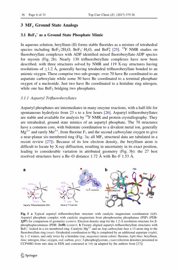
3.1.1 Aspartyl Trifluoroberyllates
Aspartyl phosphates are intermediates in many enzyme reactions, with a half-life for
spontaneous hydrolysis from 23 s to a few hours [26]. Aspartyl trifluoroberyllates
are stable and available for analysis by 19F NMR and protein crystallography. They
are tetrahedral, ground state mimics of an aspartyl phosphate. The 70 structures
have a common core, with bidentate coordination to a divalent metal ion, generally
Mg2? and rarely Mn2?, from fluorine F1 and the second carboxylate oxygen to give
a near-planar six-membered ring (Fig. 3a; all MFx structural data are tabulated in a
recent review [27]). Because of its low electron density, the beryllium atom is
difficult to locate by X-ray diffraction, resulting in uncertainty in its exact position,
leading to considerable variation in attributed geometry (Fig. 3b): the 27 best
resolved structures have a Be–O distance 1.72 A with Be–F 1.53 A.
Fig. 3 a Typical aspartyl trifluoroberyllate structure with catalytic magnesium coordination (left).Aspartyl phosphate complex with catalytic magnesium from phosphoserine phosphatase (PSP) (PDB:1j97) for comparison of geometry (center). Electron density map for the 1.2-A resolution structure for b-phosphoglucomutase (PDB: 2wf8) (center). b Twenty aligned aspartyl–trifluoroberyllate structures withBeF3
– locked in a six-membered ring. Catalytic Mg2? and an Asp carboxylate fuse a 13-atom ring to thefluoroberyllate ring (rear). Octahedral coordination to Mg is completed by an additional aspartate (right),by 1–2 waters, and only twice by a histidine (top, magenta) (atom colors: fluorine, light blue; beryllium,lime; nitrogen, blue; oxygen, red; carbon, grey; 3-phosphoglycerate, cyan) (electron densities presented inCCP4MG from mtz data in EDS and contoured at 1r) (a adapted by the authors from [27])
36 Page 6 of 31 Top Curr Chem (Z) (2017) 375:36
123

3.1.2 BeF3– Nucleotide Structures
There are 42 X-ray structures of BeF3– complexes with ADP, and six with GDP.
They are isosteric mimics of ATP and GTP (Fig. 4a) in kinases, F1 ATPase,
hydrolases, mutases, helicases, and small G proteins. Twenty structures align very
well (Fig. 4b) with Be bonded to a b-phosphate oxygen, while a catalytic Mg2? is
coordinated to F1 and to another b-phosphate oxygen.
3.1.3 Histidine Trifluoroberyllates
Various approaches to analogs of g-phosphohistidine have been explored.
Structural work on nicotinamide phosphoribosyltransferase (NAMPT) has mim-
icked phosphorylation of an active-site histidine using trifluoroberyllate. Crystal
structures of NAMPT for reactant and product complexes (PDB: 3dhf; Fig. 5b)
have a covalent His247�BeF3-, and in contrast to all other trifluoroberyllate
structures, magnesium is coordinated to one fluorine without any direct linkage to
His247 [28].
Fig. 4 a Typical nucleoside diphosphate trifluoroberyllate structure (left) with catalytic magnesiumcoordination for comparison of geometry with the nucleoside triphosphate (right). b The BeF3
– moiety in20 aligned ADP�trifluoroberyllate structures is in a six-membered ring (center) with Mg2? coordinatingF1 and O
3B. c-Phosphate coordination to an Arg and a Lys is also common. c Biphasic normal distributionof the location of the nucleophilic water, Ow, relative to the bond from ADP–O3B to beryllium in 16ADP�BeF3
– ground state complexes. Major group Ow–Be–O3B angle C165 (orange); minor group Ow–Be–O3B angle 176 C170 (blue)
Top Curr Chem (Z) (2017) 375:36 Page 7 of 31 36
123

3.1.4 Structural Conclusions
The significant ability of beryllium (II) fluorides to complete tetrahedral coordi-
nation by binding to an anionic oxygen has made them good isosteric and
electrostatic GSAs of phosphate for a wide range of uses [29]. Bond lengths for Be–
F and Be–O are close to those for P–O (1.6 ± 0.5 A) and the strong ionic character
of the Be–F bond means that its fluorines readily accept H-bonds from a range of
donors and/or coordinate to Group 2 metal ions [30]. Thus, fluoroberyllates have
been used beneficially to study changes in major conformations of proteins by
crystallography, NMR, and EM, while studies on ADP�BeF3– have supported
investigations on ATPases that drive various mechanical processes at a molecular
level, particularly for myosin [31–36]. They have proved especially valuable for the
identification of near attack conformations (NACs) in enzyme mechanisms, notably
for b-phosphoglucomutase (bPGM) [37].
4 MFxin Transition State Analog Complexes
4.1 Tetrafluoroaluminate TS Complexes—AlF4–
Aluminum (III) forms stable fluorides in water, the mixture of octahedral species
including AlF2?�4H2O, AlF3�3H2O, AlF4
–�2H2O, and AlF5
=�H2O, depending on the
concentration of fluoride [38, 39]. Crystal structures for octahedral GDP�AlF4–
protein complexes [18–20] were prompted by the discovery that aluminum plus
fluoride stimulates the activity of small G proteins in the presence of GDP [40],
while 19F NMR analysis of a GDP�AlFx complex for G1a [41] confirmed that they
Fig. 5 a Structure of BeF3– complex for bPGK (PDB: 4axx). Beryllium (lime green) is ‘‘in-line’’
between a O3B of ADP and 3PG. b Nicotinamide phosphoribosyl transferase (PDB: 3dhf) catalysesdisplacement of pyrophosphate from C1 of ribose 5-phosphate. Structures of two overlaid complexesshow BeF3
- bound to Ng of His247 and one fluorine coordinating Mg2? (green sphere). PRPP reactantC1’ (cyan sphere) moves 1.8 A to bond to nicotinamide N1 (atom colors: fluorine, light blue; beryllium,lime; nitrogen, blue; oxygen, red; protein residues are in grey; nucleotides in cyan) (a is reproduced from[27])
36 Page 8 of 31 Top Curr Chem (Z) (2017) 375:36
123
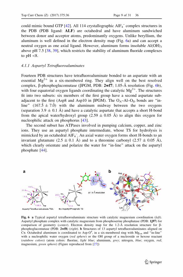
could mimic bound GTP [42]. All 114 crystallographic AlF4– complex structures in
the PDB (PDB ligand: ALF) are octahedral and have aluminum sandwiched
between donor and acceptor atoms, predominantly oxygens. Unlike beryllium, the
aluminum is well defined in the electron density map (Fig. 6a) and can accept a
neutral oxygen as one axial ligand. However, aluminum forms insoluble Al(OH)3above pH 7.5 [38, 39], which restricts the stability of aluminum fluoride complexes
to pH\8.
4.1.1 Aspartyl Tetrafluoroaluminates
Fourteen PDB structures have tetrafluoroaluminate bonded to an aspartate with an
essential Mg2? in a six-membered ring. They align well on the best resolved
complex, b-phosphoglucomutase (bPGM, PDB: 2wf7; 1.05-A resolution (Fig. 6b),
with four equatorial oxygen ligands coordinating the catalytic Mg2?. The structures
fit into two subsets: six members of the first group have a second aspartate sub-
adjacent to the first (Asp8 and Asp10 in bPGM). The OA–Al–OD bonds are ‘‘in-
line’’ (167.5 ± 7.0) with the aluminum midway between the two oxygens
(separation 3.9 ± 0.1 A) and have a catalytic aspartate that accepts a short H-bond
from the apical water/hydroxyl group (2.59 ± 0.05 A) to align this oxygen for
nucleophilic attack on phosphorus [43].
The second subset has ATPases involved in pumping calcium, copper, and zinc
ions. They use an aspartyl phosphate intermediate, whose TS for hydrolysis is
mimicked by an octahedral AlF4–. An axial water oxygen forms short H-bonds to an
invariant glutamate (2.5 ± 0.1 A) and to a threonine carbonyl (2.57 ± 0.05 A),
which clearly orientate and polarize the water for ‘‘in-line’’ attack on the aspartyl
phosphate [44].
Fig. 6 a Typical aspartyl tetrafluoroaluminate structure with catalytic magnesium coordination (left).Aspartyl phosphate complex with catalytic magnesium from phosphoserine phosphatase (PDB: 1j97) forcomparison of geometry (center). Electron density map for the 1.2-A resolution structure for b-phosphoglucomutase (PDB: 2wf8) (right). b Structures of 13 aspartyl tetrafluoroaluminates aligned onCa. Octahedral aluminum is coordinated to Asp-O4, in a six-membered ring with Mgcat and ‘‘in-line’’with a nucleophilic water oxygen (red sphere) or the OH group of a nucleoside or hexose reactant(rainbow colors) (atom colors: fluorine, light blue; aluminum, grey; nitrogen, blue; oxygen, red;magnesium, green sphere) (Figure reproduced from [27])
Top Curr Chem (Z) (2017) 375:36 Page 9 of 31 36
123

4.1.2 Nucleotide Guanosine Diphosphate (GDP) Tetrafluoroaluminates
GDP forms 50 AlF4– complexes that constitute isoelectronic but non-isosteric
mimics of GTP in a broad range of proteins. The best resolved 21 align remarkably
well (Fig. 7a), with aluminum bonded to O3B on GDP and the Mgcat coordinated to
F1 and O1B in a six-membered ring. The guanosine base and ribose usually occupy a
common conformation (Fig. 7a). The geometry of the AlF4– moiety is regularly
octahedral, with ‘‘in-line’’ OA–Al–OD angle 172.8 ± 7.1. All structures have an
axial oxygen ligand (Fig. 7a, red spheres) coordinated to aluminum that is trigonal
planar with respect to two H-bond acceptors: the backbone carbonyls of a threonine
and a glutamine side-chain (occasionally a water) (Fig. 7a, lower right, red spheres).
4.1.3 Nucleotide Adenosine Diphosphate (ADP) Tetrafluoroaluminates
Forty-nine octahedral structures have AlF4– bonded to a terminal oxygen of ADP
(O3B) to mimic ATP in the TS. They are found in kinases, hydrolases, isomerases,
ATPases, myosins, helicases, transporter pumps, and nitrogenase. The 31 that are
best resolved have an axial OA–Al–OD distance of 4.05 ± 0.03 A with an ‘‘in-line’’
angle of 170 ± 8 and most have water as the second oxygen ligand with a catalytic
Mg2? coordinating one of the fluorines. This is illustrated for F1 ATPase (PDB:
1h8e) (Fig. 8a). In contrast to the uniform conformation for complexes with GDP
(Fig. 7), complexes with ADP show a great variety of conformations, as illustrated
for 16 well-resolved structures (Fig. 8b).
4.2 Octahedral Aluminum Trifluoride Phosphate TS Mimics
An aluminum trifluoride moiety accepts three oxygens to give an octahedral, six-
coordination TSA complex in three examples. In the small G protein Rab5a, the
Fig. 7 a Twenty GDP�AlF4– structures aligned on a-carbon atoms of the invariant hexapeptide (in PDB:
2gj8). AlF4– is locked in a six–membered ring (center) with Mgcat (green spheres) coordinating F1 and a
PB oxygen. Octahedral coordination to Mg2? is provided by a second PB oxygen, two waters, a Thrhydroxyl (right), and a Ser/Thr hydroxyl (top). PB,G oxygens H-bond to a Lys (center). b Structures ofhGBP1 with a GMP�AlF3
0 complex (cyan) aligned with a GDP�AlF4– complex (green) showing occupancy
of the catalytic site by the AlF30 mimic of PB (magenta sphere) and by the AlF4
– mimic of PG (greysphere) (atom colors: GDP, cyan; GMP, green; magnesium, green; fluorine, light blue; amino acids,silver; nitrogen, blue; oxygen, red)
36 Page 10 of 31 Top Curr Chem (Z) (2017) 375:36
123

mutation A30P enables addition of the side chain hydroxyl of Ser29 to aluminum
trifluoride (PDB: 1n6k). In the case of hPGK, the K219A mutant has a water as the
fourth ligand coordinated to the aluminum [45]. Thirdly, for a bacterial dUTPase,
aluminum trifluoride takes the place of the PB in dUTP with coordination to two
oxygens from the b-phosphoryl group and to the water nucleophile to complete the
octahedral array (Fig. 9a, b). This significant structure provides a unique example
where nucleophilic attack is directed at a non-terminal nucleotide phosphorus [46].
5 MF3 Improved Geometry Transition State Mimics
5.1 MgF3–, Trifluoromagnesate
Magnesium does not form mixtures of stable fluorides in water at sub-molar
concentration: only one resonance for magnesium fluoride is seen in 19F NMR
Fig. 8 a F1 ATPase TSA complex (PDB: 1h8e) with ADP�AlF4– showing local charge balance for five
1ve and five -ve charges. b 16 ADP�AlF4– complexes aligned for C5’, PA, PB, and Al show great variety
in ATP analog conformations (atom colors: adenosines, cyan; magnesium, green spheres; fluorine, lightblue; aluminum, gold; amino acids and second substrates, grey; nitrogen, blue; oxygen, red)
Fig. 9 a Aluminum trifluoride structure for dUTPase (PDB: 4di8). UMP (cyan) coordinates aluminum(grey) with in-line water (red) and with PO4
= adjacent to the leaving O3A. Two magnesiums (greenspheres) are located by coordination to the reactants and to four carboxylate residues (amino acids ingrey). b Cartoon showing octahedral aluminum trifluoride sharing the tbp coordination of the true TS fora phosphoryl group (colors: nucleoside, cyan; magnesium, green sphere; aluminum, grey; sodium,purple; amino acids, silver; nitrogen, blue; oxygen, red) (Figure adapted from [27])
Top Curr Chem (Z) (2017) 375:36 Page 11 of 31 36
123
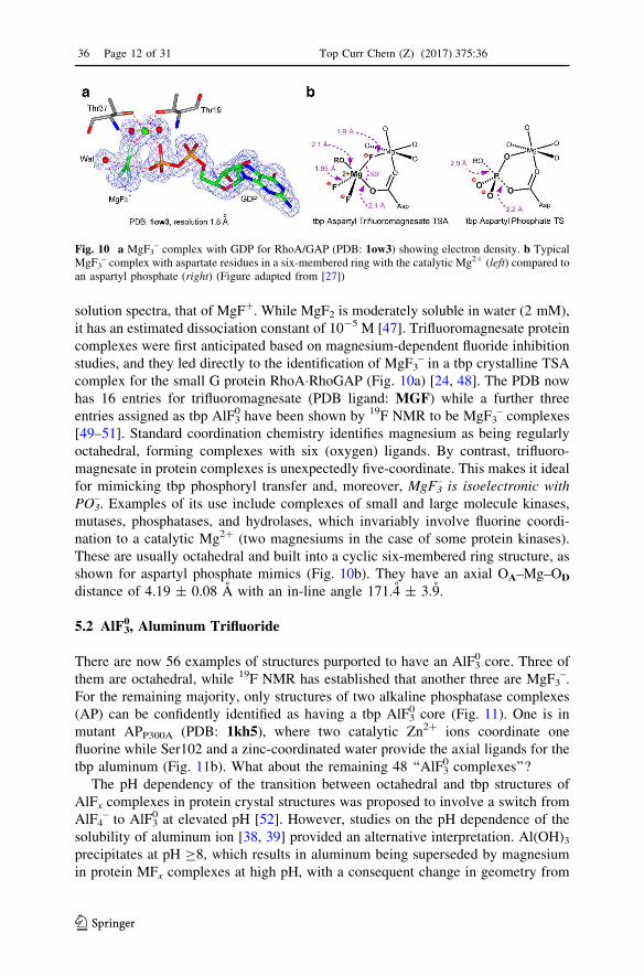
solution spectra, that of MgF?. While MgF2 is moderately soluble in water (2 mM),
it has an estimated dissociation constant of 10-5 M [47]. Trifluoromagnesate protein
complexes were first anticipated based on magnesium-dependent fluoride inhibition
studies, and they led directly to the identification of MgF3– in a tbp crystalline TSA
complex for the small G protein RhoA�RhoGAP (Fig. 10a) [24, 48]. The PDB now
has 16 entries for trifluoromagnesate (PDB ligand: MGF) while a further three
entries assigned as tbp AlF30 have been shown by 19F NMR to be MgF3
– complexes
[49–51]. Standard coordination chemistry identifies magnesium as being regularly
octahedral, forming complexes with six (oxygen) ligands. By contrast, trifluoro-
magnesate in protein complexes is unexpectedly five-coordinate. This makes it ideal
for mimicking tbp phosphoryl transfer and, moreover, MgF3– is isoelectronic with
PO3–. Examples of its use include complexes of small and large molecule kinases,
mutases, phosphatases, and hydrolases, which invariably involve fluorine coordi-
nation to a catalytic Mg2? (two magnesiums in the case of some protein kinases).
These are usually octahedral and built into a cyclic six-membered ring structure, as
shown for aspartyl phosphate mimics (Fig. 10b). They have an axial OA–Mg–OD
distance of 4.19 ± 0.08 A with an in-line angle 171.4 ± 3.9.
5.2 AlF30, Aluminum Trifluoride
There are now 56 examples of structures purported to have an AlF30 core. Three of
them are octahedral, while 19F NMR has established that another three are MgF3–.
For the remaining majority, only structures of two alkaline phosphatase complexes
(AP) can be confidently identified as having a tbp AlF30 core (Fig. 11). One is in
mutant APP300A (PDB: 1kh5), where two catalytic Zn2? ions coordinate one
fluorine while Ser102 and a zinc-coordinated water provide the axial ligands for the
tbp aluminum (Fig. 11b). What about the remaining 48 ‘‘AlF30 complexes’’?
The pH dependency of the transition between octahedral and tbp structures of
AlFx complexes in protein crystal structures was proposed to involve a switch from
AlF4– to AlF3
0 at elevated pH [52]. However, studies on the pH dependence of the
solubility of aluminum ion [38, 39] provided an alternative interpretation. Al(OH)3precipitates at pH C8, which results in aluminum being superseded by magnesium
in protein MFx complexes at high pH, with a consequent change in geometry from
Fig. 10 a MgF3– complex with GDP for RhoA/GAP (PDB: 1ow3) showing electron density. b Typical
MgF3– complex with aspartate residues in a six-membered ring with the catalytic Mg2? (left) compared to
an aspartyl phosphate (right) (Figure adapted from [27])
36 Page 12 of 31 Top Curr Chem (Z) (2017) 375:36
123

octahedral to tbp. That conclusion has now been validated by pH-dependent 19F
NMR analyses for several enzymes [50, 53]. In some marginal cases, e.g., protein
kinase A (cAPK) and PSP, there is mixed occupancy of the active site by tbp and
octahedral complexes in the crystal [43, 50, 51]. In geometric terms, ‘‘AlF30’’ tbp
complexes closely map on those of trifluoromagnesates: axial OA–M–OD bonds
4.29 ± 0.39 A (Fig. 12b), and M–F bonds 1.75 ± 0.12 A. It seems likely that some,
or many, of these ‘‘AlF30’’ complexes are trifluoromagnesates: a conclusion
supported by geometric analysis for both families of complex.
5.3 A Combined MgF32- and AlF3
0 Structural Analysis
A statistical analysis of the structures of AlF30 and MgF3
- complexes contributes to
the resolution of this compositional uncertainty. The near-invariant geometry of
Fig. 11 a Structure of the catalytic center for alkaline phosphatase complexed to AlF3 (PDB: 1kh5).b Cartoon of the coordination organization in the active site with transferring phosphoryl group (blue) andnucleophilic water (red) (Figure adapted from [27])
Fig. 12 a Overlay of five GDP�MgF3– (yellow) and eight GDP�AlF3
0 (cyan) complexes to show thegeometric uniformity of the two sets of TSA structures. b Normal distribution plots for the OA–M–OD
distance for this set of 13 structures (red) and the corresponding OA–Al–OD distance for 18 GDP�AlF4–
TSA complexes (blue)
Top Curr Chem (Z) (2017) 375:36 Page 13 of 31 36
123

octahedral AlF4- complexes for GDP makes them a useful set for comparison with
the corresponding set of tbp MF3 complexes. Thus, eight GDP ‘‘AlF30’’ structures for
small G proteins align very well with those for five MgF3- complexes (Fig. 12a).
The axial separation for the donor and acceptor oxygens in these combined 13
GDP�MF3 TSAs is 4.38 ± 0.20 A, significantly distinct from the corresponding
average for 19 GDP�AlF4– complexes, 4.02 ± 0.14 A, and clearly supported by
normal distribution analysis (Fig. 12b). The conclusion is: For ‘‘AlF30’’ read MgF3
–!
Taking ‘‘AlF30’’ together with trifluoromagnesates, a common general pattern of
axial ligands emerges. The MF3 species requires at least one anionic oxygen. b-
Oxygens from ADP (33 structures) and GDP (24 structures) provide the
overwhelming majority of examples while aspartate (11 structures) is also
significant. Water (27 structures) is the dominant neutral axial ligand while serine
and threonine hydroxyls appear less frequently. Significantly, there is no example of
both axial ligand positions being occupied by two neutral ROH groups.
5.4 MgF4=, Tetrafluoromagnesate
There are several structures for the Ca2? pump ATPase that have been assigned as
tetrahedral MgF4= moieties without objective experimental validation. Magnesium is
only exceptionally four-coordinate and then it usually has sterically bulky ether
oxygens as ligands [54]. The tetrahedral ‘‘MgF4=’’ moiety in all PDB examples is
remote from ADP, is coordinated to a second magnesium, and has one or more of its
four ‘‘fluorine’’ atoms in close contact with a backbone carbonyl oxygen, as shown
for PDB: 1wpg (Fig. 13a) [55]. Such ‘‘MgF4=’’ behavior closely resembles the six-
membered ring tbp structures common for MgF3– complexes of aspartate (Fig. 10).
Crystallographic re-refinement, with MgF3– replacing MgF4
= for 1wpg, can produce
an equally valid structure. Thus, unless established by further measurements, a more
consistent chemical interpretation for all such ‘‘MgF4=’’ situations is that they are
trifluoromagnesates that mimic the TS for hydrolysis of an aspartyl phosphate.
Subsequent work has described a similar tetrahedral moiety for the Na?/K? pump
ATPase (PDB: 2zxe) [56].
Fig. 13 a Structure of Ca2? pump ATPase with MgF4= (PDB: 1wpg). Coordination for MgF4
= is typical ofan aspartyl trifluoromagnesate complex (colors: fluorine, light blue; magnesium, green; nitrogen, blue,oxygen, red; carbons, silver). b Structure of hPPIP5K2 (PDB: 2q9p) to show the ‘‘Mg4F9’’ clusteradjacent to phosphates 4 and 5 of Ins6P
36 Page 14 of 31 Top Curr Chem (Z) (2017) 375:36
123
Finally, the most remarkable MFx structure is that of a human diphosphoinositol
phosphatase, co-crystallized with myo-inositol hexakis-phosphate and then soaked
with sodium fluoride (PDB: 2q9p) [57]. This complex has four octahedral
magnesiums with nine ligands assigned as fluorines in a complex that embraces
MgF2, MgF3, MgF4, and MgF5 species in a single block. It also offers the first
example of octahedral MgFx (Fig. 13b). Its core appears related to the Rutile
structure of MgF2, which is characterized by octahedral magnesium and trigonal
planar fluorine [58].
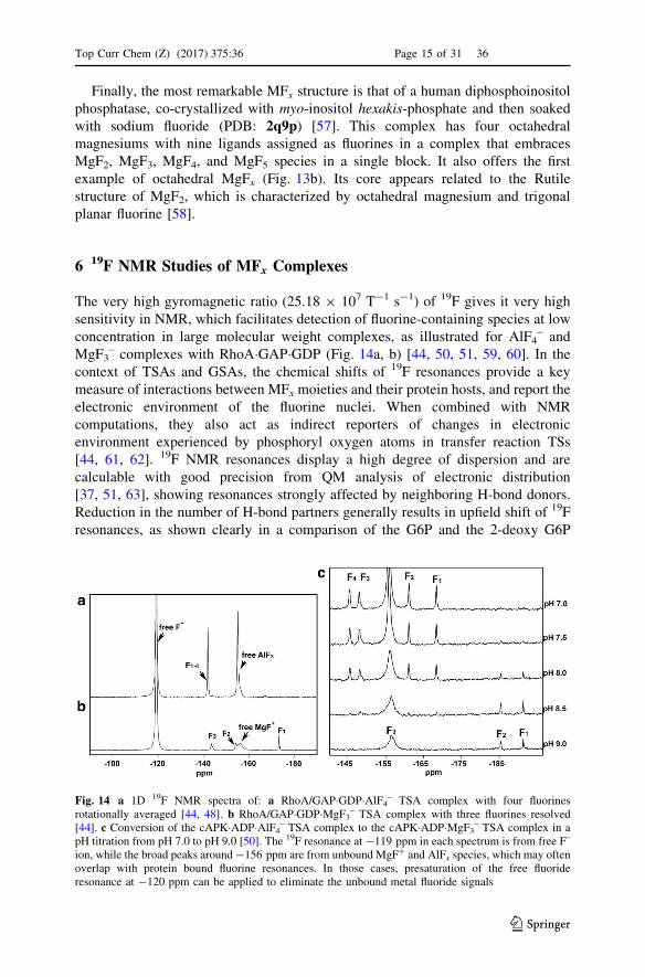
6 19F NMR Studies of MFxComplexes
The very high gyromagnetic ratio (25.18 9 107 T-1 s-1) of 19F gives it very high
sensitivity in NMR, which facilitates detection of fluorine-containing species at low
concentration in large molecular weight complexes, as illustrated for AlF4– and
MgF3– complexes with RhoA�GAP�GDP (Fig. 14a, b) [44, 50, 51, 59, 60]. In the
context of TSAs and GSAs, the chemical shifts of 19F resonances provide a key
measure of interactions between MFx moieties and their protein hosts, and report the
electronic environment of the fluorine nuclei. When combined with NMR
computations, they also act as indirect reporters of changes in electronic
environment experienced by phosphoryl oxygen atoms in transfer reaction TSs
[44, 61, 62]. 19F NMR resonances display a high degree of dispersion and are
calculable with good precision from QM analysis of electronic distribution
[37, 51, 63], showing resonances strongly affected by neighboring H-bond donors.
Reduction in the number of H-bond partners generally results in upfield shift of 19F
resonances, as shown clearly in a comparison of the G6P and the 2-deoxy G6P
Fig. 14 a 1D 19F NMR spectra of: a RhoA/GAP�GDP�AlF4– TSA complex with four fluorines
rotationally averaged [44, 48]. b RhoA/GAP�GDP�MgF3– TSA complex with three fluorines resolved
[44]. c Conversion of the cAPK�ADP�AlF4– TSA complex to the cAPK�ADP�MgF3
– TSA complex in apH titration from pH 7.0 to pH 9.0 [50]. The 19F resonance at -119 ppm in each spectrum is from free F–
ion, while the broad peaks around-156 ppm are from unbound MgF? and AlFx species, which may oftenoverlap with protein bound fluorine resonances. In those cases, presaturation of the free fluorideresonance at -120 ppm can be applied to eliminate the unbound metal fluoride signals
Top Curr Chem (Z) (2017) 375:36 Page 15 of 31 36
123
complexes of bPGM (-18.1 ppm) [53]. In general, the resonance of the fluorine
coordinated to a catalytic magnesium is always the most upfield, because of
depletion of H-bond coordination [44, 45, 50, 51, 53, 60, 62]. Proton distribution
near fluorine nuclei can be further assessed through the quantitation of 19F-1H NOEs
using perdeuterated enzyme in protonated buffer to suppress the 1H–1H spin
diffusion [49, 51], while resonance assignment of exchangeable 1H nuclei in the
protein enables unambiguous assignment of individual 19F NMR resonances. The
number of H-bond donors can also be assigned based on solvent induced hydrogen/
deuterium primary isotope shifts (SIIS) of 19F NMR resonances. For NH���F and
OH���F H–bonds to MFx moieties, the SIIS size reflects local proton densities [64],
and this has been used to assign FA, FB, and FC in a bPGM�MgF3–�G6P TSA
complex [62].
Scalar coupling between nuclei involved with N–H���F H-bonds is an additional
parameter that shows details of the coordination of the MFx moiety by the protein.1JHF and
2JNF couplings have been reported for individual NH���F pairs, with values
up to 59 and 36 Hz, respectively [62]. All the effects described above, SIIS, NOE,
chemical shifts, and scalar couplings, correlate closely with H-bonding orientations
and distances obtained from high resolution crystal structure analysis. 19F chemical
shifts are invariant over the pH range 6.5–9.5, they signal that there is no
detectable change in protonation state of the enzyme in the environment of the TS
complex, but the pH dependence of 19F NMR resonances and multiplicity can
identify a switch from AlF4– to MgF3
– complexes above pH 8, as illustrated for
cAPK (Fig. 14c) [57].
NMR measurements of 19F nuclei in the active site of MFx TSA complexes thus
provide a picture of charge distribution between the phosphoryl group mimic and
the protein. The good relationship between 19F NMR chemical shifts and SIIS
values illustrates the dominant influence that very localized H-bonds have on
shaping charge density on MFx moieties.
7 Computational Analyses of MFxComplexes
7.1 Balancing Accuracy of Energy/Structure and Conformational Sampling
A computational simulation of the structure and bonding of a biochemical system at
atomic resolution has two demanding features:
1. The solution of accurate molecular energies, ideally with as little parameter-
ization as possible;
2. The exhaustive consideration of relevant conformations of macromolecules.
For the simulation of biomolecules, it is unavoidable that both criteria must be
approximated to varying degrees. In practice, different computational methods put
different emphasis on one or the other of these two features. Any useful calculation
must meet both criteria adequately. Solutions of the energy of a macromolecule, and
thence its structure, should be made for each conformer of the molecule. Hence, the
36 Page 16 of 31 Top Curr Chem (Z) (2017) 375:36
123

task of achieving reliable energies severely raises the cost of the computation. This
constraint therefore drives down the number of conformers to be computed, with the
risk that the program may fail to examine the specific conformation most relevant
for the reaction under investigation.
To attain a compromise between these two features, the methodology used has to
strike a balance between defining a central quantum mechanics (QM) zone and a
molecular mechanics (MM) zone dealing with the major part of the macromolecule
and environment. The combination of the two regions is called a QM/MM
calculation. A QM description is necessary to describe bond–breaking–making
processes or electronic excited states because molecular mechanics cannot describe
these phenomena. Different balances between these two features are achieved by
different choices in the apportionment of resource to the QM region. These include
Kohn–Sham density functional theory (KS-DFT) [65–69] and empirical valence
bond (EVB) [70, 71], while similar choices exist for the MM zone. However, the
QM zone is the priority region.
7.2 Tradeoff in Accuracy of Energy/Structure: Parameterization
Simplification vs. Mathematical Complexity
Accurate molecular energies can be obtained in an unbiased, systematically
correctable manner [72–74] to get the desired accuracy. However, the computa-
tional resource required is very expensive, and is usually unacceptable because
resource must be apportioned to adequate conformational sampling. In general,
either an approximate QM method such as KS-DFT is used, or a heavily
parameterized model is designed for a specific system such as EVB. Briefly,
parameterization can tailor a QM method specifically to that molecule under
analysis—and thereby eliminate many mathematical degrees of freedom. Hence, the
calculation can be performed rapidly and can incorporate greater conformational
sampling, but it must rely on the assumption that the reduced mathematical form
faithfully represents the true quantum mechanics. By contrast, the various KS-DFT
forms have parameters which are fixed by the design of the functions, and are
completely independent of that particular biomolecule under investigation. Thus,
the application of KS-DFT to a specific biomolecule has no freedom to change
parameters to suit the target. Hence, KS-DFT deploys a more general mathematical
framework, and more faithfully echoes exact quantum mechanics within budget.
7.3 Tradeoffs in Conformational Sampling: Dynamics vs. Statics
In order to balance the budget of the computation program, a choice has to be made
between dynamics and statics. On the one hand, a dynamics description delivers an
explicit femtosecond-by-femtosecond time evolution of the atoms, boosted by
metadynamics [67]. On the other hand, a statics analysis of a few discrete critical
points along the reaction identifies TSs and/or intermediates as maxima/minima
along the reaction coordinate. Each has its strengths and weaknesses.
A dynamics computation shows the true time-evolution of the molecular system,
especially how atoms re-arrange to move along all possible reaction paths, step-by-
Top Curr Chem (Z) (2017) 375:36 Page 17 of 31 36
123

step. All possible chemical reactions/conformations are sampled in due frequency
with the Boltzmann distribution of states. The computation does not ‘‘target’’ a
specific reaction path. TSs are rare-events, require long simulations or metady-
namics [67], and so demand a smaller QM zone to allow an adequately fast
calculation. This reduction of the QM zone, relative to that for statics described
below, makes possible the conformational sampling needed to find the right state. A
balance has to be struck between faithfully computing dynamics or prioritizing
accurate energy calculations.
The choice for statics in following a reaction path, selected a priori, enables easy
identification of the TSs for bond-breaking-making using standard quantum
chemistry algorithms. Mathematical properties of energy maxima (TSs) and
minima (intermediates) can be sought automatically. Users can seek out any desired
pathway, but they have to sacrifice an understanding of the relative values of each
path. This requires minimal computational resource compared to that required for a
dynamics calculation, and so can accept a much larger QM region and/or a more
accurate QM calculation. However, the a priori choice of the conformation is risky:
it depends strongly on the accuracy of choice of the true TS conformation, which
may or may not be found among existing crystal structures in the PDB.
Take for example the first mechanistic step of the hairpin ribozyme. This is
cleavage of the bond from the 30-phosphate of A-12 to the 50-oxygen of G13 to form
a 20,30-cyclic phosphate which has been modeled as a pentacoordinate vanadate
TSA structure (PDB: 1m5o, 2.2-A resolution; Fig. 15a). The two proximate
nucleobases are G8’ and A57’ whose catalytic roles are controversial: there is good
support for protonation of A57’-N1 but some debate whether G8’ is deprotonated on
N1 or not. The computation accepted formation of an intermediate pentacoordinated
phosphorane and then posed the question: ‘‘How and when is the proton removed
from A8-O2’ and transferred to PO2A?’’ A thorough benchmark study of
O
O
P O
O
Ade12
O
O
O
H
OO
HO
Gua13
N N
N
N
O
H
N
HH
N
N
N
N
N
H
HH
Ade57'
Gua8'
Ab ini�oMechanism 1 KS-DFT Mechanism 2Vanadate TSA structure 1m5o
O
O
P O
O
Ade12
O
O
O
H
OO
HO
Gua13
N N
N
N
O
N
HH
N
N
N
N
N
H
HH
Ade57'
Gua8'a b c
Fig. 15 a Ab initio mechanism for the first step of the hammerhead ribozyme reaction showing PT fromAde12-O2’ to PO2 in the formation of a transient pentaoxyphosphorane species. b Crystal structure of thehammerhead ribozyme as a tbp vanadate complex (PDB: 1m5o). c DFT computed mechanism for PT tothe anionic Gua8’ preceding bond formation from P to Ade12-O2’ (colors: carbon, green; nitrogen, blue;oxygen, red; vanadium, grey; H-bonds, red dashes)
36 Page 18 of 31 Top Curr Chem (Z) (2017) 375:36
123
comparative QM/MM methods has been applied to this mechanism [75], comparing
an ab initio method (i.e., no parameterization) with a KS-DFT method (a small
number of fixed parameters). As the energy differences for the two paths should be
comparable, a careful QM analysis was necessary. Ab initio energy barrier
prediction matched experimental estimates, giving good support to Mechanism 1
(Fig. 15b) with a direct four-center proton transfer (PT), not involving a neutral
G8’.3 However, it was observed that the flow of atoms predicted by the
parameterized method was inconsistent with benchmark calculations. The authors
therefore employed umbrella sampling in a KS-DFT analysis to achieve dynamics
convergence, and found Mechanism 2 to be preferred with the anionic G8’ acting as
a base to abstract the proton from Ado12-O2’ (Fig. 15c).
7.4 KS-DFT as a QM Region Description
The most commonly chosen methodology for describing the QM portion is KS-
DFT4 functionals. This makes a practical compromise between precision and cost of
computation (Sect. 7.2). KS-DFT dispersion-corrected functionals can now describe
molecular geometries to within 0.02 A [76–78], and are particularly valuable
because their description of energies and geometries is unbiased. They have been
shown to describe basic bond-breaking behavior, H-bonding, and energetics
[77–81]. In a QM/MM calculation, the boundary region at the interface between the
QM zone and the MM zone can be problematic because the energies on the QM side
need not be the same as those on the MM side. Any mismatch between kinetic and
potential energies across the boundary leads to un-physical behavior. This boundary
problem can be eliminated by depriving large portions of the macromolecule of an
MM force field, which calls for a tradeoff between:
1. A more faithful representation of long-range chemical interactions and a
potentially problematic boundary between zones introducing artifacts; and
2. Neglect of long-range chemical interactions altogether, with no un-physical
artifacts introduced by the QM/MM boundary.
Either choice is problematic, and a case-by-case decision must be made. In
some cases, to avoid boundary complexities exclusively QM calculations have
been used, usually KS-DFT. They usually rely wholly on experimental data from
the structure of a TS mimic, thereby obviating the need for a conformational
search, and allowing full investment of the computational resource to maximize
the size of the QM zone. For example, a recent study of GTP hydrolysis by RhoA/
GAP to identify the reaction mechanism employed a large QM calculation [44].
The KS-DFT zone was large enough to embrace the reacting methyl triphosphate,
its coordinating magnesium and nucleophilic water, and also residues from some
3 NB In other computational studies, such four-center PTs for phosphoryl reactions have been deemed to
be very high energy.4 In literature meant for using DFT in organic, biological, or inorganic applications, ‘‘KS-DFT’’ and
‘‘DFT’’ are used largely interchangeably. Theoreticians draw a distinction between these terms; KS-DFT
is a subset of DFT for the given selection of expressing the kinetic energy in terms of orbitals.
Top Curr Chem (Z) (2017) 375:36 Page 19 of 31 36
123

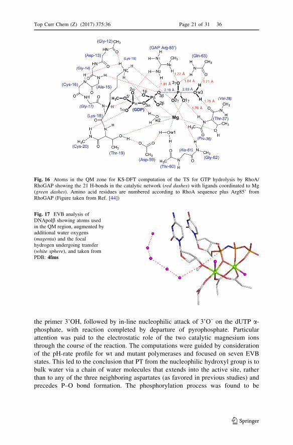
18 additional amino acids that contribute to the stability of a network of 21
H-bonds which deliver the conformation of the TS for water attack on PG.
Successive rounds of DFT computing established that contributions from atoms in
the third solvation shell of the transferring phosphoryl group were required to
deliver stability. The result was a QM region of 91 heavy atoms (181 total atoms)
(Fig. 16). Because the starting TSA structure (PDB: 1ow3) was of sufficiently
high resolution (1.8 A) to give confidence that the study was based on a reliable
model of the TS, the addition of an MM contribution was bypassed, obviating the
need for a QM/MM boundary. However, this limited the computational output to
geometric and spectroscopic features. The absence of conformational sampling,
sacrificed because of the large and very expensive QM region, also limits
comment on activation energies.
The iterative computational procedure delivered a mechanism in which the
nucleophilic water is doubly protonated with H-bonds to carbonyl oxygens of both
T37 and Q63 residues until after the TS for bond making/breaking, thereby
orientating the nucleophilic water for good orbital overlap with the antibonding
O3B-PG r* orbital. PTs are not seen in the TS, but occur subsequently.
7.5 EVB as a QM Region Description
The Empirical Valence Bond method deploys a simplified mathematical
framework to achieve the most rigorous possible conformational sampling. In
essence, the EVB framework is largely a molecular mechanics based method,
with the exception of its representation of a single ‘‘orbital’’ for each molecule,
identified as involved in the bond–breaking–making reaction. No other electrons/
orbitals are represented explicitly. This framework thus imposes the presumption
that only a single orbital is involved in the bond-reorganization for a reaction.
The EVB parameterization process is fundamentally chemistry-imposed: it
identifies, a priori, what orbitals are involved and dictates chemistry-based
molecular mechanics energy functions. This is in sharp contrast to a KS-DFT
prescription of a QM region, which is fundamentally agnostic of chemistry, not
defining bonds or selecting orbitals targeted for reaction, but merely defining a
total number of electrons and nuclei involved, with no presumption of chemistry.
As a result of the EVB simplifications, larger-scale changes in molecular
conformation can be observed. In this way, the initial conditions of the
experimental crystal structure are not a trap; the computational protocol allows
the biomolecule to move freely.
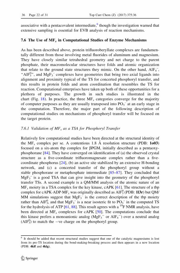
A study of the mechanism of DNA polymerase b provides a good example of the
application of the EVB methodology [82]. The questions under examination were
(i) the destination and timing of PT from the nucleophilic 30-OH, with three
aspartates and water as potential acceptors, and (ii) the concerted or stepwise nature
of phosphorus migration.5 The starting structure was native DNApolb (PDB: 2fms,
2.0 A resolution) and some 70 heavy atoms were included in the QM zone (Fig. 17),
linked to the assumption that the reaction takes place in the three steps: a PT from
5 A previous study favored a concerted reaction path (Lin et al. [83]).
36 Page 20 of 31 Top Curr Chem (Z) (2017) 375:36
123
the primer 3’OH, followed by in-line nucleophilic attack of 3’O– on the dUTP a-
phosphate, with reaction completed by departure of pyrophosphate. Particular
attention was paid to the electrostatic role of the two catalytic magnesium ions
through the course of the reaction. The computations were guided by consideration
of the pH-rate profile for wt and mutant polymerases and focused on seven EVB
states. This led to the conclusion that PT from the nucleophilic hydroxyl group is to
bulk water via a chain of water molecules that extends into the active site, rather
than to any of the three neighboring aspartates (as favored in previous studies) and
precedes P–O bond formation. The phosphorylation process was found to be
Fig. 16 Atoms in the QM zone for KS-DFT computation of the TS for GTP hydrolysis by RhoA/RhoGAP showing the 21 H-bonds in the catalytic network (red dashes) with ligands coordinated to Mg(green dashes). Amino acid residues are numbered according to RhoA sequence plus Arg85’ fromRhoGAP (Figure taken from Ref. [44])
Fig. 17 EVB analysis ofDNApolb showing atoms usedin the QM region, augmented byadditional water oxygens(magenta) and the focalhydrogen undergoing transfer(white sphere), and taken fromPDB: 4fms
Top Curr Chem (Z) (2017) 375:36 Page 21 of 31 36
123
associative with a pentacovalent intermediate,6 though the investigation warned that
extensive sampling is essential for EVB analysis of reaction mechanisms.
7.6 The Use of MFxin Computational Studies of Enzyme Mechanisms
As has been described above, protein trifluoroberyllate complexes are fundamen-
tally different from those involving metal fluorides of aluminum and magnesium.
They have closely similar tetrahedral geometry and net charge to the parent
phosphate, their macromolecular structures have folds and atomic organization
that relate to the ground state structures they mimic. On the other hand, AlF4–,
‘‘AlF30’’, and MgF3
– complexes have geometries that bring two axial ligands into
alignment and proximity typical of the TS for concerted phosphoryl transfer, and
this results in protein folds and atom coordination that resembles the TS for
reaction. Computational enterprises have taken up both of these opportunities for a
plethora of purposes. The growth in such studies is illustrated in the
chart (Fig. 18). In practice, the three MFx categories converge for the majority
of computer purposes as they are usually transposed into PO3– at an early stage in
the computation. Therefore, the major part of the following description of
computational studies on mechanisms of phosphoryl transfer will be focused on
the target protein.
7.6.1 Validation of MFx as a TSA for Phosphoryl Transfer
Relatively few computational studies have been directed at the structural identity of
the MFx complex per se. A contentious 1.8 A resolution structure (PDB: 1o03)
focused on a six-atom tbp complex for bPGM, initially described as a pentaoxy-
phosphorane [84]. They have converged on identification of (a) the observed crystal
structure as a five-coordinate trifluoromagnesate complex rather than a five-
coordinate phosphorus [24], (b) an active site stabilized by an extensive H-bonding
network, and (c) a concerted transfer of the phosphoryl group without a
stable phosphorane or metaphosphate intermediate [85–87]. They concluded that
MgF3– is a good TSA that can give insight into the geometry of the phosphoryl
transfer TSs. A second example is a QM/MM analysis of the atomic nature of an
MFx moiety in a TSA complex for the key kinase, cAPK [61]. The structure of a tbp
complex for cAPK�ADP�MFx was originally described as AlF30 (PDB: 1l3r) but QM/
MM simulations suggest that MgF3– is the correct description of the tbp moiety
rather than AlF30, and that MgF3
– is a near isosteric fit to PO3– in the computed TS
for the hydrolysis of ATP [61, 88]. This result agrees with a 19F NMR analysis, have
been directed at MFx complexes for cAPK [50]. The computations conclude that
this kinase prefers a monoanionic analog (MgF3- or AlF4
-) over a neutral analog
(AlF30) to match the -ve charge on the phosphoryl group.
6 It should be added that recent structural studies suggest that one of the catalytic magnesiums is lost
from its pre-TS location during the bond-making-breaking process and then appears in a new location
(PDB: 4klf and 4klg).
36 Page 22 of 31 Top Curr Chem (Z) (2017) 375:36
123

7.6.2 Studies Linking Reaction Mechanisms from Model Systems to MFx Enzyme
Complexes
Early QM studies on phosphoryl transfer analyzed the hydrolysis of methyl
phosphate [89] and methyl pyrophosphate [90], added magnesium [91], and then
transposed the results into the context of the Ras GTPase active site. The result does
not match well to the MFx structure for Ras�RasGAP (PDB: 1wq1) because (i) the
computed OA—OD separation lies in the region 4.7–5.5 A and in the MFx structure
is 4.4 A. (ii) The computation calls for a second water to facilitate PT [92], however,
in those (few) instances where a second water is seen in high-resolution MFxstructures for Ras, it occupies the site vacated by a displaced or missing Gln61
residue, and is in no position to deliver the proposed catalysis (Fig. 7a).
7.7 Computations Transposing GDP�MFxinto GTP Enzyme Complexes
7.7.1 Ras Family and GTP Hydrolysis
The use of MFx TSA structures to identify the TS for hydrolysis of GTP by Ras
proteins has been the basis of many computations. Several studies have used PDB:
1wq1 [22], the 2.5 A-resolution structure of Ras�RasGAP�GDP�AlF30 as starting
point, and have employed both QM/MM [92–99] and EVB approaches [100, 101].
Some of these have aroused expert criticism of limitations inherent in the QM/MM
approach [102]. The results have varied widely, from a two-step reaction
mechanism with bond breaking preceding bond making (i.e. a dissociative process;
Scheme 1a) [100], to exclusion of water by the arginine finger [98], tautomeric
catalysis [17], electrostatic catalysis [101], a two-water mechanism [92], and sundry
rationalizations of the adverse effects of mutations [97, 99, 101]. The QM zone has
generally been limited to 30–40 heavy atoms and, in consequence, has not examined
the role of the function of several amino acids in contact with the reactants, most
Fig. 18 Growth of computational publications since 1994 showing QM results on AlF4– leading with
[AlF30? MgF3
–] comparable to studies based on BeF3- complexes
Top Curr Chem (Z) (2017) 375:36 Page 23 of 31 36
123
especially the extensive H-bonding network (as in Fig. 16). By contrast, an
alternative computational approach using Kohn–Sham DFT analysis for RhoA�R-
hoGAP hydrolysis of GTP employed a QM zone of 91 heavy atoms, embracing a
network of 21 H-bonds, and has attributed catalysis to orbital orientation determined
by protein control of H-bonds donated by the nucleophilic water (Fig. 16) [44]. The
same study validated the high relevance of MgF3– as a TSA by back-computing its
structure from that of the calculated structure for the true TS complex for GTP
hydrolysis.
7.7.2 Other GTPases and GTP Hydrolysis
A study on the structures of a GMP�AlF30 complex (PDB: 2b8w) and a GDP�AlF4
-
complex (PDB: 2b92) for hGBP1, has linked a mechanism for the hydrolysis of
methyl triphosphate (MTP) to the two-step hydrolysis of GTP to GDP and thence to
GMP by this interferon-activated human GTPase (Fig. 7b). The computation
employed dated ab initio QM/MM molecular dynamics to simulate the hydrolysis of
both GTP and of MTP as a reference system [103]. The study proposes that GTP
hydrolysis involves an indirect, substrate-assisted catalysis mechanism, identifying
the nearest general base as Glu99, which is 6.2 A from the nucleophilic water in the
TSA complex. This separation problem was resolved by invoking transmission of
base catalysis via one water to Ser73, and thence via a second water to the
nucleophilic water. These bridging waters are not present in the substantive (3.2 A
resolution) TSA complex but appear to be imported from a structure of hGBP1 with
b, c-imino-GTP that is clearly an NAC complex (PDB: 2bc9; 2.8 A resolution).
This investigation merits a cautionary comment on the frailties of a computational
analysis based on structures of poor resolution, under-informed by an adequate
grasp of mechanisms of phosphoryl transfer.
7.8 Computations Transposing ADP�MFxinto ATP Enzyme Complexes
7.8.1 ATP Hydrolysis by Myosin
Myosins are a family of ATP-dependent motor proteins whose role in muscle
contraction is driven by ATP hydrolysis. Multiple structures of Mg�ADP�MFx exist,
including BeF3– (PDB: 1w9i and 1mmd) and AlF4
– (PDB: 1w9l and 1wj9). These
structures have been used to identify the catalytic amino acids and locate key water
molecules, especially the nucleophilic water that attacks in-line at PG (Fig. 8a). A
recent QM/MM computation of the hydrolysis of ATP used a DFT method with
B3LYP functional and a 6–31G(d,p) basis set to treat 84 atoms in the active site for
the Mg�ADP�BeF3– structure of the myosin II head group (PDB: 1mmd), with ATP
modeled by replacing BeF3 with a phosphate (cf. Fig. 4a) [104]. Although the
starting structure for the computation (Fig. 19a) has the nucleophilic water (Wa) in a
NAC, as defined by its H-bond proximity to F2 (2.7 A) and out-of-line angle for the
attack on Be (152), the simulations delivered a H-bond network that lead to the final
product, H2PGO4
-. The proposed mechanism involves formation of a
36 Page 24 of 31 Top Curr Chem (Z) (2017) 375:36
123
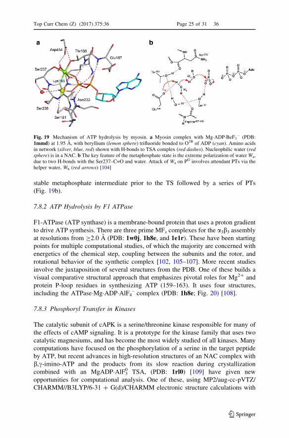
stable metaphosphate intermediate prior to the TS followed by a series of PTs
(Fig. 19b).
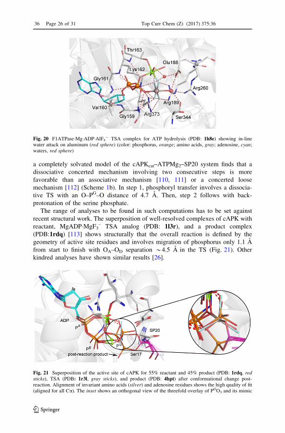
7.8.2 ATP Hydrolysis by F1 ATPase
F1-ATPase (ATP synthase) is a membrane-bound protein that uses a proton gradient
to drive ATP synthesis. There are three prime MFx complexes for the a3b3 assembly
at resolutions from C2.0 A (PDB: 1w0j, 1h8e, and 1e1r). These have been starting
points for multiple computational studies, of which the majority are concerned with
energetics of the chemical step, coupling between the subunits and the rotor, and
rotational behavior of the synthetic complex [102, 105–107]. More recent studies
involve the juxtaposition of several structures from the PDB. One of these builds a
visual comparative structural approach that emphasizes pivotal roles for Mg2? and
protein P-loop residues in synthesizing ATP (159–163). It uses four structures,
including the ATPase�Mg�ADP�AlF4– complex (PDB: 1h8e; Fig. 20) [108].
7.8.3 Phosphoryl Transfer in Kinases
The catalytic subunit of cAPK is a serine/threonine kinase responsible for many of
the effects of cAMP signaling. It is a prototype for the kinase family that uses two
catalytic magnesiums, and has become the most widely studied of all kinases. Many
computations have focused on the phosphorylation of a serine in the target peptide
by ATP, but recent advances in high-resolution structures of an NAC complex with
b,c-imino-ATP and the products from its slow reaction during crystallization
combined with an MgADP�AlF30 TSA, (PDB: 1rl0) [109] have given new
opportunities for computational analysis. One of these, using MP2/aug-cc-pVTZ/
CHARMM//B3LYP/6-31 ? G(d)/CHARMM electronic structure calculations with
Fig. 19 Mechanism of ATP hydrolysis by myosin. a Myosin complex with Mg�ADP�BeF3- (PDB:
1mmd) at 1.95 A, with beryllium (lemon sphere) trifluoride bonded to O2B of ADP (cyan). Amino acidsin network (silver, blue, red) shown with H-bonds to TSA complex (red dashes). Nucleophilic water (redsphere) is in a NAC. b The key feature of the metaphosphate state is the extreme polarization of waterWa,due to two H-bonds with the Ser237–C=O and water. Attack of Wa on PG involves attendant PTs via thehelper water, Wh (red arrows) [104]
Top Curr Chem (Z) (2017) 375:36 Page 25 of 31 36
123
a completely solvated model of the cAPKcat–ATPMg2–SP20 system finds that a
dissociative concerted mechanism involving two consecutive steps is more
favorable than an associative mechanism [110, 111] or a concerted loose
mechanism [112] (Scheme 1b). In step 1, phosphoryl transfer involves a dissocia-
tive TS with an O–PG–O distance of 4.7 A. Then, step 2 follows with back-
protonation of the serine phosphate.
The range of analyses to be found in such computations has to be set against
recent structural work. The superposition of well-resolved complexes of cAPK with
reactant, MgADP�MgF3– TSA analog (PDB: 1l3r), and a product complex
(PDB:1rdq) [113] shows structurally that the overall reaction is defined by the
geometry of active site residues and involves migration of phosphorus only 1.1 A
from start to finish with OA–OD separation *4.5 A in the TS (Fig. 21). Other
kindred analyses have shown similar results [26].
Fig. 20 F1ATPase�Mg�ADP�AlF4- TSA complex for ATP hydrolysis (PDB: 1h8e) showing in-line
water attack on aluminum (red sphere) (color: phosphorus, orange; amino acids, gray; adenosine, cyan;waters, red sphere)
Fig. 21 Superposition of the active site of cAPK for 55% reactant and 45% product (PDB: 1rdq, redsticks), TSA (PDB: 1r3l, gray sticks), and product (PDB: 4hpt) after conformational change post-reaction. Alignment of invariant amino acids (silver) and adenosine residues shows the high quality of fit(aligned for all Ca). The inset shows an orthogonal view of the threefold overlay of PGO3 and its mimic
36 Page 26 of 31 Top Curr Chem (Z) (2017) 375:36
123

7.9 Thoughts from Computations
The first phase of computational studies for phosphoryl transfer was largely focused
on finding a close match between computed activation energies and the
experimental ones. However, recent advances in computational methodology have
cast a shadow on earlier methods, where the energy error might easily lie in the
range of 2–10 kcal mol-1, with error spreading as large as 30 kcal mol-1
[78, 114, 115]. Current protocols enable a much larger number of heavy atoms to
be embraced in the QM zone, leading to computer results that hinge on geometry of
the TS and the H-bond network that it embraces [44, 108, 111]. Such analyses have
identified the propensity of nucleotide analogs, particularly b,c-iminoATP, b,c-
methyleneATP, and their GTP counterparts, to deliver NACs for phosphoryl
transfer processes, which is now recognized in recent computational studies as
capable of generating small but highly significant conformational changes in kinases
and GTPases [44, 108]. Lastly, the belief that enzymes work by optimizing reaction
mechanisms that work slowly in solution, as Knowles put it ‘‘Not different, Just
better’’ [116], is proving to be wide of the mark for phosphate reactions. There is
growing evidence that phosphoryl transfer takes place in a desolvated environment
to enable full protein control of the catalytic region. Water is rigorously excluded to
avoid disruption of H-bond networks that are essential for the organization of
catalysis.
8 Conclusions
Trifluoroberyllate, tetrafluoroaluminate, and trifluoromagnesate are the primary
anionic MFx species that can mimic the phosphoryl group. Structural, spectroscopic,
and computational methods have combined to validate their use as surrogates for
PO3– in ground state and transition state analog complexes for many enzymes. Their
use has delivered details of phosphoryl transfer at atomic resolution and supported
investigations of protein folding and aggregation for tertiary structure problems. In
particular, their analysis has confirmed existing concepts, introduced new ideas, and
set new goals, of which the following comprise a brief summary:
• In-line stereochemistry and concertedness for SN2(P) reactions has been
established at atomic resolution;
• Relative priority of charge over geometry in transition state organization is well
supported;
• Subtle conformational differences between NAC and TS conformations of
amino acid functions are increasingly apparent;
• The role of H-bond networks to give structural coherence to proteins in
transition states is burgeoning;
• The propensity of anionic phosphate oxygens to H-bond to ROH nucleophiles
explains the need for solvent exclusion from the TS, while its impedance offers a
new interpretation of general base catalysis.
Top Curr Chem (Z) (2017) 375:36 Page 27 of 31 36
123

Acknowledgments The authors thank Professors Nigel Richards (Cardiff University) and Jon Waltho
(Manchester University) for critical comments on and many contributions to this work and Dr. Christian
Roth (University of York) for advice on interpretation of some protein structures. We were supported by
BBSRC Grant BB/M021637/1 and the Universities of York and Sheffield, UK. Y.J. is funded by ERC
Advanced Grant AdG-322942. Multiple structural figures have used data taken from the Protein Data
Bank. The use of illustrations from Refs. [27] and [44] has been appreciatively acknowledged where
appropriate.
Open Access This article is distributed under the terms of the Creative Commons Attribution 4.0
International License (http://creativecommons.org/licenses/by/4.0/), which permits unrestricted use, dis-
tribution, and reproduction in any medium, provided you give appropriate credit to the original
author(s) and the source, provide a link to the Creative Commons license, and indicate if changes were
made.
References
1. Todd AR (1981) Where there’s life, there’s phosphorus. In: Kageyama M, Nakamura K, Oshima T
(eds) Japan Science Society Press, Tokyo, pp 275–279
2. Westheimer F (1987) Science 235:1173–1178
3. Lad C, Williams NH, Wolfenden R (2003) Proc Natl Acad Sci USA 100:5607–5610
4. Cohn M (1953) J Biol Chem 201:735–750
5. Knowles JR (1980) Annu Rev Biochem 49:877–919
6. Frey PA (1989) Chiral phosphorothioates: stereochemical analysis of enzymatic substitution at
phosphorus. Adv Enzymol Relat Areas Mol Biol 62:119–201
7. Lowe G (1983) Acc Chem Res 16:244–251
8. Blackburn GM, Cherfils J, Moss PR, Nigel GJ, Waltho JP, Williams NH, Wittinghofer A (2017)
Pure App Chem 89 (In press)
9. Todd SA (1959) Proc Natl Acad Sci USA 45:1389–1397
10. Mildvan AS (1997) Proteins 29:401–416
11. Cleland WW, Hengge AC (2006) Chem Rev 106:3252–3278
12. Lassila JK, Zalatan JG, Herschlag D (2011) Annu Rev Biochem 80:669–702
13. Smith JD, Traut RR, Blackburn GM, Monro RE (1965) J Mol Biol 13:617–628
14. Grisham CM, Mildvan AS (1974) J Biol Chem 249:3187–3197
15. Rao BD, Buttlaire DH, Cohn M (1976) J Biol Chem 251:6981–6986
16. Milburn MV, Tong L, Devos AM, Brunger A, Yamaizumi Z, Nishimura S, Kim SH (1990) Science
247:939–945
17. Pai EF, Krengel U, Petsko GA, Goody RS, Kabsch W, Wittinghofer A (1990) EMBO J
9:2351–2359
18. Coleman DE, Berghuis AM, Lee E, Linder ME, Gilman AG, Sprang SR (1994) Science
265:1405–1412
19. Sondek J, Lambright DG, Noel JP, Hamm HE, Sigler PB (1994) Nature 372:276–279
20. Fisher AJ, Smith CA, Thoden JB, Smith R, Sutoh K, Holden HM, Rayment I (1995) Biochemistry
34:8960–8972
21. Xu Y-W, Morera S, Janin J, Cherfils J (1997) Proc Natl Acad Sci USA 94:3579–3583
22. Scheffzek K, Ahmadian MR, Kabsch W, Wiesmuller L, Lautwein A, Schmitz F, Wittinghofer A
(1997) Science 277:333–338
23. Schlichting I, Reinstein J (1997) Biochemistry 36:9290–9296
24. Graham DL, Lowe PN, Grime GW, Marsh M, Rittinger K, Smerdon SJ, Gamblin SJ, Eccleston JF
(2002) Chem Biol 9:375–381
25. Mesmer RE, Baes CF (1969) Inorg Chem 8:618–626
26. Dai J, Finci L, Zhang C, Lahiri S, Zhang G, Peisach E, Allen KN, Dunaway-Mariano D (2009)
Biochemistry 48:1984–1995
27. Blackburn GM, Jin Y, Richards NG, Waltho JP (2016) Angew Chem Int Ed. doi:10.1002/anie.
201606474
36 Page 28 of 31 Top Curr Chem (Z) (2017) 375:36
123
28. Burgos ES, Ho MC, Almo SC, Schramm VL (2009) Proc Natl Acad Sci USA 106:13748–13753
29. Blackburn GM (1981) Phosphonates as analogues of biological phosphates. Chem Ind (London)
7:134–138
30. Pauling L (1960) The nature of the chemical bond, E3, vol E3. Cornell University Press, New York
31. Kowalinski E, Schuller A, Green R, Conti E (2015) Structure 23:1336–1343
32. Park AK, Lee JH, Chi YM, Park H (2016) Biochem Biophys Res Commun 473:625–629
33. Sheftic SR, White E, Gage DJ, Alexandrescu AT (2014) Biochemistry 53:311–322
34. Maruta S, Uyehara Y, Aihara T, Katayama E (2004) J Biochem 136:57–64
35. Hilbert BJ, Hayes JA, Stone NP, Duffy CM, Sankaran B, Kelch BA (2015) Proc Natl Acad Sci USA
112:E3792–E3799
36. Pylypenko O, Attanda W, Gauquelin C, Lahmani M, Coulibaly D, Baron B, Hoos S, Titus MA,
England P, Houdusse AM (2013) Proc Natl Acad Sci USA 110:20443–20448
37. Griffin JL, Bowler MW, Baxter NJ, Leigh KN, Dannatt HRW, Hounslow AM, Blackburn GM,
Webster CE, Cliff MJ, Waltho JP (2012) Proc Natl Acad Sci USA 109:6910–6915
38. Bruce Martin R (1988) Biochem Biophys Res Commun 155:1194–1200
39. Bruce Martin R (1996) Coord Chem Rev 149:23–32
40. Sternweis PC, Gilman AG (1982) Proc Natl Acad Sci USA 79:4888–4891
41. Higashijima T, Graziano MP, Suga H, Kainosho M, Gilman AG (1991) J Biol Chem
266:3396–3401
42. Bigay J, Deterre P, Pfister C, Chabre M (1987) The EMBO J 6:2907–2913
43. Wang W, Cho HS, Kim R, Jancarik J, Yokota H, Nguyen HH, Grigoriev IV, Wemmer DE, Kim SH
(2002) J Mol Biol 319:421–431
44. Jin Y, Molt RW, Waltho JP, Richards NGJ, Blackburn GM (2016) Angew Chem Int Ed
55:3318–3322
45. Cliff MJ, Bowler MW, Varga A, Marston JP, Szabo J, Hounslow AM, Baxter NJ, Blackburn GM,
Vas M, Waltho JP (2010) J Am Chem Soc 132:6507–6516
46. Hemsworth Glyn R, Gonzalez-Pacanowska D, Wilson Keith S (2013) Biochem J 456:81–88
47. Fovet Y, Gal J-Y (2000) Talanta 53:617–626
48. Graham DL, Eccleston JF, Chung CW, Lowe PN (1999) Biochemistry 38:14981–14987
49. Baxter NJ, Olguin LF, Golicnik M, Feng G, Hounslow AM, Bermel W, Blackburn GM, Hollfelder
F, Waltho JP, Williams NH (2006) Proc Natl Acad Sci USA 103:14732–14737
50. Jin Y, Cliff MJ, Baxter NJ, Dannatt HRW, Hounslow AM, Bowler MW, Blackburn GM, Waltho JP
(2012) Angew Chem Int Ed 51:12242–12245
51. Baxter NJ, Blackburn GM, Marston JP, Hounslow AM, Cliff MJ, Bermel W, Williams NH,
Hollfelder F, Wemmer DE, Waltho JP (2008) J Am Chem Soc 130:3952–3958
52. Schlichting I, Reinstein J (1999) Nat Struct Biol 6:721–723
53. Baxter NJ, Hounslow AM, Bowler MW, Williams NH, Blackburn GM, Waltho JP (2009) J Am
Chem Soc 131:16334–16335
54. Bock CW, Kaufman A, Glusker JP (1994) Inorg Chem 33:419–427
55. Toyoshima C, Nomura H, Tsuda T (2004) Nature 432:361–368
56. Shinoda T, Ogawa H, Cornelius F, Toyoshima C (2009) Nature 459:446–450
57. Thorsell A-G, Persson C, Graslund S, Hammarstrom M, Busam RD, Hallberg BM (2009) Proteins.
Struct Func Bioinfo 77:242–246
58. Baur WH (1956) Acta Crystallogr A 9:515–520
59. Xiaoxia L, Marston JP, Baxter NJ, Hounslow AM, Yufen Z, Blackburn GM, Cliff MJ, Waltho JP
(2011) J Am Chem Soc 133:3989–3994
60. Jin Y, Bhattasali D, Pellegrini E, Forget SM, Baxter NJ, Cliff MJ, Bowler MW, Jakeman DL,
Blackburn GM, Waltho JP (2014) Proc Natl Acad Sci USA 111:12384–12389
61. Leigh KN, Webster CE (2014) Dalton Trans 43:3039–3043
62. Baxter NJ, Bowler MW, Alizadeh T, Cliff MJ, Hounslow AM, Wu B, Berkowitz DB, Williams NH,
Blackburn GM, Waltho JP (2010) Proc Natl Acad Sci USA 107:4555–4560
63. Oldfield E (2005) Phil Trans R Soc B 360:1347–1361
64. Sosnicki JG, Langaard M, Hansen PE (2007) J Org Chem 72:4108–4116
65. Kohn W, Sham LJ (1965) Phys Rev 140:A1133–A1138
66. Hohenberg P, Kohn W (1964) Phys Rev 136:B864–B871
67. Cramer C (2008) Essentials of computational chemistry, 2nd edn. Wiley, West Sussex, pp 249–301
68. Martin RM (2004) Electronic structure, 1st edn. Cambridge University Press, Cambridge,
pp 119–184
Top Curr Chem (Z) (2017) 375:36 Page 29 of 31 36
123
69. Parr RG, Tao W (1994) Density-functional theory of atoms and molecules, 1st edn. Oxford
University Press, Oxford
70. Aqvist J, Warshel A (1993) Chem Rev 93:2523–2544
71. Hwang JK, King G, Creighton S, Warshel A (1988) J Am Chem Soc 110:5297–5311
72. Bennie SJ, van der Kamp MW, Pennifold RCR, Stella M, Manby FR, Mulholland AJ (2016) J
Chem Theory Comput 12:2689–2697
73. Bartlett RJ, Shavitt I (2009) Many-body methods in chemistry and physics. Cambridge University
Press, Cambridge, pp 251–340
74. Iii GDP, Bartlett RJ (1982) J Chem Phys 76:1910–1918
75. Mlynsky V, Banas P, Sponer J, van der Kamp MW, Mulholland AJ, Otyepka M (2014) J Chem
Theory Comput 10:1608–1622
76. Peverati R, Truhlar DG (2011) J Phys Chem Lett 2:2810–2817
77. Zhao Y, Truhlar DG (2008) Theor Chem Acc 120:215–241
78. Goerigk L, Grimme S (2011) Phys Chem Chem Phys 13:6670–6688
79. Grimme S (2004) J Comput Chem 25:1463–1473
80. Grimme S, Antony J, Ehrlich S, Krieg H (2010) J Chem Phys 132:154104
81. Moellmann J, Grimme S (2014) J Phys Chem C 118:7615–7621
82. Matute RA, Yoon H, Warshel A (2016) Proteins. Struct Funct Bioinf 84:1644–1657
83. Lin P, Batra VK, Pedersen LC, Beard WA, Wilson SH, Pedersen LG (2007) Proc Natl Acad Sci
USA 105:5670–5674
84. Lahiri SD, Zhang G, Dunaway-Mariano D, Allen KN (2003) Science 299:2067–2071
85. Webster CE (2004) J Am Chem Soc 126:6840–6841
86. Marcos E, Field MJ (2010) Crehuet R 78:2405–2411
87. Berente I, Beke T, Naray-Szabo G (2007) Theor Chem Acc 118:129–134
88. Ribeiro AJM, Ramos MJ, Fernandes PA, Russo N (2013) Chem Phys Lett 571:66–70
89. Florian J, Warshel A (1998) J Phys Chem B 102:719–734
90. Kamerlin SCL, Florian J, Warshel A (2008) Chem Phys Chem 9:1767–1773
91. Klahn M, Rosta E, Warshel A (2006) J Am Chem Soc 128:15310–15323
92. Rp B (2013) Plotnikov NV, Lameira J, Warshel A. Proc Natl Acad Sci USA 110:20509–20514
93. Grigorenko BL, Nemukhin AV, Topol IA, Cachau RE, Burt SK (2005) Proteins. Struct Funct Bioinf
60:495–503
94. Khrenova MG, Grigorenko BL, Kolomeisky AB, Nemukhin AV (2015) J Phys Chem B
119:12838–12845
95. Mironov VA, Khrenova MG, Lychko LA, Nemukhin AV (2015) Proteins. Struct Funct Bioinf
83:1046–1053
96. te Heesen H, Gerwert K, Schlitter J (2007) FEBS Lett 581:5677–5684
97. Khrenova MG, Grigorenko BL, Mironov VA, Nemukhin AV (2015) Proteins. Struct Funct Bioinf
83:2091–2099
98. Rudack T, Xia F, Schlitter J, Kotting C, Gerwert K (2012) Proc Natl Acad Sci USA
109:15295–15300
99. Khrenova MG, Mironov VA, Grigorenko BL, Nemukhin AV (2014) Biochemistry 53:7093–7099
100. Topol IA, Cachau RE, Nemukhin AV, Grigorenko BL, Burt SK (2004) Biochim Biophys Acta
1700:125–136
101. Shurki A, Warshel A (2004) Proteins. Struct Funct Bioinf 55:1–10
102. Kamerlin SC, Sharma PK, Prasad RB, Warshel A (2013) Q Rev Biophys 46:1–132
103. Tripathi R, Glaves R, Marx D (2017) Chem Sci 8:371–380
104. Kiani FA, Fischer S (2014) Proc Natl Acad Sci USA 111:E2947–E2956
105. Martın-Garcıa F, Mendieta-Moreno JI, Marcos-Alcalde I, Gomez-Puertas P, Mendieta J (2013)
Biochemistry 52:959–966
106. Ito Y, Ikeguchi M (2014) Biophys J 108:85–97
107. Kleinekathofer U, Isralewitz B, Dittrich M, Schulten K (2011) J Phys Chem A 115:7267–7274
108. Blum DJ, Ko YH, Pedersen PL (2012) Biochemistry 51:1532–1546
109. Bastidas AC, Wu J, Taylor SS (2015) Biochemistry 54:2–10
110. Perez-Gallegos A, Garcia-Viloca M, Gonzalez-Lafont A, Lluch JM (2014) J Comput Aided Mol
Des 28:1077–1091
111. Perez-Gallegos A, Garcia-Viloca M, Gonzalez-Lafont A, Lluch JM (2015) ACS Catal 5:4897–4912
112. Perez-Gallegos A, Garcia-Viloca M, Gonzalez-Lafont A, Lluch JM (2015) Phys Chem Chem Phys
17:3497–3511
36 Page 30 of 31 Top Curr Chem (Z) (2017) 375:36
123

113. Gerlits O, Tian J, Das A, Langan P, Heller WT, Kovalevsky A (2015) J Biol Chem
290:15538–15548
114. Grimme S (2006) J Comput Chem 27:1787–1799
115. Molt RW, Watson T, Bazante AP, Bartlett RJ, Richards NGJ (2016) Phys Chem Chem Phys
18:26069–26077
116. Knowles JR (1991) Nature 350:121–124
Top Curr Chem (Z) (2017) 375:36 Page 31 of 31 36
123
Related Documents