Metal Enhanced Fluorescence on Silicon Wafer Substrates I. Gryczynski 1,2,* , E.G. Matveeva 1 , P. Sarkar 1 , S. Bharill 1 , J. Borejdo 1 , W. Mandecki 3 , I. Akopova 1 , and Z. Gryczynski 1,2 1 Center for Commercialization of Fluorescence Technologies, Dept. of Molecular Biology and Immunology, UNTHSC, Fort Worth, TX 76107 2 Dept. of Cell Biology and Genetics, UNTHSC, Fort Worth, TX 76107 3 PharmaSeq, Inc., 11 Deer Park Dr., Suite 104, Monmouth Jct., NJ 08852 Abstract We report on the fluorescence enhancement induced by silver island film (SIF) deposited on a silicon wafer. The model immunoassay was studied on silvered and unsilvered wafers. The fluorescence brightness of Rhodamine Red X increased about 300% on the SIF, while the lifetime was reduced by several fold and the photostability increased substantially. We discuss potential uses of silicon wafer substrates in multiplex assays in which the fluorescence is enhanced due to the SIF, and the multiplexing is achieved by using micro transponders. 1. Introduction Fluorescence enhancement on colloidal surfaces was observed a few decades ago [1–3]. Recently however, due to the strong demand for a more sensitive detection of biomarkers, the interest in enhanced fluorescence has been revitalized. A number of research groups have been investigating the emission of fluorescent dyes on surfaces coated with various silver nanostructures. These include Colloids [4,5], Silver Island films (SIFs) [6–8], Roughened Electrodes [9], Structures made by vapor deposition [10], and Electrochemically deposited fractals [11–13]. The dependence of the enhancement on the distance of the fluorophore from the silvered surface was studied for different dyes, and the optimal fluorophore-surface distance was found to be about 100 Å [4,14]. At shorter distances, the quenching by metal dominates the fluorophore- silver interaction. At longer distances, the fluorescence enhancement progressively decreases, although the exact dependence is not yet established. Two phenomena should be taken into account while describing the fluorescence enhancement on silver nanostructures. The first is the enhanced local field which provides stronger excitation. The local field could be extremely high as observed in Surface Enhanced Raman Scattering (SERS) [15–17]. The second is the interaction of the excited molecule with silver nanoparticles which results in a rapid radiation of the excitation energy. This effect increases the quantum yield of the fluorophore and decrease the lifetime [2,18]. The total gain in brightness is the product of these two phenomena. *Corresponding author. Department of Molecular Biology and Immunology, University of North Texas Health Science Center, 3500 Camp Bowie Boulevard, Fort Worth TX – 76107, United States. Tel: 1-817- 735 5471/0148. Fax: 1-817-735- 2118. E- mail – [email protected]. Publisher's Disclaimer: This is a PDF file of an unedited manuscript that has been accepted for publication. As a service to our customers we are providing this early version of the manuscript. The manuscript will undergo copyediting, typesetting, and review of the resulting proof before it is published in its final citable form. Please note that during the production process errors may be discovered which could affect the content, and all legal disclaimers that apply to the journal pertain. NIH Public Access Author Manuscript Chem Phys Lett. Author manuscript; available in PMC 2009 January 10. Published in final edited form as: Chem Phys Lett. 2008 October ; 462(4-6): 327–330. doi:10.1016/j.cplett.2008.08.008. NIH-PA Author Manuscript NIH-PA Author Manuscript NIH-PA Author Manuscript

Welcome message from author
This document is posted to help you gain knowledge. Please leave a comment to let me know what you think about it! Share it to your friends and learn new things together.
Transcript

Metal Enhanced Fluorescence on Silicon Wafer Substrates
I. Gryczynski1,2,*, E.G. Matveeva1, P. Sarkar1, S. Bharill1, J. Borejdo1, W. Mandecki3, I.Akopova1, and Z. Gryczynski1,21 Center for Commercialization of Fluorescence Technologies, Dept. of Molecular Biology andImmunology, UNTHSC, Fort Worth, TX 761072 Dept. of Cell Biology and Genetics, UNTHSC, Fort Worth, TX 761073 PharmaSeq, Inc., 11 Deer Park Dr., Suite 104, Monmouth Jct., NJ 08852
AbstractWe report on the fluorescence enhancement induced by silver island film (SIF) deposited on a siliconwafer. The model immunoassay was studied on silvered and unsilvered wafers. The fluorescencebrightness of Rhodamine Red X increased about 300% on the SIF, while the lifetime was reducedby several fold and the photostability increased substantially. We discuss potential uses of siliconwafer substrates in multiplex assays in which the fluorescence is enhanced due to the SIF, and themultiplexing is achieved by using micro transponders.
1. IntroductionFluorescence enhancement on colloidal surfaces was observed a few decades ago [1–3].Recently however, due to the strong demand for a more sensitive detection of biomarkers, theinterest in enhanced fluorescence has been revitalized. A number of research groups have beeninvestigating the emission of fluorescent dyes on surfaces coated with various silvernanostructures. These include Colloids [4,5], Silver Island films (SIFs) [6–8], RoughenedElectrodes [9], Structures made by vapor deposition [10], and Electrochemically depositedfractals [11–13].
The dependence of the enhancement on the distance of the fluorophore from the silvered surfacewas studied for different dyes, and the optimal fluorophore-surface distance was found to beabout 100 Å [4,14]. At shorter distances, the quenching by metal dominates the fluorophore-silver interaction. At longer distances, the fluorescence enhancement progressively decreases,although the exact dependence is not yet established. Two phenomena should be taken intoaccount while describing the fluorescence enhancement on silver nanostructures. The first isthe enhanced local field which provides stronger excitation. The local field could be extremelyhigh as observed in Surface Enhanced Raman Scattering (SERS) [15–17]. The second is theinteraction of the excited molecule with silver nanoparticles which results in a rapid radiationof the excitation energy. This effect increases the quantum yield of the fluorophore and decreasethe lifetime [2,18]. The total gain in brightness is the product of these two phenomena.
*Corresponding author. Department of Molecular Biology and Immunology, University of North Texas Health Science Center, 3500Camp Bowie Boulevard, Fort Worth TX – 76107, United States. Tel: 1-817- 735 5471/0148. Fax: 1-817-735- 2118. E- mail –[email protected]'s Disclaimer: This is a PDF file of an unedited manuscript that has been accepted for publication. As a service to our customerswe are providing this early version of the manuscript. The manuscript will undergo copyediting, typesetting, and review of the resultingproof before it is published in its final citable form. Please note that during the production process errors may be discovered which couldaffect the content, and all legal disclaimers that apply to the journal pertain.
NIH Public AccessAuthor ManuscriptChem Phys Lett. Author manuscript; available in PMC 2009 January 10.
Published in final edited form as:Chem Phys Lett. 2008 October ; 462(4-6): 327–330. doi:10.1016/j.cplett.2008.08.008.
NIH
-PA Author Manuscript
NIH
-PA Author Manuscript
NIH
-PA Author Manuscript
Very recently, a new technology was proposed for multiplex assays. This technology is basedon micro-transponders (MTP): 100 micron thin, sub-millimeter size silicon chips. The micro-transponder is an integrated circuit composed of photocells, memory, clock and an antenna. Itstores information identifying the sequence of an attached oligonucleotide probe in itselectronic, 64 bit memory. The memory capacity allows for approximately 1017 different IDs.The photocells, when illuminated by light, provide power for electronic circuits on the chip.The purpose of the antenna is to transmit the ID through a variable magnetic field created closeto the tag as a result of modulated current in the antenna [19,20]. MTPs can be covered with apolymer coating that optimizes covalent linking of DNA probes. Probe-coated MTPs are stablefor over one year at −20°C, making them ideal for use in medical research and commercialassays.
We initiated a project to implement metal-enhanced fluorescence technologies on the surfaceof the micro-transponder to develop highly sensitive multiplex assays. In this manuscript wereport the first observations of fluorescence enhancement on silicon wafer substrates.
2. Experimental2.1. Materials
For silver deposition on the wafers silver nitrate, D(+)glucose, and sodium hydroxide, obtainedfrom Sigma-Aldrich (MO), and ammonium hydroxide, obtained from Fisher Scientific (PA),were used. Rabbit and goat immunoglobulins (IgGs) (95% pure) were purchased from Sigma.Rhodamine Red-X labeled (Rh Red-X) anti-rabbit IgG, 2mg/ml protein with 3.0:1.0dye:protein ratio, was purchased from Invitrogen (CA). Buffer components and salts used inthe assay (such as bovine serum albumin, poly-L-lysine, sucrose, and sodium phosphate) werepurchased from Sigma-Aldrich (MO). Milli-Q® purified water was used for all solutions.
2.2. Preparation of the wafers coated with silver island films (SIFs)The SIF was prepared using the procedure described in the literature [21,22]. First, half of eachwafer surface was coated by depositing SIF through chemical reduction of silver nitrate bywet-chemical process using D (+) glucose, while the other half was left uncoated. Next, theMTP wafers were dried in air and a rectangular (14 × 12 mm) reaction well was prepared oneach half (sliver coated and uncoated) of wafer surface by covering rest of the area withinsulation tape.
2.3. Immunoassay procedureModel immunoassays were performed on the MTP wafer surface in the wells as describedearlier [3]. First, the surface of the well was coated with poly-lysine for better proteinadsorption. Approximately 0.5 ml of freshly prepared poly-L-lysine solution (0.01% poly-L-lysine in 5mM Na-Phosphate buffer, pH 7.2) was added to each slide, incubated for 30 min atroom temperature and finally then rinsed with water. Next, rabbit IgG was non-covalentlyimmobilized on the “sample” slide, or goat IgG on the “control” slide, by physical adsorption(overnight incubation of the rabbit or goat IgG solution of 40 μg/mL in 50 mM Na-phosphatebuffer, pH 7.2, 200 μL per well, at room temperature). Then, all remaining protein bindingsites were blocked by adding 200 μL of blocking buffer (1% bovine serum albumin, 1%sucrose, 0.05% NaN3, 0.05% Tween-20 in 50 mM Na-phosphate buffer, pH 7.3 per well andincubating it for 2 hrs at room temperature. After washing, 150 μL per well labeled reporterconjugate, anti-rabbit antibody (at 5 μg/mL in blocking buffer) was added, followed byincubation of 1 hr at room temperature. The labeled antibody supernatants were removed, andthe surfaces were rinsed, covered with 50 mM Na-phosphate buffer, pH 7.3, and stored at +4°C until fluorescence measurements (spectrum, lifetime or photobleaching) were done.
Gryczynski et al. Page 2
Chem Phys Lett. Author manuscript; available in PMC 2009 January 10.
NIH
-PA Author Manuscript
NIH
-PA Author Manuscript
NIH
-PA Author Manuscript
Since the physical properties of the surfaces are different and we immobilized the antigen byphysical adsorption, it is possible that binding percentage may be different for the uncoatedand SIF-coated wafer surfaces. Therefore, the binding effectiveness was estimated bymeasuring the labeled antibody concentrations before and after incubation on each surface (bycollecting the emission spectra of the supernatants removed from the reaction wells). We foundthat the binding efficiency on the SIF-coated wafers was the same as on bare wafers, about50% of the labeled antibodies from the supernatant.
2.4. Fluorescence measurementsFluorescence measurements were carried on Fluo200 (PicoQuant, Inc.) fluorometer equippedwith a monochromator and a R3809U-50 (Head-on, 45 mm) microchannel plate (Hamamatsu,Inc.) on detection side. In addition, we used a 495 nm long wave pass cut-off filter. Theexcitation was from pulsed 475 nm laser diode with a pulse with 68 ps. For the photostabilitymeasurements we used 568 nm illumination from a argon/krypton small frame laser. Theintensity decays were analyzed in term of multi-exponential model using fitting programFluoFit4 (PicoQuant, Inc.). All fluorescence measurements were done in a ‘front-face‘configuration.
2.5. Atomic force microscopy measurementsAtomic force microscopy (AFM) images were collected by scanning dry sample wafers withan Atomic Force Microscope (TMX 2100 Explorer SPM, Veeco), equipped with AFM dryscanner, over 100 μm. The AFM scanner was calibrated using a standard calibration grid aswell as 100 nm diameter gold nanoparticles, from Ted Pella. Images were analyzed usingSPMLab software.
3. Results and DiscussionOn visual detection reliable SIF deposition was observed. The wafers are rather fragile andnon-transparent. Photographs on Figure 1, top show the half-coated silicon wafer slide. Thesilvered area is clearly visible, and the color depends on the angle of observation (Fig. 1A andB. These observations suggests of surfaces that are active in MEF study. The AFM image(Figure 1, bottom) shows that silver nanoparticles are partially clustered on the surface, whichwe found to be beneficial for fluorescence enhancements [5]. As silicon MTP wafers are nottransparent, direct absorption measurements cannot be done. To circumvent this problem, weproduced SIF surfaces on similar size glass substrates simultaneously during the preparation.These silvered glass slides showed absorption characteristic for SIF with optical density ofabout 0.4. We assume that similar SIFs were deposited on silicon wafer substrates.
3.1. Fluorescence spectra and brightnessFluorescence spectra were measured the same excitation/observation conditions for both,silvered and unsilvered areas. Figure 2 shows both spectra using 475 nm excitation. Althoughthis is not a preferable excitation for Rh Red-X dye, we did not notice any problem with spectraldata collection. It should be noted that the background from silicon wafer slides was minimal,well below 1%. The control measurement with non-specific antigen shows only about 5%signal proving that non-specific binding is not significant.
As expected, no difference in the emission spectra were recorded, and estimated enhancementin fluorescence brightness is above 300%, which is close to enhancement observed on a glasscover slip for Rhodamine-phalloidin (4 fold) [23].
Gryczynski et al. Page 3
Chem Phys Lett. Author manuscript; available in PMC 2009 January 10.
NIH
-PA Author Manuscript
NIH
-PA Author Manuscript
NIH
-PA Author Manuscript

The difference in the brightness is demonstrated on the photographs in Figure 3. Thesephotographs were taken using, 532 nm laser (2mW) with an expanded beam for the excitationand a 570 nm long wave pass filter on observation.
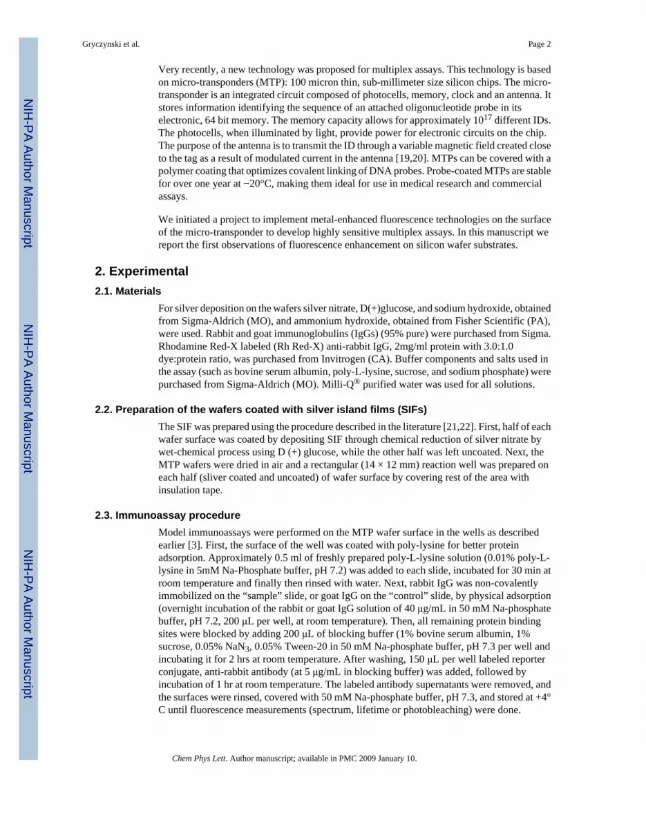
3.2. Fluorescence lifetimesThe interaction of excited molecules with silver nanoparticles modifies (increases) the radiativerate of fluorophore deactivation. The consequence is a higher quantum yield and shorterlifetime. Usually, the deactivation processes modify (increases) the nonradiative rate, whichdecreases both, quantum yield and lifetime. The intensity decays measured on silvered andunsilvered areas of the silicon wafer slide are presented in Figure 4. The decays werenormalized to compare the changes in the lifetimes. The decay parameters are summarized inTable 1.
The lifetime of Rh Red-X is significantly shorter in the presence of SIFs. The intensity decaybecomes also more heterogeneous. This is expected because fluorophores on the surface aredistributed in a slightly different plasmonic environment, i.e., at a different distance from eachother and having a different orientation to nearby nanoparticles such that “hot” micro-regionsexhibiting very strong interactions and enhancement are interspersed among areas where theenhancement is only modest.
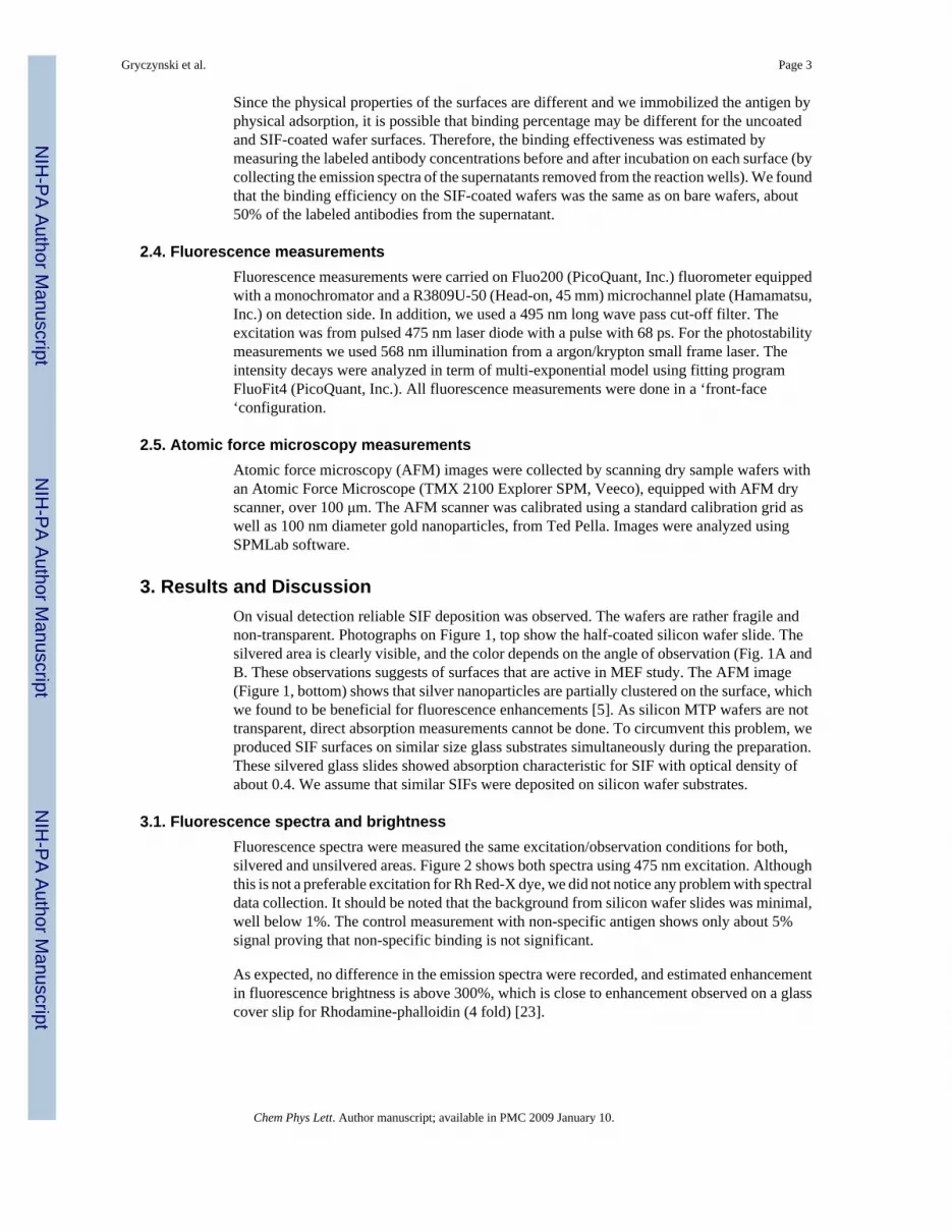
3.3. PhotostabilityWe previously observed increased photostability of fluorophores deposited on silvernanoparticels [23–27]. Both, decrease in the fluorescence lifetime and increase in the quantumyield lead to a significant reduction of photodegradation. First, decrease in lifetime decreasesthe bleaching; as shorter lifetime decreases the opportunity of free oxygen radicals to damagethe fluorophore. Second, the increase in quantum yield diminishes bleaching because it allowsa decrease of the excitation light intensity needed to maintain the same signal intensity. Acomparison of photostabilities observed on SIFs and on uncoated silicon wafer is shown inFigure 5. It is clear that the presence of silver nanoparticles allows extraction of larger numberof emitted photons from the studied system.
4. ConclusionsWe demonstrated that silver nanostructures such as SIFs can be deposited on silicon wafersubstrates and observed a three-fold fluorescence enhancement due to the presence of the SIFin a model immunoassay. A silicon surface is very attractive solid support in immunoassaybecause of its very low fluorescence background in addition to its suitability for fluorescenceenhancement approaches. In addition, the increase of the photostability of fluorophores onsilver nanostructures should be beneficial in future advanced silicon-based sensing devices formultiplex assays for genomics and drug screening.
AcknowledgmentsThis work was supported by NIH grants, R01 HG004364 and R43 CA 132547 to W.M., and by Texas EmergingTechnologies Fund grants (CCFT).
References1. Leitner A, Lippitsch ME, Draxler E, Rigler M, Aussenegg FR. Appl Phys B: Photophys Laser Chem
1985;36:105.2. Aussenegg FR, Leitner A, Lippitsch ME, Reinish H, Rigler M. Surf Sci 1987;139:935.3. Das P, Metju H. J Phys Chem 1985;89:4680.4. Sokolov K, Chumanov G, Cotton TM. Anal Chem 1987;70:3898. [PubMed: 9751028]
Gryczynski et al. Page 4
Chem Phys Lett. Author manuscript; available in PMC 2009 January 10.
NIH
-PA Author Manuscript
NIH
-PA Author Manuscript
NIH
-PA Author Manuscript

5. Lukomska J, Malicka J, Gryczynski I, Lakowicz JR. J Fluorescence 2004;14:417.6. Stich N, Gandhum A, Matushin V, Mayer C, Bauer G, Schalkhammer T. J Nanosci Nanotechnol
2001;1:397. [PubMed: 12914081]7. Lakowicz JR, Shen Y, D’Auria S, Malicka J, Fang J, Gryczynski Z, Gryczynski I. Anal Biochem
2002;301:261. [PubMed: 11814297]8. Lochner N, Lobmaier Ch, Wirth M, Leitner A, Pittner F, Gabor F. Eur J Pharm Biopharm 2003;56:469.
[PubMed: 14602192]9. Geddes CD, Parfenov A, Roll D, Gryczynski I, Malicka J, Lakowicz JR. Spectrochimica Acta Part A
2004;60:1977.10. Zhang J, Matveeva EG, Gryczynski I, Leonenko Z, Lakowicz JR. J Phys Chem 2005;109:7969.11. Kim W, Safonov VP, Shalaev VM, Armstrong RL. Phys Rev Lett 1999;82:4811.12. Goldys EM, Drozdowicz-Tomsia K, Xie F, Shtoyko T, Matveeva E, Gryczynski I, Gryczynski Z.
JACS 2007;129:12117.13. Shtoyko T, Matveeva EG, Fen Chang I, Gryczynski Z, Goldys EM, Gryczynski I. Anal Chem
2008;80:1962. [PubMed: 18288816]14. Malicka J, Gryczynski I, Gryczynski Z, Lakowicz JR. Anal Biochem 2003;315:57. [PubMed:
12672412]15. Fleischmann M, Hendra PJ, McQuillan AJ. Chem Phys Lett 1974;26:163.16. Jeanmaire DL, Van Duyne RP. J Electroanal Chem 1977;84:1.17. Kneipp K, Kneipp H, Itzkan I, Dasari RR, Feld MS. Curr Sci 1999;77:915.18. Lakowicz JR. Anal Biochem 2001;298:1. [PubMed: 11673890]19. Mandecki W, Ardelt B, Coradetti T, Davidowitz H, Flint J, Huang Z, Kopacka W, Lin X, Wang Z,
Darzynkiewicz Z. Cytometry Part A 2006;69A:1097.20. Lin X, Flint J, Azaro M, Coradetti T, Kopacka W, Streck D, Wang Z, Dermody J, Mandecki W. Clin
Chem 2007;53:1372. [PubMed: 17510306]21. Matveeva EG, Gryczynski Z, Malicka J, Gryczynski I, Lakowicz JR. Anal Biochem 2004;334:303.
[PubMed: 15494138]22. Matveeva EG, Gryczynski Z, Lakowicz JR. J Immunol Methods 2005;302:26. [PubMed: 15996681]23. Muthu P, Gryczynski I, Gryczynski Z, Talent J, Akopova I, Jain K, Borejdo J. Anal Biochem
2007;366:228. [PubMed: 17531183]24. Malicka J, Gryczynski I, Fang J, Kusba J, Lakowicz JR. J Fluorescence 2002;12:439.25. Parfenov A, Gryczynski I, Malicka J, Geddes CD, Lakowicz JR. J Phys Chem B 2003;107:8829.26. Malicka J, Gryczynski I, Lakowicz JR. Biopolymers 2004;74:263. [PubMed: 15150802]27. Muthu P, Gryczynski I, Gryczynski Z, Talent JM, Akopova I, Borejdo J. J Biomed Optics 2008;13:1.
Gryczynski et al. Page 5
Chem Phys Lett. Author manuscript; available in PMC 2009 January 10.
NIH
-PA Author Manuscript
NIH
-PA Author Manuscript
NIH
-PA Author Manuscript

Fig 1.Top: The silicon wafers half coated with silver island films (SIFs). The color observed at theSIF region depends on the observation angle as seen in photographs A and B. Bottom: TheAFM image of SIF coated silicon wafer with IgG.
Gryczynski et al. Page 6
Chem Phys Lett. Author manuscript; available in PMC 2009 January 10.
NIH
-PA Author Manuscript
NIH
-PA Author Manuscript
NIH
-PA Author Manuscript

Fig 2.Emission spectra of immobilized Rh-Red-X labeled IgG on silvered and unsilvered siliconwafers
Gryczynski et al. Page 7
Chem Phys Lett. Author manuscript; available in PMC 2009 January 10.
NIH
-PA Author Manuscript
NIH
-PA Author Manuscript
NIH
-PA Author Manuscript

Fig. 3.Photographs of fluorescent spots on silver coated area (left) and on bare Silicon wafer. Entireslide (silvered and unsilvered) was coated with Rh-Red-X labeled IgG. The excitation wasfrom a low power 532 laser. The photographs were taken through a long wave pass 570 nmfilter.
Gryczynski et al. Page 8
Chem Phys Lett. Author manuscript; available in PMC 2009 January 10.
NIH
-PA Author Manuscript
NIH
-PA Author Manuscript
NIH
-PA Author Manuscript
Fig. 4.normalized fluorescent intensity decays of Rh-Red-X labeled IgG deposited on silvered andunsilvered silicon wafer slide.
Gryczynski et al. Page 9
Chem Phys Lett. Author manuscript; available in PMC 2009 January 10.
NIH
-PA Author Manuscript
NIH
-PA Author Manuscript
NIH
-PA Author Manuscript
Fig. 5.Dependence of fluorescence intensity on the time of exposure to 568 nm Argon/Krypton laserillumination.
Gryczynski et al. Page 10
Chem Phys Lett. Author manuscript; available in PMC 2009 January 10.
NIH
-PA Author Manuscript
NIH
-PA Author Manuscript
NIH
-PA Author Manuscript

NIH
-PA Author Manuscript
NIH
-PA Author Manuscript
NIH
-PA Author Manuscript
Gryczynski et al. Page 11
Tabl
e 1
Mul
ti ex
pone
ntia
l ana
lysi
s of f
luor
esce
nce
inte
nsity
dec
ays o
f Rh-
Red
-X la
bele
d Ig
G in
abs
ence
and
pre
senc
e of
SIF
s
cond
ition
sτ 1
(ns)
α 1τ 2
(ns)
α 2T
3 (ns
)α 3
τ̄ (n
s)<τ
> (n
s)χ2
Silic
on w
afer
onl
y0.
140.
387
1.07
0.47
83.
600.
135
2.19
a1.
05b
1.02
Silic
on W
afer
-SIF
0.03
0.73
40.
330.
222
1.38
0.04
40.
690.
161.
06
a b
Chem Phys Lett. Author manuscript; available in PMC 2009 January 10.
Related Documents


