REVIEW ARTICLE published: 15 July 2014 doi: 10.3389/fnagi.2014.00138 Metal and complementary molecular bioimaging in Alzheimer’s disease Nady Braidy 1 , Anne Poljak 1,2,3 , Christopher Marjo 4 , Helen Rutlidge 4 , Anne Rich 4 , Tharusha Jayasena 1 , Nibaldo C. Inestrosa 5 and Perminder Sachdev 1,6 * 1 Faculty of Medicine, Centre for Healthy Brain Ageing, School of Psychiatry, University of New South Wales, Sydney, NSW, Australia 2 Bioanalytical Mass Spectrometry Facility, Mark Wainwright Analytical Centre, University of New South Wales, Sydney, NSW, Australia 3 Faculty of Medicine, School of Medical Sciences, University of New South Wales, Sydney, NSW, Australia 4 Solid State and Elemental Analysis Unit, Mark Wainwright Analytical Centre, University of New South Wales, Sydney, NSW, Australia 5 Faculty of Biological Sciences, Centre for Ageing and Regeneration, P. Catholic University of Chile, Santiago, Chile 6 Euroa Centre, Neuropsychiatric Institute, Prince of Wales Hospital, Sydney, NSW, Australia Edited by: Roger S. Chung, Macquarie University, Australia Reviewed by: Junming Wang, University of Mississippi Medical Center, USA Carlos Beas-Zarate, Universidad de Guadalajra Mexico, Mexico *Correspondence: Perminder Sachdev, NPI, Euroa Centre, UNSW School of Psychiatry, Prince of Wales Hospital, Barker Street, Randwick, Sydney, NSW 2031, Australia e-mail: [email protected] Alzheimer’s disease (AD) is the leading cause of dementia in the elderly, affecting over 27 million people worldwide. AD represents a complex neurological disorder which is best understood as the consequence of a number of interconnected genetic and lifestyle variables, which culminate in multiple changes to brain structure and function. These can be observed on a gross anatomical level in brain atrophy, microscopically in extracellular amyloid plaque and neurofibrillary tangle formation, and at a functional level as alterations of metabolic activity. At a molecular level, metal dyshomeostasis is frequently observed in AD due to anomalous binding of metals such as Iron (Fe), Copper (Cu), and Zinc (Zn), or impaired regulation of redox-active metals which can induce the formation of cytotoxic reactive oxygen species and neuronal damage. Metal chelators have been administered therapeutically in transgenic mice models for AD and in clinical human AD studies, with positive outcomes. As a result, neuroimaging of metals in a variety of intact brain cells and tissues is emerging as an important tool for increasing our understanding of the role of metal dysregulation in AD. Several imaging techniques have been used to study the cerebral metallo-architecture in biological specimens to obtain spatially resolved data on chemical elements present in a sample. Hyperspectral techniques, such as particle-induced X-ray emission (PIXE), energy dispersive X-ray spectroscopy (EDS), X-ray fluorescence microscopy (XFM), synchrotron X-ray fluorescence (SXRF), secondary ion mass spectrometry (SIMS), and laser ablation inductively coupled mass spectrometry (LA-ICPMS) can reveal relative intensities and even semi-quantitative concentrations of a large set of elements with differing spatial resolution and detection sensitivities. Other mass spectrometric and spectroscopy imaging techniques such as laser ablation electrospray ionization mass spectrometry (LA ESI-MS), MALDI imaging mass spectrometry (MALDI-IMS), and Fourier transform infrared spectroscopy (FTIR) can be used to correlate changes in elemental distribution with the underlying pathology in AD brain specimens. Taken together, these techniques provide new techniques to probe the pathobiology of AD and pave the way for identifying new therapeutic targets. The current review aims to discuss the advantages and challenges of using these emerging elemental and molecular imaging techniques, and highlight clinical achievements in AD research using bioimaging techniques. Keywords: LA-ICPMS, metals, Alzheimer’s disease, bioimaging, MALDI, FTIR INTRODUCTION Alzheimer’s disease (AD) is the most common progressive age-related neurodegenerative disorder, affecting about 2% of the population in the developed world (Mattson, 2004). Clinically, AD is characterized by devastating effects such as memory loss and decline in other cognitive abilities resulting in loss of independent functioning (Teri et al., 1989; Baddeley et al., 1991; Terry et al., 1991). Pathologically, AD is characterized by two main pathological hallmarks. These include extracellular amyloid plaques composed of insoluble amyloid beta (Aβ) protein produced by irregular cleavage of the amyloid precursor protein (APP), and intra-neuronal neurofibrillary tangles (NFTs) containing hyperphosphorylated tau protein (Khachaturian, 1985; Joachim et al., 1987; Selkoe et al., 1987; Mirra et al., 1991; Brun and Englund, 2002). Although the exact function of Aβ and APP remains unclear, recent studies suggest that APP may Frontiers in Aging Neuroscience www.frontiersin.org July 2014 | Volume 6 | Article 138 | 1 AGING NEUROSCIENCE

Welcome message from author
This document is posted to help you gain knowledge. Please leave a comment to let me know what you think about it! Share it to your friends and learn new things together.
Transcript
REVIEW ARTICLEpublished: 15 July 2014
doi: 10.3389/fnagi.2014.00138
Metal and complementary molecular bioimaging inAlzheimer’s diseaseNady Braidy1, Anne Poljak1,2,3, Christopher Marjo4, Helen Rutlidge4, Anne Rich4, Tharusha Jayasena1,
Nibaldo C. Inestrosa5 and Perminder Sachdev1,6*
1 Faculty of Medicine, Centre for Healthy Brain Ageing, School of Psychiatry, University of New South Wales, Sydney, NSW, Australia2 Bioanalytical Mass Spectrometry Facility, Mark Wainwright Analytical Centre, University of New South Wales, Sydney, NSW, Australia3 Faculty of Medicine, School of Medical Sciences, University of New South Wales, Sydney, NSW, Australia4 Solid State and Elemental Analysis Unit, Mark Wainwright Analytical Centre, University of New South Wales, Sydney, NSW, Australia5 Faculty of Biological Sciences, Centre for Ageing and Regeneration, P. Catholic University of Chile, Santiago, Chile6 Euroa Centre, Neuropsychiatric Institute, Prince of Wales Hospital, Sydney, NSW, Australia
Edited by:
Roger S. Chung, MacquarieUniversity, Australia
Reviewed by:
Junming Wang, University ofMississippi Medical Center, USACarlos Beas-Zarate, Universidad deGuadalajra Mexico, Mexico
*Correspondence:
Perminder Sachdev, NPI, EuroaCentre, UNSW School of Psychiatry,Prince of Wales Hospital, BarkerStreet, Randwick, Sydney,NSW 2031, Australiae-mail: [email protected]
Alzheimer’s disease (AD) is the leading cause of dementia in the elderly, affecting over27 million people worldwide. AD represents a complex neurological disorder whichis best understood as the consequence of a number of interconnected genetic andlifestyle variables, which culminate in multiple changes to brain structure and function.These can be observed on a gross anatomical level in brain atrophy, microscopicallyin extracellular amyloid plaque and neurofibrillary tangle formation, and at a functionallevel as alterations of metabolic activity. At a molecular level, metal dyshomeostasis isfrequently observed in AD due to anomalous binding of metals such as Iron (Fe), Copper(Cu), and Zinc (Zn), or impaired regulation of redox-active metals which can induce theformation of cytotoxic reactive oxygen species and neuronal damage. Metal chelatorshave been administered therapeutically in transgenic mice models for AD and in clinicalhuman AD studies, with positive outcomes. As a result, neuroimaging of metals in avariety of intact brain cells and tissues is emerging as an important tool for increasingour understanding of the role of metal dysregulation in AD. Several imaging techniqueshave been used to study the cerebral metallo-architecture in biological specimens toobtain spatially resolved data on chemical elements present in a sample. Hyperspectraltechniques, such as particle-induced X-ray emission (PIXE), energy dispersive X-rayspectroscopy (EDS), X-ray fluorescence microscopy (XFM), synchrotron X-ray fluorescence(SXRF), secondary ion mass spectrometry (SIMS), and laser ablation inductively coupledmass spectrometry (LA-ICPMS) can reveal relative intensities and even semi-quantitativeconcentrations of a large set of elements with differing spatial resolution and detectionsensitivities. Other mass spectrometric and spectroscopy imaging techniques such aslaser ablation electrospray ionization mass spectrometry (LA ESI-MS), MALDI imagingmass spectrometry (MALDI-IMS), and Fourier transform infrared spectroscopy (FTIR) canbe used to correlate changes in elemental distribution with the underlying pathology inAD brain specimens. Taken together, these techniques provide new techniques to probethe pathobiology of AD and pave the way for identifying new therapeutic targets. Thecurrent review aims to discuss the advantages and challenges of using these emergingelemental and molecular imaging techniques, and highlight clinical achievements in ADresearch using bioimaging techniques.
Keywords: LA-ICPMS, metals, Alzheimer’s disease, bioimaging, MALDI, FTIR
INTRODUCTIONAlzheimer’s disease (AD) is the most common progressiveage-related neurodegenerative disorder, affecting about 2% of thepopulation in the developed world (Mattson, 2004). Clinically,AD is characterized by devastating effects such as memoryloss and decline in other cognitive abilities resulting in lossof independent functioning (Teri et al., 1989; Baddeley et al.,1991; Terry et al., 1991). Pathologically, AD is characterized by
two main pathological hallmarks. These include extracellularamyloid plaques composed of insoluble amyloid beta (Aβ)protein produced by irregular cleavage of the amyloid precursorprotein (APP), and intra-neuronal neurofibrillary tangles (NFTs)containing hyperphosphorylated tau protein (Khachaturian,1985; Joachim et al., 1987; Selkoe et al., 1987; Mirra et al., 1991;Brun and Englund, 2002). Although the exact function of Aβ
and APP remains unclear, recent studies suggest that APP may
Frontiers in Aging Neuroscience www.frontiersin.org July 2014 | Volume 6 | Article 138 | 1
AGING NEUROSCIENCE

Braidy et al. Bioimaging in Alzheimer’s disease
play a crucial role in modulating neuronal survival, neuriteoutgrowth, synaptic plasticity and cell adhesion (Mattson, 1997).NFTs are not restricted to AD, and are also present in otherneurodegenerative diseases such as fronto-temporal dementia(Filley et al., 1994).
AD is a complex multifactorial disorder associated with irreg-ular protein aggregation (Pimplikar et al., 2010). Interestingly,accumulation of Aβ protein has been observed in cognitivelynormal brain, and sometimes an absence of Aβ deposits hasbeen noted in some postmortem in patients who had beenclinically diagnosed with AD (Edison et al., 2007). Moreover,various pathobiological mechanisms that are un-related toamyloid accumulation have been associated with the devel-opment and progression of AD. For instance, familial muta-tions in APP and presenilin-1 have been shown to induceautophagic dysfunction and impaired lysosomal proteolysis, cere-bral hypoperfusion, and AD (Lee et al., 2010; Pimplikar et al.,2010; Wong and Cuervo, 2010). Furthermore, excess or defi-ciency in several nutritional, environmental or genetic factorsmay also potentiate AD-like pathology, making the etiologyof this debilitating disorder difficult to elucidate (Russ et al.,2012).
Metals have a diversity of roles in medical biology encompass-ing both health and disease states (Olanow and Arendash, 1994;Oteiza et al., 2004; Farina et al., 2013; Jellinger, 2013; Grubmanet al., 2014). Metals such as lead and mercury cause well estab-lished neuropathologies. By contrast several types of metal ions,such as potassium, sodium and calcium are vital for normalnerve cell function. Several other metals (copper, zinc, iron, mag-nesium, manganese, cobalt) have functional roles in enzymesand proteins (Yokel, 2006; Molina-Holgado et al., 2007; Farinaet al., 2013). For example, brain iron is used by lipid and choles-terol synthesizing enzymes (Bartzokis, 2004) and up to 70% ofbrain iron is found in association with myelin (de los Monteroset al., 2000; Bartzokis, 2004). However, the careful control ofmetal ion compartmentalization and usage in the brain is crit-ical, so that metal associated toxicity is avoided. The etiologyof several neuropathologies includes a dysfunctional associationbetween otherwise important trace elements and particular pro-teins or peptides (Table 1). Consequently the pathophysiology ofmetal-protein interactions in neurodegenerative diseases gener-ally and in AD specifically is an area of growing interest. Divalentmetal cations accumulate in plaque deposits and the inflamma-tory and oxidative processes which are well documented in ADmay be mediated through chemistries involving metals (Table 2).However, the biochemistry of metal-protein interactions, sourcesof accumulating metals and chelation mechanisms are yet to befully explored in AD.
The toxicity of Aβ is linked to changes in its structure from thesoluble α-helical form to the insoluble β-pleated sheet form withconsequent plaque formation, in which metals such as copper,zinc and iron are sequestered (Lovell et al., 1998a). It is not clearwhat molecular events trigger plaque formation, a process whichmay begin much earlier in life than the clinical symptoms of AD(Almkvist and Winblad, 1999). However, dissolution of plaquewith metal chelating agents such as clioquinol is a potential newtreatment (Cherny et al., 2000, 2001), highlighting the significant
role that metals play in the etiology of this disease (Richardson,2004).
Metal ions, such as those sequestered in plaques, also par-ticipate in oxidation and free radical production (Figure 1)(Multhaup et al., 1996). These processes are well documentedin AD as are inflammatory processes, mediated by the presenceof activated microglia and astrocytes, which generate high lev-els of Aβ (Busciglio et al., 1993). Metals such as copper, zinc,iron and aluminum have been implicated as possible contribu-tors to neurodegenerative processes. In a few cases, well estab-lished links between metals and the function of specific proteinshave been demonstrated (Table 1). However, as a subset of allthe proteins studied in neuropathology, the metalloproteins areunder-represented (Dobson, 2001). Since metal containing activesites of proteins are often involved in oxidation reactions and/orfree radical generation, alterations to their biochemistry maybe of particular interest in neurodegenerative conditions. Linksbetween protein dysfunction and the role of metals in AD areemerging; (i) divalent metal cations are sequestered in Aβ plaques,(ii) oxidative processes are well documented in AD and metalcations, particularly iron, are a potential source of reactive species.Though metals are likely to play a significant role in AD andother inflammatory diseases, relatively little is known about theirsources, mechanisms of transport and chelation, biochemistryand interactions with proteins.
Apart from redox active metals associated with the pathologi-cal hallmarks of AD, the presence of other trace metals may alsobe related to impaired cognitive function in AD. Several toxicheavy metals, including arsenic, lead, mercury, and cadmium arepresent in the environment due to their high industrial demand(Park et al., 2014). These metals serve no biological function, andtheir accumulation in the brain is attributed to contact betweenhumans and the environment (Chowdhury and Chandra, 1987).Exposure to arsenic induces neuropathological and behavioralabnormalities similar to clinical features reported in AD and otherrelated neurodegenerative disorders (Gong and O’Bryant, 2010).Lead, which is a well-established neurotoxic pollutant, can inducetau hyperphosphorylation, white matter degeneration, cellularapoptosis, and changes in cellular morphology, and impairedneuronal function (Yun and Hoyer, 2000; Rahman et al., 2012).While cadmium can induce hepatic and renal toxicity, cadmiumand lead can also disrupt cholinergic transmission by reducing theturnover of the essential neurotransmitter, acetylcholine (Websterand Valois, 1981; Costa and Fox, 1983; Patra et al., 1999; Singhet al., 2012b). Inorganic mercury can mimic all the pathologi-cal hallmarks of AD in animal models (Saxe et al., 1999; Rusinaet al., 2006; Mutter et al., 2010). Under normal physiological con-ditions, sequestration of arsenic, lead, cadmium, and mercuryby the lateral choroid plexus represents a protective mechanismto prevent the influx of heavy metals from the blood and intothe brain. However, elevated levels of cadmium and mercury candirectly damage the choroid plexus, thus limiting the function ofthis endogenous defense mechanism (Gerhardsson et al., 2011).The toxicity of these metals in human neurodegenerative dis-orders is dependent on the concentration of the environmentalcontaminant, and chronic exposure to heavy metals can inducetoxicity at relatively low levels (Llobett et al., 2003).
Frontiers in Aging Neuroscience www.frontiersin.org July 2014 | Volume 6 | Article 138 | 2

Braidy et al. Bioimaging in Alzheimer’s disease
Table 1 | Metal Protein Interactions in Neurodegenerative Diseases.
Neurodegenerative disease Metal/s Metal binding protein with a link to
neurodegeneration
References
AD Zn2+, Cu2+, Fe2+, Al3+ Zn2+, Cu2+, Fe2+ are sequestered by Aβ
fibrils and oligomers leading to oxidativestress.Al3+ is potentially involved in the formationof NFTs
Rodella et al., 2008; Thinnes, 2010;Savelieff et al., 2013; Watt et al., 2013
Down’s syndrome Zn2+, Cu2+, Fe2+ Aβ fragment of the amyloid precursorprotein associates with a number of divalentmetals resulting in amyloid plaque formation
Kedziora et al., 1978; Prasher et al., 1998;Savelieff et al., 2013
Amyotrophic lateral sclerosis(Motor Neuron Disease)
Cu2+, Zn2+ Mutations in the metalloprotein superoxidedismutase (SOD) are associated with MND
Ince et al., 1994; Divers et al., 2006
Spongiform encephalopathies Cu2+ Prion Protein (Sc) Basu et al., 2007; Singh et al., 2009,2012a; Singh and Singh, 2010
Wilson’s disease Cu2+ Mutations in ATP7B, a putative Cu2+transporting gene product, leads todecrease in ceruloplasmin and consequentCu2+ accumulation
Peng et al., 2012; Walshe, 2012; Liggiet al., 2013; Ni et al., 2013
Friedreich’s ataxia Fe2+ Deficiency of mitochondrial protein frataxinis linked to altered Fe2+ homeostasis
Michael et al., 2006; Koeppen et al., 2007;Popescu et al., 2007; Lim et al., 2008
NBIA1(Hallerverden-SpatzSyndrome)
Fe2+ Brain Fe2+ deposition possibly inassociation with the protein synuclein
Valentin et al., 2006
Parkinson’s disease Fe2+Zn2+ Aggregates of α-synuclein form and releaseH2O2 in the presence of Fe2+Increased localized brain Ferritin levels
Dashdorj et al., 2012; Lucas, 2012; Binolfiand Fernandez, 2013; Björkblom et al.,2013
Aceruloplasminemia Cu2+, Fe2+ Mutations in the Cu2+ bindingmetalloprotein ceruloplasmin gene result inaccumulation of Fe2+ in neurons
Dunaief et al., 2005; Kono et al., 2006;Oide et al., 2006; Gonzalez-Cuyar et al.,2008
Effects of Mn2+ in otherneurodegenerative diseases
Mn2+ Manganism can lead to Huntington’sdisease and Parkinsonian-like symptoms.The precise mechanism how manganesecan damage the CNS is unclear
Bowman et al., 2011
The presence of sequestered biometals such as copper, zinc,and iron in β-amyloid plaques of AD-affected brain tissue, andthe presence of toxicological metals as potential pathologicalcofactors in AD, has led to a focus on metal imaging (Hutchinsonet al., 2005; Lelie et al., 2011; Pithadia and Lim, 2012; Stavitskiet al., 2013). We should note that not only metals, but a widerange of elements may be imaged, down to ultratrace levels, andat length scales from micron to tens of nanometers. In certaincases isotopes, and even oxidation state and the coordinationenvironment around specific elements can be imaged, potentiallyincreasing the scope of trace element research in neurologi-cal disease beyond what has been studied to date. Visualizingchanges in element concentration and matching them to anatom-ical and pathological features enhances our traditional approachto exploring the role of metal ions in neurological disease. Reviews
on metal imaging in neurobiology have been presented recently(Bourassa and Miller, 2012) and a comprehensive range of instru-mental techniques is available from McRae et al. (2009). However,the field continues to expand rapidly as spatially resolved ele-mental analysis is now a well-recognized method to investigatechemical changes associated with pathology in biological tissues.The experimental techniques used to obtain elemental informa-tion from tissues are quite diverse, with a range of differentcapabilities in spatial resolution, sensitivity and quantification.This review provides an overview of common instrumental tech-niques and examples of biological imaging with an emphasis onAlzheimer’s studies. Elemental imaging is the main topic of thisreview; although a selection of molecular imaging examples arepresented to demonstrate how these techniques can supplementthe elemental bioimaging. Selective colorimetric and fluorescent
Frontiers in Aging Neuroscience www.frontiersin.org July 2014 | Volume 6 | Article 138 | 3
Braidy et al. Bioimaging in Alzheimer’s disease
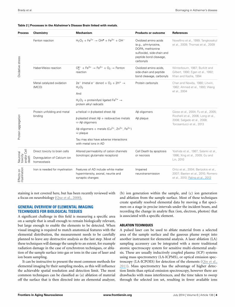
Table 2 | Processes in the Alzheimer’s Disease Brain linked with metals.
Process Chemistry Mechanism Products or outcome References
Oxi
dativ
est
ress
Fenton reaction H2O2 + Fe2+ → OH• + Fe3+ + OH− Oxidized amino acids(e.g., o/m-tyrosine,DOPA, methioninesulfoxide), side-chain andpeptide bond cleavage,carbonyls
Novellino et al., 1999; Tangkosakulet al., 2009; Thomas et al., 2009
Haber-Weiss reaction O•−2 + Fe3+ → Fe2+ + O2 → Fenton
reactionOxidized amino acids,side-chain and peptidebond cleavage, carbonyls
Winterbourn, 1987; Burkitt andGilbert, 1990; Egan et al., 1992;Khan and Kasha, 1994
Metal catalyzed oxidation(MCO)
2e− (metal e− donor) + O2 + 2H+ →H2O2
And
H2O2 + protein(lys) ligated Fe2+ →protein alkyl radical/s
Protein carbonyls Chan and Newby, 1980; Litwin,1982; Ahmed et al., 1993; Wanget al., 2004
Prot
ein
aggr
egat
ion
Protein unfolding and metalbinding
α-helical ➯ β-pleated sheet Aβ
β-pleated sheet Aβ + redox-active metals➯ Aβ oligomers
Aβ oligomers + metals (Cu2+, Zn2+, Fe2+)➯ plaque
Tau may also have adverse interactionswith metal ions in AD
Aβ oligomers
Aβ plaque
Giese et al., 2004; Fu et al., 2005;Ricchelli et al., 2006; Long et al.,2008; Salgado et al., 2008;Torosantucci et al., 2013
Neu
rona
lto
xici
tyan
d/or
Cel
lde
ath
Direct toxicity to brain cells
Dysregulation of Calcium ionhomeostasis
Altered permeability of cation channels(ionotropic glutamate receptors)
Cell Death by apoptosisor necrosis
Yoshida et al., 1987; Salanki et al.,1996; Xing et al., 2009; Gu andLin, 2010
Effe
cts
onm
yelin
atio
n Iron is needed for myelination Features of AD include white matterhyperintensity, axonal, neurite andsynaptic changes
Impairedneurotransmission
Ortiz et al., 2004; Bartzokis et al.,2007; Baeten et al., 2010; Romeroet al., 2010; Paling et al., 2012
staining is not covered here, but has been recently reviewed witha focus on neurobiology (Que et al., 2008).
GENERAL OVERVIEW OF ELEMENTAL IMAGINGTECHNIQUES FOR BIOLOGICAL TISSUESA significant challenge in this field is measuring a specific areaon a sample that is small enough to remain biologically relevant,but large enough to enable the elements to be detected. Whenvisual imaging is required to match anatomical features with theelemental distribution, the measurement needs to be carefullyplanned to leave any destructive analysis as the last step. Most ofthese techniques will damage the sample to an extent, for exampleradiation damage in the case of synchrotron techniques, or abla-tion of the sample surface into gas or ions in the case of laser andion beam sampling.
It can be instructive to present the most common methods forelemental imaging by their sampling modes, as this will influencethe achievable spatial resolution and detection limit. The mostcommon techniques can be classified as (a) ablation of materialoff the surface that is then directed into an elemental analyzer,
(b) ion generation within the sample, and (c) ion generationand ablation from the sample surface. Most of these techniquescreate spatially resolved elemental data by moving a flat speci-men on a stage in precise intervals under the incident beam, andrecording the change in analyte flux (ion, electron, photon) thatis associated with a specific element.
ABLATION TECHNIQUESA pulsed laser can be used to ablate material from a selectedarea of the sample surface and the gaseous plume swept intoanother instrument for elemental analysis. A laser ablation (LA)sampling accessory can be integrated with a more traditionalatomic spectroscopy system for sensitive multi-elemental analy-sis. These are usually inductively coupled plasma (ICP) systemsusing mass spectrometry (LA-ICPMS), or optical emission spec-troscopy (LA-ICPOES) for detection of the elements (Qin et al.,2011). Mass spectrometry has the advantage of higher detec-tion limits than optical emission spectroscopy, however there aredrawbacks with mass interferences, and the time taken to sweepthrough the selected ion set, resulting in fewer available ions
Frontiers in Aging Neuroscience www.frontiersin.org July 2014 | Volume 6 | Article 138 | 4

Braidy et al. Bioimaging in Alzheimer’s disease
FIGURE 1 | Involvement of metal dyshomeostasis in AD pathology.
Aggregation of Aβ can bind redox active metals such as copper,iron, and zinc in amyloid plaques. Sequestration of these biometalson Aβ fibrils and oligomers can potentiate synaptic dysfunction.Redox cycling of Cu2+/Cu+ and Fe3+/Fe2+ in the amyloid plaques
are capable of producing hydrogen peroxide (H2O2), which can enterthe cell. Through Fenton chemistry this can lead to the productionof hydroxyl radical (OH•) capable of inducing oxidative modificationsto both extracellular (i.e., proteins and lipids) as well as intracellular(DNA) macromolecules.
in order to create an image within a practical length of time.For example, imaging 6 metals across a 4 × 4 mm tissue sec-tion with a step size of 30 micron might take 12–24 h (Ketolaand Mauriala, 2012). Nevertheless, LA-ICPMS is by far the morecommon technique for elemental imaging of biometals and tox-icological metals in tissues than LA-ICPOES. Another variationon laser sampling is to detect the atomic excitation spectrumdirectly from the ablated plume, a technique known as laserinduced breakdown spectroscopy (LIBS) (Pareja et al., 2013). LAtechniques provide excellent analytical sensitivity in atmosphericor relatively low vacuum conditions. However, it is a destruc-tive technique, and delivering sufficient energy to the sample toallow detection tends to limit the spatial resolution. As a result,LA techniques are well suited for analysis of whole tissue sec-tions, but individual cells or pathological features such as amyloidplaques ∼20 micron are represented in an image as a single mea-sured point (Hare et al., 2010, 2011; Lear et al., 2012; Chou et al.,2014). Metal imaging of an individual cell or plaque requires thehigher resolution available from some of the techniques describedbelow.
SAMPLE IONIZATION TECHNIQUESHighly focused X-rays, electrons, or proton ion beams in a high-vacuum chamber can be used to eject an electron from the coreshell of an atom in the sample (Fahrni, 2007). The energy ofthe ejected electron can be measured using X-ray photoelectronspectroscopy (XPS) to determine the element from which it orig-inated. In certain cases, XPS is able to provide information onoxidation states and the chemical environment around an ele-ment, although spatial resolution is limited to 5–50 micron, anddetection limits are relatively poor (around 0.1 atomic%), virtu-ally ruling out the technique for trace metal studies (Pauneskuet al., 2006). The majority of sample ionization techniques uti-lize the secondary process where outer shell electrons fill the coreshell hole in the ion fluorescing X-rays with a characteristic wave-length for each element in the sample. When atoms are ionizedusing an electron beam, usually in an electron microscope, thetechnique is known as energy dispersive X-ray spectroscopy (EDXor EDS), sometimes referred to as electron photon micro analysis(EPMA). If ionization is achieved using an X-ray beam, the tech-nique is X-ray fluorescence microscopy (XFM) or synchrotron
Frontiers in Aging Neuroscience www.frontiersin.org July 2014 | Volume 6 | Article 138 | 5

Braidy et al. Bioimaging in Alzheimer’s disease
radiation micro-X-ray fluorescence (SR-μXRF) (Paunesku et al.,2006; Ralle and Lutsenko, 2009). Ionization can also be per-formed using a focused beam of protons in a technique calledparticle induced X-ray emission (PIXE). All of these techniquesare performed in high-vacuum environments, so steps such ascryopreservation or careful drying must be taken to protectbiological samples or specimen degassing that can reduce the per-formance of the instrument (de Silva et al., 2006; George et al.,2011; Ramsay et al., 2011; Weekley et al., 2013).
SECONDARY IONIZATION TECHNIQUESAblation-ionization directs a highly focused beam of ions, such asoxygen or cesium in the case of secondary ionization mass spec-trometry (SIMS), onto a tissue surface under vacuum (Altelaarand Piersma, 2010). This is a destructive process that resultsin ions being ejected from the surface. The ions are usuallydetected with a magnetic sector (NanoSIMS) or time-of-flight(TOF SIMS) mass spectrometer (Pacholski and Winograd, 1999;Eller et al., 2013; Fernandez-Lima et al., 2013). A recent review isavailable detailing the general capabilities of mass spectrometry-ablation techniques such as SIMS (Amstalden van Hove et al.,2010). The ability to focus ion beams down to very small spot sizesenables excellent spatial resolution, with features of 50 nanome-ters having been reported in the case of the NanoSIMS. However,micron to submicron imaging is more common since, in order togenerate sufficient secondary ions for detection with a very smallspot size, the ablation depth needs to increase. Submicron imag-ing at hundreds of nanometers is more common, and is sufficientfor cellular differentiation or observing small pathological fea-tures (Quintana et al., 2007; Musat et al., 2012). It is notable thatthe mass spectrometry techniques also enable more specializedimaging of isotopes across a surface, as well as providing moregeneral elemental imaging.
OTHER TECHNIQUESElectron energy loss spectroscopy (EELS) measures the energyloss due to scattering processes when a low energy, monoen-ergetic electron beam interacts with a sample. When used ina transmission electron microscope, EELS can provide atomic-scale resolution with excellent detection limits although biologicalapplications are limited (Quintana et al., 2000; Terada et al.,2002). There are a variety of X-ray techniques that have evolved asa result of the high-intensity X-ray sources available at numeroussynchrotron facilities around the world. X-ray Absorption NearEdge Structure (XANES), also known as Near edge X-ray absorp-tion fine structure (NEXAFS), is a technique where the elementcomposition change the absorption spectrum of the X-ray beam,providing information on elemental oxidation state and coordi-nation geometry around metal ions (Bourassa and Miller, 2012).Although potentially powerful, imaging of biological materialsusing this technique is still in development.
Magnetic resonance imaging (MRI) remains the most widelyused metal imaging technique in the clinical setting (Helpernet al., 2004). Although recent advances in MRI have made itpossible to detect the levels of iron at physiological concentra-tions, copper and manganese are still not widely detectable, sincethey are present in low concentrations in the brain (Schenck
and Zimmerman, 2004). Current MRI techniques exhibit lowerspatial resolution compared to elemental imaging techniquesmentioned above, but demonstrate the advantage of imaginglive patients rather than cryo-cut postmortem tissue sections(Schenck and Zimmerman, 2004).
Positron emission tomography (PET) is another techniquewhich facilitates in vivo medical imaging, usually of smallmolecules including glucose and more recently Aβ plaques usingPittsburg Compound B (PiB PET). More recently a novel metalimaging PET approach has been developed, using radioactivecoordination bis(thiosemicarbazonato)copper complex of 64Cu.This targets copper homeostasis and has been designed to bindselectively to amyloid plaques (Hickey et al., 2013). Copper radi-olabels are essential for increasing our understanding on of themechanisms of copper dyshomeostasis in AD.
COMBINED BIOIMAGING TECHNIQUES IN TISSUESECTIONSComplementary information regarding the role, uptake, trans-port, and storage of redox active metals associated with irregularprotein abnormalities can be obtained using a combination ofelemental imaging techniques, such as LA-ICPMS, and otherbiomolecular mass spectrometry imaging techniques such aslaser ablation coupled with electrospray ionization mass spec-trometry (LA-ESI-MS) or MALDI-IMS. While LA-ICPMS canbe employed to identify the specific protein-bound metals, ESI-MS/MALDI enables the identification of the structure, dynamicsand biological function of metal-protein complexes (Becker et al.,2008; Dobrowolska et al., 2008; Jakubowski et al., 2008; Wu et al.,2009).
ESI-MS is an ionization technique that is employed to detectpolar compounds within a biological specimen (Fenn et al.,1989). This method is used to identify molecules that do not con-tain an intrinsic ionizable site through formation of adduct ions.Molecules which exhibit sufficient dipole potential to interactwith a small anion or cation can be readily ionized and detectedusing ESI-MS. It is useful for the detection of triacylglycerols(TAGs) which contain long chain fatty acids. These moleculescan be ionized and quantified with sensitivity in the low pico-mole range due to the formation of lithiated adducts which areformed when chelated lithium ions non-covalently bond withthe carbonyl structures that are present in the infused solution(Han et al., 2000; Han and Gross, 2001). The benefits of usingESI-MS include more accurate quantification of lipid classes andsubclasses, a greater signal-to-noise ratio in comparison to othermass spectrometry techniques, and an almost linear relationshipbetween the relative intensities of molecular ions and the mass ofindividual lipids (Han and Gross, 1994).
MALDI-IMS allows the analysis of a diversity of biopolymerswith a variety of mass ranges. This approach has a lower spa-tial resolution but much higher mass range than TOF-SIMS,which is limited to identification of analytes with a molecu-lar mass of less than 1 kDa (McArthur et al., 2004). A vari-ety of analytes can be examined using MALDI-IMS, includingmetabolites, lipids, proteins, peptides, carbohydrates, and drugs.However, this method is limited by signal suppression effects.For instance, some analytes are more efficiently ionized during
Frontiers in Aging Neuroscience www.frontiersin.org July 2014 | Volume 6 | Article 138 | 6

Braidy et al. Bioimaging in Alzheimer’s disease
MALDI-IMS. These artifacts are not only due to their uniquechemical structure, but also to relative amounts present in thebiological tissue (Knochenmuss et al., 1998). Alternatively, pro-teins can be extracted from the tissue section using hydropho-bic materials, while preserving their specific location (Chaurandet al., 2004). Adaptation of MALDI-TOF to 2D and even 3Dtissue imaging applications has necessitated use of rapid firelong lived lasers, such as the 2 kHz Nd-YAG, to accommodatethe need to acquire 1000s of spectra across a tissue section.High end MALDI imaging mass spectrometers currently com-bine high mass resolution of 40,000 (1 ppm mass accuracy), widermass range (50–300,000 Da), spatial resolution down to 10 μm,and TOFTOF capabilities for peptide sequencing. This combi-nation of features allows detailed characterization of a diversityof tissue constituents, top-down sequencing of proteins as wellas the more commonly used bottom-up techniques involvingenzymatic/tryptic digestion and peptide sequencing, analysis ofposttranslational modifications such as glycosylation. A grow-ing body of literature recognizes the power of this approach(Cornett et al., 2007; Schuerenberg et al., 2007). A combina-tion of mass spectrometry imaging techniques using LA-ICPMSand detailed proteomics analysis can be performed using thincryo-cut sections of brain. MALDI-IMS is a relatively non-destructive technique so the tissue remaining after initial pro-teomic, metabolomic or lipidomic analysis can then be analyzedfor elemental composition using LA-ICPMS.
Fourier transform infrared spectroscopy (FTIR) is anothermolecular imaging tool that can be combined with LA-ICPMS.These tools have been used to image the secondary structure ofmetal-protein complexes (Haris and Severcan, 1999). FTIR is anon-destructive technique, allowing further analyses to providecomplementary information and to show spatial relationshipsbetween diverse analytes and/or functional groups, which mayprovide insight into biological/functional relatedness. The pro-tein’s FTIR consists of two main features: the Amide I band(∼1650 cm−1) which arises from the C=O stretching vibra-tion, and the Amide II band (∼1540 cm−1) which is due tothe N-H bending and C-N stretching vibrations of the peptidebackbone. The vibrational frequency of an aggregated protein isabout 1620–1625 cm−1, owing to its hydrophobic environment(Goormaghtigh et al., 2006; Miller et al., 2006). Apart from exam-ining the protein structure in vitro, FTIR can also be used todirectly investigate irregular protein misfolding and aggregationboth in vitro and in vivo. Protein aggregates are generally small,ranging from nanometers, to 20–30 μm for larger aggregates. Aswell, the spectral differences related to changes to protein confor-mation are subtle, requiring spectra with high signal to noise ratio(Choo et al., 1996; Miller et al., 2006). These difficulties have beenresolved using the greater brightness of a synchrotron infraredsource to directly assess protein aggregation and misfolding in ADtissue.
RECENT APPLICATIONS OF BIOIMAGING IN ALZHEIMER’SRESEARCHMetals have been shown to be associated with the pathogenesisof AD for over 50 years since the discovery of significant irondeposition in postmortem AD brain tissue using Prussian blue
stain (Goodman, 1953). Since then, other redox active metals havebeen implicated in AD, including copper, zinc, and aluminum.Several metal bioimaging strategies have been utilized to examinethe distribution of metals in human clinical AD brain tissue andAD mouse models to better understand the relationship betweenmetal dyshomeostasis and the etiology and progression of AD.
METALS AND Aβ PLAQUESIt has been well established that Aβ plaques are rich in metal ions(Opazo et al., 2002). These relatively high concentrations of met-als within the plaques compared to adjacent tissue have been reaf-firmed using a variety of bioimaging techniques. PIXE and XFMhas been used to show that both the outer and central regions ofthe Aβ plaques contain elevated levels of iron, copper and zincin human AD brain specimens (Lovell et al., 1998a,b). Althoughcopper and zinc binding sites are present on the Aβ peptide,iron does not appear to directly interact with Aβ (Atwood et al.,2000; Bush, 2003; Roberts et al., 2012). Recently, synchrotronX-ray absorption, diffraction, and tomography techniques havebeen used to identify the presence of biogenic magnetite and/ormaghematite in the plaque cores, implicating the likely role of anovel biomineralization process to account for the accumulationof iron in Aβ plaques (Collingwood et al., 2005, 2008).
Transgenic mouse models have provided additional advan-tages over postmortem human clinical AD specimens in the con-trol of both genetics and onset of AD-like symptoms. Using XFM,no abnormal increase in copper or iron were reported in with dis-ease progression in the PSAPP double transgenic mouse whichexpresses a chimeric mouse/human amyloid precursor protein(Mo/HuAPP695swe) and a mutant human presenilin 1 (PS1-dE9) both directed to CNS neurons. This mouse model developsamyloid pathology as well as learning and memory deficits by6 months of age, independent of signs of neurodegeneration(Leskovjan et al., 2011). Moreover, only a slight upregulation inzinc concentrations was reported at the late stages of the dis-ease. By contrast, the CRND mouse which expresses two familialmutations in the human Swedish (K595N/M596L) and Indiana(V717F) APP gene exhibited a 2–3-fold increase in the concen-tration of iron, copper, and zinc in the plaques after 6 monthsof age using PIXE. This unique mouse model develops diffuseand compact plaques by 10 weeks of age and Aβ depositioncontinues with advanced age (Rajendran et al., 2009). Similarfindings have been reported using LA-ICPMS analysis of plaquespresent in the brains of TASTPM mice, which carry both the APPK670N/M671L and PS1M146V mutation and develop plaques by4 months of age (Hutchinson et al., 2005).
METAL DYSHOMEOSTASIS IN AGING AND ADSince ageing is a major risk factor for the development of AD,examining the age-related changes in metal distribution is criticalfor understanding the role that metals play during pathologi-cal and physiological conditions. Using LA-ICPMS, one studyshowed that iron levels were increased in the “physiologically”aged brain of a non-transgenic mice (14 months) compared to ayoung (2 month) mice (Becker et al., 2010). These increases wereobserved in the substantia nigra, thalamus, and the CA1 regionof the hippocampus which are associated with development of
Frontiers in Aging Neuroscience www.frontiersin.org July 2014 | Volume 6 | Article 138 | 7
Braidy et al. Bioimaging in Alzheimer’s disease
neuropathologies. Remarkably, zinc levels remained unchangedand zinc-enrichment in the CA3 of the hippocampus was alreadydetected in young mice. This may be associated with the impor-tant role of zinc as an essential neuro-co-transmitter that isreleased from synaptic vesicles (Becker et al., 2010).
Evidence of metal dyshomeostasis has also been reported inAD. Studies using PIXE have shown increased levels of zinc inthe amygdala, hippocampus and neuropils of human AD brains(Danscher et al., 1997; Lovell et al., 1998a,b). This is likely to beassociated with the increased distribution of zinc enriched neu-rons (ZEN) which are located in these regions. ZENs maintainintracellular pools of zinc which is necessary as a neuromod-ulator and neuro-co-transmitter. One hypothesis suggests thatzinc released from these neurons can interact with Aβ and pro-mote aggregation (Bush et al., 1994; Frederickson et al., 2005).Zinc deficiency can also lead to excitotoxicity and neurodegener-ation (Sensi et al., 2009). Moreover, zinc reuptake is an energydependent process, and mitochondrial dysfunction can lead toincreased free zinc which can interact with Aβ and lead to furtherneurotoxicity (Mony et al., 2009).
Altered iron levels have also been suggested to play a promi-nent role in ageing and AD. Iron levels have been shown toincrease in the substantia nigra, motor rotex, hippocampus, basalganglia, putamen, cerebellum and cortex of human normal sub-jects during ageing (Connor et al., 1992; Deibel et al., 1996;Bartzokis et al., 2000). A similar increase was also reportediron, copper and zinc content was also reported in the PSAPPmouse model in the cortex and hippocampus, and coincided withincreased plaque formation using XFM (Leskovjan et al., 2011).Ferritin, the main protein responsible for iron storage, has beenshown to increase in the coronal region of human AD plaquesusing TEM and NanoSIMS (Quintana et al., 2006). It is likely thatferritin, which stores inactive iron (III) under normal physiolog-ical conditions may bind redox active iron (II) in the AD brainleading to cell death via oxidative stress.
METALS AND NFTsMetal dyshomeostasis may also play a role in the formation ofNFTs. A 10-fold increase in iron and a 6-fold increase in cop-per, with a smaller increase in zinc, have been previously reportedin NFTs (Morawski et al., 2005). Furthermore, hyperphospho-rylated tau, which forms paired helical filaments (PHFs) thatlead to NFTs, contains several binding domains which demon-strate some affinity to copper, and the presence of copper canenhance the formation of NFTs (Ma et al., 2006). Iron (III) canalso induce NFT formation similar to copper (Yamamoto et al.,2003). Apart from copper, iron and zinc, aluminum has also beenassociated with the development of AD since it was first identifiedin neurons with NFTs (Perl and Brody, 1980). However, increasedaluminum is also present in non-diseased brain tissue fixed withosmium tetroxide, which contains aluminum (Tokutake et al.,1995; Makjanic et al., 1997). Further work is warranted to validatethe involvement of aluminum in AD.
LIPIDOMIC STUDIES USING ESI/MSESI-MS techniques have been used to investigate the lipidomein patients with dementia. These studies have demonstrated
specific changes to the lipidome in the postmortem gray andwhite matter in the frontal, temporal and parietal cortex at theearliest clinically-recognizable stage of AD compared to cogni-tively normal control (Han et al., 2001, 2002). Specifically, plas-menylethanolamine (PlsEtn) mass was reduced by up to 40 mol%of total plasmalogens, in white matter in early AD subjects com-pared to age-matched controls. PlsEtn mass levels were depletedby 10% in the gray matter in patients with severe AD. Sulfatides,which form specialized components in the myelin sheath whichencapsulate neurons, were depleted by 93 and 58 mol% in grayand white matter, respectively, in AD patients in all brain regionsthat were investigated (Han et al., 2001, 2002). Additionally, a sig-nificant increase (>3 fold) in ceramide content was observed inthe white matter of all investigated brain regions during early AD.No significant changes have been observed in the levels of otherlipid classes, including phosphatidylglycerols, phosphatidylinos-itols, phosphatidylserines, and phosphatidic acids in early stagesof AD although significant reduction (∼15 mol%) of these lipidsoccurred in severe AD cases (Han et al., 2001, 2002). Takentogether, these results suggest that changes to the lipidome mayplay a vital role in the pathogenesis of AD and may be asso-ciated with early molecular and cellular events which occur inthe development of AD, such as neurodegeneration and synapticdysfunction.
MALDI-MS IMAGING IN ADRecently, MALDI-MS has been used to examine the spatial distri-bution and molecular contents of Aβ plaques. One study showedthat Aβ-(1–40) and Aβ-(1–42) are the most abundant amy-loid peptides in APP23 transgenic mice encoding the hAPP751with Swedish mutation (Rohner et al., 2005). In support of thiswork, other studies have shown that vascular amyloid is primar-ily composed of Aβ-(1–40) and Aβ-(1–42) (Miller et al., 1993).Additionally, Aβ-(1–40) is the major peptide that is found inaqueous cerebral cortical extracts from AD brains. By contrast,the insoluble amyloid Aβ-(1–42) peptide is primarily localized inthe senile plaque cores. Therefore, MALDI-MS can not only beused to identify known targets, but also facilitates mapping of thedifferent peptide targets with high precision and accuracy, thatis otherwise not possible when examining whole-brain extracts(Rohner et al., 2005).
FTIR SPECTROSCOPIC IMAGING IN ADIn AD, Aβ plaques are formed by the transformation of Aβ froma soluble form through to an oligomeric intermediate, culmi-nating in the formation of an aggregated, fibrillary structure (Ii,1995). The molecular mechanism which mediates the structuralchanges and cytotoxicity of Aβ during the aggregative processhas been previously investigated in several in vitro studies usingdichroism (CD) and nuclear magnetic resonance (NMR) to showthe structural conversion of Aβ from a soluble α-helical proteinto a fibrillar β-sheet protein (Barrow et al., 1992; Zhang andRich, 1997). FTIR spectroscopy has been essential to examine thespecific alignment of β-sheet strands within Aβ fibrils. A studyby Petty and Decatur (2005) showed that β-sheets are antipar-allel and in alignment across all strands (Petty and Decatur,2005). Recently, it has been suggested that oligomeric Aβ is more
Frontiers in Aging Neuroscience www.frontiersin.org July 2014 | Volume 6 | Article 138 | 8
Braidy et al. Bioimaging in Alzheimer’s disease
neurotoxic than Aβ fibrils and can form pore-like structures inlipid membranes that can disrupt ion homeostasis, culminatingin cell death. FTIR spectroscopy has shown that Aβ oligomersexhibit an antiparallel β-sheet structure, which is closely related tothat of bacterial outer membrane porins (Komatsu et al., 2007).
Apart from the Aβ protein, FTIR spectroscopy has also beenused to gain a greater understanding of the structural confor-mation of the tau protein, which is hyperphosphorylated in AD,leading to the formation of NFTs. In vitro FTIR analysis pro-vided confirmatory evidence that soluble tau protein is nativelyunfolded and composed of random coil structures, whilst PHFswhich are present in the AD brain have a greater level of β-structure (Berriman et al., 2003). These results provide evidenceto support the hypothesis that the repeat domain of tau (whichis located within the core of PHFs) displays an enhanced levelof β-structure during aggregation, while the N- and C-terminaldomains which venture away from the central PHF core arelargely random coils (Barghorn et al., 2004).
CONCLUSIONMetal imaging techniques are currently primed to facilitate anunderstanding of the pathobiology of AD, as well as identify-ing novel diagnostics and therapeutics. Bioimaging techniquesare important for elucidating the role of metals in neurodegen-erative diseases generally and AD in particular. Advancementsin methodology and improved spatial resolution and detectionsensitivities are essential for greater insight into the localiza-tion and distribution of metal ions at the cellular and tissuelevel, and their role in disease development and progression.A combination of other imaging techniques such as ESI- andMALDI-IMS, FTIR spectroscopy, and clinical techniques allow-ing in vivo analysis, such as MRI and PET, are invaluable inobtaining further understanding on the molecular mechanismsinvolved in the pathogenesis of AD and to confirm the diagno-sis of AD through the identification of unique biomarkers presentin the metabolome, lipidome and/or proteome. Additionally, thetechniques described in this review have the potential to followdisease progression in AD patients from early to severe stages andassess the effect of novel therapeutic interventions which mayretard, stop or reverse progressive neurodegeneration, the ulti-mate goal being a cure for this debilitating neurodegenerativedisorder.
AUTHOR CONTRIBUTIONSNady Braidy, Christopher Marjo, Anne Poljak, Tharusha Jayasena,Nibaldo C. Inestrosa, and Perminder Sachdev wrote the draft,reviewed and interpreted the bioimages. Helen Rutlidge and AnneRich processed the images. Nibaldo C. Inestrosa and PerminderSachdev provided the conceptual foundation of the review,writing of drafts and interpretation of data.
ACKNOWLEDGMENTSThis work was supported by a National Health and MedicalResearch Council of Australia Capacity Building Grant and aUNSW Faculty of Medicine Research Grant. Nady Braidy isthe recipient of an Alzheimer’s Australia Viertel FoundationPostdoctoral Research Fellowship, and the NHMRC Early Career
Postdoctoral Research Fellowship at the University of New SouthWales. Tharusha Jayasena is a recipient of the University of NewSouth Wales Postgraduate Award (UPA). The authors thank theRebecca Cooper Medical Research Foundation for their ongoingfinancial support.
REFERENCESAhmed, N., Liggins, J., and Furth, A. J. (1993). Metal-catalysed oxidation and post-
Amadori reactions of serum albumin and other model proteins. Biochem. Soc.Trans. 21:93S.
Almkvist, O., and Winblad, B. (1999). Early diagnosis of Alzheimer dementia basedon clinical and biological factors. Eur. Arch. Psychiatry Clin. Neurosci. 249(Suppl.3), 3–9. doi: 10.1007/PL00014171
Altelaar, A. F., and Piersma, S. R. (2010). Cellular imaging using matrix-enhancedand metal-assisted SIMS. Methods Mol. Biol. 656, 197–208. doi: 10.1007/978-1-60761-746-4_11
Amstalden van Hove, E. R., Smith, D. F., and Heeren, R. M. (2010). A concisereview of mass spectrometry imaging. J. Chromatogr. A 1217, 3946–3954. doi:10.1016/j.chroma.2010.01.033
Atwood, C. S., Scarpa, R. C., Huang, X. D., Moir, R. D., Jones, W. D., Fairlie,D. P., et al. (2000). Characterization of copper interactions with Alzheimeramyloid beta peptides: identification of an attomolar-affinity copper bindingsite on amyloid beta 1-42. J. Neurochem. 75, 1219–1233. doi: 10.1046/j.1471-4159.2000.0751219.x
Baddeley, A. D., Bressi, S., Della Sala, S., Logie, R., and Spinnler, H. (1991). Thedecline of working memory in Alzheimer’s disease. A longitudinal study. Brain114(pt 6), 2521–2542. doi: 10.1093/brain/114.6.2521
Baeten, K., Adriaensens, P., Hendriks, J., Theunissen, E., Gelan, J., Hellings, N.,et al. (2010). Tracking of myelin-reactive T cells in Experimental AutoimmuneEncephalomyelitis (EAE) animals using small particles of iron oxide and MRI.NMR Biomed. 23, 601–609. doi: 10.1002/nbm.1501
Barghorn, S., Davies, P., and Mandelkow, E. (2004). Tau paired helical fil-aments from Alzheimer’s disease brain and assembled in vitro are basedon beta-structure in the core domain. Biochemistry 43, 1694–1703. doi:10.1021/bi0357006
Barrow, C. J., Yasuda, A., Kenny, P. T., and Zagorski, M. G. (1992). Solution con-formations and aggregational properties of synthetic amyloid beta-peptides ofAlzheimer’s disease. Analysis of circular dichroism spectra. J. Mol. Biol. 225,1075–1093. doi: 10.1016/0022-2836(92)90106-T
Bartzokis, G. (2004). Age-related myelin breakdown: a developmental model ofcognitive decline and Alzheimer’s disease. Neurobiol. Aging 25, 5–18; authorreply 49–62. doi: 10.1016/j.neurobiolaging.2003.03.001
Bartzokis, G., Lu, P. H., Tishler, T. A., Fong, S. M., Oluwadara, B., Finn, J. P.,et al. (2007). Myelin breakdown and iron changes in Huntington’s disease:pathogenesis and treatment implications. Neurochem. Res. 32, 1655–1664. doi:10.1007/s11064-007-9352-7
Bartzokis, G., Sultzer, D., Cummings, J., Holt, L. E., Hance, D. B., Henderson,V. W., et al. (2000). In vivo evaluation of brain iron in Alzheimer diseaseusing magnetic resonance imaging. Arch. Gen. Psychiatry 57, 47–53. doi:10.1001/archpsyc.57.1.47
Basu, S., Mohan, M. L., Luo, X., Kundu, B., Kong, Q. Z., and Singh, N.(2007). Modulation of proteinase K-resistant prion protein in cells and infec-tious brain homogenate by redox iron: implications for prion replication anddisease pathogenesis. Mol. Biol. Cell 18, 3302–3312. doi: 10.1091/mbc.E07-04-0317
Becker, J. S., Dobrowolska, J., Zoriy, M., and Matusch, A. (2008). Imaging of ura-nium on rat brain sections using laser ablation inductively coupled plasmamass spectrometry: a new tool for the study of critical substructures affinedto heavy metals in tissues. Rapid Commun. Mass Spectrom. 22, 2768–2772. doi:10.1002/rcm.3673
Becker, J. S., Matusch, A., Palm, C., Salber, D., Morton, K. A., and Becker, S.(2010). Bioimaging of metals in brain tissue by laser ablation inductively cou-pled plasma mass spectrometry (LA-ICP-MS) and metallomics. Metallomics 2,104–111. doi: 10.1039/b916722f
Berriman, J., Serpell, L. C., Oberg, K. A., Fink, A. L., Goedert, M., and Crowther,R. A. (2003). Tau filaments from human brain and from in vitro assembly ofrecombinant protein show cross-beta structure. Proc. Natl. Acad. Sci. U.S.A. 100,9034–9038. doi: 10.1073/pnas.1530287100
Frontiers in Aging Neuroscience www.frontiersin.org July 2014 | Volume 6 | Article 138 | 9
Braidy et al. Bioimaging in Alzheimer’s disease
Binolfi, A., and Fernandez, C. O. (2013). “Interactions of alpha-Synuclein withmetal ions: new insights into the structural biology and bioinorganic chemistryof Parkinson’s disease,” in Brain Diseases and Metalloproteins, ed D. R. Brown(Pan Stanford Publishing), 327–366. doi: 10.4032/9789814364072
Björkblom, B., Adilbayeva, A., Maple-Grødem, J., Piston, D., Ökvist, M., Xu,X. M., et al. (2013). Parkinson disease protein DJ-1 binds metals and pro-tects against metal-induced cytotoxicity. J. Biol. Chem. 288, 22809–22820. doi:10.1074/jbc.M113.482091
Bourassa, M. W., and Miller, L. M. (2012). Metal imaging in neurodegenerativediseases. Metallomics 4, 721–738. doi: 10.1039/c2mt20052j
Bowman, A. B., Kwakye, G. F., Hernandez, E. H., and Aschner, M. (2011). Role ofmanganese in neurodegenerative diseases. J. Trace Elem. Med. Biol. 25, 191–203.doi: 10.1016/j.jtemb.2011.08.144
Brun, A., and Englund, E. (2002). Regional pattern of degeneration in Alzheimer’sdisease: neuronal loss and histopathological grading. Histopathology 41, 40–55.doi: 10.1046/j.1365-2559.2002.148910.x
Burkitt, M. J., and Gilbert, B. C. (1990). Model studies of the iron-catalysed Haber-Weiss cycle and the ascorbate-driven Fenton reaction. Free Radic. Res. Commun.10, 265–280. doi: 10.3109/10715769009149895
Busciglio, J., Gabuzda, D. H., Matsudaira, P., and Yankner, B. A. (1993). Generationof beta-amyloid in the secretory pathway in neuronal and nonneuronal cells.Proc. Natl. Acad. Sci. U.S.A. 90, 2092–2096. doi: 10.1073/pnas.90.5.2092
Bush, A. I. (2003). The metallobiology of Alzheimer’s disease. Trends Neurosci. 26,207–214. doi: 10.1016/S0166-2236(03)00067-5
Bush, A. I., Pettingell, W. H., Multhaup, G., Paradis, M. D., Vonsattel, J. P., Gusella,J. F., et al. (1994). Rapid induction of Alzheimer a-beta amyloid formation byzinc. Science 265, 1464–1467. doi: 10.1126/science.8073293
Chan, H. W., and Newby, V. K. (1980). Haemoprotein- and transition metal ion-catalysed oxidation of linoleic acid. Selectivity of the position of oxygenation.Biochim. Biophys. Acta 617, 353–362. doi: 10.1016/0005-2760(80)90001-6
Chaurand, P., Schwartz, S. A., Billheimer, D., Xu, B. G. J., Crecelius, A., andCaprioli, R. M. (2004). Integrating histology and imaging mass spectrometry.Anal. Chem. 76, 1145–1155. doi: 10.1021/ac0351264
Cherny, R. A., Atwood, C. S., Xilinas, M. E., Gray, D. N., Jones, W. D., McLean, C.A., et al. (2001). Treatment with a copper-zinc chelator markedly and rapidlyinhibits beta-amyloid accumulation in Alzheimer’s disease transgenic mice.Neuron 30, 665–676. doi: 10.1016/S0896-6273(01)00317-8
Cherny, R. A., Barnham, K. J., Lynch, T., Volitakis, I., Li, Q. X., McLean, C. A.,et al. (2000). Chelation and intercalation: complementary properties in a com-pound for the treatment of Alzheimer’s disease. J. Struct. Biol. 130, 209–216. doi:10.1006/jsbi.2000.4285
Choo, L. P., Wetzel, D. L., Halliday, W. C., Jackson, M., LeVine, S. M., and Mantsch,H. H. (1996). In situ characterization of beta-amyloid in Alzheimer’s diseasedtissue by synchrotron Fourier transform infrared Microspectroscopy. Biophys. J.71, 1672–1679. doi: 10.1016/S0006-3495(96)79411-0
Chou, J., Austin, C., Doble, P., Ben-Nissan, B., and Milthorpe, B. (2014). Traceelemental imaging of coralline hydroxyapatite by laser-ablation inductively cou-pled plasma-mass spectroscopy. J. Tissue Eng. Regen. Med. 8, 515–520. doi:10.1002/term.1544
Chowdhury, B. A., and Chandra, R. K. (1987). Biological and health implicationsof toxic heavy metal and essential trace element interactions. Prog. Food Nutr.Sci. 11, 55–113.
Collingwood, J. F., Chong, R. K. K., Kasama, T., Cervera-Gontard, L., Dunin-Borkowski, R. E., Perry, G., et al. (2008). Three-dimensional tomographicimaging and characterization of iron compounds within Alzheimer’s plaquecore material. J. Alzheimers Dis. 14, 235–245.
Collingwood, J. F., Mikhaylova, A., Davidson, M. R., Batich, C., Streit, W. J., Eskin,T., et al. (2005). High-resolution x-ray absorption spectroscopy studies of metalcompounds in neurodegenerative brain tissue. J. Phys. Conf. Ser. 17, 54–60. doi:10.1088/1742-6596/17/1/009
Connor, J. R., Snyder, B. S., Beard, J. L., Fine, R. E., and Mufson, E. J.(1992). Regional distribution of iron and iron-regulatory proteins in thebrain in aging and Alzheimers-disease. J. Neurosci. Res. 31, 327–335. doi:10.1002/jnr.490310214
Cornett, D. S., Reyzer, M. L., Chaurand, P., and Caprioli, R. M. (2007). MALDIimaging mass spectrometry: molecular snapshots of biochemical systems. Nat.Methods 4, 828–833. doi: 10.1038/nmeth1094
Costa, L. G., and Fox, D. A. (1983). A selective decrease of cholinergic mus-carinic receptors in the visual cortex of adult rats following developmental leadexposure. Brain Res. 276, 259–266. doi: 10.1016/0006-8993(83)90733-3
Danscher, G., Jensen, K. B., Frederickson, C. J., Kemp, K., Andreasen, A., Juhl,S., et al. (1997). Increased amount of zinc in the hippocampus and amygdalaof Alzheimer’s diseased brains: a proton-induced X-ray emission spectroscopicanalysis of cryostat sections from autopsy material. J. Neurosci. Methods 76,53–59. doi: 10.1016/S0165-0270(97)00079-4
Dashdorj, U., Tserensodnom, B., Bold, B., Chimedregzen, U., Komatsu, F., andKagawa, Y. (2012). Association of cumulative some heavy metal exposure withParkinson’s disease. Mov. Disord. 27, S1. doi: 10.1002/mds.25052
Deibel, M. A., Ehmann, W. D., and Markesbery, W. R. (1996). Copper, iron,and zinc imbalances in severely degenerated brain regions in Alzheimer’s dis-ease: possible relation to oxidative stress. J. Neurol. Sci. 143, 137–142. doi:10.1016/S0022-510X(96)00203-1
de los Monteros, A. E., Korsak, R. A., Tran, T., Vu, D., de Vellis, J., andEdmond, J. (2000). Dietary iron and the integrity of the developing rat brain:a study with the artificially-reared rat pup. Cell. Mol. Biol. (Noisy-le-grand) 46,501–515.
de Silva, R., Gutierrez, L. F., Raval, A. N., McVeigh, E. R., Ozturk, C., andLederman, R. J. (2006). X-ray fused with magnetic resonance imaging (XFM)to target endomyocardial injections: validation in a swine model of myocardialinfarction. Circulation 114, 2342–2350. doi: 10.1161/CIRCULATIONAHA.105.598524
Divers, T. J., Cummings, J. E., de Lahunta, A., Hintz, H. F., and Mohammed, H.O. (2006). Evaluation of the risk of motor neuron disease in horses fed a dietlow in vitamin E and high in copper and iron. Am. J. Vet. Res. 67, 120–126. doi:10.2460/ajvr.67.1.120
Dobrowolska, J., Dehnhardt, M., Matusch, A., Zoriy, M., Palomero-Gallagher, N.,Koscielniak, P., et al. (2008). Quantitative imaging of zinc, copper and lead inthree distinct regions of the human brain by laser ablation inductively coupledplasma mass spectrometry. Talanta 74, 717–723. doi: 10.1016/j.talanta.2007.06.051
Dobson, J. (2001). Nanoscale biogenic iron oxides and neurodegenerative disease.FEBS Lett. 496, 1–5. doi: 10.1016/S0014-5793(01)02386-9
Dunaief, J. L., Richa, C., Franks, E. P., Schultze, R. L., Aleman, T. S., Schenck, J.F., et al. (2005). Macular degeneration in a patient with aceruloplasminemia, adisease associated with retinal iron overload. Ophthalmology 112, 1062–1065.doi: 10.1016/j.ophtha.2004.12.029
Edison, P., Archer, H. A., Hinz, R., Hammers, A., Pavese, N., Tai, Y. F.,et al. (2007). Amyloid, hypometabolism, and cognition in Alzheimer dis-ease: an [11C]PIB and [18F]FDG PET study. Neurology 68, 501–508. doi:10.1212/01.wnl.0000244749.20056.d4
Egan, T. J., Barthakur, S. R., and Aisen, P. (1992). Catalysis of the Haber-Weissreaction by iron-diethylenetriaminepentaacetate. J. Inorg. Biochem. 48, 241–249.doi: 10.1016/0162-0134(92)84051-N
Eller, M. J., Verkhoturov, S. V., Della-Negra, S., and Schweikert, E. A. (2013). SIMSinstrumentation and methodology for mapping of co-localized molecules. Rev.Sci. Instrum. 84, 103706. doi: 10.1063/1.4824199
Fahrni, C. J. (2007). Biological applications of X-ray fluorescence microscopy:exploring the subcellular topography and speciation of transition met-als. Curr. Opin. Chem. Biol. 11, 121–127. doi: 10.1016/j.cbpa.2007.02.039
Farina, M., Avila, D. S., da Rocha, J. B., and Aschner, M. (2013). Metals, oxida-tive stress and neurodegeneration: a focus on iron, manganese and mercury.Neurochem. Int. 62, 575–594. doi: 10.1016/j.neuint.2012.12.006
Fenn, J. B., Mann, M., Meng, C. K., Wong, S. F., and Whitehouse, C. M. (1989).Electrospray ionization for mass-spectrometry of large biomolecules. Science246, 64–71. doi: 10.1126/science.2675315
Fernandez-Lima, F. A., Debord, J. D., Schweikert, E. A., Della-Negra, S.,Kellersberger, K. A., and Smotherman, M. (2013). Surface characterization ofbiological nanodomains using NP-ToF-SIMS. Surf. Interface Anal. 45, 1–8. doi:10.1002/sia.4901
Filley, C. M., Kleinschmidt-De Masters, B. K., and Gross, K. F. (1994). Non-Alzheimer fronto-temporal degenerative dementia. A neurobehavioral andpathologic study. Clin. Neuropathol. 13, 109–116.
Frederickson, C. J., Koh, J. Y., and Bush, A. I. (2005). The neurobiology ofzinc in health and disease. Nat. Rev. Neurosci. 6, 449–462. doi: 10.1038/nrn1671
Fu, L., Blakely, E., Marcus, M., Bjornstad, K., Due, B., Rehrmann, M., et al.(2005). Alzheimer’s disease β-amyloid (Aβ) mediates metal-dependent humanlens protein aggregation and light scattering. Investig. Ophthalmol. Vis. Sci. 46,2906–B459.
Frontiers in Aging Neuroscience www.frontiersin.org July 2014 | Volume 6 | Article 138 | 10
Braidy et al. Bioimaging in Alzheimer’s disease
George, A. K., Sonmez, M., Lederman, R. J., and Faranesh, A. Z. (2011). Robustautomatic rigid registration of MRI and X-ray using external fiducial mark-ers for XFM-guided interventional procedures. Med. Phys. 38, 125–141. doi:10.1118/1.3523621
Gerhardsson, L., Lundh, T., Londos, E., and Minthon, L. (2011). Cerebrospinalfluid/plasma quotients of essential and non-essential metals in patients withAlzheimer’s disease. J. Neural Transm. 118, 957–962. doi: 10.1007/s00702-011-0605-x
Giese, A., Levin, J., Bertsch, U., and Kretzschmar, H. (2004). Effect of metal ionson de novo aggregation of full-length prion protein. Biochem. Biophys. Res.Commun. 320, 1240–1246. doi: 10.1016/j.bbrc.2004.06.075
Gong, G., and O’Bryant, S. E. (2010). The arsenic exposure hypothesisfor Alzheimer disease. Alzheimer Dis. Assoc. Disord. 24, 311–316. doi:10.1097/WAD.0b013e3181d71bc7
Gonzalez-Cuyar, L. F., Perry, G., Miyajima, H., Atwood, C. S., Riveros-Angel, M.,Lyons, P. F., et al. (2008). Redox active iron accumulation in aceruloplasmine-mia. Neuropathology 28, 466–471. doi: 10.1111/j.1440-1789.2008.00901.x
Goodman, L. (1953). Alzheimer’s disease; a clinico-pathologic analysis of twenty-three cases with a theory on pathogenesis. J. Nerv. Ment. Dis. 118, 97–130. doi:10.1097/00005053-195308000-00001
Goormaghtigh, E., Ruysschaert, J. M., and Raussens, V. (2006). Evaluation ofthe information content in infrared spectra for protein secondary structuredetermination. Biophys. J. 90, 2946–2957. doi: 10.1529/biophysj.105.072017
Grubman, A., White, A. R., and Liddell, J. R. (2014). Mitochondrial metals asa potential therapeutic target in neurodegeneration. Br. J. Pharmacol. 171,2159–2173. doi: 10.1111/bph.12513
Gu, Q., and Lin, R. L. (2010). Heavy metals zinc, cadmium, and copper stimulatepulmonary sensory neurons via direct activation of TRPA1. J. Appl. Physiol. 108,891–897. doi: 10.1152/japplphysiol.01371.2009
Han, X. L., Abendschein, D. R., Kelley, J. G., and Gross, R. W. (2000). Diabetes-induced changes in specific lipid molecular species in rat myocardium. Biochem.J. 352, 79–89. doi: 10.1042/0264-6021:3520079
Han, X. L., and Gross, R. W. (1994). Electrospray-ionization mass spectroscopicanalysis of human erythrocyte plasma-membrane phospholipids. Proc. Natl.Acad. Sci. U.S.A. 91, 10635–10639. doi: 10.1073/pnas.91.22.10635
Han, X. L., and Gross, R. W. (2001). Quantitative analysis and molecular speciesfingerprinting of triacylglyceride molecular species directly from lipid extractsof biological samples by electrospray ionization tandem mass spectrometry.Anal. Biochem. 295, 88–100. doi: 10.1006/abio.2001.5178
Han, X. L., Holtzman, D. M., and McKeel, D. W. (2001). Plasmalogen deficiencyin early Alzheimer’s disease subjects and in animal models: molecular charac-terization using electrospray ionization mass spectrometry. J. Neurochem. 77,1168–1180. doi: 10.1046/j.1471-4159.2001.00332.x
Han, X. L., Holtzman, D. M., McKeel, D. W., Kelley, J., and Morris, J. C. (2002).Substantial sulfatide deficiency and ceramide elevation in very early Alzheimer’sdisease: potential role in disease pathogenesis. J. Neurochem. 82, 809–818. doi:10.1046/j.1471-4159.2002.00997.x
Hare, D., Austin, C., Doble, P., and Arora, M. (2011). Elemental bio-imaging oftrace elements in teeth using laser ablation-inductively coupled plasma-massspectrometry. J. Dent. 39, 397–403. doi: 10.1016/j.jdent.2011.03.004
Hare, D. J., George, J. L., Grimm, R., Wilkins, S., Adlard, P. A., Cherny, R. A.,et al. (2010). Three-dimensional elemental bio-imaging of Fe, Zn, Cu, Mn andP in a 6-hydroxydopamine lesioned mouse brain. Metallomics 2, 745–753. doi:10.1039/c0mt00039f
Haris, P. I., and Severcan, F. (1999). FTIR spectroscopic characterization of pro-tein structure in aqueous and non-aqueous media. J. Mol. Catal. B Enzym. 7,207–221. doi: 10.1016/S1381-1177(99)00030-2
Helpern, J. A., Jensen, J., Lee, S. P., and Falangola, M. F. (2004). QuantitativeMRI assessment of Alzheimer’s disease. J. Mol. Neurosci. 24, 45–48. doi:10.1385/JMN:24:1:045
Hickey, J. L., Lim, S., Hayne, D. J., Paterson, B. M., White, J. M., Villemagne, V. L.,et al. (2013). Diagnostic imaging agents for Alzheimer’s disease: copper radio-pharmaceuticals that target Aβ plaques. J. Am. Chem. Soc. 135, 16120–16132.doi: 10.1021/ja4057807
Hutchinson, R. W., Cox, A. G., McLeod, C. W., Marshall, P. S., Harper, A., Dawson,E. L., et al. (2005). Imaging and spatial distribution of beta-amyloid peptide andmetal ions in Alzheimer’s plaques by laser ablation-inductively coupled plasma-mass spectrometry. Anal. Biochem. 346, 225–233. doi: 10.1016/j.ab.2005.08.024
Ii, K. (1995). The role of beta-amyloid in the development of Alzheimer’s disease.Drugs Aging 7, 97–109. doi: 10.2165/00002512-199507020-00004
Ince, P. G., Shaw, P. J., Candy, J. M., Mantle, D., Tandon, L., Ehmann, W. D.,et al. (1994). Iron, selenium and glutathione-peroxidase activity are elevated insporadic motor-neuron disease. Neurosci. Lett. 182, 87–90. doi: 10.1016/0304-3940(94)90213-5
Jakubowski, N., Waentig, L., Hayen, H., Venkatachalam, A., von Bohlen, A., Roos, P.H., et al. (2008). Labelling of proteins with 2-(4-isothiocyanatobenzyl)-1,4,7,10-tetraazacyclododecane-1,4,7,10-tetraacetic acid and lanthanides and detectionby ICP-MS. J. Anal. Atom. Spectrom. 23, 1497–1507. doi: 10.1039/b800346g
Jellinger, K. A. (2013). The relevance of metals in the pathophysiology of neu-rodegeneration, pathological considerations. Int. Rev. Neurobiol. 110, 1–47. doi:10.1016/B978-0-12-410502-7.00002-8
Joachim, C. L., Morris, J. H., Selkoe, D. J., and Kosik, K. S. (1987). Tau epitopes areincorporated into a range of lesions in Alzheimer’s disease. J. Neuropathol. Exp.Neurol. 46, 611–622. doi: 10.1097/00005072-198711000-00001
Kedziora, J., Witas, H., Bartosz, G., Leyko, W., Jeske, J., and Rozynkowa, D. (1978).Down syndrome - transferrin parallels plasma iron changes. Experientia 34,712–713. doi: 10.1007/BF01947275
Ketola, R. A., and Mauriala, T. (2012). Mass spectrometric tools for cell and tissuestudies. Eur. J. Pharm. Sci. 46, 293–314. doi: 10.1016/j.ejps.2012.03.011
Khachaturian, Z. S. (1985). Diagnosis of Alzheimer’s disease. Arch. Neurol. 42,1097–1105. doi: 10.1001/archneur.1985.04060100083029
Khan, A. U., and Kasha, M. (1994). Singlet molecular oxygen in theHaber-Weiss reaction. Proc. Natl. Acad. Sci. U.S.A. 91, 12365–12367. doi:10.1073/pnas.91.26.12365
Knochenmuss, R., Karbach, V., Wiesli, U., Breuker, K., and Zenobi, R. (1998).The matrix suppression effect in matrix-assisted laser desorption/ionization:application to negative ions and further characteristics. Rapid Commun. MassSpectrom. 12, 529–534.
Koeppen, A. H., Michael, S. C., Knutson, M. D., Haile, D. J., Qian, J., Levi, S., et al.(2007). The dentate nucleus in Friedreich’s ataxia: the role of iron-responsiveproteins. Acta Neuropathol. 114, 163–173. doi: 10.1007/s00401-007-0220-y
Komatsu, H., Liu, L., Murray, I. V., and Axelsen, P. H. (2007). A mechanis-tic link between oxidative stress and membrane mediated amyloidogenesisrevealed by infrared spectroscopy. Biochim. Biophys. Acta 1768, 1913–1922. doi:10.1016/j.bbamem.2007.05.026
Kono, S., Suzuki, H., Takahashi, K., Takahashi, Y., Shirakawa, K., Murakawa, Y.,et al. (2006). Hepatic iron overload associated with a decreased serum cerulo-plasmin level in a novel clinical type of aceruloplasminemia. Gastroenterology131, 240–245. doi: 10.1053/j.gastro.2006.04.017
Lear, J., Hare, D. J., Fryer, F., Adlard, P. A., Finkelstein, D. I., and Doble, P. A. (2012).High-resolution elemental bioimaging of Ca, Mn, Fe, Co, Cu, and Zn employ-ing LA-ICP-MS and hydrogen reaction gas. Anal. Chem. 84, 6707–6714. doi:10.1021/ac301156f
Lee, J. H., Yu, W. H., Kumar, A., Lee, S., Mohan, P. S., Peterhoff, C. M.,et al. (2010). Lysosomal proteolysis and autophagy require presenilin 1 andare disrupted by Alzheimer-related PS1 mutations. Cell 141, 1146–1158. doi:10.1016/j.cell.2010.05.008
Lelie, H. L., Liba, A., Bourassa, M. W., Chattopadhyay, M., Chan, P. K., Gralla, E.B., et al. (2011). Copper and zinc metallation status of copper-zinc superoxidedismutase from amyotrophic lateral sclerosis transgenic mice. J. Biol. Chem. 286,2795–2806. doi: 10.1074/jbc.M110.186999
Leskovjan, A. C., Kretlow, A., Lanzirotti, A., Barrea, R., Vogt, S., and Miller,L. M. (2011). Increased brain iron coincides with early plaque forma-tion in a mouse model of Alzheimer’s disease. Neuroimage 55, 32–38. doi:10.1016/j.neuroimage.2010.11.073
Liggi, M., Murgia, D., Civolani, A., Demelia, E., Sorbello, O., and Demelia, L.(2013). The relationship between copper and steatosis in Wilson’s disease. Clin.Res. Hepatol. Gastroenterol. 37, 36–40. doi: 10.1016/j.clinre.2012.03.038
Lim, C. K., Kalinowski, D. S., and Richardson, D. R. (2008). Protection againsthydrogen peroxide-mediated cytotoxicity in Friedreich’s ataxia fibroblasts usingnovel iron chelators of the 2-pyridylcarboxaldehyde isonicotinoyl hydrazoneclass. Mol. Pharmacol. 74, 225–235. doi: 10.1124/mol.108.046847
Litwin, J. A. (1982). Transition metal-catalysed oxidation of 3,3’-diaminobenzidine[DAB] in a model system. Acta Histochem. 71, 111–117. doi: 10.1016/S0065-1281(82)80023-8
Llobett, J. M., Falco, G., Casas, C., Teixido, A., and Domingo, J. L. (2003).Concentrations of arsenic, cadmium, mercury, and lead in common foods and
Frontiers in Aging Neuroscience www.frontiersin.org July 2014 | Volume 6 | Article 138 | 11
Braidy et al. Bioimaging in Alzheimer’s disease
estimated daily intake by children, adolescents, adults, and seniors of Catalonia,Spain. J. Agric. Food Chem. 51, 838–842. doi: 10.1021/jf020734q
Long, X. F., Zhang, C. H., Cheng, J. J., and Bi, S. P. (2008). A novel method forstudy of the aggregation of protein induced by metal ion aluminum(III) usingresonance Rayleigh scattering technique. Spectrochim. Acta A 69, 71–77. doi:10.1016/j.saa.2007.03.011
Lovell, M. A., Robertson, J. D., Teesdale, W. J., Campbell, J. L., and Markesbery,W. R. (1998a). Copper, iron and zinc in Alzheimer’s disease senile plaques.J. Neurol. Sci. 158, 47–52. doi: 10.1016/S0022-510X(98)00092-6
Lovell, M. A., Robertson, J. D., Teesdale, W. J., Campbell, J. L., and Markesbery,W. R. (1998b). Use of micro-pixe in the analysis of zinc, copper and iron inAlzheimer’s disease senile plaques. Abstr. Pap. Am. Chem. Soc. 215, U969–U969.
Lucas, H. R. (2012). “Amyloids and copper biochemistry: effects of metal dyshome-ostasis in Alzheimer’s and Parkinson’s disease,” in Abstract Paper Presented atthe 244th ACS National Meeting and Exposition (Philadelphia, PA: PennsylvaniaConvention Center).
Ma, Q. F., Li, Y. M., Du, J. T., Liu, H. D., Kanazawa, K., Nemoto, T., et al. (2006).Copper binding properties of a tau peptide associated with Alzheimer’s dis-ease studied by CD, NMR, and MALDI-TOF MS. Peptides 27, 841–849. doi:10.1016/j.peptides.2005.09.002
Makjanic, J., McDonald, B., and Watt, F. (1997). Nuclear microscopy study of neu-rofibrillary tangles in Alzheimer’s disease. Nucl. Instrum. Methods Phys. Res. Sect.B 130, 439–443. doi: 10.1016/S0168-583X(97)00236-X
Mattson, M. P. (1997). Cellular actions of beta-amyloid precursor protein and itssoluble and fibrillogenic derivatives. Physiol. Rev. 77, 1081–1132.
Mattson, M. P. (2004). Pathways towards and away from Alzheimer’s disease.Nature 430, 631–639. doi: 10.1038/nature02621
McArthur, S. L., Vendettuoli, M. C., Ratner, B. D., and Castner, D. G. (2004).Methods for generating protein molecular ions in ToF-SIMS. Langmuir 20,3704–3709. doi: 10.1021/la0358419
McRae, R., Bagchi, P., Sumalekshmy, S., and Fahrni, C. J. (2009). In situ imaging ofmetals in cells and tissues. Chem. Rev. 109, 4780–4827. doi: 10.1021/cr900223a
Michael, S., Petrocine, S. V., Qian, J., Lamarche, J. B., Knutson, M. D., Garrick, M.D., et al. (2006). Iron and iron-responsive proteins in the cardiomyopathy ofFriedreich’s ataxia. Cerebellum 5, 257–267. doi: 10.1080/14734220600913246
Miller, D. L., Papayannopoulos, I. A., Styles, J., Bobin, S. A., Lin, Y. Y., Biemann, K.,et al. (1993). Peptide compositions of the cerebrovascular and senile plaque coreamyloid deposits of Alzheimers-disease. Arch. Biochem. Biophys. 301, 41–52.doi: 10.1006/abbi.1993.1112
Miller, L. M., Wang, Q., Telivala, T. P., Smith, R. J., Lanzirotti, A., and Miklossy,J. (2006). Synchrotron-based infrared and X-ray imaging shows focalized accu-mulation of Cu and Zn co-localized with beta-amyloid deposits in Alzheimer’sdisease. J. Struct. Biol. 155, 30–37. doi: 10.1016/j.jsb.2005.09.004
Mirra, S. S., Heyman, A., McKeel, D., Sumi, S. M., Crain, B. J., Brownlee, L.M., et al. (1991). The consortium to establish a registry for Alzheimer’s dis-ease (CERAD). Part II. standardization of the neuropathologic assessment ofAlzheimer’s disease. Neurology 41, 479–486. doi: 10.1212/WNL.41.4.479
Molina-Holgado, F., Hider, R. C., Gaeta, A., Williams, R., and Francis, P.(2007). Metals ions and neurodegeneration. Biometals 20, 639–654. doi:10.1007/s10534-006-9033-z
Mony, L., Kew, J. N. C., Gunthorpe, M. J., and Paoletti, P. (2009). Allostericmodulators of NR2B-containing NMDA receptors: molecular mechanisms andtherapeutic potential. Br. J. Pharmacol. 157, 1301–1317. doi: 10.1111/j.1476-5381.2009.00304.x
Morawski, M., Meinecke, C., Reinert, T., Dorffel, A. C., Riederer, P., Arendt,T., et al. (2005). Determination of trace elements in the human substan-tia nigra. Nucl. Instrum. Methods Phys. Res. Sect. B 231, 224–228. doi:10.1016/j.nimb.2005.01.061
Multhaup, G., Schlicksupp, A., Hesse, L., Beher, D., Ruppert, T., Masters, C. L.,et al. (1996). The amyloid precursor protein of Alzheimer’s disease in thereduction of copper(II) to copper(I). Science 271, 1406–1409. doi: 10.1126/sci-ence.271.5254.1406
Musat, N., Foster, R., Vagner, T., Adam, B., and Kuypers, M. M. (2012).Detecting metabolic activities in single cells, with emphasis on nanoSIMS.FEMS Microbiol. Rev. 36, 486–511. doi: 10.1111/j.1574-6976.2011.00303.x
Mutter, J., Curth, A., Naumann, J., Deth, R., and Walach, H. (2010). Doesinorganic mercury play a role in Alzheimer’s disease? A systematic reviewand an integrated molecular mechanism. J. Alzheimers Dis. 22, 357–374. doi:10.3233/JAD-2010-100705
Ni, W., Dong, Q. Y., Zhang, Y., and Wu, Z. Y. (2013). Zinc monotherapy anda low-copper diet are beneficial in patients with Wilson disease after livertransplantation. CNS Neurosci. Ther. 19, 905–907. doi: 10.1111/cns.12167
Novellino, L., Napolitano, A., and Prota, G. (1999). 5,6-dihydroxyindoles in theFenton reaction: a model study of the role of melanin precursors in oxidativestress and hyperpigmentary processes. Chem. Res. Toxicol. 12, 985–992. doi:10.1021/tx990020i
Oide, T., Yoshida, K., Kaneko, K., Ohta, M., and Arima, K. (2006). Ironoverload and antioxidative role of perivascular astrocytes in aceruloplas-minemia. Neuropathol. Appl. Neurobiol. 32, 170–176. doi: 10.1111/j.1365-2990.2006.00710.x
Olanow, C. W., and Arendash, G. W. (1994). Metals and free radicals in neurode-generation. Curr. Opin. Neurol. 7, 548–558. doi: 10.1097/00019052-199412000-00013
Opazo, C., Huang, X., Cherny, R. A., Moir, R. D., Roher, A. E., White, A. R.,et al. (2002). Metalloenzyme-like activity of Alzheimer’s disease beta-amyloid.Cu-dependent catalytic conversion of dopamine, cholesterol, and biologicalreducing agents to neurotoxic H(2)O(2). J. Biol. Chem. 277, 40302–40308. doi:10.1074/jbc.M206428200
Ortiz, E., Pasquini, J. M., Thompson, K., Felt, B., Butkus, G., Beard, J., et al. (2004).Effect of manipulation of iron storage, transport, or availability on myelin com-position and brain iron content in three different animal models. J. Neurosci.Res. 77, 681–689. doi: 10.1002/jnr.20207
Oteiza, P. I., Mackenzie, G. G., and Verstraeten, S. V. (2004). Metals in neurode-generation: involvement of oxidants and oxidant-sensitive transcription factors.Mol. Aspects Med. 25, 103–115. doi: 10.1016/j.mam.2004.02.012
Pacholski, M. L., and Winograd, N. (1999). Imaging with mass spectrometry.Chem. Rev. 99, 2977–3006. doi: 10.1021/cr980137w
Paling, D., Tozer, D., Wheeler-Kingshott, C., Kapoor, R., Miller, D. H., and Golay,X. (2012). Reduced R ‘(2) in multiple sclerosis normal appearing white matterand lesions may reflect decreased myelin and iron content. J. Neurol. Neurosurg.Psychiatry 83, 785–792. doi: 10.1136/jnnp-2012-302541
Pareja, J., Lopez, S., Jaramillo, D., Hahn, D. W., and Molina, A. (2013).Laser ablation-laser induced breakdown spectroscopy for the measurementof total elemental concentration in soils. Appl. Opt. 52, 2470–2477. doi:10.1364/AO.52.002470
Park, J. H., Lee, D. W., Park, K. S., and Joung, H. (2014). Serum trace metal levelsin Alzheimer’s disease and normal control groups. Am. J. Alzheimer’s Dis. OtherDemen. 29, 76–83. doi: 10.1177/1533317513506778
Patra, R. C., Swarup, D., and Senapati, S. K. (1999). Effects of cadmium on lipidperoxides and superoxide dismutase in hepatic, renal and testicular tissue ofrats. Vet. Hum. Toxicol. 41, 65–67.
Paunesku, T., Vogt, S., Maser, J., Lai, B., and Woloschak, G. (2006). X-ray fluo-rescence microprobe imaging in biology and medicine. J. Cell. Biochem. 99,1489–1502. doi: 10.1002/jcb.21047
Peng, F. Y., Lutsenko, S., Sun, X. K., and Muzik, O. (2012). Imaging coppermetabolism imbalance in Atp7b (-/-) knockout mouse model of Wilson’s dis-ease with PET-CT and orally administered (CuCl2)-Cu-64. Mol. Imaging Biol.14, 600–607. doi: 10.1007/s11307-011-0532-0
Perl, D. P., and Brody, A. R. (1980). Alzheimers-disease - X-ray spectrometric evi-dence of aluminum accumulation in neurofibrillary tangle-bearing neurons.Science 208, 297–299. doi: 10.1126/science.7367858
Petty, S. A., and Decatur, S. M. (2005). Experimental evidence for the reorganiza-tion of beta-strands within aggregates of the a beta(16-22) peptide. J. Am. Chem.Soc. 127, 13488–13489. doi: 10.1021/ja054663y
Pimplikar, S. W., Nixon, R. A., Robakis, N. K., Shen, J., and Tsai, L. H.(2010). Amyloid-independent mechanisms in Alzheimer’s disease pathogenesis.J. Neurosci. 30, 14946–14954. doi: 10.1523/JNEUROSCI.4305-10.2010
Pithadia, A. S., and Lim, M. H. (2012). Metal-associated amyloid-betaspecies in Alzheimer’s disease. Curr. Opin. Chem. Biol. 16, 67–73. doi:10.1016/j.cbpa.2012.01.016
Popescu, B. F., Pickering, I. J., George, G. N., and Nichol, H. (2007). The chem-ical form of mitochondrial iron in Friedreich’s ataxia. J. Inorg. Biochem. 101,957–966. doi: 10.1016/j.jinorgbio.2007.03.004
Prasher, V. P., Gosling, P., and Blair, J. (1998). Role of iron in Alzheimer-typedementia in Down syndrome. Int. J. Geriatr. Psychiatry 13, 818–819.
Qin, Z., Caruso, J. A., Lai, B., Matusch, A., and Becker, J. S. (2011). Trace metalimaging with high spatial resolution: applications in biomedicine. Metallomics3, 28–37. doi: 10.1039/c0mt00048e
Frontiers in Aging Neuroscience www.frontiersin.org July 2014 | Volume 6 | Article 138 | 12

Braidy et al. Bioimaging in Alzheimer’s disease
Que, E. L., Domaille, D. W., and Chang, C. J. (2008). Metals in neurobiology:probing their chemistry and biology with molecular imaging. Chem. Rev. 108,1517–1549. doi: 10.1021/cr078203u
Quintana, C., Bellefqih, S., Laval, J. Y., Guerquin-Kern, J. L., Wu, T. D., Avila, J.,et al. (2006). Study of the localization of iron, ferritin, and hemosiderin inAlzheimer’s disease hippocampus by analytical microscopy at the subcellularlevel. J. Struct. Biol. 153, 42–54. doi: 10.1016/j.jsb.2005.11.001
Quintana, C., Lancin, M., Marhic, C., Perez, M., Martin-Benito, J., Avila, J.,et al. (2000). Initial studies with high resolution TEM and electron energyloss spectroscopy studies of ferritin cores extracted from brains of patientswith progressive supranuclear palsy and Alzheimer disease. Cell Mol. Biol. 46,807–820.
Quintana, C., Wu, T. D., Delatour, B., Dhenain, M., Guerquin-Kern, J. L.,and Croisy, A. (2007). Morphological and chemical studies of pathologicalhuman and mice brain at the subcellular level: correlation between light,electron, and nanosims microscopies. Microsc. Res. Tech. 70, 281–295. doi:10.1002/jemt.20403
Rahman, A., Khan, K. M., Al-Khaledi, G., Khan, I., and Attur, S. (2012). Early post-natal lead exposure induces tau phosphorylation in the brain of young rats. ActaBiol. Hung. 63, 411–425. doi: 10.1556/ABiol.63.2012.4.1
Rajendran, R., Ren, M. Q., Ynsa, M. D., Casadesus, G., Smith, M. A., Perry, G.,et al. (2009). A novel approach to the identification and quantitative elementalanalysis of amyloid deposits-Insights into the pathology of Alzheimer’s disease.Biochem. Biophys. Res. Commun. 382, 91–95. doi: 10.1016/j.bbrc.2009.02.136
Ralle, M., and Lutsenko, S. (2009). Quantitative imaging of metals in tissues.Biometals 22, 197–205. doi: 10.1007/s10534-008-9200-5
Ramsay, S. C., Cassidy, N., de Jonge, M. D., Howard, D. L., Paterson, D., andKetheesan, N. (2011). Examination of trafficking of phagocytosed colloid par-ticles in neutrophils using synchrotron-based X-ray fluorescence microscopy(XFM). J. Biol. Phys. 37, 493–506. doi: 10.1007/s10867-011-9233-9
Ricchelli, F., Buggio, R., Drago, D., Salmona, M., Forloni, G., Negro, A., et al.(2006). Aggregation/fibrillogenesis of recombinant human prion protein andGerstmann-Straussler-Scheinker disease peptides in the presence of metal ions.Biochemistry. 45, 6724–6732. doi: 10.1021/bi0601454
Richardson, D. R. (2004). Novel chelators for central nervous system disorders thatinvolve alterations in the metabolism of iron and other metal ions. Ann. N.Y.Acad. Sci. 1012, 326–341. doi: 10.1196/annals.1306.026
Roberts, B. R., Ryan, T. M., Bush, A. I., Masters, C. L., and Duce, J. A. (2012).The role of metallobiology and amyloid-ss peptides in Alzheimer’s disease.J. Neurochem. 120, 149–166. doi: 10.1111/j.1471-4159.2011.07500.x
Rodella, L. F., Ricci, F., Borsani, E., Stacchiotti, A., Foglio, E., Favero, G., et al.(2008). Aluminium exposure induces Alzheimer’s disease-like histopathologicalalterations in mouse brain. Histol. Histopathol. 23, 433–439.
Rohner, T. C., Staab, D., and Stoeckli, M. (2005). MALDI mass spectrometricimaging of biological tissue sections. Mech. Ageing Dev. 126, 177–185. doi:10.1016/j.mad.2004.09.032
Romero, M. D. C., Pliego-Rivero, F. B., Altamirano, B. M., and Otero,G. A. (2010). Effect of postlactation iron deficiency on the compositionof fatty acids of whole brain myelin. Nutr. Neurosci. 13, 237–244. doi:10.1179/147683010X12611460764606
Rusina, R., Matej, R., Urban, P., Cabelkova, Z., Nerudova, J., and Cikrt, M. (2006).Regional mercury concentration in Alzheimer’s disease brains. Eur. J. Neurol.13, 169–294. doi: 10.1111/j.1468-1331.2006.01622.x
Russ, T. C., Batty, G. D., Hearnshaw, G. F., Fenton, C., and Starr, J. M. (2012).Geographical variation in dementia: systematic review with meta-analysis. Int.J. Epidemiol. 41, 1012–1032. doi: 10.1093/ije/dys103
Salanki, J., Carpenter, D. O., Gyori, J., and Rubakhin, S. (1996). Toxic heavy met-als potentiate ligand-gated C1-currents in gastropoda neurons. Neurobiology 4,277–278.
Salgado, E. N., Lewis, R. A., Faraone-Mennella, J., and Tezcan, F. A. (2008). Metal-mediated self-assembly of protein superstructures: influence of secondary inter-actions on protein oligomerization and aggregation. J. Am. Chem. Soc. 130,6082. doi: 10.1021/ja8012177
Savelieff, M. G., Lee, S., Liu, Y., and Lim, M. H. (2013). Untangling amyloid-beta, tau, and metals in Alzheimer’s disease. ACS Chem. Biol. 8, 856–865. doi:10.1021/cb400080f
Saxe, S. R., Wekstein, M. W., Kryscio, R. J., Henry, R. G., Cornett, C. R., Snowdon,D. A., et al. (1999). Alzheimer’s disease, dental amalgam and mercury. J. Am.Dent. Assoc. 130, 191–199. doi: 10.14219/jada.archive.1999.0168
Schenck, J. F., and Zimmerman, E. A. (2004). High-field magnetic resonanceimaging of brain iron: birth of a biomarker? NMR Biomed. 17, 433–445. doi:10.1002/nbm.922
Schuerenberg, M., Luebbert, C., Deininger, S. O., Ketterlinus, R., and Suckau, D.(2007). MALDI tissue imaging: mass spectrometric localization of biomarkersin tissue slices. Nat. Methods 4, 3–4.
Selkoe, D. J., Bell, D. S., Podlisny, M. B., Price, D. L., and Cork, L. C. (1987).Conservation of brain amyloid proteins in aged mammals and humans withAlzheimer’s disease. Science 235, 873–877. doi: 10.1126/science.3544219
Sensi, S. L., Paoletti, P., Bush, A. I., and Sekler, I. (2009). Zinc in the physiology andpathology of the CNS. Nat. Rev. Neurosci. 10, 780–791. doi: 10.1038/nrn2734
Singh, A., Mohan, M. L., Isaac, A. O., Luo, X., Petrak, J., Vyoral, D., et al. (2009).Prion protein modulates cellular iron uptake: a novel function with implica-tions for prion disease pathogenesis. PLoS ONE 4:e4468. doi: 10.1371/jour-nal.pone.0004468
Singh, A., Qing, L. T., Kong, Q. Z., and Singh, N. (2012a). Change in the char-acteristics of ferritin induces iron imbalance in prion disease affected brains.Neurobiol. Dis. 45, 930–938. doi: 10.1016/j.nbd.2011.12.012
Singh, N., and Singh, A. (2010). “Redox-iron and prion disease pathogenesis,” inCongress Abstract Presented at PRION 2010, Abstract Ppo5-18: Basic Mechanismsof Neurodegeneration and Pathology (Salzburg: Salzburg Congress Center).
Singh, P. K., Baxi, D., Diwedi, R., and Ramachandran, A. V. (2012b). Prior cadmiumexposure improves glucoregulation in diabetic rats but exacerbates effects onmetabolic dysregulation, oxidative stress, and hepatic and renal toxicity. DrugChem. Toxicol. 35, 167–177. doi: 10.3109/01480545.2011.589450
Stavitski, E., Smith, R. J., Bourassa, M. W., Acerbo, A. S., Carr, G. L., and Miller,L. M. (2013). Dynamic full-field infrared imaging with multiple synchrotronbeams. Anal. Chem. 85, 3599–3605. doi: 10.1021/ac3033849
Tangkosakul, T., Tantimongcolwat, T., Isarankura-Na-Ayudhya, C., Mejare, M.,Bulow, L., and Prachayasittikul, V. (2009). Native and chimeric metal-bindinglactate dehydrogenase as detection and protection tools for oxidative stressinduced by Fenton’s reaction. Excli J. 8, 1–11.
Terada, S., Aoyama, T., Yano, F., and Mitsui, Y. (2002). Time-resolved acquisitiontechnique for spatially-resolved electron energy-loss spectroscopy by energy-filtering TEM. J. Electron Microsc. 51, 291–296. doi: 10.1093/jmicro/51.5.291
Teri, L., Borson, S., Kiyak, H. A., and Yamagishi, M. (1989). Behavioral distur-bance, cognitive dysfunction, and functional skill. Prevalence and relationshipin Alzheimer’s disease. J. Am. Geriatr. Soc. 37, 109–116.
Terry, R. D., Masliah, E., Salmon, D. P., Butters, N., DeTeresa, R., Hill, R., et al.(1991). Physical basis of cognitive alterations in Alzheimer’s disease: synapseloss is the major correlate of cognitive impairment. Ann. Neurol. 30, 572–580.doi: 10.1002/ana.410300410
Thinnes, F. P. (2010). Opening cell membrane-standing type-1 VDAC/porinchannels by trivalent aluminium-a factor in amyotrophic lateral scle-rosis and Alzheimer’s disease? Mol. Genet. Metab. 101, 299–300. doi:10.1016/j.ymgme.2010.06.004
Thomas, C., Mackey, M. M., Diaz, A. A., and Cox, D. P. (2009). Hydroxyl radical isproduced via the Fenton reaction in submitochondrial particles under oxidativestress: implications for diseases associated with iron accumulation. Redox Rep.14, 102–108. doi: 10.1179/135100009X392566
Tokutake, S., Nagase, H., Morisaki, S., and Oyanagi, S. (1995). Aluminum detectedin senile plaques and neurofibrillary tangles is contained in lipofuscin gran-ules with silicon, probably as aluminosilicate. Neurosci. Lett. 185, 99–102. doi:10.1016/0304-3940(94)11234-A
Torosantucci, R., Sharov, V. S., van Beers, M., Brinks, V., Schoneich, C., andJiskoot, W. (2013). Identification of oxidation sites and covalent cross-linksin metal catalyzed oxidized interferon beta-1a: potential implications forprotein aggregation and immunogenicity. Mol. Pharm. 10, 2311–2322. doi:10.1021/mp300665u
Valentin, A., Lin, J., Samuel, M., Hulse, G., Alarcon, H., Dervos, H., et al. (2006).Micro-electrode recordings from Globus Pallidus Internus (GPi) using generalanaesthesia in Neurodegeneration with Brain Iron Accumulation 1 (NBIA1).Mov. Disord. 21, S614–S708. doi: 10.1002/mds.21249
Walshe, J. M. (2012). Serum ‘free’ copper in Wilson disease. QJM 105, 419–423.doi: 10.1093/qjmed/hcr229
Wang, X., Zhang, Q., Guo, Q., Lou, Y., Yang, L., and Wang, Y. (2004). Iron-catalysedpropylene epoxidation by nitrous oxide: dramatic shift of allylic oxidationto epoxidation by the modification with alkali metal salts. Chem. Commun.1396–1397. doi: 10.1039/b402839b
Frontiers in Aging Neuroscience www.frontiersin.org July 2014 | Volume 6 | Article 138 | 13
Braidy et al. Bioimaging in Alzheimer’s disease
Watt, A. D., Villemagne, V. L., and Barnham, K. J. (2013). Metals, membranes,and amyloid-beta oligomers: key pieces in the Alzheimer’s disease puzzle?J. Alzheimer’s Dis. 33(Suppl. 1), S283–S293. doi: 10.3233/JAD-2012-129017
Webster, W. S., and Valois, A. A. (1981). The toxic effects of cadmium onthe neonatal mouse CNS. J. Neuropathol. Exp. Neurol. 40, 247–257. doi:10.1097/00005072-198105000-00003
Weekley, C. M., Aitken, J. B., Finney, L., Vogt, S., Witting, P. K., and Harris, H.H. (2013). Selenium metabolism in cancer cells: the combined application ofXAS and XFM techniques to the problem of selenium speciation in biologicalsystems. Nutrients 5, 1734–1756. doi: 10.3390/nu5051734
Winterbourn, C. C. (1987). The ability of scavengers to distinguish OH. productionin the iron-catalyzed Haber-Weiss reaction: comparison of four assays for OH.Free Rad. Biol. Med. 3, 33–39. doi: 10.1016/0891-5849(87)90037-2
Wong, E., and Cuervo, A. M. (2010). Autophagy gone awry in neurodegenerativediseases. Nat. Neurosci. 13, 805–811. doi: 10.1038/nn.2575
Wu, B., Zoriy, M., Chen, Y., and Becker, J. S. (2009). Imaging of nutrientelements in the leaves of Elsholtzia splendens by laser ablation inductivelycoupled plasma mass spectrometry (LA-ICP-MS). Talanta 78, 132–137. doi:10.1016/j.talanta.2008.10.061
Xing, X., Du, M., Xu, X., Rui, Q., and Wang, D. (2009). Exposure to met-als induces morphological and functional alteration of AFD neurons innematode Caenorhabditis elegans. Environ. Toxicol. Pharm. 28, 104–110. doi:10.1016/j.etap.2009.03.006
Yamamoto, A., Shin, R. W., Hasegawa, K., Naiki, H., Sato, H., Yoshimasu,F., et al. (2003). Iron(III) induces aggregation hyperphosphorylated tauand its of reduction to iron(II) reverses the aggregation: implications inthe formation of neurofibrillary tangles of Alzheimer’s disease (vol 82, pg1137, 2002). J. Neurochem. 86, 1568–1568. doi: 10.1046/j.1471-4159.2002.01061.x
Yokel, R. A. (2006). Blood-brain barrier flux of aluminum, manganese, iron andother metals suspected to contribute to metal-induced neurodegeneration.J. Alzheimer’s Dis. 10, 223–253.
Yoshida, S., Yase, Y., Iwata, S., Yoshida, H., and Mizumoto, Y. (1987). Trace-metals and its relationship to early pathological changes in motor neurons inamyotrophic lateral sclerosis. Rinsho Shinkeigaku 27, 518–527.
Yun, S. W., and Hoyer, S. (2000). Effects of low-level lead on glycolytic enzymes andpyruvate dehydrogenase of rat brain in vitro: relevance to sporadic Alzheimer’sdisease? J. Neural Transm. 107, 355–368. doi: 10.1007/s007020050030
Zhang, S. G., and Rich, A. (1997). Direct conversion of an oligopeptide from abeta-sheet to an alpha-helix: a model for amyloid formation. Proc. Natl. Acad.Sci. U.S.A. 94, 23–28. doi: 10.1073/pnas.94.1.23
Conflict of Interest Statement: The authors declare that the research was con-ducted in the absence of any commercial or financial relationships that could beconstrued as a potential conflict of interest.
Received: 02 December 2013; accepted: 09 June 2014; published online: 15 July 2014.Citation: Braidy N, Poljak A, Marjo C, Rutlidge H, Rich A, Jayasena T, Inestrosa NCand Sachdev P (2014) Metal and complementary molecular bioimaging in Alzheimer’sdisease. Front. Aging Neurosci. 6:138. doi: 10.3389/fnagi.2014.00138This article was submitted to the journal Frontiers in Aging Neuroscience.Copyright © 2014 Braidy, Poljak, Marjo, Rutlidge, Rich, Jayasena, Inestrosa andSachdev. This is an open-access article distributed under the terms of the CreativeCommons Attribution License (CC BY). The use, distribution or reproduction in otherforums is permitted, provided the original author(s) or licensor are credited and thatthe original publication in this journal is cited, in accordance with accepted academicpractice. No use, distribution or reproduction is permitted which does not comply withthese terms.
Frontiers in Aging Neuroscience www.frontiersin.org July 2014 | Volume 6 | Article 138 | 14
Related Documents