Title Metabolic Phenotype of Stage IV Lung Adenocarcinoma: relationship with epidermal growth factor receptor mutation Author(s) Lee, EYP; Khong, PL; Lee, VHF; Qian, W; Yu, X; Wong, MP Citation Clinical Nuclear Medicine, 2015, v. 40 n. 3, p. e190-e195 Issued Date 2015 URL http://hdl.handle.net/10722/215257 Rights This is a non-final version of an article published in final form in Clinical Nuclear Medicine, 2015, v. 40 n. 3, p. e190-e195; This work is licensed under a Creative Commons Attribution- NonCommercial-NoDerivatives 4.0 International License.

Welcome message from author
This document is posted to help you gain knowledge. Please leave a comment to let me know what you think about it! Share it to your friends and learn new things together.
Transcript

Title Metabolic Phenotype of Stage IV Lung Adenocarcinoma:relationship with epidermal growth factor receptor mutation
Author(s) Lee, EYP; Khong, PL; Lee, VHF; Qian, W; Yu, X; Wong, MP
Citation Clinical Nuclear Medicine, 2015, v. 40 n. 3, p. e190-e195
Issued Date 2015
URL http://hdl.handle.net/10722/215257
Rights
This is a non-final version of an article published in final form inClinical Nuclear Medicine, 2015, v. 40 n. 3, p. e190-e195; Thiswork is licensed under a Creative Commons Attribution-NonCommercial-NoDerivatives 4.0 International License.
Title
Metabolic phenotype of stage IV lung adenocarcinoma: relationship with epidermal
growth factor receptor mutation
Abstract
Purpose Epidermal growth factor receptor (EGFR) mutation status is important in
treatment stratification of stage IV lung adenocarcinoma. We evaluated the relationship
between the maximum standardized uptake value (SUVmax) measured on PET/CT and
EGFR mutations; and the value of SUVmax in predicting EGFR mutations.
Patients and methods: Seventy-one stage IV lung adenocarcinoma patients with verified
EGFR mutations (48 EGFR-mutant, 23 EGFR-wild type) having pre-treatment PET/CT
were retrospectively reviewed. SUVmax of the primary tumors (n=71), nodal (n=246)
and distant metastases (n=618) were compared between EGFR-mutant and EGFR-wild
type adenocarcinoma by Mann-Whitney U-test. The receiver operating characteristics
(ROC) curve and logistic regression were performed for factors, SUVmax, age, sex and
smoking status. The significant predictors were assessed individually and in combination
in discriminating EGFR mutation status. Statistical significance was assumed at p<0.05
Results: The metastases in EGFR-mutant adenocarcinoma had lower SUVmax than
EGFR-wild type adenocarcinoma (nodal SUVmax 3.4 vs. 5.5, distant metastasis
SUVmax 3.4 vs. 4.7 respectively; both p<0.001). No statistical significant difference was
observed in the primary tumors SUVmax between the two groups (SUVmax 7.4 vs. 8.1,
p=0.311). A ROC-derived SUVmax ≦7.2 in metastasis could separate EGFR-mutant
from EGFR-wild type adenocarcinoma (area under the curves, AUC, 0.71-0.74, p<0.05).
SUVmax was a significant independent predictor and when combined with age, sex and
smoking status, were highly predictive of EGFR mutation status (AUC 0.90)
Conclusion: Low SUVmax in the metastasis favors the presence of EGFR mutations in
stage IV lung adenocarcinoma and SUVmax is an independent predictor of EGFR
mutations.
Keywords: EGFR mutations; adenocarcinoma; stage IV; PET/CT; SUVmax

Introduction
Tyrosine kinase inhibitors (TKIs) have revolutionized the treatment of stage IV non-
small cell lung cancer (NSCLC), especially in adenocarcinoma. The drug efficacy is
dependent on the presence of epidermal growth factor receptor (EGFR) mutations that
confer favorable response to TKIs. Though demographic characteristics such as age, sex
and smoking status were correlated factors with the presence of EGFR mutations, they
were insufficiently sensitive to select individual for TKI therapy. Hence, the current
recommendation remains in prioritizing EGFR mutation testing over other molecular
predictive tests in all advanced stage lung adenocarcinoma 1. Obtaining sufficient tumor
material of good quality to allow for EGFR mutations testing remains challenging in
advanced disease when the primary tumor is not resectable 2.
18-fluoro-deoxyglucose (18F-FDG) positron-emission tomography/computed tomography
(PET/CT) forms an essential staging tool for NSCLC. The glucose metabolism has been
found to be associated with disease aggressiveness and cell proliferation 3. Given that
EGFR signaling transduction pathway is responsible for cell survival and proliferation 4,
previous studies have explored the relationship between the metabolic uptake and EGFR
mutations. These studies showed correlations of opposite trends between pre-treatment
maximum standardized uptake value (SUVmax) of the primary tumor and the presence of
EGFR mutations and one reported no correlation 5-9. There was significant design
heterogeneity among these studies, which included patients with different stages of
disease and of various histological subtypes, thus difficult to draw conclusive results
from these studies.
Herein, we aim to evaluate the metabolic signatures of the primary tumors and metastases
in a Chinese cohort of stage IV lung adenocarcinoma in association to their EGFR
mutation status and the value of SUVmax in predicting EGFR mutations.

Materials and methods
Patients
EGFR mutations testing started at our hospital in 2009. We retrospectively identified all
newly diagnosed therapy-naive patients with NSCLC who underwent staging PET/CT
from January 2009 to January 2014. Inclusion criteria were (a) patients with histological
confirmation of adenocarcinoma, (b) stage IV (both M1a and M1b) disease demonstrated
either by PET/CT or proven by histology and (c) EGFR mutation status determined.
Staging was based on the new 7th revised edition for lung cancer staging by the
International Staging Committee of the International Association of the Study of Lung
Cancer (IASLC) 10. The study was approved by the institutional review board and the
need for written informed consent was waived.
Eighty-nine stage IV NSCLC patients were identified but EGFR mutation status was not
verified in 17 of them due to insufficient tissue material. One PET/CT was excluded due
to technical error that prevented retrospective quantitative analysis. Thus, the study
population comprised of 71 patients. The patients’ demographics characteristics; age, sex
and smoking history were collected. Non-smokers were defined as those who never
smoked or smoked less than 100 cigarettes in their lifetime, while patients who gave up
smoking more than one year at the time of diagnosis were considered ex-smokers. The
rest were categorized as current smokers5.
EGFR mutation status
EGFR mutations were tested on genomic DNA from frozen tumor tissues using Sanger
sequencing of exons 18 to 21, or DNA extracted from formalin-fixed, paraffin-embedded
tumors using allele-specific PCR (amplification refractory mutation system) (EGFR RGQ
PCR Kit, Qiagen) according to previously described protocols 11, 12. Tumors harboring
EGFR mutations on these exons were labeled as EGFR-mutant and those without were
labeled as EGFR-wild type.

PET/CT acquisition and analysis
PET/CT examinations were performed using dedicated PET/CT scanner (Discovery
VCT, 64-multislice CT, GE Healthcare Bio-Sciences Corp., Piscataway, New Jersey,
USA). Patients were required to fast 6 hours prior to the examination and serum glucose
was maintained below 180mg/dl before 370MBq 18F-FDG injection. An hour following 18F-FDG injection, either a low-dose CT (field of view, 50 cm; pixel size, 3.91 mm; 0.5
s/CT rotation, pitch 0.984:1; 2.5 mm intervals; 120 kVp; 80–200 mA) or contrast
enhanced CT (same parameters but with 200-400mA, 1.5ml/kg intravenous contrast at a
rate of 2.0 ml/sec) was performed for anatomical correlation and attenuation correction,
covering from skull base to the upper thighs. This was followed by PET emission scan,
taking approximately 3-4 min per bed position and 5-6 bed positions per patient. PET
images were reconstructed using 14 subsets and two iterations based on an ordered-subset
expectation maximization iterative algorithm.
All the examinations were retrospectively reviewed on dedicated ADW4.3 workstation
(GE Healthcare, Milwaukee, Wisconsin, USA). Reviewers were blinded to the EGFR
mutations at the time of review. Volume of interest (VOI) was placed to encompass the
entire primary tumor, lymph node or metastasis, but carefully excluding tissue outside of
the measured lesion by WSQ and XY to derive the SUVmax. Radiologist EL (3 years
experience in PET/CT with special interest in thoracic imaging) subsequently verified all
lesions and VOI contoured. Metastatic lymph nodes were defined as lymph nodes with
increased metabolic activity compared to background mediastinal blood pool based on
visual qualitative analysis. Only lesions with the longest axis equal or more than 1.0 cm
were included in the analysis to avoid partial volume effect. The SUVmax was corrected
based on lean body mass. In the presence of multiple metastatic lesions, one lymph node
and one distant metastasis with the highest SUVmax in each patient were selected for
subgroup analyses. The lesions that were not biopsied were verified by follow-up
imaging by either PET/CT or CT based on EORTC and RECIST 1.1 criteria
respectively13, 14. Tumors that responded in concordant fashion as the overall disease in
the form of complete response, partial response, stable disease or disease progression
were considered true positive tumors; whereas tumors that responded different from the

overall disease were considered false positive tumors and would be excluded from
analysis.
Statistics
Descriptive statistics were used for demographic data. Median value was expressed with
ranges. Non-parametric Mann-Whitney U test was used to compare the difference in
SUVmax between EGFR-mutant and EGFR-wild type adenocarcinoma. Receiver
operating characteristics (ROC) curve was constructed to derive the optimal cut-off value
for SUVmax in predicting EGFR mutation status. Demographic features (age, sex,
smoking status) and SUVmax with p-value <0.05 in the univariate analysis were further
analyzed by multivariate logistic regression to identify significant predictors for EGFR
mutations. The SUVmax was dichotomized by the ROC-derived cut-off value and age
was treated as continuous variable for both univariate and multivariate analyses. ROC
curves were constructed for individual predictor and combined factors in predicting
EGFR mutations. Null hypothesis was rejected when p-value <0.05 and statistical
significance was assumed. All analyses were performed using SPSS (version 20.0,
Chicago, IL, USA).
Results
Patients and disease characteristics
The median age of the study population was 65 years old (range 35-85 years-old). The
median age of patients with EGFR-mutant adenocarcinoma (median 70 years-old, range
41-85 years-old) was higher than patients with EGFR-wild type adenocarcinoma (median
57 years-old, range 35-79 years-old) (p<0.001). Further clinical characteristics were
tabulated in Table 1. The follow-up PET/CT or CT was performed at a median of 9.2
months (1.1-44.8 months). Five patients had shorter follow-up period of less than 3
months due to rapid disease progression given that our study cohort was stage IV
adenocarcinoma with poor prognosis.

There were 48 patients with EGFR-mutant adenocarcinoma (with 4 patients having
double EGFR mutations, Table 1) and 23 patients with EGFR-wild type adenocarcinoma
(Figures 1A and 1B). Forty-eight patients (30 EGFR-mutant and 18 EGFR-wild type) had
nodal metastases with 246 metastatic lymph nodes evaluated. There were 618 distant
metastases evaluated in 68 patients (45 EGFR-mutant and 23 EGFR-wild type). Three
patients had their brain metastases resected at the time of initial diagnosis of underlying
NSCLC prior to staging PET/CT, therefore not evaluated.
18F-FDG avidity of tumors
There was no difference in the SUVmax between the EGFR-mutant and EGFR-wild type
primary tumors (p=0.311) (Figure 2, Table 2).
The SUVmax of the EGFR-mutant lymph nodes was lower than EGFR-wild type
adenocarcinoma (p<0.001) (Table 2). In subgroup analysis based on the highest nodal
SUVmax, the metabolic uptake remained significantly lower in the EGFR-mutant lymph
nodes, SUVmax 3.5 (1.1-10.5) than EGFR-wild type lymph nodes, SUVmax 7.1 (2.4-
19.1) (p=0.005, Figure 3A).
The EGFR-mutant distant metastases had lower 18F-FDG avidity (p<0.001) (Table 2).
The SUVmax of the most avid distant metastasis was lower in EGFR-mutant
adenocarcinoma, SUVmax 5.8 (2.6-16.6) than EGFR-wild type metastasis, 8.4 (3.0-18.1)
(p=0.006, Figure 3B).
ROC curve analysis based on the most 18F-FDG-avid metastases
When attempting to optimize the sensitivity and maintaining a high specificity (>80%),
SUVmax ≦7.2 in both nodal and distant metastases could predict EGFR-mutant status.
In lymph node categorization, the accuracy (Acc) 73%, sensitivity (Sen) 50%, specificity
(Spec) 87%, positive predictive value (PPV) 69%, negative predictive value (NPV) 74%,
area under the curve (AUC) 0.74, p=0.005 were achieved; whereas in distant metastasis,
the diagnostic characteristics were Acc 72%, Sen 57%, Spec 80%, PPV 59%, NPV 78%,
AUC 0.71, p=0.006 (Figure 4).
Prediction of EGFR mutation status
The SUVmax was dichotomized at SUVmax 7.2. In the univariate analysis, all factors
tested (age, sex, smoking status and SUVmax) were significantly correlated with EGFR
mutation status (all p<0.001). Subsequent multivariate logistic regression analysis
demonstrated all factors were significant predictors (all p<0.001). ROC curves analysis
showed that each individual factor could predict EGFR mutation status with AUC
ranging from 0.58-0.74. When combining all 4 factors, they were highly predictive of
EGFR mutations (AUC 0.90, Sen 82%, Spec 79%, PPV 85%, NPV 76%) (Figure 5) 15, 16.
Discussion
In this study, we demonstrated that the metastases, but not the primary tumor, from stage
IV EGFR-mutant adenocarcinoma had significantly lower SUVmax than EGFR-wild
type adenocarcinoma and that SUVmax was a significant independent predictor for
EGFR mutations. When SUVmax was combined with easily accessible demographic
parameters, namely age, sex and smoking status, these were highly predictive of EGFR
mutations with an AUC 0.90 15, 16.
The EGFR mutation status is important in selecting NSCLC for TKI therapy. The clinical
challenge remains with obtaining adequate tumor tissue of good quality, often from small
samples acquired from bronchoscopic fine needle aspiration or core needle biopsies, for
EGFR mutation analysis. Even with sensitive PCR approaches, insufficient DNA
extraction from these small samples can result in artifacts that preclude confident
interpretation 17, as shown in 19% of our patients whom could not have their EGFR status
verified and had to be excluded from the study cohort. This is often the limiting step in
initiation of personalized treatment with TKIs.
The low SUVmax in EGFR-mutant adenocarcinoma in our study may seem
counterintuitive, given the role of EGFR in modulating cell survival. We propose that the
cellular metabolism of the metastasis may have been altered following a series of
complex cell-biological events, forming the invasion-metastasis cascade 18. It is plausible
that the metabolic phenotype of EGFR-mutant primary tumor may differ from that of
metastases. This biological adaptation supports our results in that the nodal and distant
metastases from EGFR-mutant adenocarcinoma had lower SUVmax but not the primary
tumor. The lower metabolic uptake in EGFR-mutant adenocarcinoma could be related to
the lower proportion of GLUT-1 overexpression in mutant-EGFR adenocarcinoma, 23%
compared to 58% in EGFR-wild type adenocarcinoma 19.
Our study cohort was made up of all stage IV lung adenocarcinoma when compared to
others published studies that included various stages and different histological subtypes
of NSCLC (Table 3) 5-9. The inclusion of different histological subtypes is likely going to
impact upon the semi-quantification of SUVmax, as squamous cell carcinoma is known
to be more FDG avid than adenocarcinoma 20. Some studies concentrated in analyzing the
primary tumors but not the metastases, therefore potentially masking the different
metabolic phenotypes expressed by the EGFR-mutant and EGFR-wild type metastases 6,
9. Huang et al. drew different conclusion from ours and suggested that SUVmax > 9.5
was more likely to harbor EGFR mutations in 77 patients 5. The discrepancy maybe
related to the evaluation protocol that only included SUVmax from the primary tumors
and mediastinal nodal metastases but not distant metastases. Furthermore, the number of
metastases per patient may have also affected the results. Higher SUVmax in NSCLC
with EGFR-overexpression was observed in stage I resected primary tumors but the
results could be again confounded by high proportion (24.3%) of squamous cell origin in
the cohort 9.
Despite statistical significant difference in the SUVmax between the two groups,
substantial overlap was observed in the lower ranges of SUVmax of the metastases
(Figure 3A and 3B) and tissue molecular confirmation should be performed whenever
possible. In our study, identifying the EGFR mutation status was not feasible in 19% of
the identified cases, indicating the clinical challenge in determining EGFR based on
tissue molecular testing. Thus, SUVmax maybe a useful adjunct to demographic features
in predicting EGFR mutations.
This was a retrospective study that may introduce selection bias, likely explaining a high
proportion of EGFR-mutant adenocarcinoma (68%) in our cohort. Our findings may be
less applicable to populations that have lower incidence of EGFR mutation. The study
only evaluated stage IV lung adenocarcinoma and would require further validation of our
results in less advanced stage NSCLC. As clinically impractical and unethical, not all
lesions were biopsied and analyzed individually for EGFR mutation status; this may
introduce bias in the molecular analysis given disease heterogeneity and validation
through follow-up imaging could be imprecise. Thus, we could have included
inflammatory and reactive lymph nodes.
Conclusion
In conclusion, the metastases of EGFR-mutant stage IV adenocarcinoma have lower
metabolic phenotype compared to EGFR-wild type metastases and SUVmax is a
significant independent predictor of EGFR mutations.
Disclosure
No conflict of interest is reported with this article.
Acknowledgments
This study was support by the Seed Funding Program for Basic Research granted by the
University of Hong Kong, Hong Kong. We wish to acknowledge the support given by the
HKU PET/CT unit in retrieving the data for analysis.
References
1. Lindeman NI, Cagle PT, Beasley MB et al.: Molecular testing guideline for
selection of lung cancer patients for EGFR and ALK tyrosine kinase inhibitors: guideline

from the College of American Pathologists, International Association for the Study of
Lung Cancer, and Association for Molecular Pathology. J Mol Diagn. 2013; 15: 415-453.
2. Dacic S: EGFR assays in lung cancer. Adv Anat Pathol. 2008; 15: 241-247.
3. Vesselle H, Schmidt RA, Pugsley JM et al.: Lung cancer proliferation correlates
with [F-18]fluorodeoxyglucose uptake by positron emission tomography. Clin Cancer
Res. 2000; 6: 3837-3844.
4. Siegelin MD, Borczuk AC: Epidermal growth factor receptor mutations in lung
adenocarcinoma. Lab Invest. 2014; 94: 129-137.
5. Huang CT, Yen RF, Cheng MF et al.: Correlation of F-18 fluorodeoxyglucose-
positron emission tomography maximal standardized uptake value and EGFR mutations
in advanced lung adenocarcinoma. Med Oncol. 2010; 27: 9-15.
6. Na, II, Byun BH, Kim KM et al.: 18F-FDG uptake and EGFR mutations in
patients with non-small cell lung cancer: a single-institution retrospective analysis. Lung
Cancer. 2010; 67: 76-80.
7. Mak RH, Digumarthy SR, Muzikansky A et al.: Role of 18F-fluorodeoxyglucose
positron emission tomography in predicting epidermal growth factor receptor mutations
in non-small cell lung cancer. Oncologist. 2011; 16: 319-326.
8. Chung HW, Lee KY, Kim HJ et al.: FDG PET/CT metabolic tumor volume and
total lesion glycolysis predict prognosis in patients with advanced lung adenocarcinoma.
J Cancer Res Clin Oncol. 2014; 140: 89-98.
9. Lee Y, Lee HJ, Kim YT et al.: Imaging characteristics of stage I non-small cell
lung cancer on CT and FDG-PET: relationship with epidermal growth factor receptor
protein expression status and survival. Korean J Radiol. 2013; 14: 375-383.
10. Goldstraw P, Crowley J, Chansky K et al.: The IASLC Lung Cancer Staging
Project: proposals for the revision of the TNM stage groupings in the forthcoming
(seventh) edition of the TNM Classification of malignant tumours. J Thorac Oncol. 2007;
2: 706-714.
11. Lee VH, Tin VP, Choy TS et al.: Association of exon 19 and 21 EGFR mutation
patterns with treatment outcome after first-line tyrosine kinase inhibitor in metastatic
non-small-cell lung cancer. J Thorac Oncol. 2013; 8: 1148-1155.

12. Tam IY, Chung LP, Suen WS et al.: Distinct epidermal growth factor receptor and
KRAS mutation patterns in non-small cell lung cancer patients with different tobacco
exposure and clinicopathologic features. Clin Cancer Res. 2006; 12: 1647-1653.
13. Young H, Baum R, Cremerius U et al.: Measurement of clinical and subclinical
tumour response using [18F]-fluorodeoxyglucose and positron emission tomography:
review and 1999 EORTC recommendations. European Organization for Research and
Treatment of Cancer (EORTC) PET Study Group. Eur J Cancer. 1999; 35: 1773-1782.
14. Eisenhauer EA, Therasse P, Bogaerts J et al.: New response evaluation criteria in
solid tumours: revised RECIST guideline (version 1.1). Eur J Cancer. 2009; 45: 228-247.
15. Mok TS, Wu YL, Thongprasert S et al.: Gefitinib or carboplatin-paclitaxel in
pulmonary adenocarcinoma. N Engl J Med. 2009; 361: 947-957.
16. Ueno T, Toyooka S, Suda K et al.: Impact of age on epidermal growth factor
receptor mutation in lung cancer. Lung Cancer. 2012; 78: 207-211.
17. Angulo B, Conde E, Suarez-Gauthier A et al.: A comparison of EGFR mutation
testing methods in lung carcinoma: direct sequencing, real-time PCR and
immunohistochemistry. PLoS One. 2012; 7: e43842.
18. Valastyan S, Weinberg RA: Tumor metastasis: molecular insights and evolving
paradigms. Cell. 2011; 147: 275-292.
19. Sasaki H, Shitara M, Yokota K et al.: Overexpression of GLUT1 correlates with
Kras mutations in lung carcinomas. Mol Med Rep. 2012; 5: 599-602.
20. Vesselle H, Salskov A, Turcotte E et al.: Relationship between non-small cell
lung cancer FDG uptake at PET, tumor histology, and Ki-67 proliferation index. J Thorac
Oncol. 2008; 3: 971-978.
Legends
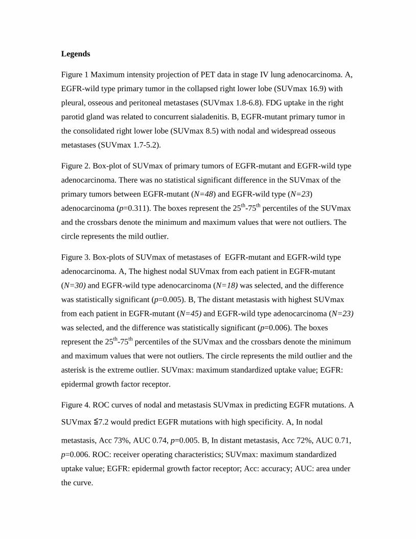
Figure 1 Maximum intensity projection of PET data in stage IV lung adenocarcinoma. A,
EGFR-wild type primary tumor in the collapsed right lower lobe (SUVmax 16.9) with
pleural, osseous and peritoneal metastases (SUVmax 1.8-6.8). FDG uptake in the right
parotid gland was related to concurrent sialadenitis. B, EGFR-mutant primary tumor in
the consolidated right lower lobe (SUVmax 8.5) with nodal and widespread osseous
metastases (SUVmax 1.7-5.2).
Figure 2. Box-plot of SUVmax of primary tumors of EGFR-mutant and EGFR-wild type
adenocarcinoma. There was no statistical significant difference in the SUVmax of the
primary tumors between EGFR-mutant (N=48) and EGFR-wild type (N=23)
adenocarcinoma (p=0.311). The boxes represent the 25th-75th percentiles of the SUVmax
and the crossbars denote the minimum and maximum values that were not outliers. The
circle represents the mild outlier.
Figure 3. Box-plots of SUVmax of metastases of EGFR-mutant and EGFR-wild type
adenocarcinoma. A, The highest nodal SUVmax from each patient in EGFR-mutant
(N=30) and EGFR-wild type adenocarcinoma (N=18) was selected, and the difference
was statistically significant (p=0.005). B, The distant metastasis with highest SUVmax
from each patient in EGFR-mutant (N=45) and EGFR-wild type adenocarcinoma (N=23)
was selected, and the difference was statistically significant (p=0.006). The boxes
represent the 25th-75th percentiles of the SUVmax and the crossbars denote the minimum
and maximum values that were not outliers. The circle represents the mild outlier and the
asterisk is the extreme outlier. SUVmax: maximum standardized uptake value; EGFR:
epidermal growth factor receptor.
Figure 4. ROC curves of nodal and metastasis SUVmax in predicting EGFR mutations. A
SUVmax ≦7.2 would predict EGFR mutations with high specificity. A, In nodal
metastasis, Acc 73%, AUC 0.74, p=0.005. B, In distant metastasis, Acc 72%, AUC 0.71,
p=0.006. ROC: receiver operating characteristics; SUVmax: maximum standardized
uptake value; EGFR: epidermal growth factor receptor; Acc: accuracy; AUC: area under
the curve.

Figure 5. ROC curves of individual predictors and combined predictive factors
(SUVmax, age, sex and smoking status) in determining EGFR mutation status. ROC:
receiver operating characteristics; AUC: area under the curve, followed by 95%
confidence intervals in brackets.
Table 1. The clinical characteristics of the study population in respect to EGFR mutation
status and the frequencies of EGFR mutation types. EGFR: epidermal growth factor
receptor; N: number of patients.
Table 2. The metabolic parameters of the primary tumors and metastases in association
with EGFR mutation status. EGFR: epidermal growth factor receptor; n: number of
lesions; *: statistical significance.
Table 3. Summary of the studies evaluated the relationship between metabolic uptake and
EGFR mutation status. N: total number; n: number in subgroups; ADC: adenocarcinoma;
EGFR: epidermal growth factor receptor; LN: lymph node; SUV: standardized uptake
value; †only mutations in exons 19 and 21; ‡EGFR status determined by EGFR-
overexpression on immunohistochemistry.

TABLE 1. The clinical characteristics of the study population in respect to EGFR
mutation status and the frequencies of EGFR mutation types. EGFR: epidermal growth
factor receptor; N: number of patients.
EGFR-mutant
(N)
EGFR-wild type
(N)
Total
(N)
Smoking
status
Non-smoker 39 12 51
Smoker/
ex-smoker
8 11 19
Undetermined 1 0 1
Sex
Female 31 7 38
Male 17 16 33
EGFR
mutations
Exon 21 27
52 Exon 20 5
Exon 19 15
Exon 18 5
Negative 23 23

TABLE 2. The metabolic parameters of the primary tumors and metastases in association
with EGFR mutation status. EGFR: epidermal growth factor receptor; n: number of
lesions; *: statistical significance.
EGFR-mutant EGFR-wild
type
p-value
Primary tumor SUVmax
Median (range)
n=48 n=23
7.4
(2.5-15.2)
8.1
(1.3-22.5)
0.311
Nodal metastasis
SUVmax
Median (range)
n=118 n=128
3.4
(1.1-10.5)
5.5
(1.3-19.1)
<0.001*
Distant metastasis
SUVmax
Median (range)
n=389 n=229
3.4
(0.9-16.6)
4.7
(1.4-18.1)
<0.001*

TABLE 3. Summary of the studies evaluated the relationship between metabolic uptake and EGFR mutation status. N: total number;
n: number in subgroups; ADC: adenocarcinoma; EGFR: epidermal growth factor receptor; LN: lymph node; SUV: standardized
uptake value; †only mutations in exons 19 and 21; ‡EGFR status determined by EGFR-overexpression on immunohistochemistry.
Studies N Disease stage (n)
Histology EGFR mutant: wild type (n)
Lesions measured
Metabolic parameters
Findings
Huang et al.5
77 IIIB/IV (15/62)
ADC 49:28
Primary tumor, mediastinal LN
SUVmax High SUVmax was predictive of EGFR mutations
Na et al.6 100 I/II/III/IV (39/18/38/5)
ADC, non-ADC
21:79† Primary tumor
SUVmax Low SUV was predictive of EGFR mutations
Mak et al.7 100 I-IV (32/8/18/42)
ADC, non-ADC
24:76
Primary tumor, one LN, one metastasis
SUVmax Low SUV was associated with EGFR mutations
Chung et al.8
106 I-IV (12/7/27/60)
ADC 42:64
Primary tumor, one metastasis
SUVmax Not predictive of EGFR mutations
All MTV, TLG
Lee et al.9 167 I (167)
ADC, non-ADC
42:125‡
Primary tumor
SUVmax High SUVmax was associated with EGFR overexpression

Figure 1
Figure 2

Figure 3
Figure 4
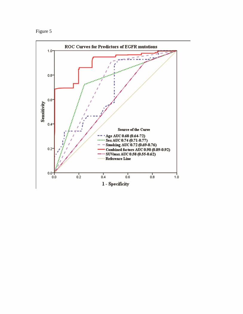
Figure 5
Related Documents