Metabolic Interactions Between Glutamatergic and Dopaminergic Neurotransmitter Systems Are Mediated Through D 1 Dopamine Receptors Tiago B. Rodrigues, 1,2 Noelia Granado, 3 Oskar Ortiz, 3 Sebastia ´n Cerda ´n, 1 * and Rosario Moratalla 3 1 Instituto de Investigaciones Biome ´dicas ‘‘Alberto Sols,’’ C.S.I.C./U.A.M., Madrid, Spain 2 Departamento de Bioquı ´mica, Centro de Neurocie ˆncias e Biologia Celular e Espectroscopia RMN, Faculdade de Cie ˆncias e Tecnologia, Universidade de Coimbra, Coimbra, Portugal 3 Instituto Cajal, C.S.I.C., Madrid, Spain Interactions between the dopaminergic and glutamatergic neurotransmission systems were investigated in the adult brain of wild-type (WT) and transgenic mice lacking the dopamine D 1 or D 2 receptor subtypes. Activity of the glutamine cycle was evaluated by using 13 C NMR spectroscopy, and striatal activity was assessed by c-Fos expression and motor coordination. Brain extracts from (1,2- 13 C 2 ) acetate-infused mice were prepared and ana- lyzed by 13 C NMR to determine the incorporation of the label into the C4 and C5 carbons of glutamate and gluta- mine. D 1 R –/– mice showed a significantly higher concentra- tion of cerebral (4,5- 13 C 2 ) glutamine, consistent with an increased activity of the glutamate-glutamine cycle and of glutamatergic neurotransmission. Conversely, D 2 R –/– mice did not show any significant changes in (4,5- 13 C 2 ) gluta- mate or (4,5- 13 C 2 ) glutamine, suggesting that alterations in glutamine metabolism are mediated through D 1 receptors. This was confirmed with D 1 R –/– and WT mice treated with reserpine, a dopamine-depleting drug, or with reserpine followed by L-DOPA, a dopamine precursor. Exposure to reserpine increased (4,5- 13 C 2 ) glutamine in WT to levels similar to those found in untreated D 1 R –/– mice. These val- ues were the same as those reached in the reserpine- treated D 1 R –/– mice. Treatment of WTanimals with L-DOPA returned (4,5- 13 C 2 ) glutamine levels to normal, but this was not verified in D 1 R –/– animals. Reserpine impaired motor coordination and decreased c-Fos expression, whereas L- DOPA restored both variables to normal values in WT but not in D 1 R –/– . Together, our results reveal novel neurometa- bolic interactions between glutamatergic and dopaminergic systems that are mediated through the D 1 , but not the D 2 , dopamine receptor subtype. V V C 2007 Wiley-Liss, Inc. Key words: dopamine; glutamate; dopamine D 1 and D 2 receptors; 13 C-NMR; Parkinson’s disease, striatum Precise integration of the different neurotransmitter signalling pathways is necessary to provide efficient and coordinated responses to internal and external stimuli in the central nervous system (CNS). Glutamate is the prin- cipal neurotransmitter in the excitatory circuits of the CNS, participating in many neuronal activities (Conn, 2003; Lipsky and Goldman, 2003; Malenka, 2003). Sim- ilarly, dopamine is implicated in motor control, cogni- tion, motivation, and emotional processing. Notably, indirect pharmacological and behavioral evidence revealed that glutamatergic neurotransmission is altered in several pathological disorders traditionally associated with abnormal dopamine signalling (Kalivas et al., 2003; Sanacora et al., 2003; Tamminga et al., 2003; Coyle, 2006). These findings indicate the existence of interac- tions between dopaminergic and glutamatergic neuro- transmission. Such interactions may underlie not only the adaptive and plastic responses to pathophysiological states such as Parkinson’s disease (Lange et al., 1997; Chase and Oh, 2000), schizophrenia (de Bartolomeis et al., 2005), drug abuse (Lapish et al., 2006), and per- Contract grant sponsor: Spanish Ministerio de Educacion y Ciencia; Contract grant number: SAF 2001-2245 (to S.C.); Contract grant num- ber: SAF 2004-03197 (to S.C.); Contract grant number: NAN 2004- 09125-C07-03 (to S.C.); Contract grant number: SAF 2003-4864 (to R.M.); Contract grant number: GEN2003-C06-02/NAC (to R.M.); Contract grant sponsor: Instituto de Salud Carlos III; Contract grant number: FISss C03/08 (to S.C.); Contract grant number: G03/155 C03/ 10 (to S.C.); Contract grant number: PI051530 (to S.C.); Contract grant sponsor: Spanish Ministerio de Sanidad y Consumo; Contract grant num- ber: FIS 03/1004 (to R.M.); Contract grant number: RTA (to R.M.); Contract grant number: G03/05 (to R.M.); Contract grant number: PNSD (to R.M.). *Correspondence to: Dr. Sebastia ´n Cerda ´n, Laboratory for Imaging and Spectroscopy by Magnetic Resonance (LISMAR), Instituto de Investiga- ciones Biome ´dicas, ‘‘Alberto Sols’’ C.S.I.C./U.A.M., Calle Arturo Dupe- rier 4, E-28029 Madrid, Spain. E-mail: [email protected] Received 1 November 2006; Revised 17 January 2007; Accepted 25 January 2007 Published online 23 April 2007 in Wiley InterScience (www. interscience.wiley.com). DOI: 10.1002/jnr.21302 Journal of Neuroscience Research 85:3284–3293 (2007) ' 2007 Wiley-Liss, Inc.

Welcome message from author
This document is posted to help you gain knowledge. Please leave a comment to let me know what you think about it! Share it to your friends and learn new things together.
Transcript
Metabolic Interactions BetweenGlutamatergic and DopaminergicNeurotransmitter Systems Are MediatedThrough D1 Dopamine Receptors
Tiago B. Rodrigues,1,2 Noelia Granado,3 Oskar Ortiz,3 Sebastian Cerdan,1*
and Rosario Moratalla3
1Instituto de Investigaciones Biomedicas ‘‘Alberto Sols,’’ C.S.I.C./U.A.M., Madrid, Spain2Departamento de Bioquımica, Centro de Neurociencias e Biologia Celular e Espectroscopia RMN,Faculdade de Ciencias e Tecnologia, Universidade de Coimbra, Coimbra, Portugal3Instituto Cajal, C.S.I.C., Madrid, Spain
Interactions between the dopaminergic and glutamatergicneurotransmission systems were investigated in the adultbrain of wild-type (WT) and transgenic mice lacking thedopamine D1 or D2 receptor subtypes. Activity of theglutamine cycle was evaluated by using 13C NMRspectroscopy, and striatal activity was assessed by c-Fosexpression and motor coordination. Brain extracts from(1,2-13C2) acetate-infused mice were prepared and ana-lyzed by 13C NMR to determine the incorporation of thelabel into the C4 and C5 carbons of glutamate and gluta-mine. D1R
–/– mice showed a significantly higher concentra-tion of cerebral (4,5-13C2) glutamine, consistent with anincreased activity of the glutamate-glutamine cycle and ofglutamatergic neurotransmission. Conversely, D2R
–/– micedid not show any significant changes in (4,5-13C2) gluta-mate or (4,5-13C2) glutamine, suggesting that alterations inglutamine metabolism are mediated through D1 receptors.This was confirmed with D1R
–/– and WT mice treated withreserpine, a dopamine-depleting drug, or with reserpinefollowed by L-DOPA, a dopamine precursor. Exposure toreserpine increased (4,5-13C2) glutamine in WT to levelssimilar to those found in untreated D1R
–/– mice. These val-ues were the same as those reached in the reserpine-treated D1R
–/– mice. Treatment of WTanimals with L-DOPAreturned (4,5-13C2) glutamine levels to normal, but this wasnot verified in D1R
–/– animals. Reserpine impaired motorcoordination and decreased c-Fos expression, whereas L-DOPA restored both variables to normal values in WT butnot in D1R
–/–. Together, our results reveal novel neurometa-bolic interactions between glutamatergic and dopaminergicsystems that are mediated through the D1, but not the D2,dopamine receptor subtype. VVC 2007Wiley-Liss, Inc.
Key words: dopamine; glutamate; dopamine D1 andD2 receptors; 13C-NMR; Parkinson’s disease, striatum
Precise integration of the different neurotransmittersignalling pathways is necessary to provide efficient andcoordinated responses to internal and external stimuli in
the central nervous system (CNS). Glutamate is the prin-cipal neurotransmitter in the excitatory circuits of theCNS, participating in many neuronal activities (Conn,2003; Lipsky and Goldman, 2003; Malenka, 2003). Sim-ilarly, dopamine is implicated in motor control, cogni-tion, motivation, and emotional processing. Notably,indirect pharmacological and behavioral evidencerevealed that glutamatergic neurotransmission is alteredin several pathological disorders traditionally associatedwith abnormal dopamine signalling (Kalivas et al., 2003;Sanacora et al., 2003; Tamminga et al., 2003; Coyle,2006). These findings indicate the existence of interac-tions between dopaminergic and glutamatergic neuro-transmission. Such interactions may underlie not onlythe adaptive and plastic responses to pathophysiologicalstates such as Parkinson’s disease (Lange et al., 1997;Chase and Oh, 2000), schizophrenia (de Bartolomeiset al., 2005), drug abuse (Lapish et al., 2006), and per-
Contract grant sponsor: Spanish Ministerio de Educacion y Ciencia;
Contract grant number: SAF 2001-2245 (to S.C.); Contract grant num-
ber: SAF 2004-03197 (to S.C.); Contract grant number: NAN 2004-
09125-C07-03 (to S.C.); Contract grant number: SAF 2003-4864 (to
R.M.); Contract grant number: GEN2003-C06-02/NAC (to R.M.);
Contract grant sponsor: Instituto de Salud Carlos III; Contract grant
number: FISss C03/08 (to S.C.); Contract grant number: G03/155 C03/
10 (to S.C.); Contract grant number: PI051530 (to S.C.); Contract grant
sponsor: Spanish Ministerio de Sanidad y Consumo; Contract grant num-
ber: FIS 03/1004 (to R.M.); Contract grant number: RTA (to R.M.);
Contract grant number: G03/05 (to R.M.); Contract grant number:
PNSD (to R.M.).
*Correspondence to: Dr. Sebastian Cerdan, Laboratory for Imaging and
Spectroscopy by Magnetic Resonance (LISMAR), Instituto de Investiga-
ciones Biomedicas, ‘‘Alberto Sols’’ C.S.I.C./U.A.M., Calle Arturo Dupe-
rier 4, E-28029 Madrid, Spain. E-mail: [email protected]
Received 1 November 2006; Revised 17 January 2007; Accepted 25
January 2007
Published online 23 April 2007 in Wiley InterScience (www.
interscience.wiley.com). DOI: 10.1002/jnr.21302
Journal of Neuroscience Research 85:3284–3293 (2007)
' 2007 Wiley-Liss, Inc.
sonality disorders (Karasawa et al., 2006) but also theoperation of the normal brain under physiological condi-tions (Lee et al., 2002; Salter, 2003; Sesack et al., 2003;West et al., 2003; Pei et al., 2004). However, despitethe neurobiological and clinical relevance of these inter-actions, the underlying mechanisms remain poorlyunderstood (Abekawa et al., 2000; David and Abraini,2001). In particular, the dopamine receptor subtype thatmediates communication between glutamatergic and do-paminergic systems has not been identified.
The use of 13C NMR and the 13C-enriched pre-cursors 13C glucose or 13C acetate has provided exten-sive information on glutamate metabolism in neuronsand glia as well as the interactions between these celltypes through glutamate-glutamine cycling and glutama-tergic neurotransmission (Rothman et al., 2003;Rodrigues and Cerdan, 2005). This approach can beused to reveal glutamate turnover, and it is sensitive toperturbations at the glutamate receptor and transport lev-els as well as in the transport of glucose or acetate pre-cursors (Waagepetersen et al., 2001; Garcia-Espinosaet al., 2004). On the other hand, transgenic animals alsoprovide an excellent tool for investigating the effects ofgene and protein ablation on their function in vivo(Chen and Zhuang, 2003).
On these grounds, we employed 13C NMR(Rothman et al., 2003; Rodrigues and Cerdan, 2005) toinvestigate the metabolism of glutamate and glutamine inthe brain of transgenic mice that lack dopamine D1 orD2 receptors (Xu et al., 1994; Baik et al., 1995; Kellyet al., 1998). We confirmed these results using a com-plementary pharmacological approach. Accordingly, WTand D1R
–/– mice were treated with reserpine, a dopa-mine-depleting drug (Graybiel et al., 1990; Slatteryet al., 2004), and also with reserpine and L-DOPA, anacute antiparkinsonian treatment (Carlsson et al., 1957;Schildkraut et al., 1965; Hornykiewicz, 2002; Pavonet al., 2006), to investigate the effects of these drugs onthe glutamate-glutamine cycle and on glutamatergic neu-rotransmission. Finally, we correlated the informationobtained by 13C NMR with tests of motor coordinationand with the expression of the c-Fos protein, a markerof dopaminergic activity in neurons. Together, ourresults indicate that the interactions observed betweenglutamatergic and dopaminergic neurotransmitter systemsare mediated through the D1 but not the D2 dopaminereceptors and that the lack of dopamine or lack of D1
receptors results in an increase in glutamatergic activity.
MATERIALS AND METHODS
Animals, Drug Treatments, and Infusion Techniques
The experimental protocols used in this study wereapproved by the appropriate institutional review committeesand met the guidelines of the appropriate government agency.All efforts were made to minimize animal suffering. Experi-ments were carried out in male dopamine D1 and D2 receptorknockout (D1R
–/–, D2R–/–) mice (Baik et al., 1995; Kelly
et al., 1998) as well as in their heterozygous (+/–) and wild-
type (+/+) littermates. Mice were used at 2-3 months of age,and the genotype of each mouse was determined by genomicSouthern blot analysis as described previously (Chen et al.,1999). The mice were housed at the Cajal Institute, in a hu-midity- and temperature-controlled room on a 12-hr light/dark cycle, receiving water and food ad libitum.
In the first series of experiments, two groups of micewere used without any kind of pharmacological intervention:group 1 included D1R
+/+, D1R–/+, and D1R
–/– mice; group2 included D2R
+/+ and D2R–/– mice (n ¼ 6 per genotype).
The involvement of D1 receptors was further investigated byanalyzing the effects of dopamine depletion in WT andD1R
–/– mice (n ¼ 12 for each genotype). Half of these ani-mals (n ¼ 6 per genotype) were given the dopamine-deplet-ing drug reserpine (5 mg/kg, i.p.) 18 hr before the infusion ofthe 13C label (group 3). The other half (n ¼ 6 per genotype)also received an intraperitoneal injection of L-DOPA (50 mg/kg) combined with benserazide-HCl (20 mg/kg), a DOPA-decarboxylase inhibitor, 18 hr after reserpine administrationand 60 min before the infusion of the 13C label (group 4).This reserpine treatment protocol has previously been shownto lower striatal dopamine content by 81–96% and to producechanges in behavior and in IEG expression (Graybiel et al.,1990; LaHoste and Marshall, 1992; Moratalla et al., 1996b;Day et al., 2006). Before the infusion of the 13C label, theanimals were deeply anesthetised by administering 1–2% ofisofluorane in 1 liter/min O2 through a nose cap. Body tem-perature was maintained at approximately 378C with a ther-mostatic blanket and a temperature-regulated circulating waterbath, and it was measured with a rectal probe (Panlab, Barce-lona, Spain). The physiological state of the animal was moni-tored by a Biotrig physiological monitor (Bruker MedicalGmbh, Ettlingen, Germany), measuring the respiratory rate andthe body temperature. The right jugular vein was dissected andcannulated, and (1,2-13C2) acetate (0.24 lmol min–1 g–1) wasinfused for 60 min. At the end of this infusion, the head of theanimal was funnel-frozen, and perchloric acid extracts of thebrain, excluding the cerebellum, were prepared prior to per-forming high-resolution 13C NMR spectroscopy as describedpreviously (Cerdan et al., 1990; Chapa et al., 1995).
13C NMR Spectroscopy
High-resolution proton-decoupled 13C NMR spectra ofbrain extracts were obtained at 11.9 Tesla (125.13 MHz,258C, pH 7.2) with a Bruker AVANCE 500WB NMR spec-trometer using a commercial (5 mm) triple- resonance probe(1H, 13C, 2H) optimized for direct 13C NMR detection. Theacquisition conditions were: p/3 pulses, 30.0 kHz spectralwidth, 1.09 sec acquisition time, 64k words data table, and6.0 sec recycling time. Proton decoupling was gated only dur-ing the acquisition using a broad-band composite pulse decou-pling (CPD) sequence, and chemical shifts were calibratedwith an external reference of dioxane (10% v/v, 67.4 ppm).Resonance assignments were based on literature values and onthe addition of internal standards (Cerdan et al., 1990). Spectradeconvolution and multiplet structures were analyzed by usinga PC-based (Intel Centrino platform) NMR program, NUTS(Acorn, Freemont, CA). All 13C NMR resonance areas were
Glutamate-Dopamine Interactions 3285
Journal of Neuroscience Research DOI 10.1002/jnr
normalized relative to the unchanged myo-inositol resonanceareas of the corresponding perchloric acid extracts. This waspossible because myo-inositol had a low turnover and did notbecome enriched after the 60-min (1,2-13C2) acetate infusion,providing a robust internal reference from which all 13C reso-nances can be derived (Bouzier et al., 1998).
Behavioral Experiments (Motor Coordination,Rota-Rod)
Motor coordination was measured by using an accelerat-ing rota-rod apparatus (Hugo Basile, Rome, Italy). Twogroups of adult mice were used, D1R
–/– and D1R+/+ mice
(n ¼ 6 per group). On the first day, the mice were subjectedto a practice trial, placing the animal on the rota-rod withoutrotation for 60 sec, followed by a 1-min period during whichthe rod rotated at a constant speed of 4 r.p.m. After this, themice were returned to their cages, and 20 min later they weretrained to use the rota-rod with six sessions of 5 min each at aconstant acceleration and with an interval of 20 min betweentrials. On the following day, mice were tested on the acceler-ated rota-rod, and the time to fall from the rod was measured,with a cutoff time established at 5 min. Animals were testedunder basal conditions or 3 days after administration of reser-pine (5 mg/kg) or 30 min after L-DOPA administration(20 mg/kg) given 3 days after the reserpine.
c-Fos Immunocytochemical Staining andQuantification Experiments
At the end of the experiments, animals were anesthe-tized with pentobarbital and transcardially perfused with 4%paraformaldehyde in 0.1% phosphate buffer (PB), pH 7.4. Af-ter perfusion, the brains were dissected out and immersedovernight in the same fixative solution. The brains were thencryoprotected in a solution of 30% sucrose for 1 day. Coronalbrain sections (30 lm) throughout the striatal area wereobtained on a slicing microtome (Leica, Madrid, Spain) andwere kept in PB solution at 48C until use. Immunostainingwas carried out on free-floating sections with a standard avi-din-biotin immunocytochemical protocol previously described(Rivera et al., 2002; Grande et al., 2004; Pavon et al., 2006).Striatal sections were incubated overnight with specific c-Fosantisera (Chemicon, Temecula, CA), and, after three washes,the sections were incubated for 2 hr with the secondary anti-sera at room temperature. Peroxidase reactions were devel-oped with 0.06% of diaminobenzidine (DAB) in PB buffercontaining 1% nickel ammonium sulfate and 5 ll H2O2, andthe reaction was monitored every 5 min with an opticalmicroscope. After being washed, the sections were mountedon gelatin-coated slides, air dried, dehydrated in ascending se-ries of ethanol, cleared with xylene, and coverslipped withPermount. Quantification of c-Fos striatal expression was per-formed with the aid of an image analysis system (AIS; ImagingResearch Inc., Linton, United Kingdom) using a 310 lens.Before counting, a standardized threshold was established inthe images at the gray-scale level, empirically determined bytwo different observers, to permit the detection of stainednuclei of low to high intensity and to suppress the very lightlystained nuclei. Thus, the number of positive nuclei for c-Foswas determined and expressed as number of positive nuclei
per micrometer. Counting was performed on five sectionsfrom each animal and on three different mice.
Statistical Analysis
Statistical analysis was performed with the SPSS package(SAS Institute Inc., Cary, NC). The results are expressed asmeans 6 SD. When indicated, the statistical significance of thedifferences was assessed by Student’s t-test. Statistical analysesfor gene expression and motor coordination between mice ofdifferent genotypes and drug treatments were performed bytwo-way ANOVA, followed by post hoc Tukey’s test. Thethreshold for statistical significance was set at P < 0.05.
Materials
Reserpine, L-DOPA, benserazide-HCl, and d-ampheta-mine sulfate were obtained from Sigma-Aldrich (Madrid,Spain). (1,2-13C2) acetate (99.9% 13C) was obtained fromCambridge Isotope Laboratories (Andover, MA). 2H2O(99.9% 2H) was obtained from Apollo Scientific Ltd. All theother items were of the highest purity available commerciallyfrom Sigma-Aldrich.
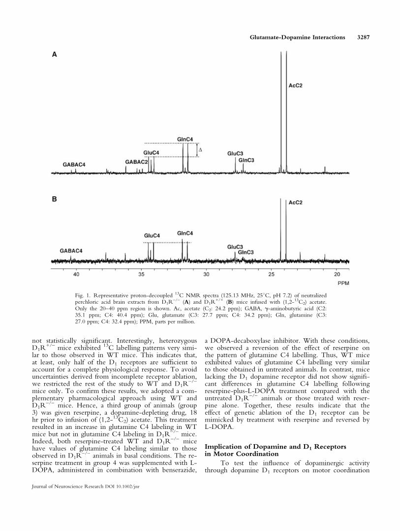
RESULTS13C NMR Spectroscopy
High-resolution 13C NMR spectra from perchloricacid brain extracts from a homozygous mutant mouse(D1R
–/–, Fig. 1A) and from a wild-type (D1R+/+, Fig.
1B) were obtained 60 min after infusion of (1,2-13C2)acetate. The spectra display different characteristics, indi-cating that the loss of D1 receptor has significant conse-quences on acetate metabolism in the brain. After(1,2-13C2) acetate infusion, glutamate, glutamine, and g-aminobutyric acid (GABA) are labelled in different car-bon positions depending on the metabolic pathwaysconnecting these intermediates (Cerdan et al., 1990;Rodrigues and Cerdan, 2005). Glutamate and glutamine(Glx) are both labelled contiguously at C4 and C5 dur-ing the first turn of the cycle, because these carbons arederived directly from a (1,2-13C2) acetyl-CoA precursor.It is readily apparent that mice lacking D1 dopaminereceptors were characterized by a significant increase in13C incorporation into the cerebral glutamine C4 reso-nance doublet compared with the glutamate precursorC4 (dotted lines).
The incorporation of 13C into the resonance dou-blet for cerebral glutamate C4 and glutamine C4 in thefour different groups examined in our experiments andrelative to the natural abundance of 13C inositol C1-C3resonance is summarized in Figure 2. There were nodetectable changes in glutamate C4 labelling, whenboth D1 and D2 receptor expressions were geneticallydeleted. However, glutamine C4 labelling increased sig-nificantly in D1R
–/– mice compared with the valuesobtained from the WT and heterozygous counterparts(group 1). In contrast, complete ablation of D2 receptorsappeared to provoke only a small increase in the incor-poration of 13C in glutamine C4, although when bothgroups of animals were compared this difference was
3286 Rodrigues et al.
Journal of Neuroscience Research DOI 10.1002/jnr
not statistically significant. Interestingly, heterozygousD1R
+/– mice exhibited 13C labelling patterns very simi-lar to those observed in WT mice. This indicates that,at least, only half of the D1 receptors are sufficient toaccount for a complete physiological response. To avoiduncertainties derived from incomplete receptor ablation,we restricted the rest of the study to WT and D1R
–/–
mice only. To confirm these results, we adopted a com-plementary pharmacological approach using WT andD1R
–/– mice. Hence, a third group of animals (group3) was given reserpine, a dopamine-depleting drug, 18hr prior to infusion of (1,2-13C2) acetate. This treatmentresulted in an increase in glutamine C4 labeling in WTmice but not in glutamine C4 labeling in D1R
–/– mice.Indeed, both reserpine-treated WT and D1R
–/– micehave values of glutamine C4 labeling similar to thoseobserved in D1R
–/– animals in basal conditions. The re-serpine treatment in group 4 was supplemented with L-DOPA, administered in combination with benserazide,
a DOPA-decaboxylase inhibitor. With these conditions,we observed a reversion of the effect of reserpine onthe pattern of glutamine C4 labelling. Thus, WT miceexhibited values of glutamine C4 labelling very similarto those obtained in untreated animals. In contrast, micelacking the D1 dopamine receptor did not show signifi-cant differences in glutamine C4 labelling followingreserpine-plus-L-DOPA treatment compared with theuntreated D1R
–/– animals or those treated with reser-pine alone. Together, these results indicate that theeffect of genetic ablation of the D1 receptor can bemimicked by treatment with reserpine and reversed byL-DOPA.
Implication of Dopamine and D1 Receptorsin Motor Coordination
To test the influence of dopaminergic activitythrough dopamine D1 receptors on motor coordination
Fig. 1. Representative proton-decoupled 13C NMR spectra (125.13 MHz, 258C, pH 7.2) of neutralizedperchloric acid brain extracts from D1R
–/– (A) and D1R+/+ (B) mice infused with (1,2-13C2) acetate.
Only the 20–40 ppm region is shown. Ac, acetate (C2: 24.2 ppm); GABA, g-aminobutyric acid (C2:35.1 ppm; C4: 40.4 ppm); Glu, glutamate (C3: 27.7 ppm; C4: 34.2 ppm); Gln, glutamine (C3:27.0 ppm; C4: 32.4 ppm); PPM, parts per million.
Glutamate-Dopamine Interactions 3287
Journal of Neuroscience Research DOI 10.1002/jnr
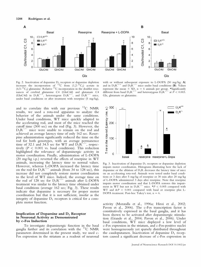
and to correlate this with our previous 13C NMRresults, we used a rota-rod apparatus to analyze thebehavior of the animals under the same conditions.Under basal conditions, WT mice quickly adapted tothe accelerating rod, and most of the mice reached thecutoff time (300 sec) on the rod (Fig. 3). However, theD1R
–/– mice were unable to remain on the rod andachieved an average latency time of only 162 sec. Reser-pine administration significantly reduced the time on therod for both genotypes, with an average permanencetime of 32.1 and 34.5 sec for WT and D1R
–/–, respec-tively (P < 0.001 vs. basal conditions). This reductionhighlighted the relevance of dopaminergic activity inmotor coordination. Finally, administration of L-DOPA(20 mg/kg i.p.) reverted the effects of reserpine in WTanimals, increasing the latency time to normal values.However, whereas L-DOPA increased the latency timeon the rod for D1R
–/– animals (from 34 to 128 sec), thisincrease did not completely restore motor coordinationto the level of WT mice. Indeed, the average time onthe rod of 128 sec for D1R
–/– animals after L-DOPAtreatment was similar to the latency time obtained underbasal conditions (average 162 sec; Fig. 3). These resultsindicate that dopamine is necessary for proper motorcoordination but that it is not sufficient, insofar as theintegrity of dopamine D1 receptors is critical for a com-plete motor function.
Implication of Dopamine and D1 Receptorin Neuronal Activity as Demonstratedby c-Fos Induction
To investigate dopaminergic function in the basalganglia further and its correlation with the 13C NMRparameters determined in the present study, we used c-Fos expression in the striatum as a readout of neuronal
activity (Moratalla et al., 1996a; Hiroi et al., 2002;Pavon et al., 2006). The c-Fos transcription factor isconstitutively expressed in the basal ganglia, and it hasbeen shown to be activated after dopaminergic stimula-tion (Grande et al., 2004; Pavon et al., 2006). Underbasal conditions, WT mice displayed a low level ofc-Fos expression in the striatum, and c-Fos-positive nucleiwere homogeneously yet sparsely distributed throughoutthe caudoputamen. Inactivation of dopamine D1 recep-tors caused a significant decrease of c-Fos expression in
Fig. 3. Inactivation of dopamine D1 receptors or dopamine depletionimpairs motor coordination. Histograms illustrating how the lack ofdopamine or the ablation of D1R decreases the latency time of miceon an accelerating rota-rod. Animals were tested under basal condi-tions or 3 days after 5 mg/kg of reserpine or 30 min after 20 mg/kgof L-DOPA administered 3 days after reserpine. Note that reserpineimpairs motor coordination and that L-DOPA restores this impair-ment in WT but not in D1R
–/– mice. *P < 0.005 compared withWT and &P < 0.001 compared with basal or reserpine plus L-DOPA treatment. Post-hoc Tukey’s test, n ¼ 6.
Fig. 2. Inactivation of dopamine D1 receptors or dopamine depletionincreases the incorporation of 13C from (1,2-13C2) acetate in(4,5-13C2) glutamine. Relative 13C incorporation in the doublet reso-nances of cerebral glutamine C4 (GlnC4d) and glutamate C4(GluC4d) in D1R
+/+, heterozygous D1R+/–, and D1R
–/– mice,under basal conditions or after treatment with reserpine (5 mg/kg),
with or without subsequent exposure to L-DOPA (50 mg/kg; A)and in D2R
+/+ and D2R–/– mice under basal conditions (B). Values
represent the mean 6 SD, n ¼ 6 animals per group. *Significantlydifferent from basal D1R
+/+ and heterozygous D1R+/– at P < 0.003.
Glx, glutamate or glutamine.
3288 Rodrigues et al.
Journal of Neuroscience Research DOI 10.1002/jnr

the striatum (P ¼ 0.001, WT vs. D1R–/–; Fig. 4). Re-
serpine treatment significantly diminished c-Fos expres-sion in the caudoputamen of WT mice but did not alterthe already weak expression in D1R
–/– mice. This lossof c-Fos in WT mice was observed mainly in the middleof the caudoputamen. However, administering L-DOPAafter reserpine treatment resulted in a significant increasein c-Fos expression in WT but not in D1R
–/– mice, inwhich the level of c-Fos expression was similar to thelevels found under basal conditions (P < 0,001, WT vs.D1R
–/– mice; Fig. 4, Table I). These results indicatethat the constitutive c-Fos gene expression found in thecaudoputamen of WT mice was due to dopaminergicactivity via D1 receptor activation, insofar as the absenceof dopamine or dopamine D1 receptors significantlyreduced c-Fos expression. In summary, the decrease inc-Fos expression revealed diminished dopaminergic ac-tivity, and this correlated well with the increase in the(4,5-13C2) glutamine labelling and glutamatergic activitydetected by 13C NMR.
DISCUSSION
Our results confirm that an interaction exists be-tween the dopaminergic and the glutamatergic neuro-transmission systems, demonstrating that functional D1
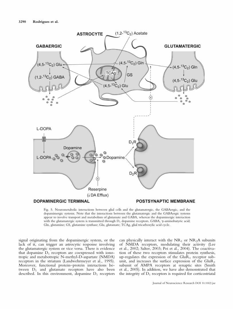
dopamine receptors are required for adequate glutama-tergic neurotransmission. Based on the data presentedhere regarding the metabolism of (1,2-13C2) acetate, wepresent an overview of the neurometabolic interactionsbetween the dopaminergic and the glutamatergic orGABAergic systems (see Fig. 5). In brain metabolism,(1,2-13C2) acetate is incorporated mainly through theglial compartment (Waniewski and Martin, 1998; Cruzand Cerdan, 1999; Lebon et al., 2002). The (4,5-13C2)glutamine produced by glutamine synthase, an enzymefound exclusively in glia, is exported to glutamatergicneurons. It is in these neurons where the neurotransmit-ter glutamate is stored in vesicles and eventually releasedto the synaptic cleft during the action potential. Synapticglutamate is recaptured by the astrocytes and transformedinto glutamine, completing the glutamate-glutaminecycle that supports glutamatergic neurotransmission. InGABAergic neurons, glial glutamate is transformed intoGABA by the GABA decarboxylase isozymes, and it iseventually released following GABAergic stimulation.Released GABA is recaptured by astrocytes and inacti-vated through GABA transaminase and semialdehydesuccinic dehydrogenase isozymes. Thus, astrocytic gluta-mate and glutamine play a crucial role during glutama-tergic or GABAergic neurotransmission, providing acommon metabolic precursor that supports both neuro-transmission systems.
The present study shows that the supporting roleof astrocytes can be further extended to the dopami-nergic system, to which no direct metabolic links areimmediately apparent to our knowledge. Dopaminergicneurons release dopamine to the synaptic cleft, and thisdopamine eventually binds to the postsynaptic dopamineD1 or D2 receptors. Our results show that D1R
–/– micedisplay significant increases in (4,5-13C2) glutamine,revealing an increase in glutamine synthase activity.Therefore, the question that arises is how a metabolic
Fig. 4. Inactivation of dopamine D1 receptors or dopamine depletionresults in a drastic reduction of c-Fos expression in the striatum. Pho-tomicrographs of coronal sections from the striatum of D1R
+/+ andD1R
–/– mice illustrating c-Fos expression under basal conditions(A,B), after treatment with 5 mg/kg of reserpine (C,D) and reser-pine plus 20 mg/kg of L-DOPA (E,F). Note the constitutive expres-sion of c-Fos nuclei in D1R
+/+ mice in basal conditions comparedwith D1R
–/–. Treatment with reserpine diminishes c-Fos expressionand treatment with L-DOPA restores c-Fos expression in the WT,but not in D1R
–/– animals. Res, reserpine. Scale bar ¼ 200 lm.
TABLE I. Quantification of c-fos-Positive Nuclei Expression per
Square Millimeter{
Number of positive nucleus c-fos
WT D1R�/�
Basal conditions 83.16 6 10.56 3.11 6 1.18**
Reserpine 15.86 6 4.26*** 13.71 6 3.12*
Reserpine + L-DOPA 292.33 6 24.61**** 9.92 6 2.97**
{Immunostained cell nuclei were counted in striatal sections from 3 dif-
ferent animals per group, 5 sections each. Data are expressed as mean of
c-fos-positive nuclei per mm2 (mean 6 SEM).*P < 0.05 compared with basal condition in D1R
�/�.**P < 0.001 compared with WT mice.***P < 0.001 compared with basal conditions in WT mice.****P < 0.001 compared with reserpine and basal conditions in WT
mice; post hoc Tukey test.
Glutamate-Dopamine Interactions 3289
Journal of Neuroscience Research DOI 10.1002/jnr
signal originating from the dopaminergic system, or thelack of it, can trigger an astrocytic response involvingthe glutamatergic system or vice versa. There is evidencethat dopamine D1 receptors are coexpressed with iono-tropic and metabotropic N-methyl-D-aspartate (NMDA)receptors in the striatum (Landwehrmeyer et al., 1995).Moreover, functional protein–protein interactions be-tween D1 and glutamate receptors have also beendescribed. In this environment, dopamine D1 receptors
can physically interact with the NR1 or NR2A subunitsof NMDA receptors, modulating their activity (Leeet al., 2002; Salter, 2003; Pei et al., 2004). The coactiva-tion of these two receptors stimulates protein synthesis,up-regulates the expression of the GluR1 receptor sub-unit, and increases the surface expression of the GluR1
subunit of AMPA receptors at synaptic sites (Smithet al., 2005). In addition, we have also demonstrated thatthe integrity of D1 receptors is required for corticostriatal
Fig. 5. Neurometabolic interactions between glial cells and the glutamatergic, the GABAergic, and thedopamimergic systems. Note that the interactions between the glutamatergic and the GABAergic systemsappear to involve transport and metabolism of glutamate and GABA, whereas the dopaminergic interactionwith the glutamatergic system is transmitted through D1 dopamine receptors. GABA, g-aminobutyric acid;Gln, glutamine; GS, glutamine synthase; Glu, glutamate; TCAg, glial tricarboxylic acid cycle.
3290 Rodrigues et al.
Journal of Neuroscience Research DOI 10.1002/jnr
and hippocampal LTP (Centonze et al., 2003; Granadoet al., 2007), a phenomenon known to be dependent onglutamate receptors. Therefore, these two classes ofreceptors could interact with each other to modulateglutamatergic or dopaminergic neurotransmission. Inagreement with previous in vivo microdialysis studies,adaptive changes in the glutamatergic system have beenobserved in rats and in reserpinized mice, as in the pres-ent study, probably as a feedback mechanism to increasesynaptic activity after lesion (Meshul et al., 1999; Jonkerset al., 2002; Day et al., 2006). Moreover, decreases inthe level of cerebral glutamate have been reported fol-lowing dopaminergic treatments (Yamamoto and Davy,1992). In summary, the simultaneous presence of dopa-mine D1 receptors in glutamatergic neurons in the cor-tex or the coexpression of D1 and glutamate receptors inthe caudoputamen opens the possibility that these twoneurotransmitter systems interact and influence eachother. Thus, pathologic disturbances or genetic ablationof either of these partners can induce an increase in theactivity of the other in an attempt to reestablish normalsynaptic activity.
In the present study, we also correlated the in vivoneuronal glutamatergic activity defined in 13C labellingstudies with motor coordination and the expression ofmarkers of neuronal activity in selected brain regions inwild-type mice, dopamine-depleted animals, and dopa-mine D1R
–/– mice. Our results show that dopaminedepletion or inactivation of dopamine D1 receptors inmice augments the activity of the glutamine cycle, pro-ducing impairment in motor coordination and a signifi-cant decrease in c-Fos expression in the striatum.Replacing dopamine in animals treated with reserpineby administering L-DOPA normalizes glutamatergicactivity, motor coordination, and c-Fos activity in thestriatum of WT but not D1R
–/– mice. All these resultssuggest that the increase in glutamatergic activity that wefound in dopamine-depleted animals or in D1R
–/– miceis inversely correlated with c-Fos expression and withmotor coordination. Thus, we think that the increase in(4,5-13C2) glutamine, consistent with an increase in theactivity of the glutamine cycle, is the result of a com-pensatory mechanism in the glutamatergic system tocope with the loss of dopamine or D1R function. How-ever, this increase in glutamatergic activity appears notto be sufficient to restore normal motor coordination, asrevealed by the rota-rod experiments, or normal c-Fosexpression. On the other hand, we have found repeat-edly over the years that D1R
–/– mice have enhancedhorizontal motor activity (Xu et al., 1994; Centonzeet al., 2003; Granado et al., 2007), a result that couldreflect the increased glutamatergic activity we reporthere. In addition, our results also show that the reduc-tion in motor coordination observed in D1R
–/– micefurther decreased after exposure to reserpine and that itwas restored by L-DOPA administration. These dataclearly indicate that components of the dopaminergicsystem besides D1 dopamine receptors are implicated inmotor coordination. Among the components potentially
implicated in this effect, the dopamine D2 receptorappears most likely to be involved, because the lack ofD2 receptors in the mutant mice impairs motor activity(Baik et al., 1995; Kelly et al., 1998). Thus, the presentresults disclose novel implications of the 13C NMRlabelling patterns, revealing motor coordination or local-ized c-Fos expression.
The results of the present study invoke additionalimplications to be able to interpret the 13C NMR spec-tra of brain metabolism in vivo and in vitro. A variety of13C NMR studies have revealed significant alterations inglutamate and glutamine metabolism during neuro-degenerative diseases, ischemia, or psychiatric disorders(Pascual et al., 1998; Krystal et al., 2002; Lin et al.,2003; Ross et al., 2003). Our work indicates that thesealterations may include, embedded, the contributions ofaltered dopaminergic neurotransmission. Moreover, cur-rent therapeutic protocols for Parkinson’s disease arebased mainly on substitution therapies with dopamineanalogues or precursors such as L-DOPA. The presentresults indicate that more adequate therapeutic regimesshould also be considered, including the use of inhibitorsof glutamatergic neurotransmission.
Finally, it is possible that the neurometabolic inter-actions of the glutamatergic neurotransmission systeminvolve not only the dopaminergic system, as disclosedin the present study, but additional neurotransmitterssuch as the cholinergic or serotoninergic systems. Moreexperiments in this area will be necessary, but it can beenvisioned now that strong coupling mechanisms mayexist among the different neurotransmission systems toensure a coordinated response in the brain under physio-logical or pathological conditions.
ACKNOWLEDGMENTS
T.B.R. was supported by a fellowship from theFundacao para a Ciencia e Tecnologia/Ministerio daCiencia e Ensino Superior, Portugal; N.G. and O.O.were supported by fellowships from Ministerio de Edu-cacion y Ciencia and the Basque Country, respectively.The authors thank Mr. Javier Perez for his careful workon the illustrations and Ms. Patricia Sanchez and Ms.Emilia Rubio for excellent technical assistance.
REFERENCES
Abekawa T, Ohmori T, Ito K, Koyama T. 2000. D1 dopamine receptor
activation reduces extracellular glutamate and GABA concentrations in
the medial prefrontal cortex. Brain Res 867:250–254.
Baik JH, Picetti R, Saiardi A, Thiriet G, Dierich A, Depaulis A, Le
Meur M, Borrelli E. 1995. Parkinsonian-like locomotor impairment in
mice lacking dopamine D2 receptors. Nature 377:424–428.
Bouzier AK, Goodwin R, de Gannes FM, Valeins H, Voisin P, Canioni
P, Merle M. 1998. Compartmentation of lactate and glucose metabo-
lism in C6 glioma cells. A 13C and 1H NMR study. J Biol Chem
273:27162–27169.
Carlsson A, Lindqvist M, Magnusson T. 1957. 3,4-Dihydroxyphenylala-
nine and 5-hydroxytryptophan as reserpine antagonists. Nature 180:
1200.
Glutamate-Dopamine Interactions 3291
Journal of Neuroscience Research DOI 10.1002/jnr
Centonze D, Grande C, Saulle E, Martin AB, Gubellini P, Pavon N,
Pisani A, Bernardi G, Moratalla R, Calabresi P. 2003. Distinct roles of
D1 and D5 dopamine receptors in motor activity and striatal synaptic
plasticity. J Neurosci 23:8506–8512.
Cerdan S, Kunnecke B, Seelig J. 1990. Cerebral metabolism of
[1,2-13C2]acetate as detected by in vivo and in vitro 13C NMR. J Biol
Chem 265:12916–12926.
Chapa F, Kunnecke B, Calvo R, Escobar del Rey F, Morreale de Esco-
bar G, Cerdan S. 1995. Adult-onset hypothyroidism and the cerebral
metabolism of (1,2-13C2) acetate as detected by 13C nuclear magnetic
resonance. Endocrinology 136:296–305.
Chase TN, Oh JD. 2000. Striatal dopamine- and glutamate-mediated
dysregulation in experimental parkinsonism. Trends Neurosci 23:S86–
S91.
Chen JF, Huang Z, Ma J, Zhu J, Moratalla R, Standaert D, Moskowitz
MA, Fink JS, Schwarzschild MA. 1999. A2A adenosine receptor defi-
ciency attenuates brain injury induced by transient focal ischemia in
mice. J Neurosci 19:9192–9200.
Chen L, Zhuang X. 2003. Transgenic mouse models of dopamine defi-
ciency. Ann Neurol 54:S91–S102.
Conn PJ. 2003. Physiological roles and therapeutic potential of metabo-
tropic glutamate receptors. Ann N Y Acad Sci 1003:12–21.
Coyle JT. 2006. Glutamate and schizophrenia: beyond the dopamine
hypothesis. Cell Mol Neurobiol 26:363–382.
Cruz F, Cerdan S. 1999. Quantitative 13C NMR studies of metabolic
compartmentation in the adult mammalian brain. NMR Biomed 12:
451–462.
David HN, Abraini JH. 2001. The group I metabotropic glutamate
receptor antagonist S-4-CPG modulates the locomotor response pro-
duced by the activation of D1-like, but not D2-like, dopamine recep-
tors in the rat nucleus accumbens. Eur J Neurosci 13:2157–2164.
Day M, Wang Z, Ding J, An X, Ingham CA, Shering AF, Wokosin D,
Ilijic E, Sun Z, Sampson AR, Mugnaini E, Deutch AY, Sesack SR,
Arbuthnott GW, Surmeier DJ. 2006. Selective elimination of glutama-
tergic synapses on striatopallidal neurons in Parkinson disease models.
Nat Neurosci 9:251–259.
de Bartolomeis A, Fiore G, Iasevoli F. 2005. Dopamine-glutamate inter-
action and antipsychotics mechanism of action: implication for new
pharmacological strategies in psychosis. Curr Pharm Des 11:3561–3594.
Garcia-Espinosa MA, Rodrigues TB, Sierra A, Benito M, Fonseca C,
Gray HL, Bartnik BL, Garcia-Martin ML, Ballesteros P, Cerdan S.
2004. Cerebral glucose metabolism and the glutamine cycle as detected
by in vivo and in vitro 13C NMR spectroscopy. Neurochem Int
45:297–303.
Granado N, Ortiz O, Suarez LM, Martın ED, Cena V, Solıs JM, Mora-
talla R. 2007. D1 but not D5 dopamine receptors are critical for LTP,
spatial learning and LTP-induced arc and zif268 expression in the hip-
pocampus. Cereb Cortex (in press).
Grande C, Zhu H, Martin AB, Lee M, Ortiz O, Hiroi N, Moratalla R.
2004. Chronic treatment with atypical neuroleptics induces striosomal
FosB/DeltaFosB expression in rats. Biol Psychiatry 55:457–463.
Graybiel AM, Moratalla R, Robertson HA. 1990. Amphetamine and
cocaine induce drug-specific activation of the c-fos gene in striosome-
matrix compartments and limbic subdivisions of the striatum. Proc Natl
Acad Sci U S A 87:6912–6916.
Hiroi N, Martin AB, Grande C, Alberti I, Rivera A, Moratalla R. 2002.
Molecular dissection of dopamine receptor signaling. J Chem Neuroa-
nat 23:237–242.
Hornykiewicz O. 2002. L-DOPA: from a biologically inactive amino
acid to a successful therapeutic agent. Amino Acids 23:65–70.
Jonkers N, Sarre S, Ebinger G, Michotte Y. 2002. MK801 suppresses the
L-DOPA-induced increase of glutamate in striatum of hemi-Parkinson
rats. Brain Res 926:149–155.
Kalivas PW, McFarland K, Bowers S, Szumlinski K, Xi ZX, Baker D.
2003. Glutamate transmission and addiction to cocaine. Ann N Y Acad
Sci 1003:169–175.
Karasawa J, Yoshimizu T, Chaki S. 2006. A metabotropic glutamate 2/3
receptor antagonist, MGS0039, increases extracellular dopamine levels
in the nucleus accumbens shell. Neurosci Lett 393:127–130.
Kelly MA, Rubinstein M, Phillips TJ, Lessov CN, Burkhart-Kasch S,
Zhang G, Bunzow JR, Fang Y, Gerhardt GA, Grandy DK, Low MJ.
1998. Locomotor activity in D2 dopamine receptor-deficient mice is
determined by gene dosage, genetic background, and developmental
adaptations. J Neurosci 18:3470–3479.
Krystal JH, Sanacora G, Blumberg H, Anand A, Charney DS, Marek G,
Epperson CN, Goddard A, Mason GF. 2002. Glutamate and GABA
systems as targets for novel antidepressant and mood-stabilizing treat-
ments. Mol Psychiatry 7:S71–S80.
LaHoste GJ, Marshall JF. 1992. Dopamine supersensitivity and D1/D2
synergism are unrelated to changes in striatal receptor density. Synapse
12:14–26.
Landwehrmeyer GB, Standaert DG, Testa CM, Penney JB Jr, Young
AB. 1995. NMDA receptor subunit mRNA expression by projection
neurons and interneurons in rat striatum. J Neurosci 15:5297–5307.
Lange KW, Kornhuber J, Riederer P. 1997. Dopamine/glutamate inter-
actions in Parkinson’s disease. Neurosci Biobehav Rev 21:393–400.
Lapish CC, Seamans JK, Judson Chandler L. 2006. Glutamate-dopamine
cotransmission and reward processing in addiction. Alcohol Clin Exp
Res 30:1451–1465.
Lebon V, Petersen KF, Cline GW, Shen J, Mason GF, Dufour S, Behar
KL, Shulman GI, Rothman DL. 2002. Astroglial contribution to brain
energy metabolism in humans revealed by 13C nuclear magnetic reso-
nance spectroscopy: elucidation of the dominant pathway for neuro-
transmitter glutamate repletion and measurement of astrocytic oxidative
metabolism. J Neurosci 22:1523–1531.
Lee FJ, Xue S, Pei L, Vukusic B, Chery N, Wang Y, Wang YT, Niznik
HB, Yu XM, Liu F. 2002. Dual regulation of NMDA receptor func-
tions by direct protein–protein interactions with the dopamine D1
receptor. Cell 111:219–230.
Lin AP, Shic F, Enriquez C, Ross BD. 2003. Reduced glutamate neuro-
transmission in patients with Alzheimer’s disease—an in vivo 13C mag-
netic resonance spectroscopy study. Magma 16:29–42.
Lipsky RH, Goldman D. 2003. Genomics and variation of ionotropic
glutamate receptors. Ann N Y Acad Sci 1003:22–35.
Malenka RC. 2003. Synaptic plasticity and AMPA receptor trafficking.
Ann N Y Acad Sci 1003:1–11.
Meshul CK, Emre N, Nakamura CM, Allen C, Donohue MK, Buckman
JF. 1999. Time-dependent changes in striatal glutamate synapses follow-
ing a 6-hydroxydopamine lesion. Neuroscience 88:1–16.
Moratalla R, Elibol B, Vallejo M, Graybiel AM. 1996a. Network-level
changes in expression of inducible Fos-Jun proteins in the striatum dur-
ing chronic cocaine treatment and withdrawal. Neuron 17:147–156.
Moratalla R, Xu M, Tonegawa S, Graybiel AM. 1996b. Cellular
responses to psychomotor stimulant and neuroleptic drugs are abnormal
in mice lacking the D1 dopamine receptor. Proc Natl Acad Sci U S A
93:14928–14933.
Pascual JM, Carceller F, Roda JM, Cerdan S. 1998. Glutamate, gluta-
mine, and GABA as substrates for the neuronal and glial compartments
after focal cerebral ischemia in rats. Stroke 29:1048–1056; discussion
1056–1057.
Pavon N, Martin AB, Mendialdua A, Moratalla R. 2006. ERK phospho-
rylation and FosB expression are associated with L-DOPA-induced
dyskinesia in hemiparkinsonian mice. Biol Psychiatry 59:64–74.
Pei L, Lee FJ, Moszczynska A, Vukusic B, Liu F. 2004. Regulation of
dopamine D1 receptor function by physical interaction with the
NMDA receptors. J Neurosci 24:1149–1158.
3292 Rodrigues et al.
Journal of Neuroscience Research DOI 10.1002/jnr
Rivera A, Cuellar B, Giron FJ, Grandy DK, de la Calle A, Moratalla R.
2002. Dopamine D4 receptors are heterogeneously distributed in the strio-
somes/matrix compartments of the striatum. J Neurochem 80:219–229.
Rodrigues TB, Cerdan S. 2005. 13C MRS: an outstanding tool for meta-
bolic studies. Concepts Magnet Reson 27A:1–16.
Ross B, Lin A, Harris K, Bhattacharya P, Schweinsburg B. 2003. Clinical
experience with 13C MRS in vivo. NMR Biomed 16:358–369.
Rothman DL, Behar KL, Hyder F, Shulman RG. 2003. In vivo NMR
studies of the glutamate neurotransmitter flux and neuroenergetics:
implications for brain function. Annu Rev Physiol 65:401–427.
Salter MW. 2003. D1 and NMDA receptors hook up: expanding on an
emerging theme. Trends Neurosci 26:235–237.
Sanacora G, Rothman DL, Mason G, Krystal JH. 2003. Clinical studies
implementing glutamate neurotransmission in mood disorders. Ann
N Y Acad Sci 1003:292–308.
Schildkraut JJ, Gordon EK, Durell J. 1965. Catecholamine metabolism in
affective disorders. I. Normetanephrine and VMA excretion in
depressed patients treated with imipramine. J Psychiatr Res 3:213–228.
Sesack SR, Carr DB, Omelchenko N, Pinto A. 2003. Anatomical sub-
strates for glutamate–dopamine interactions: evidence for specificity of
connections and extrasynaptic actions. Ann N Y Acad Sci 1003:36–52.
Slattery DA, Hudson AL, Nutt DJ. 2004. Invited review: the evolution
of antidepressant mechanisms. Fundam Clin Pharmacol 18:1–21.
Smith WB, Starck SR, Roberts RW, Schuman EM. 2005. Dopaminergic
stimulation of local protein synthesis enhances surface expression of
GluR1 and synaptic transmission in hippocampal neurons. Neuron 45:
765–779.
Tamminga CA, Lahti AC, Medoff DR, Gao XM, Holcomb HH. 2003.
Evaluating glutamatergic transmission in schizophrenia. Ann N Y Acad
Sci 1003:113–118.
Waagepetersen HS, Sonnewald U, Larsson OM, Schousboe A. 2001.
Multiple compartments with different metabolic characteristics are
involved in biosynthesis of intracellular and released glutamine and
citrate in astrocytes. Glia 35:246–252.
Waniewski RA, Martin DL. 1998. Preferential utilization of acetate by
astrocytes is attributable to transport. J Neurosci 18:5225–5233.
West AR, Floresco SB, Charara A, Rosenkranz JA, Grace AA. 2003.
Electrophysiological interactions between striatal glutamatergic and
dopaminergic systems. Ann N Y Acad Sci 1003:53–74.
Xu M, Moratalla R, Gold LH, Hiroi N, Koob GF, Graybiel AM, Tone-
gawa S. 1994. Dopamine D1 receptor mutant mice are deficient in
striatal expression of dynorphin and in dopamine-mediated behavioral
responses. Cell 79:729–742.
Yamamoto BK, Davy S. 1992. Dopaminergic modulation of glutamate
release in striatum as measured by microdialysis. J Neurochem 58:
1736–1742.
Glutamate-Dopamine Interactions 3293
Journal of Neuroscience Research DOI 10.1002/jnr
Related Documents