DOI: 10.1634/stemcells.2007-1072 published online Feb 28, 2008; Stem Cells Frank Barry and Eva Szegezdi Louise A. Mylotte, Angela M. Duffy, Mary Murphy, Timothy O'Brien, Afshin Samali, Metabolic Flexibility Permits MSC Survival in an Ischemic Environment This information is current as of March 4, 2008 http://www.StemCells.com the World Wide Web at: The online version of this article, along with updated information and services, is located on 1549-4918. Carolina, 27701. © 2008 by AlphaMed Press, all rights reserved. Print ISSN: 1066-5099. Online ISSN: North owned, published, and trademarked by AlphaMed Press, 318 Blackwell Street, Suite 260, Durham, STEM CELLS® is a monthly publication, it has been published continuously since 1983. The Journal is and genomics; translational and clinical research; technology development. embryonic stem cells; tissue-specific stem cells; cancer stem cells; the stem cell niche; stem cell genetics STEM CELLS®, an international peer-reviewed journal, covers all aspects of stem cell research: at National University of Ireland- Galway on March 4, 2008 www.StemCells.com Downloaded from

Welcome message from author
This document is posted to help you gain knowledge. Please leave a comment to let me know what you think about it! Share it to your friends and learn new things together.
Transcript

DOI: 10.1634/stemcells.2007-1072 published online Feb 28, 2008; Stem Cells
Frank Barry and Eva Szegezdi Louise A. Mylotte, Angela M. Duffy, Mary Murphy, Timothy O'Brien, Afshin Samali,
Metabolic Flexibility Permits MSC Survival in an Ischemic Environment
This information is current as of March 4, 2008
http://www.StemCells.comthe World Wide Web at:
The online version of this article, along with updated information and services, is located on
1549-4918. Carolina, 27701. © 2008 by AlphaMed Press, all rights reserved. Print ISSN: 1066-5099. Online ISSN:
Northowned, published, and trademarked by AlphaMed Press, 318 Blackwell Street, Suite 260, Durham, STEM CELLS® is a monthly publication, it has been published continuously since 1983. The Journal is
and genomics; translational and clinical research; technology development.embryonic stem cells; tissue-specific stem cells; cancer stem cells; the stem cell niche; stem cell genetics STEM CELLS®, an international peer-reviewed journal, covers all aspects of stem cell research:
at National U
niversity of Ireland- Galw
ay on March 4, 2008
ww
w.Stem
Cells.com
Dow
nloaded from

*Address correspondence to: Eva Szegezdi, Cell Stress and Apoptosis Research Group, Regenerative Medicine Institute (REMEDI), National Centre for Biomedical Engineering Science, National University of Ireland, Galway, University Rd., Galway, Ireland; Tel: +353 91 495037; Fax: +353 91 495547; e-mail: [email protected]; www.remedi.ie; Received December 17, 2007; accepted for publication February 21, 2008; first published online in Stem Cells Express February 28, 2008. ©AlphaMed Press 1066-5099/2008/$30.00/0 doi: 10.1634/stemcells.2007-1072
STEM CELLS®
TISSUE-SPECIFIC STEM CELLS Metabolic Flexibility Permits MSC Survival in an Ischemic Environment Louise A. Mylotte1, Angela M. Duffy1, Mary Murphy1, Timothy O’Brien1,3, Afshin Samali1,2, Frank Barry1 and Eva Szegezdi1,2* 1Regenerative Medicine Institute, National Centre of Biomedical Engineering Science, National University of Ireland Galway, Galway, Ireland; 2Department of Biochemistry and 3Department of Medicine, National University of Ireland Galway, Galway, Ireland Key Words. Cardiac Ischemia • Hypoxia • Glucose Deprivation • Mesenchymal Stem Cells (MSCs) • Apoptosis •
Metabolic Pathways ABSTRACT The application of mesenchymal stem cells (MSCs) for myocardial repair following ischemic injury is of strong interest but current knowledge regarding the survival and retention of differentiation potency of stem cells under ischemic conditions is limited. The present study investigated the effects of ischemia and its components (hypoxia and glucose depletion) on MSC viability and multipotency. We demonstrate that MSCs have a profoundly greater capacity to survive under conditions of ischemia compared to cardiomyocytes measured by detecting changes in cellular morphology, caspase activity and phosphatidyl serine exposure. MSCs were also resistant to exposure to hypoxia (0.5% O2) as well as inhibition of mitochondrial respiration with 2,4-dinitrophenol for 72 h, indicating that in the absence of oxygen MSCs can survive using anaerobic ATP
production. Glucose deprivation (glucose-free medium in combination with 2-deoxyglucose) induced rapid death of MSCs. Depletion of cellular ATP occurred at a lower rate during glucose deprivation than during ischemia, suggesting that glycolysis has specific pro-survival functions, independent of energy production in MSCs. After exposure to hypoxic or ischemic conditions, MSCs retained the ability to differentiate into chondrocytes and adipocytes and more importantly, retained cardiomyogenic potency. These results suggest that MSCs are characterized by metabolic flexibility which enables them to survive under conditions of ischemic stress and retain their multipotent phenotype. These results highlight the potential utility of MSCs in the treatment of ischemic disease.
INTRODUCTION Congestive heart failure (CHF) is a leading cause of morbidity and mortality in the western world. Myocardial ischemia (MI), one of the main causes of CHF, results in injury and death of cardiomyocytes due to deprivation of oxygen and nutrients. Cardiomyocytes have a high energy requirement for their contractile function, which leaves the myocardium very sensitive MI. Ischemia induces cardiomyocyte death via necrosis in the core of the ischemic region and apoptosis in the infarct border zone (for review see [1-3]). Since cardiomyocytes are terminally differentiated post-mitotic cells
[4], their loss during MI leads to permanent damage. Thus, it is important that cardiomyocyte numbers be maintained either by strategies that prevent cell death/damage or alternatively by regenerating the myocardium. Some reports have suggested that cardiac progenitor cells are present in the heart and can facilitate myocardial regeneration [5, 6]. However, their contribution appears to be insufficient as fibroblasts predominantly repopulate the ischemic zone and a non-contractile scar tissue is formed.
Stem Cells Express, published online February 28, 2008; doi:10.1634/stemcells.2007-1072
Copyright © 2008 AlphaMed Press
at National U
niversity of Ireland- Galw
ay on March 4, 2008
ww
w.Stem
Cells.com
Dow
nloaded from

Response of Mesenchymal Stem Cells to Ischemia
2
Mesenchymal stem cells (MSCs), a subset of non-hematopoietic stem cells of the bone marrow, are advantageous for tissue repair due to their ease of isolation, high proliferative activity, suitability for allogeneic transplantation, and differentiation potential [7, 8]. MSCs have the ability to differentiate into chondrocytes [9], adipocytes [10], osteocytes [11] as well as myocytes [12]. A number of studies evaluated the utility of MSCs for cardiac repair. However, there is some lack of clarity regarding the ability of MSCs to differentiate into cardiomyocytes [13, 14]. The demonstration that MSCs injected into non-infarcted murine myocardium differentiated and formed sarcomeric organization suggested that MSC therapy maybe therapeutically effective [14]. Both animal and clinical studies for the treatment of MI have reported positive results using transplanted bone marrow stem cells, with improvements in cardiac function, perfusion and remodeling [15, 16]. However, complete differentiation of MSCs could not be demonstrated in another study where MSCs were injected into infracted myocardium [15]. It is also possible that MSCs and other populations from the bone marrow exert their therapeutic effect by releasing paracrine factors which enhance cardiac recovery rather than by direct differentiation into cardiomyocytes [13, 17]. Little is known currently about the mechanisms involved in the response of MSC to the ischemic environment. Fundamental questions remain unanswered regarding the response of MSCs to the different components of ischemia. Ischemia includes both glucose and oxygen deprivation and these have varied effects depending on the cell type. In the present study we examined the effects of both glucose and oxygen deprivation on MSC viability in comparison to cardiomyocytes and fibroblasts. The objective of this study was to obtain a better understanding of how MSCs may influence post-ischemic recovery. Our results show that MSCs are more resistant to ischemia than cardiomyocytes and exposure to ischemia does not impair their differentiation potential. While cardiomyocytes were sensitive to both hypoxia and glucose deprivation, MSCs could survive hypoxic conditions for at least 72 h.
MSCs were capable of surviving hypoxia because of their ability to rely on glycolysis rather than mitochondrial respiration. This metabolic flexibility may provide the necessary protection in an ischemic environment and allow MSCs to function in a reparative or regenerative capacity.
MATERIALS & METHODS Cell isolation and culture conditions All procedures involving animal material were performed in accordance with the ethical regulations of the National University of Ireland, Galway. All reagents were from Sigma-Aldrich (Sigma-Aldrich, St. Louis, MO) unless otherwise stated. Mesenchymal Stem Cells (MSCs) were prepared as reported previously [10]. Briefly, bone marrow was flushed from femurs and tibiae from 8-12 week old female F-344 rats (Harlan, UK) with MSC growth medium (44.5% α-MEM/44.5% F12 (Gibco, Grand. Island, NY) containing 10% FBS and 1% antibiotic/antimycotic solution (Gibco, Grand. Island, NY). The marrow mononuclear cells were resuspended, centrifuged, plated and grown in MSC growth medium. The mesenchymal population was isolated based on adherence to plastic and the non-adherent hematopoietic cells were removed by regular media changes. When cultures became confluent, the adherent, spindle-shaped MSCs were detached with 0.25% trypsin-EDTA, re-plated at 5500 cells/cm2 and passaged when they reached 90% confluency. Expression of MSC cell surface markers (CD3, CD19, CD34, CD45, CD106, CD29, CD73, CD90 and CD105) were characterized by flow cytometry and summarized in Supplementary Table 1. Dermal fibroblasts were isolated from F-344 neonatal rats (1-4 days old). Briefly, the rat’s abdomen was sterilized, a section of skin was removed and cut into 4 mm strips and placed on astericks indented on 60 mm dishes. Fibroblasts grew out from the strips and onto the astericks. The fibroblasts were grown in MSC growth medium. Cells were detached with 0.25% Trypsin-EDTA and passaged every 7-8 days when 90% confluent. Neonatal cardiomyocytes were prepared and cultured as reported before [18]. Briefly, 1-4 day old F-
at National U
niversity of Ireland- Galw
ay on March 4, 2008
ww
w.Stem
Cells.com
Dow
nloaded from

Response of Mesenchymal Stem Cells to Ischemia
3
344 rats were euthanized and the hearts were excised, homogenized and subjected to overnight trypsin (Langanbach Services, Wicklow, Ireland) digestion at 4ºC. Trypsin inhibitor and collagenase (Langanbach Services, Wicklow, Ireland) were added to digest the extracellular matrix prior to differential centrifugation through a discontinuous percoll gradient. The middle layer of cells was collected, resuspended and plated at a density of 1 105 cells/ml in DMEM/F-12 supplemented with 10% newborn fetal calf serum, 100 μM 5-bromo-2-deoxyuridine, 1% insulin transferrin sodium selenite (ITS) liquid supplement media, 1 mM sodium pyruvate (Gibco, Grand. Island, NY), 1% antibiotic/antimycotic on 0.2% gelatin pre-coated flasks (Corning, One Riverfront Plaza Corning, New York, NY). The purity of the preparations was checked by cardiac α-actinin immunostaining and found to be between 85-95%. Media was changed every other day and cells were used between 7-14 days of isolation. All cells were cultured in a humidified atmosphere with 5% CO2 and 95% air at 37ºC. Treatments To mimic hypoxia, cells were cultured in a hypoxia chamber (Ruskinn Technologies, Leeds, UK) under 0.5:94.5:5.0% O2:N2:CO2 in normal growth medium. Ischemia was induced by applying hypoxia in glucose- and serum-free DMEM (Gibco, Grand. Island, NY). To inhibit glycolysis, cells were exposed to 1 mM 2-deoxyglucose (2DG) in glucose free-DMEM supplemented with 1% antibiotic/antimycotic solution for MSCs and fibroblasts, and 1 mM sodium pyruvate, 1% ITS for cardiomyocytes under normoxic conditions. To inhibit oxidative phosphorylation, 0.5, 1 and 2 μM of the mitochondrial uncoupler 2,4-dinitrophenol (DNP) was added to normal growth medium [19]. As a positive control for apoptosis, cells were treated with 4 µg/ml of Fas ligand (FasL) in combination with 5 μg/ml cyclohexamide (CHX) for 24 h; for caspase-3-like activity cells were treated with 500 nM Staurosporine (STS) for 4 h (cardiomyocytes) or 12 h (MSCs and fibroblasts).
Differentiation Assays Adipogenesis was induced as described previously [10]. Wells were stained with Oil Red O to detect lipid vacuoles. Images were acquired using an Olympus BX51 microscope (Olympus, Tokyo, Japan; http://www.olympus-global.com) at a final magnification of 400 X. Oil Red O was then extracted with isopropanol and quantified photometrically using a Wallac 1420 multilabel counter (Perkin Elmer Life and Analytical Sciences, Inc., Waltham, MA; http://las.perkinelmer.com) at 490-520 nm. Chondrogenesis was induced as described previously [10]. At day 21, pellets were digested with 200 μl papain (1 μg/ml in 50 mM sodium phosphate, pH6.5, 2 mM N-acetyl cysteine, 2 mM EDTA) overnight at 65°C. Glycosaminoglycan (GAG) content was measured with 1,9-dimethylmethylene blue (DMMB, 35) by spectrophotometry at 595 nm using chondroitin sulfate as standard. DNA quantitation was carried out using the PicoGreen® dsDNA quantitation kit (Molecular Probes, Eugene, OR) with phage lambda DNA as standard. Chondrogenic differentiation was expressed as μg GAG/μg DNA. To induce cardiomyogenesis, MSCs were seeded at 4 × 10⁴ cells/ml and exposed to 10 μM 5-azacytidine for 24 h. 5-azacytidine containing medium was then removed, washed off with PBS, replaced with MSC growth medium and the cells were allowed to grow for 14 days, when they were harvested and total RNA was isolated. Induction of cardiomygenic genes Nkx2.5, α−myosin heavy chain and cardiac troponin I were detected by RT-PCR. Caspase enzyme activity assay The ability of caspases to cleave the tetrapeptide, DEVD-AMC, as a measure of apoptosis was assayed as previously described [20]. Enzyme activity was measured kinetically with a Wallac 1420 multilabel counter using 355 nm excitation and 460 nm emission wavelengths and was expressed as nmole AMC liberated by 1 mg total cellular protein per minute.
at National U
niversity of Ireland- Galw
ay on March 4, 2008
ww
w.Stem
Cells.com
Dow
nloaded from

Response of Mesenchymal Stem Cells to Ischemia
4
Cytological stainings Annexin-V binding assay: Externalization of phosphatidyl serine (PS) on the outer leaflet of the plasma membrane of the apoptotic cells was detected by Annexin-V as previously described [21]. Propidium iodide (PI) was added to label necrotic and late apoptotic cells with permeabilized membranes and the samples were analyzed immediately by flow cytometry (FacsCalibur, Beckton Dickinson, Biosciences, San Jose, CA; http:www.bd.com). Hematoxylin-eosin staining: Cells were seeded onto 35 mm dishes at a density of 5 104 cells/dish. Cells were fixed in 3.7% formaldehyde and the cytosol was stained with eosin Y for 4 min, rinsed in tap water and the nucleus was stained in Harris haematoxylin solution for 5 min. Images were acquired using an Olympus BX51 microscope at a final magnification of 100 X. Measurement of Intracellular ATP concentration After treatment, cells were harvested by scraping followed by centrifugation, lysed in 50 µl of 0.3% trichloroacetic acid (TCA) and snap frozen in liquid nitrogen. For measurement, the lysates were thawed, centrifuged and the pH neutralized with 250 mM Tris-acetate. ATP concentration of the lysate was then measured with the Enliten ATP detection kit according to the manufacturers instructions (Promega, Madison, WI) using a Wallac 1420 multilabel counter. The ATP concentration was expressed as nmole ATP per mg total protein. Measurement of mitochondrial inner membrane potential (ΔΨm) Membrane depolarisation was measured with 100 nM tetramethylrhodamine ethyl ester (TMRE, Molecular Probes, Eugene, OR) as described before [22]. Carbonyl cyanide m-chlorophenylhydrazone (CCCP), a proton ionophore was used a positive control for mitochondrial un-coupling. RT-PCR RNA was extracted using the RNeasy Mini kit (Qiagen GmbH, Germany. Reverse transcription was carried out using 2 μg total
RNA with oligo(dT) primers (Invitrogen Corporation, Carlsbad, CA) with SuperScript II reverse transcriptase (Invitrogen Corporation, Carlsbad, CA). cDNAs for genes of interest were amplified during 26-32 cycles of 30 s denaturing at 94oC, 30 s annealing at 55oC, and 30 s extension at 72oC, followed by a final chain extension for 7 min at 72oC with the following primers: Nkx2.5 sense: CAGAACCGCCGCTACAAG, antisense: AGTCCCCGACGCCAAAGT (product size: 325 bp), α-MHC sense: GCAGACCATCAAGGACCT, antisense: GTTGGCCTGTTCCTCCGCC (product size: 310 bp), cardiac troponin I sense : GCGAAGCAGGAGATGGAG antisense: TGCCACGCAGGTCATAGA (product size: 250 bp), and glyceraldehyde-3-phosphate dehydrogenase (GAPDH) sense: ACCACAGTCCATGCCATC, antisense: TCCACCACCCTGTTGCTG, used as an internal standard (product size: 450 bp). Statistical analysis Data are presented as mean ± SEM. Differences in adipogenic and chondrogenic potency were assessed using one-way ANOVA followed by pairwise comparisons using Tukey’s post hoc test with a significance level of P <0.05, using SPSS 14.0 for Windows.
RESULTS MSCs are more resistant to ischemia than cardiomyocytes Cardiomyocytes and fibroblasts were isolated from neonatal animals. Neonatal cardiomyocytes were chosen over adult cells as neonatal cells display a stable phenotype in vitro, do not develop calcium hypersensitivity, and beat in culture synchronously. Furthermore, the contraction profile of neonatal cardiomyocytes followed by hypoxia-reoxygenation is compatible with that of in situ hearts during ischemia-reperfusion [23]. MSCs were isolated from adult rats as Tolakov and co-workers have shown that the phenotype or differentiation capacity of rat MSCs is not altered by the age of the animal [24]. The cardiomyocyte data presented in the manuscript are derived from a number of donors, while in the case of MSCs and
at National U
niversity of Ireland- Galw
ay on March 4, 2008
ww
w.Stem
Cells.com
Dow
nloaded from

Response of Mesenchymal Stem Cells to Ischemia
5
fibroblasts, all experiments were repeated at least 3 times on two separate donors, one being male and the other female. No significant differences were observed in any experiment between the different donors. To determine the extent to which MSCs can tolerate ischemia, cardiomyocytes, MSCs and fibroblasts were subjected to a combination of serum, glucose and oxygen deprivation as described in Materials and Methods. Changes in cellular morphology were examined in hematoxylin and eosin (H&E) stained cultures. In cardiomyocyte cultures shrunken cells with condensed nuclei, indicative of apoptotic cell death, became detectable after 12 h of exposure to ischemia and by 48 h most cells were dead (Fig. 1A). On the contrary, first signs of cell death were observed in MSC cultures subjected to ischemia for 48 h (Fig.1A). Fibroblasts were resistant to ischemia in a manner similar to MSCs (Fig. 1A). To quantitate cell death induced by ischemia, the percentage of Annexin-V+/Propidium Iodide- (PI) and Annexin-V+/PI+ cells was determined using flow cytometry as described in materials and methods (Fig. 1B-D). Annexin-V+/PI- cells were identified as early apoptotic cells and Annexin-V+/PI+ cells as late apoptotic/necrotic cells (early/late). Apoptotic cardiomyocytes were detectable as early as 4 h after induction of ischemia. The percentage of dying cardiomyocytes increased gradually up to 24 h and thereafter decreased due to disintegration of the dead cells by secondary necrosis (Fig. 1B). MSCs displayed no significant Annexin-V positivity until 48 h exposure to ischemia where 16.2/2.4% early/late apoptotic cells were detected which rose to 25.8/3.2% at 72 h (Fig. 1C). Fibroblasts exhibited a 16.7/3.8% early/late cell death ratio at 4 h ischemia which was maintained up to 36 h and thereafter increased to 44.0/13.2% by 60 h. Subsequently, the level of cell death declined probably due to clearance of dead bodies by secondary necrosis (Fig. 1D). The classical apoptotic stimulus cyclohexamide (CHX) in combination with the apoptosis-inducing cytokine Fas ligand (FasL), was used as a positive control to induce apoptosis. Both MSCs and fibroblasts displayed a high
percentage of Annexin-V+ cells in response to FasL/CHX, confirming that apoptotic MSCs and fibroblasts externalize PS and thus the lack of Annexin-V positivity is a true indicator of the absence of cell death (Fig. 1C, 1D). In addition to the morphology, the apoptotic mode of cell death was confirmed by measuring caspase activity (Fig. 1E-G). Caspase activity (DEVD-cleavage activity) was detectable in cardiomyocyte cultures at 4 h to 36 h of ischemia with the activity slowly decreasing to basal levels at later times (coinciding with disintegration of dead cells, Fig. 1E). MSC cultures showed no caspase activity until 48 h when a 3.1±0.1 fold increase was detected that gradually rose to 7.9±1.6 at 72 h (Fig. 1F). Similarly, fibroblasts showed no significant caspase activity until 48 h when it rose by 13.5±1.8 fold after which it decreased again (Fig. 1G). Staurosporine (STS), a well characterized caspase activator was used as a positive control. STS treatment resulted in a 32.6±1.8 and 18.2±3.7 fold increase in caspase activity in MSCs and fibroblasts, respectively, confirming that caspases are activated in both cell types upon induction of apoptosis (Fig. 1F, 1G). Taken together, these data show that MSCs are dramatically resistant to ischemia compared to cardiomyocytes. Effect of ischemia, inhibition of glycolysis and reoxygenation on cell survival Initially, cardiomyocytes respond to ischemia by increasing the rate of glycolysis to maintain ATP levels. However, the reduced coronary blood flow causes accumulation of lactate that inhibits glycolysis through negative feedback [25, 26]. To model these in vivo conditions in vitro, ischemia (glucose and oxygen deprivation as above) was induced in the presence of 2-deoxyglucose (2DG), an inhibitor of hexokinase. While addition of 2DG did not have any significant effect on ischemia-induced cardiomyocyte death, it exacerbated the effect of ischemia on MSCs (Fig 2). H&E staining showed increased number of dying MSCs in the presence of 2DG (Fig. 2A), which was confirmed by Annexin-V/PI staining. This showed a maximum of 64.2/5.5% early/late
at National U
niversity of Ireland- Galw
ay on March 4, 2008
ww
w.Stem
Cells.com
Dow
nloaded from

Response of Mesenchymal Stem Cells to Ischemia
6
apoptotic cells in the presence of 2DG, compared with a maximum of 25.3/4.3% in the absence of 2DG (Figs. 2C and 1C respectively). Caspase activation also occurred 12 h earlier in MSCs when 2DG was added, peaking at a much higher induction level (58.7±6.4-fold increase, Fig. 2F). Fibroblasts responded to addition of 2DG in a manner similar to MSCs, displaying earlier and enhanced caspase activation and cell death (Fig. 2A, D and G). These results indicate that glycolysis is an important energy producing pathway in MSCs and fibroblasts, that contributes to prolonged survival in an ischemic environment although MSCs still displayed significant resistance to ischemia+2DG compared to cardiomyocytes. Reperfusion occurring after prolonged ischemia alters the extracellular milieu in the heart in a manner that aggravates cellular damage by inducing calcium influx and production of reactive oxygen species [26, 27]. The response of MSCs to these alterations was also tested and compared to cardiomyocytes. While reoxygenation enhanced cardiomyocyte death, it did not exacerbate the effect of ischemia in MSCs, as no increase in DEVDase activity, or appearance of apoptotic/necrotic cell morphology could be observed (Supplementary Figure 1). How ischemic stress affected the functionality of MSCs was studied next. MSCs were exposed to 6-12 h of ischemia in the presence of 2DG after which adipogenic, chondrogenic and cardiomyogenic differentiation potency was assessed. Ischemia+2DG treatment did not diminish the ability of MSCs to produce lipid vacuoles as seen with Oil Red O staining after culturing the cells in adipogenic medium (Fig. 3A). Colorimetric quantification of Oil Red O uptake showed slightly reduced values in the ischemia+2DG-treated samples compared to the control (p<0.05, Fig. 3B), which was probably due to the lower cell numbers in the ischemia-treated samples. Chondrogenic differentiation was determined by quantifying the level of sulfated GAGs secreted by MSCs cultured in chondrogenic medium using the colorimetric DMMB assay.
GAG production by MSCs treated with ischemia+2DG for 6 h or 12 h was not significantly different to that of control MSCs (p>0.05, Fig. 3C). Proteoglycan secretion by MSCs was also studied in situ by toluidine blue staining confirmed the results of the DMMB assay (Supplementary Figure 2). Finally, cardiomyogenic potency induced by 5-azacytidine was not hindered by ischemia either. MSCs exposed to ischemia for 6 h or 12 h were equally capable of inducing the expression of cardiomyogenic markers, Nkx2.5, troponin I as well as α-myosin heavy chain detected by RT-PCR analysis in total RNA samples (Figure 3D) [28]. MSCs are able to tolerate hypoxic conditions but depend on active glycolysis During ischemia several factors may contribute to cellular injury, including lack of glucose, growth/survival factors and reduced oxygen concentration [29]. In order to compare the effect of oxygen deprivation on MSCs, fibroblasts and cardiomyocytes, cells were subjected to hypoxic conditions for various times (4-72 h). H&E stained cardiomyocyte cultures showed cells with apoptotic morphology after 12 h of hypoxia (Fig. 4A). At 24 h hypoxia, numerous apoptotic cells with dark and fragmented nuclei were present (Fig. 4A). By 48 h and 72 h most cells had undergone secondary necrosis and had lifted off the plates. MSCs and fibroblasts on the other hand displayed no visible signs of cell death across the 72 h time-course examined (Fig. 4A). Analysis of PS externalization by Annexin-V confirmed the morphological data (data not shown). Cardiomyocyte cultures subjected to hypoxia displayed a 2.8±0.3-fold increase in DEVDase activity, which was maintained throughout the 72 h time course (Fig. 4B). MSCs showed no increase in DEVDase activity in response to hypoxia up to the 72 h examined (Fig. 4C). Fibroblasts exhibited a small increase in DEVDase activity in response to hypoxia (Fig. 4D) after 24 h (although statistically non-significant), which was maintained up to 72 h correlating with the phased, low level of annexin-V positivity (data not shown).
at National U
niversity of Ireland- Galw
ay on March 4, 2008
ww
w.Stem
Cells.com
Dow
nloaded from

Response of Mesenchymal Stem Cells to Ischemia
7
MSCs not only survived hypoxia but also retained their multipotency. 48 h of hypoxic treatment did not reduce the ability of MSCs to differentiate into either adipocytes, measured by Oil Red O staining of lipid vacuoles, or to chondrocytes, measured by detecting sulfated GAGs (Fig. 4E, 4F). In fact, colorimetric quantification of Oil Red O uptake showed a slightly increased value in the hypoxia-treated sample compared to the control (p<0.05, Fig. 4E). Finally, RT-PCR analysis showed that induction of markers of cardiomyogenic differentiation was not reduced by exposure to hypoxia for 48 h (Figure 4G). To examine how glucose deprivation affects MSC survival, we examined the ability of all three cell types to survive glycolytic inhibition under normoxic conditions. In order to inhibit glycolysis, cells were treated with 2DG, an inhibitor of hexokinase and exposed to glucose deprivation in serum free medium for varying periods of time. Onset of cell death was measured by cellular morphology and detection of caspase activity. H&E stained cardiomyocytes showed a substantial amount of cell death at 12 h of glucose deprivation with very few live cells remaining after 24 h (Fig. 5A). MSCs and fibroblasts were more resistant than cardiomyocytes, but also displayed numerous apoptotic-like condensed cells after 24-36 h (Fig. 5A). The apoptotic morphology induced by glucose deprivation was verified by caspase activation (Fig. 5B-D). In cardiomyocytes, glucose deprivation induced early caspase activation detectable after 4 h comparable to that induced by ischemia (Fig. 5B). MSCs and fibroblasts on the other hand responded to glucose deprivation with much stronger caspase activation, comparable to that induced by ischemia in the presence of 2DG (Fig. 5C, 5D). These data indicate that active glycolysis is essential for MSC survival. MSC survival is independent of oxidative phosphorylation As shown above, MSCs are completely resistant to hypoxia and more resistant to ischemia than the other cell types examined. We hypothesized that MSCs appear to adapt to a low oxygen concentration (0.5%) probably
by sourcing their energy requirements from glycolysis, suggesting they can survive in the absence of oxidative phosphorylation. To test this, mitochondrial respiration was uncoupled with the 2,4-dinitrophenol (DNP) [30]. Treatment with DNP resulted in the loss of mitochondrial transmembrane potential (ΔΨm) in both cardiomyocytes and MSCs. The loss of ΔΨm occurred as early as 2 h after treatment in both cell types (data not shown) and the mitochondria remained depolarized up to 72 h (Fig. 6A). Carbonyl cyanide m-chlorophenylhydrazone (CCCP) or higher concentration of DNP (up to 2 mM) induced ΔΨm loss to the same extent as 0.5 mM DNP, confirming that 0.5 mM DNP was sufficient to completely diminish the proton gradient (data not shown). Treatment with 0.5 mM DNP induced death in cardiomyocytes reflected by the increasing percentage of Annexin-V positive cells (Fig. 6B, 6C). At 48 and 72 h a PI-only positive population appeared representing the nuclear fragments remaining from the disintegrated dead cells (Fig. 6B, 6C). On the contrary, MSCs displayed no increase in cell death after DNP treatment up to the 72 h examined (Fig. 6B, 6C). Glucose deprivation significantly reduces the ATP concentration in MSCs The high sensitivity of MSCs to glucose deprivation but not to inhibition of mitochondrial ATP production suggested that MSCs source their ATP primarily from glycolysis. Thus, ATP concentration in MSCs exposed to hypoxia, ischemia, ischemia+2DG or glucose deprivation was determined. Hypoxia gradually reduced the ATP concentration from 66±17% after 12 h to 39±4% at 48 h (Fig. 7). In addition to hypoxia, depletion of glucose from the culture medium due to consumption could have also contributed to the low ATP level at 48 h. Ischemia and ischemia+2DG completely diminished ATP synthesis and the ATP concentration dropped to 6±3% after 12 h treatment (Fig. 7). Glucose deprivation resulted in an intermediate drop in ATP concentration, leveling at 20±4% between 12 h to 48 h confirming that active glycolysis is required for 80% of the total cellular ATP synthesized in MSCs (Fig. 7).
at National U
niversity of Ireland- Galw
ay on March 4, 2008
ww
w.Stem
Cells.com
Dow
nloaded from

Response of Mesenchymal Stem Cells to Ischemia
8
DISCUSSION Despite multiple positive pre-clinical and clinical studies using cell-based therapies for MI, greater clarity on the mechanisms underlying these beneficial effects is required. Therefore, this study examined the effects of different components of ischemia as well as ischemia followed by reperfusion on MSC survival and functionality in comparison to fibroblasts and, more importantly, to cardiomyocytes. To our knowledge, no complete comparative study has been carried out to date examining the effect of the components of ischemia on MSC survival and multipotentiality in comparison to cardiomyocytes. The findings of this study illustrate that MSCs are much more resistant to ischemia than cardiomyocytes, suggesting that MSC can cope with the acidic conditions developing during prolonged ischemia. MSC were also fully resistant to 6 h ischemia followed by reperfusion. This finding has a major importance, as it is well known that the reactive oxygen species generated by reperfusion are detrimental to cardiomyocytes. In agreement with the results of Zhu and colleagues [29], it was found that ischemia triggers apoptosis in MSCs, but it occurred only after exposure for 48 h compared to 4 h for cardiomyocytes. To mimic inhibition of glycolysis occurring during in vivo ischemia, the glycolytic inhibitor 2DG was included in the in vitro model [25]. Again, MSCs displayed considerable resistance compared to cardiomyocytes. At the same time, inhibition of glycolysis during ischemia exacerbated cell death in MSCs, suggesting glycolysis is a significant contributor to MSC survival in ischemic conditions. Exposure to ischemia in the presence of 2DG did not reduce the differentiation potency of MSCs showing that in ischemic stress conditions MSCs can retain multipotency. In addition to differentiation or transdifferentiation, MSCs may aid cardiac tissue recovery by secretion of cytokines, paracrine growth factors and/or anti-apoptotic factors. When the identity of these factors
becomes known, the effect of ischemic exposure on their secretion by MSCs must be examined to conclude that full MSC functionality is maintained. In case of severe acute myocardial infarction, and especially in cases with no revascularization option, it is possible that MSCs can home to and survive in the damaged area by secreting VEGF and other growth factors. MSCs may reduce scar tissue formation and/or enhance angiogenesis leading to recovery of a contractile tissue. Due to the robustness of the MSCs, combination of partial/stepwise reperfusion with cell therapy may offer a new avenue in MI therapy. Overall, these data show that MSCs may possess great potency to assist myocardium regeneration and questions the need for overexpression of high risk pro-survival or anti-apoptotic genes in MSCs to retain their viability/functionality in ischemic conditions [31-33]. To further investigate components of ischemia to which MSCs were most sensitive, the effect of hypoxia and glucose deprivation was studied. MSCs cultured in hypoxia showed no signs of apoptosis or necrosis suggesting that hypoxia had little contribution to MSC apoptosis observed after ischemic treatment. Besides viability, hypoxia can affect cell adhesion, metabolism, proliferation and growth factor secretion [34] and thus its effect on MSC multipotency was studied. MSCs exposed to 48 h hypoxia maintained the ability to differentiate into chondrocytes, adipocytes as well as into cardiomyocyte-like cells. Although presently there is no generally accepted protocol to fully differentiate MSCs to cardiomyocytes, treatment with 5-azacytidine leads to induction of a number of cardiomyogenic marker genes and thus was suitable to assess the effect of hypoxic and ischemic stress on the induction of cardiomyogenesis [28]. These results are in agreement with the studies of Ren [35] and Grayson [34, 36] who showed that human MSCs retained their ability to differentiate into osteoblasts and adipocytes after exposure to hypoxia. Lennon and colleagues [37] showed that cultured rat MSCs functioned optimally in an atmosphere of reduced oxygen concentration (5%) that more closely
at National U
niversity of Ireland- Galw
ay on March 4, 2008
ww
w.Stem
Cells.com
Dow
nloaded from
Response of Mesenchymal Stem Cells to Ischemia
9
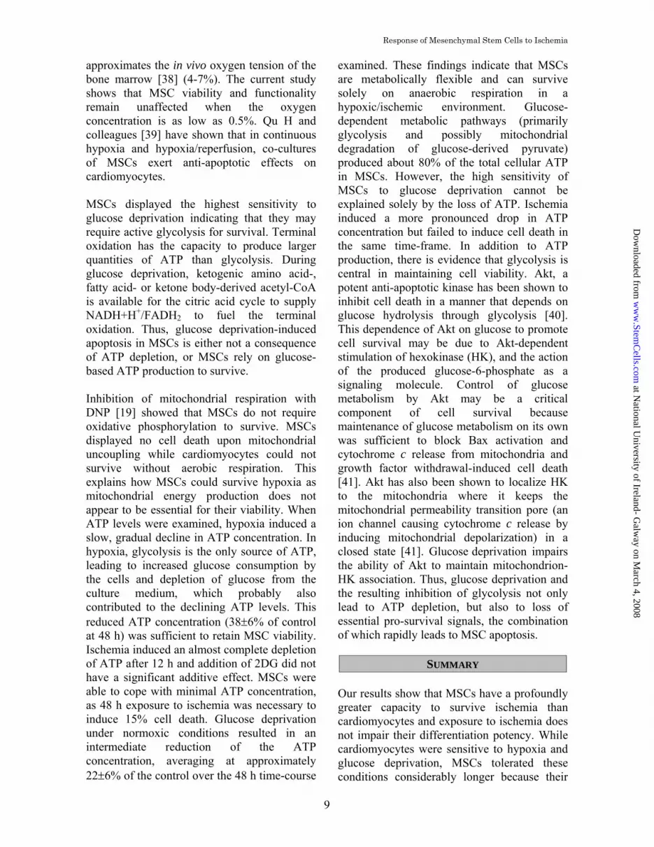
approximates the in vivo oxygen tension of the bone marrow [38] (4-7%). The current study shows that MSC viability and functionality remain unaffected when the oxygen concentration is as low as 0.5%. Qu H and colleagues [39] have shown that in continuous hypoxia and hypoxia/reperfusion, co-cultures of MSCs exert anti-apoptotic effects on cardiomyocytes. MSCs displayed the highest sensitivity to glucose deprivation indicating that they may require active glycolysis for survival. Terminal oxidation has the capacity to produce larger quantities of ATP than glycolysis. During glucose deprivation, ketogenic amino acid-, fatty acid- or ketone body-derived acetyl-CoA is available for the citric acid cycle to supply NADH+H+/FADH2 to fuel the terminal oxidation. Thus, glucose deprivation-induced apoptosis in MSCs is either not a consequence of ATP depletion, or MSCs rely on glucose-based ATP production to survive. Inhibition of mitochondrial respiration with DNP [19] showed that MSCs do not require oxidative phosphorylation to survive. MSCs displayed no cell death upon mitochondrial uncoupling while cardiomyocytes could not survive without aerobic respiration. This explains how MSCs could survive hypoxia as mitochondrial energy production does not appear to be essential for their viability. When ATP levels were examined, hypoxia induced a slow, gradual decline in ATP concentration. In hypoxia, glycolysis is the only source of ATP, leading to increased glucose consumption by the cells and depletion of glucose from the culture medium, which probably also contributed to the declining ATP levels. This reduced ATP concentration (38±6% of control at 48 h) was sufficient to retain MSC viability. Ischemia induced an almost complete depletion of ATP after 12 h and addition of 2DG did not have a significant additive effect. MSCs were able to cope with minimal ATP concentration, as 48 h exposure to ischemia was necessary to induce 15% cell death. Glucose deprivation under normoxic conditions resulted in an intermediate reduction of the ATP concentration, averaging at approximately 22±6% of the control over the 48 h time-course
examined. These findings indicate that MSCs are metabolically flexible and can survive solely on anaerobic respiration in a hypoxic/ischemic environment. Glucose-dependent metabolic pathways (primarily glycolysis and possibly mitochondrial degradation of glucose-derived pyruvate) produced about 80% of the total cellular ATP in MSCs. However, the high sensitivity of MSCs to glucose deprivation cannot be explained solely by the loss of ATP. Ischemia induced a more pronounced drop in ATP concentration but failed to induce cell death in the same time-frame. In addition to ATP production, there is evidence that glycolysis is central in maintaining cell viability. Akt, a potent anti-apoptotic kinase has been shown to inhibit cell death in a manner that depends on glucose hydrolysis through glycolysis [40]. This dependence of Akt on glucose to promote cell survival may be due to Akt-dependent stimulation of hexokinase (HK), and the action of the produced glucose-6-phosphate as a signaling molecule. Control of glucose metabolism by Akt may be a critical component of cell survival because maintenance of glucose metabolism on its own was sufficient to block Bax activation and cytochrome c release from mitochondria and growth factor withdrawal-induced cell death [41]. Akt has also been shown to localize HK to the mitochondria where it keeps the mitochondrial permeability transition pore (an ion channel causing cytochrome c release by inducing mitochondrial depolarization) in a closed state [41]. Glucose deprivation impairs the ability of Akt to maintain mitochondrion-HK association. Thus, glucose deprivation and the resulting inhibition of glycolysis not only lead to ATP depletion, but also to loss of essential pro-survival signals, the combination of which rapidly leads to MSC apoptosis.
SUMMARY Our results show that MSCs have a profoundly greater capacity to survive ischemia than cardiomyocytes and exposure to ischemia does not impair their differentiation potency. While cardiomyocytes were sensitive to hypoxia and glucose deprivation, MSCs tolerated these conditions considerably longer because their
at National U
niversity of Ireland- Galw
ay on March 4, 2008
ww
w.Stem
Cells.com
Dow
nloaded from

Response of Mesenchymal Stem Cells to Ischemia
10
metabolism could become independent of mitochondrial energy production. Of the different components of ischemia, MSCs showed the highest sensitivity to inhibition of glycolysis, and its inhibition led to accelerated cell death. In summary, MSCs showed a metabolic flexibility that may provide the necessary protection in an ischemic environment and allow MSCs to function in a reparative or regenerative capacity. These important features of MSCs may have considerable significance in improving the efficiency of stem cell therapy in cardiac disease.
ACKNOWLEDGMENTS We thank Garry Duffy, Caroline Curtin, Miriam Kearns, Aoife O’Reilly, Linda Howard and Georgina Shaw for technical assistance and Cynthia Coleman for critical reading of the manuscript. This work was supported in part by grants from the Science Foundation Ireland (SFI) and Medtronic Vascular Division, Santa Rosa, CA.
REFERENCES 1. Gill C, Mestril R, Samali A. Losing heart: the role of
apoptosis in heart disease--a novel therapeutic target? Faseb J 2002;16:135–146.
2. Logue SE, Gustafsson AB, Samali A et al. Ischemia/reperfusion injury at the intersection with cell death. J Mol Cell Cardiol 2005;38:21–33.
3. Reeve JL, Duffy AM, O'Brien T et al. Don't lose heart--therapeutic value of apoptosis prevention in the treatment of cardiovascular disease. J Cell Mol Med 2005;9:609–622.
4. Laflamme MA, Murry CE. Regenerating the heart. Nat Biotechnol 2005;23:845–856.
5. Barry FP, Murphy JM. Mesenchymal stem cells: clinical applications and biological characterization. Int J Biochem Cell Biol 2004;36:568–584.
6. Kocher AA, Schlechta B, Gasparovicova A et al. Stem cells and cardiac regeneration. Transpl Int 2007;20:731–746.
7. Prockop DJ, Gregory CA, Spees JL. One strategy for cell and gene therapy: harnessing the power of adult stem cells to repair tissues. Proc Natl Acad Sci U S A 2003;100 Suppl 1:11917–11923.
8. Spees JL, Olson SD, Ylostalo J et al. Differentiation, cell fusion, and nuclear fusion during ex vivo repair of epithelium by human adult stem cells from bone marrow stroma. Proc Natl Acad Sci U S A 2003;100:2397–2402.
9. Mackay AM, Beck SC, Murphy JM et al. Chondrogenic differentiation of cultured human mesenchymal stem cells from marrow. Tissue Eng 1998;4:415–428.
10. Neuhuber B, Gallo G, Howard L et al. Reevaluation of in vitro differentiation protocols for bone marrow stromal cells: disruption of actin cytoskeleton induces rapid morphological changes and mimics neuronal phenotype. J Neurosci Res 2004;77:192–204.
11. Friedenstein AJ, Chailakhyan RK, Gerasimov UV. Bone marrow osteogenic stem cells: in vitro cultivation and transplantation in diffusion chambers. Cell Tissue Kinet 1987;20:263–272.
12. Makino S, Fukuda K, Miyoshi S et al. Cardiomyocytes can be generated from marrow stromal cells in vitro. J Clin Invest 1999;103:697–705.
13. Noiseux N, Gnecchi M, Lopez-Ilasaca M et al. Mesenchymal stem cells overexpressing Akt dramatically repair infarcted myocardium and improve cardiac function despite infrequent cellular fusion or differentiation. Mol Ther 2006;14:840–850.
14. Toma C, Pittenger MF, Cahill KS et al. Human mesenchymal stem cells differentiate to a cardiomyocyte phenotype in the adult murine heart. Circulation 2002;105:93–98.
15. Shake JG, Gruber PJ, Baumgartner WA et al. Mesenchymal stem cell implantation in a swine myocardial infarct model: engraftment and functional effects. Ann Thorac Surg 2002;73:1919–1925; discussion 1926.
16. Wollert KC, Meyer GP, Lotz J et al. Intracoronary autologous bone-marrow cell transfer after myocardial infarction: the BOOST randomised controlled clinical trial. Lancet 2004;364:141–148.
17. Gao F, He T, Wang H et al. A promising strategy for the treatment of ischemic heart disease: Mesenchymal stem cell-mediated vascular endothelial growth factor gene transfer in rats. Can J Cardiol 2007;23:891–898.
18. Szegezdi E, Duffy A, O'Mahoney ME et al. ER stress contributes to ischemia-induced cardiomyocyte apoptosis. Biochem Biophys Res Commun 2006;349:1406–1411.
19. Mitchell P, Moyle J. Acid-base titration across the membrane system of rat-liver mitochondria. Catalysis by uncouplers. Biochem J 1967;104:588–600.
20. Reeve JL, Szegezdi E, Logue SE et al. Distinct mechanisms of cardiomyocyte apoptosis induced by doxorubicin and hypoxia converge on mitochondria and are inhibited by Bcl-xL. J Cell Mol Med 2007;11:509–520.
21. Szegezdi E, Cahill S, Meyer M et al. TRAIL sensitisation by arsenic trioxide is caspase-8
at National U
niversity of Ireland- Galw
ay on March 4, 2008
ww
w.Stem
Cells.com
Dow
nloaded from

Response of Mesenchymal Stem Cells to Ischemia
11
dependent and involves modulation of death receptor components and Akt. Br J Cancer 2006;94:398–406.
22. Holohan C, Szegezdi E, Ritter T et al. Cytokine-induced beta-cell apoptosis is no-dependent, mitochondria-mediated and inhibited by BCL-X(L). J Cell Mol Med 2007.
23. Yamashita N, Nishida M, Hoshida S et al. Induction of manganese superoxide dismutase in rat cardiac myocytes increases tolerance to hypoxia 24 hours after preconditioning. J Clin Invest 1994;94:2193–2199.
24. Tokalov SV, Gruener S, Schindler S et al. A number of bone marrow mesenchymal stem cells but neither phenotype nor differentiation capacities changes with age of rats. Mol Cells 2007;24:255–260.
25. Rovetto MJ, Lamberton WF, Neely JR. Mechanisms of glycolytic inhibition in ischemic rat hearts. Circ Res 1975;37:742–751.
26. Jennings RB, Reimer KA. The cell biology of acute myocardial ischemia. Annu Rev Med 1991;42:225–246.
27. Zhao ZQ. Oxidative stress-elicited myocardial apoptosis during reperfusion. Curr Opin Pharmacol 2004;4:159–165.
28. Zhang FB, Li L, Fang B et al. Passage-restricted differentiation potential of mesenchymal stem cells into cardiomyocyte-like cells. Biochem Biophys Res Commun 2005;336:784–792.
29. Zhu W, Chen J, Cong X et al. Hypoxia and serum deprivation-induced apoptosis in mesenchymal stem cells. Stem Cells 2006;24:416–425.
30. Ray J, Noll F, Daut J et al. Long-chain fatty acids increase basal metabolism and depolarize mitochondria in cardiac muscle cells. Am J Physiol Heart Circ Physiol 2002;282:H1495–1501.
31. Gnecchi M, He H, Noiseux N et al. Evidence supporting paracrine hypothesis for Akt-modified mesenchymal stem cell-mediated cardiac protection and functional improvement. Faseb J 2006;20:661–669.
32. Li W, Ma N, Ong LL et al. Bcl-2 Engineered MSCs Inhibited Apoptosis and Improved Heart Function. Stem Cells 2007.
33. Mangi AA, Noiseux N, Kong D et al. Mesenchymal stem cells modified with Akt prevent remodeling and restore performance of infarcted hearts. Nat Med 2003;9:1195–1201.
34. Grayson WL, Zhao F, Bunnell B et al. Hypoxia enhances proliferation and tissue formation of human mesenchymal stem cells. Biochem Biophys Res Commun 2007;358:948–953.
35. Ren H, Cao Y, Zhao Q et al. Proliferation and differentiation of bone marrow stromal cells under hypoxic conditions. Biochem Biophys Res Commun 2006;347:12–21.
36. Grayson WL, Zhao F, Izadpanah R et al. Effects of hypoxia on human mesenchymal stem cell expansion and plasticity in 3D constructs. J Cell Physiol 2006;207:331–339.
37. Lennon DP, Edmison JM, Caplan AI. Cultivation of rat marrow-derived mesenchymal stem cells in reduced oxygen tension: effects on in vitro and in vivo osteochondrogenesis. J Cell Physiol 2001;187:345–355.
38. Kofoed H, Sjontoft E, Siemssen SO et al. Bone marrow circulation after osteotomy. Blood flow, pO2, pCO2, and pressure studied in dogs. Acta Orthop Scand 1985;56:400–403.
39. Qu H, Guo YH, Zhu XJ et al. [Anti-apoptotic effects of mesenchymal stem cells on cardiac myocytes: in vitro study with rats]. Zhonghua Yi Xue Za Zhi 2007;87:271–274.
40. Rathmell JC, Fox CJ, Plas DR et al. Akt-directed glucose metabolism can prevent Bax conformation change and promote growth factor-independent survival. Mol Cell Biol 2003;23:7315–7328.
41. Majewski N, Nogueira V, Bhaskar P et al. Hexokinase-mitochondria interaction mediated by Akt is required to inhibit apoptosis in the presence or absence of Bax and Bak. Mol Cell 2004;16:819–830.
at National U
niversity of Ireland- Galw
ay on March 4, 2008
ww
w.Stem
Cells.com
Dow
nloaded from

Response of Mesenchymal Stem Cells to Ischemia
12
Figure 1: MSCs are more resistant to ischemia than cardiomyocytes Cells were subjected to hypoxia in the absence of serum and glucose for varying time points. (A) Images of H&E stained cells. The arrows indicate apoptotic bodies in cardiomyocytes, MSCs and fibroblasts (overall magnification, 100X). (B-D) Representative FACS quantitation of early apoptotic (Annexin-V+/PI-; black bar) and late apoptotic/necrotic (Annexin-V+/PI+; white bar) cells after exposure to different times of ischemia. (B) cardiomyocytes, (C) MSCs and (D) fibroblasts. CHX was used in combination with FasL to induce apoptotic cell death as a positive control. The presented graphs are representatives of three independent experiments. (E-G) Caspase-3-like activity in ischemic cardiomyocytes (E), MSCs (F) and fibroblasts (G) measured by DEVDase assay. Enzyme activity is expressed as fold activation compared to controls. STS, an inducer of apoptotic cell death was used as a positive control for caspase-3-like activity. Data are shown as representative samples or as mean ±SEM, n=3. The (*) denotes a statistically significant difference (p<0.05) compared to the control.
at National U
niversity of Ireland- Galw
ay on March 4, 2008
ww
w.Stem
Cells.com
Dow
nloaded from

Response of Mesenchymal Stem Cells to Ischemia
13
at National U
niversity of Ireland- Galw
ay on March 4, 2008
ww
w.Stem
Cells.com
Dow
nloaded from

Response of Mesenchymal Stem Cells to Ischemia
14
Figure 2: Inhibition of glycolysis with 2DG increases sensitivity of MSCs to ischemia Cells were cultured with 1 mM 2DG in the absence of serum and glucose in hypoxia (ischemia+2DG) for varying time-points. (A) Images of H&E stained cells. The arrows indicate apoptotic bodies in cardiomyocytes, MSCs and fibroblasts (overall magnification, 100X). (B-D) Representative FACS quantitation of early apoptotic (Annexin-V+/PI-; black bar) and late apoptotic/necrotic (Annexin-V+/PI+; white bar) cells after exposure to different times of ischemia+2DG. (B) cardiomyocytes, (C) MSCs and (D) fibroblasts. CHX was used in combination with FasL to induce apoptotic cell death as a positive control. The presented graphs are representatives of three independent experiments. (E-G) Caspase-3-like activity in ischemic cardiomyocytes (E), MSCs (F) and fibroblasts (G) measured by DEVDase assay. Enzyme activity is expressed as fold activation compared to controls. STS, an inducer of apoptotic cell death was used as a positive control for caspase-3-like activity. Data are shown as representative samples or as mean ±SEM, n=3. The (*) denotes a statistically significant difference (p<0.05) compared to the control.
at National U
niversity of Ireland- Galw
ay on March 4, 2008
ww
w.Stem
Cells.com
Dow
nloaded from

Response of Mesenchymal Stem Cells to Ischemia
15
at National U
niversity of Ireland- Galw
ay on March 4, 2008
ww
w.Stem
Cells.com
Dow
nloaded from

Response of Mesenchymal Stem Cells to Ischemia
16
Figure 3: MSCs retain their differentiation potential after exposure to ischemia MSCs were subjected to ischemia + 2DG for 6-12 h. Cells were then induced to differentiate along the adipogenic, chondrogenic and cardiomyogenic pathway under normoxic conditions. (A) Representative images of Oil Red O stained MSCs cultured in adipogenic medium. Overall magnification was 400x. (B) Quantitation of Oil Red O uptake by photometry. The graph shows the absorbance values at 490 nm. (C) Glycosaminoglycan production by control and ischemia+2DG -treated MSCs after induction of chondrogenesis. The graph shows the averaged GAG concentration per 1 μg DNA. The (*) denotes a statistically significant difference (p<0.05) compared to the control. (D) Effect of ischemia+2DG on the induction of cardiac troponinI (cTnI), Nkx2.5 and α-myosin heavy chain (αMHC) in MSCs exposed to 5-azacytidine treatment. Induction of cardiomyogenic marker mRNAs was detected by RT-PCR. RNA from H9c2 rat embryonic cardiomyocytes was used as a positive control. GAPDH was used to confirm equal loading. The presented images are representatives of three independent experiments.
at National U
niversity of Ireland- Galw
ay on March 4, 2008
ww
w.Stem
Cells.com
Dow
nloaded from

Response of Mesenchymal Stem Cells to Ischemia
17
Figure 4: MSCs are resistant to hypoxia Cells were subjected to hypoxia for varying time-points. (A) Images of H&E stained cells. The arrows indicate apoptotic bodies in cardiomyocyte and fibroblast cultures (overall magnification, 100X). (B-D) Caspase-3-like activity in hypoxic cardiomyocytes (B), MSCs (C) and fibroblasts (D) measured by DEVDase assay. Enzyme activity is expressed as fold activation compared to controls. STS, an inducer of apoptotic cell death was used as a positive control for caspase-3-like activity. Data are shown as mean ± SEM, n=3. (E and F) MSCs were subjected to hypoxia for 48 h, then induced to differentiate into adipocytes and chondrocytes under normoxic conditions. (E) Quantitation of Oil Red O uptake by photometry. The graph shows the absorbance values at 490 nm. (F) Glycosaminoglycan production by control and hypoxia-treated MSCs after induction of chondrogenesis. The graph shows the averaged GAG concentration per 1 μg DNA. Data are shown as representative samples or as mean ±SEM, n=3. The (*) denotes a statistically significant difference (p<0.05) compared to the control. (G) Effect of hypoxia on the induction of cardiac troponinI (cTnI), Nkx2.5 and α-myosin heavy chain (αMHC) in MSCs exposed to 5-azacytidine treatment. Induction of cardiomyogenic marker mRNAs was detected by RT-PCR. RNA from H9c2 rat embryonic cardiomyocytes was used as a positive control. GAPDH was used to confirm equal loading. The presented images are representatives of three independent experiments.
at National U
niversity of Ireland- Galw
ay on March 4, 2008
ww
w.Stem
Cells.com
Dow
nloaded from

Response of Mesenchymal Stem Cells to Ischemia
18
at National U
niversity of Ireland- Galw
ay on March 4, 2008
ww
w.Stem
Cells.com
Dow
nloaded from

Response of Mesenchymal Stem Cells to Ischemia
19
Figure 5: Effect of glucose deprivation on cardiomyocyte, MSC and fibroblast cell viability Cells were cultured with 1 mM 2DG in the absence of serum and glucose for varying times under normoxic conditions. (A) Images of H&E stained cells. The arrows indicate apoptotic bodies in cardiomyocyte, MSC and fibroblast cultures (overall magnification, 100X). (B-D) Caspase-3-like activity induced by glucose deprivation in cardiomyocytes (B), MSCs (C) and fibroblasts (D) measured by DEVDase assay. Enzyme activity is expressed as fold activation compared to controls. STS, an inducer of apoptotic cell death was used as a positive control for caspase-3-like activity. Data are shown as representative samples (A) or as mean ±SEM, n=3. The (*) denotes a statistically significant difference (p<0.05) compared to the control.
at National U
niversity of Ireland- Galw
ay on March 4, 2008
ww
w.Stem
Cells.com
Dow
nloaded from

Response of Mesenchymal Stem Cells to Ischemia
20
Figure 6: Effect of 2,4-dinitrophenol on cardiomyocyte, MSC, and fibroblast survival Cells were cultured in the presence of 0.5 μM DNP for varying times under normoxic conditions. (A) Loss of mitochondrial inner membrane potential (ΔΨm) measured with TMRE staining in cardiomyocytes (upper panel) and MSCs (lower panel) (B) Representative FACS analysis of cell death induced by 0.5 mM DNP after in cardiomyocytes (upper panel) and MSCs (lower panel). (C) Quantitation of cell death induced by 0.5 mM DNP in cardiomyocytes and MSCs with Annexin-V assay. The graph shows the percentage of live, Annexin-V-/PI- cells after increasing times of DNP (0.5 mM) treatment. Data shown are representatives of three independent experiments.
at National U
niversity of Ireland- Galw
ay on March 4, 2008
ww
w.Stem
Cells.com
Dow
nloaded from

Response of Mesenchymal Stem Cells to Ischemia
21
at National U
niversity of Ireland- Galw
ay on March 4, 2008
ww
w.Stem
Cells.com
Dow
nloaded from

Response of Mesenchymal Stem Cells to Ischemia
22
Figure 7: Effect of ischemia and its components on MSC ATP concentration MSCs were exposed to hypoxia, glucose deprivation, ischemia or ischemia+2DG for 12-48 h. After treatment cells were lysed immediately and intracellular ATP concentration was determined. The graph shows the average ATP concentration of three independent experiments as fraction of the ATP concentration in untreated cells ± SEM, n=3.
at National U
niversity of Ireland- Galw
ay on March 4, 2008
ww
w.Stem
Cells.com
Dow
nloaded from

DOI: 10.1634/stemcells.2007-1072 published online Feb 28, 2008; Stem Cells
Frank Barry and Eva Szegezdi Louise A. Mylotte, Angela M. Duffy, Mary Murphy, Timothy O'Brien, Afshin Samali,
Metabolic Flexibility Permits MSC Survival in an Ischemic Environment
This information is current as of March 4, 2008
& ServicesUpdated Information
http://www.StemCells.comincluding high-resolution figures, can be found at:
Supplementary Material http://www.StemCells.com/cgi/content/full/2007-1072/DC1
Supplementary material can be found at:
at National U
niversity of Ireland- Galw
ay on March 4, 2008
ww
w.Stem
Cells.com
Dow
nloaded from
Related Documents