Journal of Extracellular Vesicles ISSN: (Print) 2001-3078 (Online) Journal homepage: http://www.tandfonline.com/loi/zjev20 Obstacles and opportunities in the functional analysis of extracellular vesicle RNA – an ISEV position paper Bogdan Mateescu, Emma J. K. Kowal, Bas W. M. van Balkom, Sabine Bartel, Suvendra N. Bhattacharyya, Edit I. Buzás, Amy H. Buck, Paola de Candia, Franklin W. N. Chow, Saumya Das, Tom A. P. Driedonks, Lola Fernández- Messina, Franziska Haderk, Andrew F. Hill, Jennifer C. Jones, Kendall R. Van Keuren-Jensen, Charles P. Lai, Cecilia Lässer, Italia di Liegro, Taral R. Lunavat, Magdalena J. Lorenowicz, Sybren L. N. Maas, Imre Mäger, Maria Mittelbrunn, Stefan Momma, Kamalika Mukherjee, Muhammed Nawaz, D. Michiel Pegtel, Michael W. Pfaffl, Raymond M. Schiffelers, Hidetoshi Tahara, Clotilde Théry, Juan Pablo Tosar, Marca H. M. Wauben, Kenneth W. Witwer & Esther N. M. Nolte-‘t Hoen To cite this article: Bogdan Mateescu, Emma J. K. Kowal, Bas W. M. van Balkom, Sabine Bartel, Suvendra N. Bhattacharyya, Edit I. Buzás, Amy H. Buck, Paola de Candia, Franklin W. N. Chow, Saumya Das, Tom A. P. Driedonks, Lola Fernández-Messina, Franziska Haderk, Andrew F. Hill, Jennifer C. Jones, Kendall R. Van Keuren-Jensen, Charles P. Lai, Cecilia Lässer, Italia di Liegro, Taral R. Lunavat, Magdalena J. Lorenowicz, Sybren L. N. Maas, Imre Mäger, Maria Mittelbrunn, Stefan Momma, Kamalika Mukherjee, Muhammed Nawaz, D. Michiel Pegtel, Michael W. Pfaffl, Raymond M. Schiffelers, Hidetoshi Tahara, Clotilde Théry, Juan Pablo Tosar, Marca H. M. Wauben, Kenneth W. Witwer & Esther N. M. Nolte-‘t Hoen (2017) Obstacles and opportunities in the functional analysis of extracellular vesicle RNA – an ISEV position paper, Journal of Extracellular Vesicles, 6:1, 1286095, DOI: 10.1080/20013078.2017.1286095 To link to this article: http://dx.doi.org/10.1080/20013078.2017.1286095 © 2017 The Author(s). Published by Informa UK Limited, trading as Taylor & Francis Group. Published online: 07 Mar 2017. Submit your article to this journal View related articles

Welcome message from author
This document is posted to help you gain knowledge. Please leave a comment to let me know what you think about it! Share it to your friends and learn new things together.
Transcript

Journal of Extracellular Vesicles
ISSN: (Print) 2001-3078 (Online) Journal homepage: http://www.tandfonline.com/loi/zjev20
Obstacles and opportunities in the functionalanalysis of extracellular vesicle RNA – an ISEVposition paper
Bogdan Mateescu, Emma J. K. Kowal, Bas W. M. van Balkom, Sabine Bartel,Suvendra N. Bhattacharyya, Edit I. Buzás, Amy H. Buck, Paola de Candia,Franklin W. N. Chow, Saumya Das, Tom A. P. Driedonks, Lola Fernández-Messina, Franziska Haderk, Andrew F. Hill, Jennifer C. Jones, Kendall R. VanKeuren-Jensen, Charles P. Lai, Cecilia Lässer, Italia di Liegro, Taral R. Lunavat,Magdalena J. Lorenowicz, Sybren L. N. Maas, Imre Mäger, Maria Mittelbrunn,Stefan Momma, Kamalika Mukherjee, Muhammed Nawaz, D. Michiel Pegtel,Michael W. Pfaffl, Raymond M. Schiffelers, Hidetoshi Tahara, Clotilde Théry,Juan Pablo Tosar, Marca H. M. Wauben, Kenneth W. Witwer & Esther N. M.Nolte-‘t Hoen
To cite this article: Bogdan Mateescu, Emma J. K. Kowal, Bas W. M. van Balkom, SabineBartel, Suvendra N. Bhattacharyya, Edit I. Buzás, Amy H. Buck, Paola de Candia, Franklin W.N. Chow, Saumya Das, Tom A. P. Driedonks, Lola Fernández-Messina, Franziska Haderk,Andrew F. Hill, Jennifer C. Jones, Kendall R. Van Keuren-Jensen, Charles P. Lai, Cecilia Lässer,Italia di Liegro, Taral R. Lunavat, Magdalena J. Lorenowicz, Sybren L. N. Maas, Imre Mäger,Maria Mittelbrunn, Stefan Momma, Kamalika Mukherjee, Muhammed Nawaz, D. Michiel Pegtel,Michael W. Pfaffl, Raymond M. Schiffelers, Hidetoshi Tahara, Clotilde Théry, Juan Pablo Tosar,Marca H. M. Wauben, Kenneth W. Witwer & Esther N. M. Nolte-‘t Hoen (2017) Obstacles andopportunities in the functional analysis of extracellular vesicle RNA – an ISEV position paper,Journal of Extracellular Vesicles, 6:1, 1286095, DOI: 10.1080/20013078.2017.1286095
To link to this article: http://dx.doi.org/10.1080/20013078.2017.1286095
© 2017 The Author(s). Published by InformaUK Limited, trading as Taylor & FrancisGroup.
Published online: 07 Mar 2017.
Submit your article to this journal
View related articles

Full Terms & Conditions of access and use can be found athttp://www.tandfonline.com/action/journalInformation?journalCode=zjev20
Download by: [195.147.74.213] Date: 08 March 2017, At: 01:37
View Crossmark data

ORIGINAL RESEARCH ARTICLE
Obstacles and opportunities in the functional analysis of extracellular vesicle RNA– an ISEV position paperBogdan Mateescua*, Emma J. K. Kowal b*, Bas W. M. van Balkomc, Sabine Barteld, Suvendra N. Bhattacharyyae,Edit I. Buzásf, Amy H. Buckg, Paola de Candiah, Franklin W. N. Chowg, Saumya Dasi, Tom A. P. Driedonksj,Lola Fernández-Messinak, Franziska Haderkl,m, Andrew F. Hill n, Jennifer C. Jones o, Kendall R. Van Keuren-Jensenp, Charles P. Laiq, Cecilia Lässerr,s, Italia di Liegrot, Taral R. Lunavatr,s, Magdalena J. Lorenowiczu,Sybren L. N. Maasv, Imre Mägerw,x, Maria Mittelbrunny, Stefan Mommaz, Kamalika Mukherjeee, Muhammed Nawazaa,D. Michiel Pegtelab, Michael W. Pfafflac, Raymond M. Schiffelersad, Hidetoshi Taharaae, Clotilde Théryaf,Juan Pablo Tosarag, Marca H. M. Waubenj, Kenneth W. Witwer ah and Esther N. M. Nolte-‘t Hoenj
aDepartment of Biology, Swiss Federal Institute of Technology Zurich (ETH Zürich), Zurich, Switzerland; bDepartment of Biology, MassachusettsInstitute of Technology, Cambridge, MA, USA; cDepartment of Nephrology and Hypertension, UMC Utrecht, Utrecht, the Netherlands; dExperimentalAsthma Research, Priority Area Asthma& Allergy, Research Center Borstel, Leibniz-Center forMedicine and Biosciences, Airway Research Center North(ARCN), Member of the German Center for Lung Research (DZL), Borstel, Germany; eDepartment of Science and Technology, CSIR-Indian Institute ofChemical Biology, Kolkata, India; fDepartment of Genetics, Cell- and Immunobiology, Semmelweis University, Budapest, Hungary; gInstitute ofImmunology and Infection Research, Centre for Immunity, Infection and Evolution, School of Biological Sciences, University of Edinburgh, Edinburgh,UK; hIRCCS MultiMedica, Milan, Italy; iCardiovascular Research Institute, Massachusetts General Hospital, Boston, MA, USA; jDepartment ofBiochemistry & Cell Biology, Faculty of Veterinary Medicine, Utrecht University, Utrecht, the Netherlands; kImmunology Service, Hospital La Princesa,Madrid, Spain; lDepartment of Molecular Genetics, German Cancer Research Center (DKFZ), Heidelberg, Germany; mDepartment of Medicine, HelenDiller Family Comprehensive Cancer Center, UC San Francisco, San Francisco, CA, USA; nDepartment of Biochemistry and Genetics, La Trobe Institutefor Molecular Science, La Trobe University, Bundoora, Australia; oMolecular Immunogenetics & Vaccine Research Section, Vaccine Branch, CCR, NCI,Bethesda, MD, USA; pNeurogenomics Division, TGen, Phoenix, AZ, USA; qInstitute of Biomedical Engineering, National Tsing Hua University, Hsinchu,Taiwan; rDepartment of Neurology and Center for Molecular Imaging Research, Department of Radiology, Massachusetts General Hospital andNeuroDiscovery Center, Harvard Medical School, Boston, MA, USA; sKrefting Research Centre, Department of Internal Medicine and Clinical Nutrition,Sahlgrenska Academy, University of Gothenburg, Gothenburg, Sweden; tDepartment of Experimental Biomedicine and Clinical Neurosciences(BIONEC), University of Palermo, Palermo, Italy; uCenter for Molecular Medicine, University Medical Center Utrecht & Regenerative Medicine Center,Utrecht, the Netherlands; vDepartment of Neurology and Center for Molecular Imaging Research, Department of Radiology, Massachusetts GeneralHospital and NeuroDiscovery Center, Harvard Medical School, Boston, MA, USA; wDepartment of Physiology, Anatomy and Genetics, University ofOxford, Oxford, UK; xInstitute of Technology, University of Tartu, Tartu, Estonia; yInstituto de Investigación del Hospital 12 de Octubre, Madrid, Spain;zInstitute of Neurology (Edinger Institute), Frankfurt University Medical School, Frankfurt amMain, Germany; aaDepartment of Pathology and ForensicMedicine, Ribeirão Preto School of Medicine, University of Sao Paulo, Sao Paulo, Brazil; abDepartment of Pathology, Exosomes Research Group, VUUniversityMedical Center, Amsterdam, the Netherlands; acAnimal Physiology and Immunology, School of Life Sciences, Technical University ofMunich(TUM) Weihenstephan, Freising, Germany; adLaboratory Clinical Chemistry & Haematology, University Medical Center Utrecht, Utrecht, theNetherlands; aeDepartment of Cellular and Molecular Biology, Institute of Biomedical & Health Sciences, Hiroshima University, Hiroshima, Japan;afInstitut Curie, PSL Research University, INSERM U932, Paris, France; agFunctional Genomics Unit, Institut Pasteur de Montevideo, Nuclear ResearchCenter, Faculty of Science, Universidad de la República, Montevideo, Uruguay; ahDepartment of Molecular and Comparative Pathobiology andDepartment of Neurology, The Johns Hopkins University School of Medicine Baltimore, MD, USA
ABSTRACTThe release of RNA-containing extracellular vesicles (EV) into the extracellular milieu has been demon-strated in a multitude of different in vitro cell systems and in a variety of body fluids. RNA-containing EVare in the limelight for their capacity to communicate genetically encodedmessages to other cells, theirsuitability as candidate biomarkers for diseases, and their use as therapeutic agents. Although EV-RNAhas attracted enormous interest frombasic researchers, clinicians, and industry,we currently have limitedknowledge on which mechanisms drive and regulate RNA incorporation into EV and on how RNA-encoded messages affect signalling processes in EV-targeted cells. Moreover, EV-RNA research facesvarious technical challenges, such as standardisation of EV isolationmethods, optimisation ofmethodol-ogies to isolate and characterise minute quantities of RNA found in EV, and development of approachesto demonstrate functional transfer of EV-RNA in vivo. These topics were discussed at the 2015 EV-RNAworkshop of the International Society for Extracellular Vesicles. This position paper was written by theparticipants of the workshop not only to give an overview of the current state of knowledge in the field,but also to clarify that our incomplete knowledge – of the nature of EV(-RNA)s and of how to effectivelyand reliably study them – currently prohibits the implementation of gold standards in EV-RNA research.In addition, this paper creates awareness of possibilities and limitations of currently used strategies toinvestigate EV-RNA and calls for caution in interpretation of the obtained data.
ARTICLE HISTORYReceived 19 October 2016Accepted 25 December 2016
RESPONSIBLE EDITORYong Song Gho, IntercellularCommunications NetworkLab, Republic of Korea
KEYWORDSExtracellular vesicles;exosomes; non-coding RNA;mRNA; function; sorting;RNA binding proteins;quantification
CONTACT Esther N. M. Nolte-‘t Hoen [email protected] Department of Biochemistry & Cell Biology, Faculty of Veterinary Medicine, UtrechtUniversity, Yalelaan 2, 3584CM Utrecht, the Netherlands
*These authors contributed equally to this workExcept for Bogdan Mateescu, Emma Kowal, and Esther Nolte-‘t Hoen, authors are sorted alphabetically based on their last names
JOURNAL OF EXTRACELLULAR VESICLES, 2017VOL. 6, 1286095http://dx.doi.org/10.1080/20013078.2017.1286095
© 2017 The Author(s). Published by Informa UK Limited, trading as Taylor & Francis Group.This is an Open Access article distributed under the terms of the Creative Commons Attribution-NonCommercial License (http://creativecommons.org/licenses/by-nc/4.0/), whichpermits unrestricted non-commercial use, distribution, and reproduction in any medium, provided the original work is properly cited.

Introduction
Extracellular vesicles (EVs), including exosomes andmicrovesicles, are released into the extracellular spaceby many cell types. EVs carry a repertoire of bioactivemolecules, including proteins, nucleic acids, lipids andcarbohydrates.[1] Their role has been repeatedly high-lighted in cell-to-cell communication and lately theyhave been implicated in the progression of severaldiseases, including neurodegenerative, cardiovascularand infectious diseases as well as cancer.[2–6] EVs arepresent in various body fluids and since the molecularcontent of EVs reflects the type and activation status oftheir parent cell, they are regarded as potent biomar-kers for disease.[5,7,8] Additionally, EVs are beingexplored as delivery vehicles for therapeutic purposes.In the last 10 years it has become established that EVscontain RNA molecules, and thereby represent a vehi-cle through which cells may transfer geneticallyencoded messages to other cells. Although other extra-cellular carriers of RNA also exist, the purpose of thisarticle is to highlight particularities in the study of EV-associated RNA. Current research in the EV field aimsto characterise the RNA content of EVs and the detailsof its delivery in vitro and in vivo. Although this fieldhas attracted enormous interest spanning basicresearch, clinics, and industry, understanding of manyaspects of the formation and function of RNA-contain-ing EVs remains elusive. A lack of standardisation withregard to EV purification and characterisation of theirmolecular contents, as well as technical difficulties inunequivocally demonstrating that EV-RNA is a causa-tive agent in EV-mediated effects on target cells, areamong the present challenges to this field.
Following up on the first workshop organised by theInternational Society for Extracellular Vesicles (ISEV)on “Extracellular Vesicle RNA” (New York, 2012),[9]the society recently organised a second workshop toaddress the question: “EV-associated RNA: is there apurpose?”. This workshop, held in September 2015 inUtrecht, the Netherlands, brought together 70 interna-tional experts in the field – including principal inves-tigators, postdocs, and PhD students as well asrepresentatives from industry – to share knowledgeand technical expertise on how to address the natureand function of EV-associated RNA.
Below, we report on the topics that were addressedduring the workshop and substantiate them with refer-ences to recent literature. We raise awareness of var-ious factors affecting EV-RNA characterisation (e.g. EVpurity and biases in RNA sequencing methodologies),discuss the heterogeneity in RNA content of EV,describe both active and passive RNA sorting processes
which have been suggested to determine the RNAcontent of EVs, and conclude that providing undispu-table evidence that RNA mediates EV-induced effectsremains difficult with currently available methodolo-gies. In reporting this discussion we provide research-ers from both inside and outside of the EV communitywith a critical overview of the current status of the EV-RNA research field and an outlook to future challenges.
1. Purification of EVs prior to EV-RNA isolation
1.1. New insights in the heterogeneity of EV andtheir RNA content
At the time of the previous ISEV workshop on EV-RNA in 2012, the research field had already recognisedthe heterogeneous nature of vesicles present in theextracellular environment.[10] The typically presentedclassification divided EVs into two subtypes: EVs withdiameters ≤100–150 nm and buoyant densities of 1.11–1.19 g ml–1 that are formed inside multivesicular bodies(MVBs) were defined as exosomes, while EVs withdiameters ranging up to 1000 nm which presumablybud from the plasma membrane were variably calledectosomes, microvesicles, microparticles, or large onco-somes. The exact definitions varied widely betweenpublications and overlap in sizes between these twocategories was not generally commented on.
It is clear that these categories represent an over-simplification. For instance, EVs formed directly at theplasma membrane can share several biophysical prop-erties with EVs formed in MVBs, such as size, isolationby high-speed ultracentrifugation, and floatation indensity gradients at the expected 1.11–1.19 g ml–1
position.[11] Thus, EVs in the small size range likelyrepresent vesicles heterogeneous in origin, with anunknown portion coming from MVBs. The definitionof larger EVs is even less precise, and these vesiclescomprise a wide range of membrane-enclosed entities.Indeed, it is not yet clear how to divide EVs into theirrelevant subtypes, or even how many functionally dis-tinct subtypes there may be.
Several laboratories have now started characterisingthe protein composition of subtypes of EVs. EV sub-types have been isolated by a number of means, includ-ing recovery at different centrifugation speeds, throughdifferent filters, at slightly different positions in densitygradients, via immuno-isolation by different surfacemolecules, or by flow cytometric sorting.[12–20]
Extensive comparison of these results (obtained withEVs from different cellular sources) has not yet beenperformed, but the available data already indicate thatsome proteins classically regarded as “exosome
2 B. MATEESCU ET AL.

markers” are in fact present in all different EV types(e.g. heat shock proteins, flotillins and major histocom-patibility complex molecules). Such intracellular ormembrane-associated proteins can therefore be usedas EV markers, but will not define the nature of theEV subtype analysed. Furthermore, within the “exo-some” population, subsets could be defined based oncombinations of protein markers which colocalise orare co-depleted in vesicles enriched in endosomal pro-teins vs. plasma membrane proteins.[12,19] Given thedifficulty of separating subtypes of EVs with ultracen-trifugation, Thery’s group has recently chosen to referto vesicles sedimenting at 100,000 g as “small EVs”(sEVs) rather than exosomes, those pelleting at inter-mediate speed (lower than 20,000 g) as “medium EVs”(mEVs, including microvesicles, ectosomes) and thosepelleting at low-speed (e.g. 2000 g) as “large EVs”(lEVs, including large fragments of the releasing celland large apoptotic bodies). EVs of small size are alsoenriched in many studies by the use of filters of smallpore size (100 or 220 nm). These definitions are per-haps less biologically meaningful but far more experi-mentally tractable than the previous exosome/microvesicle definitions, since EV size, often deter-mined by nanoparticle tracking analysis or electronmicroscopy, is frequently reported in EV studies.Until we have stringent and robust methods for separa-tion and characterisation of MVB vs. plasma mem-brane-derived EVs, the proposed nomenclature couldincrease clarity of discussion and ease of cross-referen-cing between studies. However, consensus has not beenreached on this issue and the current nomenclature istherefore maintained until further notice.
It is still unknown whether all EVs contain RNA andhow diverse the RNA content of different EV subpopu-lations may be. Various studies indicate that the RNAcontent of EVs varies among cell types and among EVsubpopulations. For example, miR-145 is present at verylow levels in HepG2 cell-derived large EVs, whereas thesame miRNA is present at significant levels in both largeand small EVs derived from A549 cells.[21] In immunecells, the miRNA content of EVs was shown to differ byimmune cell type when comparing EVs from B- or T-celllines and primary dendritic cells (DC).[22] Anotherremarkable example of EV-RNA heterogeneity is thesex difference observed in the miRNA content of urinaryEVs.[23] With regard to the RNA content of differentEV subpopulations, it was shown that EV populationsthat separated into different fractions based on pelletingat different g-forces differed in RNA content.[24] EvenEVs sedimenting at the same g-force are heterogeneousin nature and may be further separated based on differ-ences in migration velocity in density gradients; recent
data indicate that EV subpopulations isolated based onthis parameter differ in both protein and RNA content.[20] In a recent study, the copy number of a givenmiRNA molecule was suggested to be on average lowerthan one per vesicle/particle in an EV sample.[25] If weassume that all of the detected miRNA species wereindeed EV-associated and that EV quantifications wereaccurate, one explanation for these data is that specificmiRNA sequences could be restricted to specific sub-types of EVs. This scenario would be consistent with ahigh specificity in delivery of RNA molecules to targetcells (see also section 4.1.2).
The presence of extracellular RNA circulating innon-EV-associated forms, for instance in large protein(e.g. Argonaute 2 (AGO2)) or lipoprotein complexes,adds another layer of complexity to the analysis of EV-RNA. These complexes have been shown to co-isolatewith EVs during common isolation procedures such asultracentrifugation [26] (see section 1.3.1). Thus it ispossible that, of the numerous types of nucleic acidsdescribed “in EVs” in the existing literature, some arecontained within specific subtypes of EVs and some areperhaps not present in EVs at all but exclusively inother carriers which co-isolate. A particular point ofconcern is the potential carryover of extracellular RNAoriginating from foetal bovine serum used in cell cul-ture media, which, if not effectively removed, affectsthe analysis of EV-RNA released by the cell of interest.[27,28] This urges the need for including control iso-lates from non-conditioned culture medium in theRNA analysis.
1.2. Update on EV purification methods andeffects on EV-RNA analysis
Participants of the meeting expressed their concernabout the enduring lack of standardisation with regardto the collection, storage, and processing of EV-con-taining fluids, and the large diversity of methods usedfor EV isolation. Each of these factors influences notonly the type and number of EV isolated, but also thelevel of contamination with non-EV-associated RNA inthe obtained EV preparation.
Though differential centrifugation remains the mostcommonly used method of EV isolation, several differ-ent techniques have risen in prominence since the 2012meeting on EV-RNA (Figure 1). Pros and cons ofcommonly used EV isolation techniques have beenreviewed more elaborately elsewhere (see for example[29,30]), but are briefly summarised below. Differentialcentrifugation can be followed by density gradientultracentrifugation to separate low-density EVs fromhigh-density protein aggregates that often contaminate
JOURNAL OF EXTRACELLULAR VESICLES 3

EV ultracentrifugation pellets. Size exclusion chroma-tography (SEC) is now a more widely used techniquefor EV isolation. It can be used for low volume samplesand allows separation of EV from the bulk of solubleproteins. However, since separation is purely based onparticle size, contaminating particles in the EV sizerange such as (lipo)protein complexes may be co-iso-lated. Immuno-affinity capture presents an alternativemethod for EV isolation. The method can yield pureEV subpopulations, but is highly influenced by boththe choice of affinity reagent and the ligand density on
different EV types. In addition, various commercial kitsthat make use of volume-excluding polymers such aspolyethylene glycol (PEG) are currently available forrapid EV isolation from culture media or body fluids.However, such polymers co-precipitate protein (com-plexes) that contaminate EV isolates. It was highlightedduring the meeting that the different EV isolationtechniques are based on different principles and willtherefore enrich for different subpopulations of vesi-cles. In addition, these methods co-isolate contami-nants (e.g. protein complexes and lipoproteins) to
Figure 1. Schematic illustration of commonly used EV isolation techniques. (a) Differential centrifugation is the sequential pelletingof particles with decreasing sedimentation coefficients. Typically 2000 g is used to pellet large EVs, 10,000–20,000 g to pelletmiddle-sized EVs (green), and finally ~100,000 g to pellet the smallest EVs (different EV subpopulations are indicated in grey andorange). At these high g-forces, complexes of soluble proteins (black dots) may also sediment. (b) Lipids have a density that isapproximately 1 g cm–3, while proteins and RNA have a higher density (>1.3 g cm–3). Therefore density gradients can be used toseparate subpopulations of EVs with different ratio of lipids, RNA, and proteins. Moreover, these gradients can be used to purifyvesicles away from soluble proteins, RNA, and protein–RNA complexes as the latter structures will not float at the same density asthe lipid containing EVs. (c) Size exclusion chromatography separates particles based on their size, by trapping the smallermolecules (such as proteins and protein complexes) in the pores. The larger molecules (such as EVs) are too large to enter thepores and will elute first. (d) Precipitation of EV from cell culture medium or body fluids is based on volume-excluding polymerssuch as polyethylene glycol (PEG) with which biological materials such as proteins and EVs are precipitated from the solution. (e)(Immuno-)affinity capture isolates vesicles using beads coated with antibodies or proteins (such as heparin) with affinity for an EVtransmembrane protein. Vesicles displaying the protein of interest will bind to the beads and can thereby be isolated from thevesicle-containing solution.
4 B. MATEESCU ET AL.

different degrees. Combinations of techniques, such asdensity gradient centrifugation followed by size exclu-sion or immuno-affinity capture, are being used morefrequently. Moreover, comparative studies on differenttechniques including SEC for isolation of pure EVpopulations have been published.[31] There are fewdata available on the impact of different EV isolationmethods on EV-RNA yield and purity.[32,33] In one ofthese studies, RNA was obtained from EVs isolated byultracentrifugation, density gradient centrifugation,and two commercially available precipitation-basedkits.[32] Although three to eight times less proteinand fewer particles were detected compared to thekit-based EV isolation, density gradient-based isolationof EV yielded the purest EV population, as assessed byimmunogold electron microscopy and Western blots.Importantly, with some commercial kits 100-fold moreRNA could be isolated compared to the density gradi-ent method, albeit the technical reproducibility of thekit-based isolations was often low. This study high-lights the trade-off of yield vs. purity: it is clear thatalthough some isolation techniques give higher yieldsof RNA, it comes at the cost of lower purity, which canaffect conclusions drawn from RNA analysis. Whichisolation method is optimal and which impurities areacceptable depends on the research question anddownstream analysis; in the discovery phase of EV-RNA biomarkers, when association of disease markerswith EV still needs confirmation, contamination of EVisolates may lead to erroneous conclusions. Also stu-dies to unravel the role of EV-RNA in (patho)physio-logical processes require pure EV populations.However, when detecting established EV-RNA-basedbiomarkers in low volume patient samples, increasedprotein/lipid/RNA yields at the cost of lower EV puritymay be acceptable. In addition, preferences for specificEV isolation methods will depend on the type andvolume of the starting material, the number of samplesto be analysed, and the logistical setting and laboratoryinfrastructure.
At this stage, the EV-RNA community would cer-tainly benefit from more comparative studies on theeffects of EV isolation strategies on EV-RNA yield andidentity, and from studies that critically evaluate thepotential use of kit-based assays for clinicalapplications.
1.3. Isolation of EVs from different sources prior toEV-RNA isolation and characterisation
EVs are isolated from a variety of different sources,including body fluids with highly variable composition(e.g. plasma, serum, milk, urine, nasal washes,
cerebrospinal fluid and saliva), cell culture media ofcell lines and primary cells, and tissues or tumours.Additionally, EVs from various different species arebeing investigated, ranging from humans to microbes.It is therefore difficult to provide general recommenda-tions for EV isolation and characterisation. For severalbody fluids, considerations and recommendations wereprovided after the previous EV-RNA meeting in 2012.[34] Although these are still valid, we have extendedour knowledge on the complexity of body fluids andhow this affects EV isolation. Although no gold stan-dards can be provided yet for isolation of EV from thedifferent fluids, recent data progressed our understand-ing of the nature of contaminants in EV isolates fromdifferent body fluids and of pre-analytic variables thataffect the type or purity of isolated EV (see for example[35–40]). The non-EV contaminants found in EV pre-parations differ substantially between body fluids; EVsfrom certain fluids (e.g. nasal fluid, saliva, milk andurine) can contain bacteria-derived material. Otherfluids may contain substantial amounts of biofluid-specific contaminants, such as Tamm–Horsfall glyco-protein in urine or glycosaminoglycans/proteoglycansin synovial fluid samples. Standardised and optimisedpre-analytical conditions for EV isolation should there-fore be carefully determined for each of the biologicalfluids separately.
Blood plasma is the most commonly used source ofEVs in EV-RNA analysis studies. It represents a verycomplex fluid from which EV isolation remains challen-ging. Below, we highlight recent developments in researchon EVs in plasma to exemplify the challenges we facewhen performing EV-RNA analysis in body fluids.
1.3.1. EV-RNA isolation from blood plasmaBoth serum and plasma are used for EV research andbiomarker discovery. Serum contains high numbers ofEV released by platelets in response to coagulation.Although platelet-derived EV may be considered asbiomarkers for a variety of pathological processes,[41]there is limited knowledge on differences in the RNAcontent of EV isolated from serum and plasma samplesobtained from the same donor.[42] Plasma mainlycontains EV originally present in circulating bloodand is therefore the preferred source when studying(patho)physiological functions of EV. Anticoagulationof blood samples has a major impact on the numberand composition of isolated EVs because the efficiencyof this process affects the number of platelet-derivedEVs in plasma preparations. The recommendation ofthe International Society on Thrombosis andHaemostasis is the use of citrated platelet free plasma[43] for EV isolation and analysis. However, acid-
JOURNAL OF EXTRACELLULAR VESICLES 5

citrate dextrose (ACD) has been shown to be superiorto citrate with respect to preventing in vitro generationof platelet EVs within the blood collection tube, and iscompatible with downstream RNA analysis.[44] Thus,using ACD as anticoagulant ensures isolation of EVsthat are present in circulation in vivo (and not thosereleased in vitro by platelets in the blood collectiontube).
Protein complexes may co-purify with EVs fromblood plasma and may also mimic EVs during enu-meration of vesicles.[26,45] These protein complexesinclude RNA-binding proteins such as AGO proteins,[26,46] which form complexes with miRNAs.Importantly, lipoproteins can also contaminate blood-derived EV preparations. Both LDL and HDL wereshown to transport miRNA,[47] which may be co-iso-lated with EV-associated RNA. In addition, EV-sizedchylomicrons are present in platelet-free blood plasmasamples, and can confound EV enumeration, mostprominently in the postprandial state.[39]Postprandial state also affects the levels of HDL parti-cles that co-purify with EVs.[48] HDL cannot be dis-criminated from EVs based on buoyant density (1.06–1.20 g cm–3), but may in theory be separated from EVby SEC or ultracentrifugation because of their muchsmaller size (10 nm). Other lipoproteins such as VLDLand chylomicrons may be more effectively removedusing a density gradient as they have a density<1.06 g cm–3, but are similar in size to EV (≥60 nm).
SEC was shown to allow separation of EV from con-taminating proteins and HDL present in platelet concen-trates.[49] However, a more recent study proposes thatEV-mimicking LDL particles are present in blood plasmaat almost one order of magnitude higher concentrationthan EVs and suggests that they cannot be fully removedfrom EV preparations by any of the known EV isolationand purification methods.[39] As a result, detection ofblood plasma-derived EVs based on particle countsmight strongly overestimate EV numbers, and proteomicor nucleic acid analysis of these EV preparations maycontain significant contamination from non-EV sources.
1.4. The importance of knowledge exchange andcorrect reporting
The participants stressed the importance of setting up aforum on which key issues with regard to best practicefor fluid collection, storage, processing, and for EVisolation methodologies can be discussed for each indi-vidual fluid. Arising from discussions at the UtrechtEV-RNA workshop, an initiative to meet this need wastaken at the ISEV meeting in Rotterdam 2016, wherethe “Experts Meet” sessions were introduced. In each of
these sessions, researchers with hands-on expertise onworking with particular body fluids (blood, milk,urine) met and discussed recent developments. Thismay in the future lead to renewed and refined guide-lines and also could fuel collaborative research inwhich several labs analyse the same samples to furtherdevelop standardised protocols. Ideally, researchersshould engage with biobanks to ensure that collectionof new samples will occur using the best possible pro-tocols for collection and storage of body fluids. It wasalso highlighted during the meeting that methods sec-tions of EV publications usually contain too few detailsto be able to reproduce the obtained results. Currentlythere is a strong need to develop tailored checklists fordescriptions of collection methods, storage conditions,and EV purification methods, which will improve bestpractices and reproducibility of published results.
2. Analysis of the quantity and diversity of EV-RNA
Several different types of small and long RNAs havebeen identified in EVs (reviewed in [50]). The EVisolation method of choice determines the yield andpurity of EV preparations, and as a consequence,the quantity and quality of EV-RNA.[32,51]Measuring the quantity and integrity of EV-asso-ciated RNA is challenging due to low RNA quanti-ties and a lack of standards, such as thoseestablished for cell RNA. Below, we address topicsdiscussed at the workshop concerning quantificationof EV-RNA and reliable assessment of the nature ofEV-associated RNAs.
2.1. Assessing EV-RNA quantity
The study of EV-RNA poses challenges both sharedwith and distinct from the study of cellular RNA. Manyof these stem from the fact that researchers studyingEV-RNA are typically working with very small quan-tities of RNA relative to quantities found in cells; this isgenerally true for EVs from in vitro cell cultures butespecially pertinent for those harvested from patient oranimal samples, where large sample volumes may bedifficult to obtain. Even the quantification of thesesmall amounts of RNA can be non-trivial. In contrastto cellular RNA, in which intact ribosomal RNA dom-inates the pool of RNA and detection signal, EV sam-ples are mostly devoid of intact large and smallribosomal RNA subunits. As a result, the requiredRNA quantity for specific analysis methods (e.g.sequencing, microarrays or quantitative reverse tran-scription polymerase chain reaction (RT-qPCR)) does
6 B. MATEESCU ET AL.

not necessarily match respective recommendations forcellular RNA samples. RNA quantification methodshave recently been compared and evaluated byAranda et al. [52]. In this section we review RNAquantification methods discussed at the workshop andcomment on their suitability for use in EV-RNA stu-dies (summarised in Table 1).
The Nanodrop spectrophotometer family (Nanodrop1000, 2000, or 2000c; Thermo Fisher Scientific,Wilmington, USA) measures microliter volumes ofRNA based on UV-absorbance that is accurate in therange of 3 µg µl–1 to 2 ng µl–1. As RNA obtained fromEV preparations is typically present in less than 2 ng µl–1
concentration, Nanodrop is not a suitable method formeasuring EV-RNA unless working with a highly con-centrated sample.
The Qubit RNA HS (high sensitivity) assay (ThermoFisher Scientific) is highly specific for RNA but has a limitof >0.2 ng µl–1 when using the maximum volume for thekit (20 µl of sample). Therefore, it is not convenient formeasuring EV-RNA unless using a large volume of sam-ple or a relatively concentrated sample. However, it wasshown that with the addition of spike-ins to bring sampleRNA concentration above the minimum, the Qubit RNAHS assay is able to quantify small amounts of RNA withhigh specificity, down five-fold (to 1 ng) from the assay’sprevious lower detection limit.[53] This technique maybe particularly useful if it is necessary to measure RNA inthe presence of DNA contamination.
With a lower detection limit of 50 pg µl–1, theBioanalyzer Pico chip (Agilent Technologies, Foster city,USA) is one of the most sensitive RNA quantificationmethods currently available. Notably, it requires only 1 µlof sample and gives electrophoresis-like length profileswhich are useful for estimating the size distribution of
RNA in EV samples. However, the chip and the softwarebased on the RIN algorithm are designed to assess qualitybased on the large ribosomal subunits, which are notpresent at the same level in EVs. It should also be notedthat peaks of mRNAs and long RNAs are less well dis-cerned, as they are distributed over a greater range oftranscript lengths. The small RNA chip (AgilentTechnologies) may therefore be more relevant for asses-sing EV-associated RNA content and length. It has beenreported that RNA quantification by chip-based systemsis in general error prone.[54] In both Pico and small RNAchips the RNA concentration is determined relative to asupplied RNA ladder and internal marker peaks, whichcan show variability between measurements if the chip isnot prepared meticulously. Occasionally, aggregates inthe RNA dye can cause peaks in the electrophoresisprofiles, leading to quantification errors. Furthermore,the Pico assay is sensitive to differences in salt concentra-tion, which can vary between different RNA isolation kitsand can also be affected by DNase treatment. The Nanochip is less sensitive to salt but has a quantitative detectionlimit of 25 ng µl–1 (qualitative lower limit 5 ng µl–1). Inaddition, contaminating DNA in EV-RNA isolates isdetected in any Bioanalyzer 2100 chip, as their detectionstrategy employs a dye that is not specific to RNA.Despite these caveats, the Bioanalyzer remains a popularmethod to quantify EV-RNA, particularly when it isdesirable to obtain a size profile of RNA present in thesample.
When access to specialised RNA quantitation equip-ment is restricted, an alternative is to use the Quant-iTRiboGreen RNA Assay kit (Thermo Fisher Scientific).This assay is based on a nucleic acid-specific fluores-cent dye that can be used to quantify RNA with a lineardetection range of 1–200 ng using any standard
Table 1. Suitability of RNA detection methods for quantification of EV-RNA.
Method Lower detection limit
RNA vs.DNA
specific? Remarks
Nanodrop spectrophotometerfamily (Nanodrop, ThermoFisher Scientific)
3 µg µl–1 to 2 ng µl–1 range for microlitervolumes of RNA
No Not generally suited for measuring EV-RNA due to high lowerlimit for detection.
Qubit RNA HS (highsensitivity) assay (ThermoFisher Scientific)
>0.2 ng µl–1 (initial sample concentration ifusing the maximum volume for the kit,20 µl of sample)
Yes Not generally suited for measuring EV-RNA due to high lowerlimit for detection.
Bioanalyzer Pico chip (AgilentTechnologies)
50 pg µl–1 No Most sensitive quantification method for total RNA, but prone toerror.Most relevant for assessing total RNA content and length distribution.
Bioanalyzer small RNA chip(Agilent Technologies)
50 pg µl–1 of purified miRNA or 10 ng µl–1
of total (cell) RNA in size range of 6–150 ntNo Similar properties as Pico chip.
Useful for resolving miRNA from tRNA and other small RNAspecies.
Quant-iT RiboGreen RNA Assaykit (Thermo Fisher Scientific)
Detection range of 1–200 ng (samplediluted to 1 ml)
No Less sensitive to contaminants, such as protein and phenolchloroform.
Quantitative reversetranscription polymerase chainreaction (RT-qPCR)
1 fg (~2500 copies for mRNA) of aparticular transcript
No Most sensitive quantification method overall but does notanalyse total RNA, must select primers specific to targettranscript(s) and validate to check for off-target amplification.
JOURNAL OF EXTRACELLULAR VESICLES 7

fluorescence microplate reader. This assay is sensitiveto DNA contamination, but less sensitive to proteinand phenol chloroform. The use of a standard curvethat can be adjusted for low input RNA and the customof running samples in triplicate improve suitability forlow input samples.
Finally, RT-qPCR was used by various participants ofthe workshop to measure transcript abundances in EV-RNA preparations. This technique quantifies levels of aparticular nucleic acid transcript in a sample by measur-ing its increase in concentration over time (using fluor-escent nucleotides or a fluorescent probe) when subjectedto exponential amplification by PCR.[55] Although thismethod does not directly measure total RNA, it is extre-mely sensitive, able to detect 1 fg or ~2500 copies of agiven transcript in an optimised system.[56] Given theoften material-limited nature of EV-RNA research, mea-suring a panel of individual transcripts by RT-qPCR as aproxy for total RNA content may be more experimentallyfeasible than any other quantificationmethod (see section2.4 on normalization strategies and reference transcriptsfor further discussion). RT-qPCR is sensitive to DNAcontamination, though this can be minimised by goodexperimental practice, such as running a gel to verify asingle amplicon of the expected size and designing pri-mers over exon-exon junctions in the case of mRNA.
In summary, sensitive techniques such as AgilentBioanalyzer pico chip and the Quant-iT RiboGreenRNA Assay are far more suitable for EV-RNA quanti-fication than the Nanodrop. Detection of the levels ofparticular transcripts by highly sensitive RT-qPCR maybe used as a proxy for total RNA quantity in samplescontaining a very low amount of RNA. Most techni-ques, with the exception of the Qubit RNA HS Assay,are also sensitive to DNA contamination. We therefore
recommend pre-treatment of samples with DNase foraccurate RNA quantitation (see section 2.2.1.2 forfurther discussion).
2.2. Assessing EV-RNA quality
Isolation of intact (non-degraded) RNA is of greatimportance in quantitative gene expression profilingexperiments. RNA may be degraded in many ways:by enzymes, namely ribonucleases (RNases), whichare both ubiquitous and extremely stable; by mechan-ical stress introduced by freezing, thawing or centrifu-gation; by base-catalysed hydrolysis; by heat, especiallyin the presence of divalent cations; and by UV damage.Exposure to any of these agents can cause RNA damageand influence the results obtained by downstreamquantitative applications.[57] This risk becomes morepertinent when working with small quantities of RNA,as it is more likely to become fragmented over thecourse of many handling steps required for the isola-tion procedure. However, it should also be taken intoaccount that EVs may contain processed fragments oflonger RNAs that are biologically relevant. In additionto RNA integrity, another important quality measure isthe purity of RNA. The following sections will dealwith assessment of EV-RNA purity and integrity.Methods and considerations for these experiments aresummarised in Table 2.
2.2.1. Experimental artefacts and contaminantsaffecting EV-RNA analysis2.2.1.1. Non-EV associated RNA and lab-derived con-taminations. A major source of contamination is thepresence of other RNA-containing structures in EVsamples. Potential contaminants include
Table 2. Methods for determining EV-RNA purity and integrity.Method Use Pros Cons
Agilent Bioanalyzer chips Integrity ● Small volume required● Highly sensitive● Total length profile of RNA
● Not suited for assessing small RNA integrity● Assessment based on intact 18S/28S rRNAs gener-
ally depleted from EVs● Sensitive to contaminants such as DNA
Next generationsequencing
Integrity &purity
● Detects fragmentation, for example as 3′ bias in mRNAreads after poly-A selection
● Detects presence of foreign genetic material (e.g.derived from foetal bovine serum)
● Erroneous assessment of fragments in the case ofhighly modified RNA types
● Long reads (i.e. PacBio) most useful but require lotsof material
RT-PCR and derivatives(i.e. 5′/3′ RACE)
Integrity ● Robust and sensitive, can map exact sites offragmentation
● Analysis of single transcripts only
Northern blot Integrity ● Robust and sensitive● Simultaneous detection of full length and fragmented
stretches of the same RNA
● Analysis of single transcripts only● Time-consuming
Proteinase-nucleaseprotection assay
Purity ● Rigorously determine that RNA is present in EV lumen ● Leftover nucleases may still be active at point ofvesicle lysis
Blank run Purity ● Test kits and reagents for nucleic acid contamination —
Picogreen Purity ● Test for presence of dsDNA ● Not DNA-specific in samples with RNA concen-trations over 130 ng ml–1
8 B. MATEESCU ET AL.

ribonucleoprotein complexes (RNPs), viral particles,and lipoproteins (HDL and LDL), which may originateeither from the EV source or from foetal bovine serumused in cell culture media.[27,28] Steps taken toincrease stringency of the isolation protocol, for exam-ple washing and re-pelleting EVs after centrifugation,may decrease contamination but cannot fully eliminateit, since contaminating particles present in the firstpellet may re-pellet together with EVs (see referencesin section 1.3.1 for details). RNPs may also in theorybecome non-specifically associated with the EV surface,especially after high centrifugal force is applied to thesample. To rigorously distinguish between RNA encap-sulated within EVs from RNA outside EVs, it is criticalto treat EV samples with proteinase and RNase todisrupt ribonucleoproteins exterior to vesicles (see sec-tion 3.2.3 for more details). Low RNA inputs can alsoamplify the effects of lab-derived contamination, espe-cially when performing high throughput sequencing.This was shown both for DNA [25] and RNA [58]contamination, coming from various sources includingcommercial nucleic acid extraction kits and samplecross-contamination. A blank run can be performed(e.g. sequencing a pure buffer sample processed similarto the EV-RNA samples) to control for these possibi-lities, but in general researchers should be meticulouswhen working with EV-RNA and aware of the poten-tial pitfalls.
2.2.1.2. Are DNA and rRNA naturally associated withEVs? The question of which RNA components shouldbe considered as “true” EV-RNA and which as impu-rities or contaminants has been extensively addressedduring the workshop. The presence of both DNA andrRNA in EVs, for example, is disputed. ExtracellularDNA is known to be present in various biological fluids(e.g. plasma and urine) and in culture medium as aresult of necrosis/apoptosis or active cellular secretionprocesses,[59] but it is not clear if it is also presentinside EVs. In order to determine if DNA present in anEV preparation is truly encapsulated in vesicles,researchers should perform proteinase and DNasetreatments prior to vesicle lysis (compared withDNase treatment post-lysis) and read out DNA todemonstrate protection or lack thereof.[60]
If DNA is not the intended object of study, thepresence of DNA in EV-RNA preparations can inter-fere with downstream analysis. It was mentioned abovethat ssDNA and dsDNA could interfere with RNAsignals in the Bioanalyzer small RNA and RNA picochips as well as in RT-PCR. Although RNA extractionkits usually give very low DNA contamination,[61]many EV-RNA researchers indicated that they
regularly experience DNA contamination in EV-RNApreparations. It is advisable to test the RNA extractionkits with or without DNase treatment according to thedesired downstream applications. Especially for down-stream deep sequencing analysis, it is important totreat EV-RNA samples with DNase. The most suitablekit according to workshop attendants was the AmbionTurbo-DNA free kit (Thermo Fisher Scientific). Iteffectively eliminates most DNA, and RNA can subse-quently be cleaned up from the enzyme and buffer withbeads, which is useful for very low RNA input samplesand ease of handling. Picogreen was suggested as anassay for detecting DNA contamination, as it is sensi-tive to dsDNA down to 250 pg ml–1 and selective fordsDNA over RNA for RNA concentrations less than130 ng ml–1.[62] Alternatively, in deep sequencingprotocols based on poly-A enrichment, presence ofgenomic DNA could be assessed by the percentage ofintronic reads, though this may not hold true for otherprotocols.[63]
During the round-table discussions, attendants ofthe workshop also discussed the origin of DNA andrRNA in EV-RNA preparations. It was raised that largeDNA fragments (>3 kb) could elute in EV-containingfractions obtained by SEC, either due to non-specificbinding to EV or due to similarity in size and molecu-lar weight. Similarly, large DNA fragments could pelletat high g-force if they have sufficiently high molecularweight, or become associated with EVs during centri-fugation. These considerations may also apply to ribo-somes present in extracellular fluids and explain thevariable presence of rRNA in EVs in the literature.Ultimately, demonstration of protection from nucleasesafter proteinase treatment is the only way to assert thata given nucleic acid species is encapsulated in a lipidmembrane structure and not adhered to it or simplyco-isolating.
2.2.1.3. RNA integrity and RNA fragments. Severalmethods exist to measure RNA integrity. In mostcases, research groups assess total RNA quality usingAgilent Bioanalyzer chips. The standard metric forcellular RNA quality, the RNA integrity number(RIN), determined using Agilent Bioanalyzer chips,corresponds to the presence and profile of intact 18Sand 28S ribosomal RNA subunits. However, ribosomalRNAs are generally depleted from EVs, making thisapproach ineffective as quality control for EV-RNA.Next-generation sequencing technologies allowing sin-gle-molecule RNA sequencing (e.g. Pacific Bio) may beuseful to assess the overall integrity of long EV-RNA;however, the large quantity of RNA starting materialrequired for these technologies currently impedes their
JOURNAL OF EXTRACELLULAR VESICLES 9

application to analysing EV-RNA. Alternatively, moreclassical techniques such as RT-PCR and derivatives(e.g. 5′/3′ RACE) allow the determination of integrityfor a selected set of RNAs, which could be used asquality control markers to validate an EV preparationtechnique or batch.[64,65] For example, “housekeep-ing” mRNAs or miRNAs at a range of levels of expres-sion could serve this purpose.
An interesting question rising in the field is whetherthe RNA associated with EVs is fully intact, fragmen-ted, or specifically processed. There is an increasingbody of evidence supporting the involvement ofncRNA fragments in many biological processes, includ-ing gene regulation.[66–69] Small RNA-seq analysishas indicated that EVs are associated with variousfragments derived from mRNAs and ncRNAs, includ-ing rRNA, tRNA (also called tRFs, tRNA-derived RNAfragments), YRNA, snRNA, snoRNA, lncRNA andvault RNA.[70–74] Many of these fragments evenshowed pronounced enrichment in the EVs comparedto their parental cells. The degree of fragmentation ofparticular RNAs may also depend on the protection byencapsulation in the vesicle membrane; for example, innematode-derived EVs, full length YRNAs were foundexclusively inside EVs whereas fragments were foundoutside EVs.[75]
Currently, there is no definitive proof indicatingwhether the RNA fragments found in EVs are formedby specific processing steps or whether these are arte-facts induced by handling during the EV-isolation pro-cedure. Specific RNA cleavage may occur in theparental cell cytoplasm, prior to enclosure of suchfragments into EVs; alternatively, RNA fragmentsmay be generated due to processing inside the EVs,as part of an extracellular maturation process. Forexample, EVs containing the enzyme Dicer werereported to perform cell-independent miRNA biogen-esis by cleaving pre-miRNAs shuttled into the vesicles.[76] This remains controversial, as some attendants ofthe meeting were unable to detect Dicer or other maincomponents of the RNA interference machinery in thevesicle fractions of sucrose gradient-based purifica-tions, and thus this particular pathway remains to beexplored in more detail.
Artefactual EV-RNA fragments could arise due tonon-specific degradation of ncRNAs after sample collec-tion or technical inability to amplify the full-length pre-cursors. Common reverse transcriptase enzymes used inRNA-sequencing library preparation protocols areunable to read through highly modified or structuredRNAs such as tRNAs, and fall off, producing what looksin the analysis like a fragment. However, a recent protocolbased on RNA pretreatment with E.coli AlkB (which
demethylates tRNAs and removes most of the so-called“hard-stop” modifications responsible for reverse tran-scriptase fall-off) showed a sharp increase rather than adecrease in tRNA-derived fragments,[77] which impliesthat the presence of these fragments is not necessarilyartefactual. During the meeting it was discussed thatsequencing of EV-RNA using a thermostable group IIintron reverse transcriptase [78] may also improve theefficiency with which full-length tRNA can be sequenced.In addition, Northern blotting can be used to distinguishfull length and fragmented stretches of the same RNA,since this technique is not susceptible to the above men-tioned biases. Using this method, defined and biologicallyrelevant fragmentation of YRNA was demonstrated byapplying oligo probes targeting the putative fragment(s)or full length RNA.[79,80] Additionally, RT-PCR usingvarious placements of primers along the transcript can beused to map the RNA fragmentation state. This has forexample been applied for detection of a fragmented formof 7SL RNA in HIV-1 virus-like particles, using a combi-nation of S1 nuclease mapping and analysis of RT-PCRamplicons with primers located inside or outside thefragment.[81]
2.3. Biases in RNA isolation and high-throughputRNA sequencing
Due to the enrichment of small RNAs in EV, recentsequencing studies in the field have largely focused onthe assessment of miRNAs and other small non-cod-ing RNAs. Though the discussions at the workshopfocused on biases specific to deep sequencing experi-ments, it is worth emphasising that for all experimentsanalysing quantitative gene expression, the RNAextraction strategy and any biases specific to thatmethod should be taken into consideration. Forexample, the popular RNA extraction reagent TRIzolhas been shown to exhibit strong anti-GC-contentbias in small RNA extraction from low quantities oftotal RNA, a caveat which is highly relevant for EV-RNA researchers working with similarly low RNAquantities.[82] Sources of bias in RNA extractionand sequencing discussed in this section are sum-marised in Table 3.
Downstream of RNA extraction, deep sequencinganalysis of EV-RNA is prone to biases at several dif-ferent steps in the analysis. First, sequence biases occurduring library preparation, for which many kits areavailable. For small RNA, for example, these includeNEBNext multiplex small RNA library preparation kit(New England Biolab, Ipswich, MA, USA), NEXTflexsmall RNA sequencing kit (Bioo Scientific, Austin, TX,USA), TruSeq small RNA sample preparation kit
10 B. MATEESCU ET AL.

(Illumina, San Diego, CA, USA), and others.[72,74,83,84] One study presented at the meeting alsoused the CleanTag small RNA library kit (TriLink, SanDiego, CA, USA) which has been proposed to reducebackground adapter-dimers that can negatively influ-ence the analysis of low-input samples. Most smallRNASeq kits require RNAs to be captured by ligation,rather than priming, and these ligations are prone tobiases.[85,86] Newly developed methods, such asCATS,[87] 4N adapter-based kits and the NEXTflexand SMARTer smRNA-Seq kits, have introduced stra-tegies to avoid adaptor-ligation biases. The type of kitor protocol used heavily influences library preparationand the observed RNA profile, as was for exampleshown for plasma-derived EVs.[83] One challenge ofdeep sequencing analysis is that some “medium” lengthnon-coding RNAs (e.g. snoRNAs of 60–300 nt) aredifficult to capture either by small RNA sequencingkits or long RNA sequencing kits, and therefore requirethe use of kit-free protocols.
A nearly unavoidable second source of bias is thatintroduced due to size selection after cDNA synthesisand adapter ligation. This is performed in most smallRNA sequencing protocols and thus particularly rele-vant for small non-coding RNAs. Almost all nucleicacid purification strategies have some size selectivity.For example, Zymo Clean & Concentrate kits yieldsmall RNA with one concentration of ethanol andlong RNA with another; SPRI (solid phase reversibleimmobilisation) beads isolate DNA fragments of dif-ferent lengths according to the bead:DNA ratio, andeven RNA precipitation in different volumes of ethanolwill yield different size distributions in the precipitate.By selecting for a subset of the isolated RNA, one doesnot obtain a global small RNA profile of the vesiclesunder study. This can be an advantage if focusing onthe analysis of a specific subset of small RNA, due to again in sequencing depth and thus the possibility ofidentifying low abundance RNAs, but it makes
comparisons of RNA levels across differently sizedtranscripts challenging.
A third source of bias is introduced by the sequenceplatform used (e.g. HiSeq or MiSeq systems fromIllumina, Ion Torrent or SOLiD system from LifeTechnologies, and others) and the subsequent bioinfor-matics analysis of obtained data. For example, it wasdiscussed during the workshop that the order in whichreads are mapped to multiple databases can have adramatic effect on results: this is particularly pertinentfor small RNAs, i.e. piRNA and tRNA. Because smallRNASeq reads are 30 bp or less in length, many readsfall into a category of “multi-mappers”. The sequencescannot be confidently assigned to any one RNA bio-type, but instead could be counted in many categoriesof small RNA. In general it is recommended to map toa concatenated database if possible, or, if mapping tomultiple databases with stepwise removal of mappedsequences, to state clearly which order was used.Further considerations for bioinformatic analysis ofEV-RNA sequencing data are outlined in the previousISEV position paper on “extracellular vesicle RNAanalysis and bioinformatics”.[9] As described in thatpaper, various parameters including the set-up of theanalysis pipeline and data normalisation are crucialsteps in interpreting sequencing data for EVs. Thus,processing steps applied to the raw data such as trim-ming/clipping of adapters, cut-off values and specificdatabases used for sequence annotation should beclearly described in publications in order to allowcomparison of studies. Sharing of pipelines and rawdata, as is often performed in genetics research, wouldlead to even better reproducibility and standardisationin the field.
It should above all be noted that none of the avail-able methods is completely unbiased, and therefore alllibraries in a given project should be created in aconsistent manner throughout, and expression of keytranscripts validated using a second platform, e.g. RT-qPCR. When comparing across projects from different
Table 3. Common sources of bias in RNA isolation and sequencing methods.Source Example Solution
Size selection Underrepresentation of mid-size RNAs in RNAsequencing experiments
Tailor size selectivity of RNA purification technique to size of RNA ofinterest.If analysing total RNA, perform multiple extractions for differently sizedpopulations.
Extraction reagent TRIzol induces GC content bias in small RNAs Use alternative RNA extraction reagents for comparison.
Library preparation kitor protocol
Adaptor ligation bias Use newly developed strategies to control for ligation bias, i.e. 4Nadapter-based kits.
Sequencing platform Different biases in different sequencing platforms Use of identical platforms for experiments to be directly compared.Corroborate important conclusions with a second technique.
Bioinformatics Mapping order Map to concatenated databases and clearly indicate order of steps ifmapping to multiple databases.
JOURNAL OF EXTRACELLULAR VESICLES 11

laboratories using different sequencing kits, the datashould be examined with consideration of the differingbiases that may be present. Many problems encoun-tered by EV-RNA researchers are familiar to practi-tioners of single-cell RNA sequencing, given the sharedchallenge of low input material, and it would thusperhaps be prudent to keep an eye out for solutionsfrom the single-cell field to some of these issues.
2.4. Normalisation strategies and referencetranscripts
A number of strategies are in use for normalising EV-RNAdata, as was discussed in the previous position paper.[9]Commonly used methods include normalisation over thetotal number of mapped reads or the number of readsmapping to a specific class of RNAs (e.g. miRNAs). DuringRT-qPCR validation of EV-RNA data, it is important toinclude validated reference gene transcripts, preferablythree or more.[88] Reference genes should preferably bein the same size range as the transcript of interest. Toevaluate the best and most stable reference gene combina-tion for expression normalisation, various mathematicalalgorithms are recommended, such as Genorm,Normfinder or “Pattern Recognition Analysis”.[88–90]Alternatively, the geometric mean of all mRNA ormiRNA analysed in the study (geomean) has been sug-gested to serve as an accurate normalisation factor.[88,91]Other authors recommend a combination of various algo-rithms to identify the perfect normalisers.[92] Referencetranscripts often used to normalise expression levelsbetween cellular RNA samples are not necessarily reliablefor normalising EV-RNA data. The reason for this is thatRNAs stably expressed between cells in different condi-tions could still be differentially sorted into EVs released bythese cells. Therefore, EV-RNA reference gene candidatesshould be extensively evaluated for their stable presence ineach of the different experimental conditions tested.Various researchers rely on non-coding RNAs such assnoRNA or U-RNA as endogenous reference genes fornormalisation,[92,93] although these RNAs differ substan-tially in length and stability from miRNAs. Moreover,other reports have described sno- and U-RNAs as extra-cellular biomarkers in other contexts.[94] Justification ofthe choice of particular transcripts as expression normali-sers must be included in manuscripts, following the MIQEguidelines.[55] During the workshop, RNA spike-ins werediscussed as a normalisation strategy, since these wererecommended in the previous EV-RNA position paper.Synthetic miRNA spike-ins have been used for normal-isation in several studies.[46,95,96] Addition of spike-inRNA to EV samples during RNA extraction serves tonormalise RNA isolation efficiencies between samples.
Adding a spike-in to equal amounts of isolated RNAdoes not control for technical variations in RNA quantifi-cation, but may be used to compare PCR efficienciesbetween samples.
A final factor to consider, especially when attemptingto compare relative RNA expression levels of a givenspecies between the cellular and EV sample, is the PCRefficiency in different sample types. Depending on theRNA sample composition and the levels of possible con-taminants, the PCR efficiencies of a particular primer setcan differ considerably. In these cases, the simplifiedΔΔCt method [97] can lead to erroneous results, espe-cially if cellular and EV-RNA amplification curves (andcorresponding Ct values) differ significantly. These issuesmay be addressed by determining the PCR reaction effi-ciencies using calibration curves or other relative quanti-fication methods, or by conducting absolute quantitationof studied transcripts.[98,99]
New advances in both EV isolation techniques andRNA quantification, including careful optimisation andstandardisation of existing techniques and protocols, willcertainly foster progress towards more reliable EV-RNAcharacterisation and identification of specific biomarkers.
3. Diversity in EV-RNA content andmechanisms underlying RNA sorting into EVs
As mentioned in section 1, it is currently thought thatthe RNA content of EVs likely differs according to theparent cell type, cell status, and the subcellular locationwhere the EVs were formed. Although there are severalindications that sorting of RNAs into EV is a regulatedprocess, discussions at the EV-RNA workshop sug-gested that the biological mechanisms underlying thisdistribution of RNAs among EVs are largely unknownand that the relative contributions of passive and activeloading of RNAs into EVs remain unclear. There is alsolimited information on exactly how RNA loading var-ies by cell activation status and pathological conditions,due to the technical challenges in gathering this data.As most of the common EV isolation methods are notable to distinguish between different vesicle subpopula-tions, and the exact subpopulations relevant to a parti-cular disease state are not known, differential sorting ofRNA during cell activation or diseases may be difficultto detect in heterogeneous EV mixtures.Understanding the molecular mechanisms underlyingspecific incorporation of RNAs into EVs is not onlyimportant for understanding potential biological func-tions, but is also an important prerequisite to rationa-lising their use as disease biomarkers. In the followingsection, we summarise recent developments in the fieldof EV-RNA loading, identify the major questions
12 B. MATEESCU ET AL.

within this topic, and provide suggestions for how toaddress these questions in the near future.
3.1. What is known and what is suggested byrecent findings?
3.1.1. Intracellular versus EV-associated RNA profilesIt is now well established that the activation and differ-entiation status of cells are reflected in EV-associatedRNA released by those cells. In cell culture, environmen-tal stressors such as hypoxia [100–102] and oxidativestress [103,104] alter EV-RNA profiles in a manner con-sistent with shifts to the RNA profile inside the cell.Modification of EV miRNA profiles have been associatedwith cancers,[105–110] autoimmune diseases,[111–113]asthma [114] and cardiovascular disease.[115] There isgrowing evidence that microbial infections lead tochanges in the RNA content of host EVs, and that patho-gens and commensals may modulate the host’s immunesystem via EVs. Macrophages secrete EVs with differentRNA content after infections with e.g. HIV [116] orEpstein-Barr virus.[117] Similar findings have beenobserved for Hepatitis C,[118] Enterovirus 71,[119] andMycobacterium tuberculosis.[120] Of note, the isolationof virus-free EV preparation from infected cells is chal-lenging,[121,122] and may impact the observed diversityof EV-associated RNA.
Although EV-RNA content can clearly reflect changesin the RNA profile of parent cells, the extent of thisassociation and its applicability to all or only certainRNA species remain open to debate. Independent studies,for example, have reported profound differences betweenintracellular RNA profiles of cell lines and EVs recoveredfrom these cells. Recurrent observations include anincreased relative abundance of small RNAs in EVs,[22,123] and the prevalence of ncRNA fragments (frome.g. vault RNA, Y RNA, and specific tRNAs) among smallRNA species.[70,72,73] Thus, observational studies sup-port that certain RNA subtypes are preferentially detectedin EVs over others. We will return to possible explana-tions for this in the following sections.
With regard to the specificity of miRNA inclusion inEVs, some labs have found a strong correlation(r > 0.9) between intracellular and extracellularmiRNA profiles,[46,73] while others found weaker cor-relations (r = 0.5–0.7), observing that certain EV-enriched miRNAs were common to different celltypes.[22,124] This discrepancy may be explained bydifferences in experimental procedures, causing differ-ential sensitivity for detecting low abundant RNA inEVs or cells or biases in isolation of different EVsubpopulations and RNA-containing contaminants(see section 2 for more discussion). For example, recent
data suggest that co-isolation of contaminating RNApresent in foetal bovine serum, generally used in cellculture medium, may directly affect observed abun-dances of miRNAs in EV-RNA preparations.[28]Depicted in Figure 2 and described below are molecu-lar players and pathways that are currently thought tobe involved in incorporation of RNAs into EVs.
3.1.2. RNA binding proteins are likely involved in EV-RNA sorting mechanismsAs emphasised by an overview speaker at the work-shop, RNases are ubiquitous in the cell and in thevarious extracellular spaces of the body; individualRNAs are protected from degradation chiefly throughassociation with RNA binding proteins (RBPs) andtheir molecular interaction partners. For example,while unprotected mature miRNA is destroyed withinseconds to minutes in biological settings (e.g. in blood[125]), AGO-bound but inactive miRNAs can persistfor as long as several weeks.[126] Therefore, any dis-cussion of RNA sorting is necessarily a discussion ofRNA–protein association and complexing with otherbinding partners. As noted at the workshop, until nowthe field has mostly focused on EV-associated miRNAsand their association with RBPs, including thoseinvolved in the biogenesis and function of miRNAs.
The canonical binding partners and effectors ofmiRNAs are AGO proteins, four versions of which areencoded in the human genome (AGO1–4). MaturemiRNAs are loaded into AGO during the pre-miRNAmaturation process mediated by Dicer. Once associatedwith AGO, the miRNA is “committed” and cannot easilybe competed away by excess molecules.[127] This, com-bined with the fact that an unloaded miRNA is rapidlydegraded,[128] suggests that miRNAs associated to EVmay only be functional in a recipient cell if an entireAGO-miRNA complex is transferred. As such, it is ofinterest to investigate whether the abundance and avail-ability of AGO- or miRNA-interacting proteins likehuman embryonic lethal abnormal visual (ELAV proteinsor human antigen R (HuR),[129,130] Dicer,[131] trinu-cleotide repeat-containing gene 6 (TNRC6) proteins [132]or fragile Xmental retardation syndrome-related protein 1(FXR1) [133] play a role in selecting miRNAs for EVexport. There is evidence that these AGO- and othermiRNA-interacting proteins interact with endomem-branes. MVB [134,135] and endoplasmic reticulum (ER),[136] for example, were shown to associate with compo-nents of the miRNA effector complex (AGO2, TNRC6A,Dicer) andmodulate miRNA complex assembly and activ-ity. Moreover, AGO2 and Dicer were shown to be selec-tively degraded by the autophagosome,[137] and AGO2directly interacts with a transmembrane form of the
JOURNAL OF EXTRACELLULAR VESICLES 13
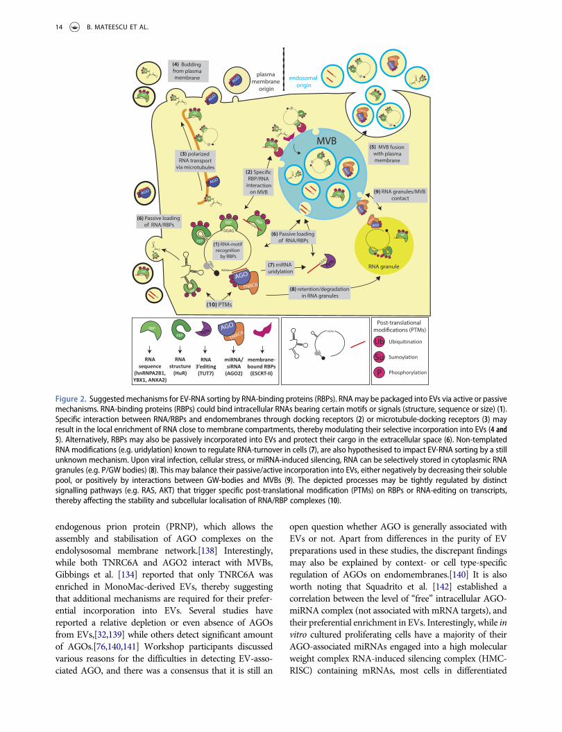
endogenous prion protein (PRNP), which allows theassembly and stabilisation of AGO complexes on theendolysosomal membrane network.[138] Interestingly,while both TNRC6A and AGO2 interact with MVBs,Gibbings et al. [134] reported that only TNRC6A wasenriched in MonoMac-derived EVs, thereby suggestingthat additional mechanisms are required for their prefer-ential incorporation into EVs. Several studies havereported a relative depletion or even absence of AGOsfrom EVs,[32,139] while others detect significant amountof AGOs.[76,140,141] Workshop participants discussedvarious reasons for the difficulties in detecting EV-asso-ciated AGO, and there was a consensus that it is still an
open question whether AGO is generally associated withEVs or not. Apart from differences in the purity of EVpreparations used in these studies, the discrepant findingsmay also be explained by context- or cell type-specificregulation of AGOs on endomembranes.[140] It is alsoworth noting that Squadrito et al. [142] established acorrelation between the level of “free” intracellular AGO-miRNA complex (not associated with mRNA targets), andtheir preferential enrichment in EVs. Interestingly, while invitro cultured proliferating cells have a majority of theirAGO-associated miRNAs engaged into a high molecularweight complex RNA-induced silencing complex (HMC-RISC) containing mRNAs, most cells in differentiated
RNA
sequence
(hnRNPA2B1,
YBX1, ANXA2)
RNA
structure
(HuR)
RNA
3’editing
(TUT7)
miRNA/
siRNA
(AGO2)
membrane-
bound RBPs
(ESCRT-II)
Sumoylation
Phosphorylation
Ubiquitination
Su
P
Figure 2. Suggestedmechanisms for EV-RNA sorting by RNA-binding proteins (RBPs). RNAmay be packaged into EVs via active or passivemechanisms. RNA-binding proteins (RBPs) could bind intracellular RNAs bearing certain motifs or signals (structure, sequence or size) (1).Specific interaction between RNA/RBPs and endomembranes through docking receptors (2) or microtubule-docking receptors (3) mayresult in the local enrichment of RNA close to membrane compartments, thereby modulating their selective incorporation into EVs (4 and5). Alternatively, RBPs may also be passively incorporated into EVs and protect their cargo in the extracellular space (6). Non-templatedRNA modifications (e.g. uridylation) known to regulate RNA-turnover in cells (7), are also hypothesised to impact EV-RNA sorting by a stillunknown mechanism. Upon viral infection, cellular stress, or miRNA-induced silencing, RNA can be selectively stored in cytoplasmic RNAgranules (e.g. P/GW bodies) (8). This may balance their passive/active incorporation into EVs, either negatively by decreasing their solublepool, or positively by interactions between GW-bodies and MVBs (9). The depicted processes may be tightly regulated by distinctsignalling pathways (e.g. RAS, AKT) that trigger specific post-translational modification (PTMs) on RBPs or RNA-editing on transcripts,thereby affecting the stability and subcellular localisation of RNA/RBP complexes (10).
14 B. MATEESCU ET AL.

mammalian tissues express AGO-miRNA as a “free” low-molecular weight complex (LMC-RISC).[143] Moreover,cell stimulation (e.g. T-cell activation), could induce themobilisation of AGOs from its LMC- toward the HMC-pool.[143] Moreover, alteration in cell signalling (e.g.oncogenic RAS) was shown to alter the pool of P-body-vs. MVB-associated AGO2, by modulating AGO phos-phorylation.[140] Altogether, these observations suggestthat AGO2 post-translational modifications and the mod-ulation of the level of endogenous miRNAs targets, maydirectly modulate the export of AGO2-loaded miRNAsinto EVs,[144,145] either by promoting the association ofAGO2 to MVBs, or by modulating the pool of AGO2 thatcould be passively or actively engulfed into EVs.
Beyond AGOs and their direct partners, other proteinshave been proposed to directly affect miRNA sorting.hnRNPA2B1 has been suggested to regulate EV sortingof miRNAs containing a specific motif [146] when mod-ified by a small ubiquitin-like modifier, SUMO, which inmono-attachment does not target proteins for degrada-tion, but instead regulates stability and subcellular localisa-tion.[147,148] Interestingly, constitutive activation ofmutated forms of KRAS, frequent in certain cancers, wasrecently shown to promote EV-associated release of spe-cific miRNAs, while inducing intracellular retention ofothers.[149] Downregulation of Annexin A2 (ANXA2), aprotein exhibiting Ca2+-dependent phospholipid-bindingprotein and RNA-binding properties,[150] was shown todecrease the level of EV-associated miRNAs, without sig-nificantly impacting the abundance of released EVs.[151]In addition, HuR protein, an ELAV RBP family protein,blocks particularmiRNAbinding sites on target transcripts[152] and facilitates RISC dissociation from target mRNA.[153] The first author of the latter study reported at theworkshop and now in publication [129] that in stressedhepatic cells, HuR also binds miR-122 after dissociating itfrom AGO2, and would promote its release at the surfaceof the MVB to favour its incorporation into EVs. A similarmechanism involving the RNA-binding protein Y-boxprotein 1, a known EV-associated protein,[154–157] wasrecently identified by Shurtleff et al. [139] using cell-freeassays for exosome biogenesis. It was shown that YBX1binds to specific miRNAs (e.g. miR-223) and promotestheir selective packaging into CD63+ EVs. Interestingly,knocking out YBX1, alone or in combination with addi-tional knockdown of its homologue YBX2, led to a strongdepletion of YBX1-bound siRNA into EVs, and theirremobilisation into cellular AGO2. Future work shouldaddress whether miRNA dissociation from AGO and sub-sequent binding by non-AGO RNA-binding proteins(such as hnRNPA2B1, Annexin A2, HuR and YBX1) is ageneral phenomenon that may contribute to EV-RNAsorting. Moreover, as these RBPs are known to bind to
other type of RNAs (including mRNA, tRNA andsnoRNAs) it will be important to decipher whether theycould also promote the packaging of these RNA biotypesinto EVs.
Finally, proteins implicated in EV biogenesis caninfluence RNA loading into EV. Modulation of endo-somal sorting complex required for transport (ESCRT)activity can impact both the number of released EVand EV-RNA abundance. For example, Vps4 wasfound to influence the secretion of oncogenicmiRNAs by human hepatoma cells.[158] Interestingly,knocking down Alix, another ESCRT protein, did notaffect the number of released EVs but induced adecrease of secreted miRNAs.[159] It should be notedthat ESCRT complex is also involved in the regulationof the RNA silencing pathway,[134] thereby complicat-ing the interpretation of these observations.
3.1.3. RNA motifs and modifications associated withEV incorporationAs intimated above, several RNA motifs have beenidentified that correlate positively with release in EVs.A GGAG motif in miRNAs, recognised byhnRNPA2B1, has been suggested to promote packaginginto EVs.[146] Studies involving analysis of transcriptsenriched in EVs over their secreting cells have alsoidentified motifs that positively correlate with RNAtargeting to EVs.[160,161]
Besides the recognition of specific RNA secretionmotifs, the addition of non-templated nucleotides hasbeen reported to determine RNA fate; 3′ adenylation ofmiRNAs has been associated with intracellular reten-tion whereas uridylated isoforms are more enriched inthe small vesicles fraction.[84] This could suggest thatthe 3′ end of the miRNA sequence contains a criticalsignal for sorting into EVs, or that the specific subcel-lular localisation of RNA-editing enzymes directlyimpacts this sorting. Alternatively, the 3′ uridylationmay indicate the presence of apoptotic bodies in theEV isolate, since this is also a signature of RNA under-going degradation during apoptosis.[162] The possibi-lity of mutating the packaging motif of specific EV-associated RNA (using CRISPR-mediated homologousrecombination) or mutation/overexpression of keyRNA editing enzymes (e.g. TUT7) may allow this ques-tion to be addressed.
In conclusion, as typified by strong recent work onmiRNA, EV-RNA sorting mechanisms are likely toinvolve RNA binding proteins and their partners,although other mechanisms may exist (Figure 2). Thefocus on miRNAs at the workshop and in this article issimply a reflection of the field; indeed, diverse otherclasses of cellular RNAs, or their fragments, have been
JOURNAL OF EXTRACELLULAR VESICLES 15

reported in EVs.[70] Below, we specify potentialresearch directions to uncover how these diverse RNAspecies are packaged into EVs.
3.2. Remaining questions and how we can answerthem
3.2.1. How can the varying correlations betweencellular and EV-associated RNA contents beexplained?As previously mentioned for the case of miRNAs,different studies report differing degrees of correlationbetween intracellular and EV-RNA content.[22,46,73,124,163] At the most basic level, all EV-RNAs originate in cells, and thus any EV-RNAs mustalso exist at least transiently in the parent cell. We canreadily envision four distinct but non-exclusive scenar-ios that might contribute to observed relationshipsbetween cellular and EV-associated RNA:
(1) Cellular RNAs are randomly included in the vesiclelumen during EV biogenesis, yielding an EV-RNAprofile representative of the relative RNA abun-dance at close proximity of the EV luminal engulf-ment site. Note that this scenario does notnecessarily mean that EV and cellular RNA wouldcorrelate perfectly. Also, random RNA packagingmight be limited by the length of prospective pack-aged RNAs and the size of RNPs. In a pure exampleof this case, the EV-RNA profile would be roughlyidentical to that of cellular membrane-associatedRNAs;[164] for example, those near MVBs, orendosome-like domains of the plasma membrane.The recent development of a high throughputmethod to determine subcellular localisation ofRNA in situ [165] may facilitate the identificationof such bias in the future.
(2) Cellular RNAs are included in the vesicle lumenbased on their proximity to the site of engulfment,but with enrichment provided by affinity of RNAmotifs and/or RNP components for protein andlipid elements preferentially found at sites of EVbiogenesis.[139] Here, EV-RNAs would approxi-mate a subset of membrane-associated RNAs,enriched in a particular sequence or proteinassociation.
(3) RNAs harbouring specific structural or sequencesignals are loaded into EVs in an energy-depen-dent, i.e. active, manner. Interestingly, someRNAs might “bring their own” ATP, as ATP-binding motifs have been identified.[166]Depleting ATP from parent cells would presum-ably be an experimental approach to identify
active loading processes, although careful con-trols would be necessary. However, workshopparticipants were unaware of any reportedATP dependence in EV-RNA sorting pathways.
(4) Finally, it remains a possibility that recyclingmechanisms operating in cells (e.g. RNautophagy)would lead to continuous EV incorporation of alarge range of cellular RNAs at a given time.[167,168]
As discussed, it is anticipated that more than one of theabove are likely to define the relationship of cellular andEV-RNA, complicating the picture substantially.Resolving the relative contributions of different scenariosin different contexts is an area of much-needed research.
3.2.2. Can further research into RBPs and theirpartners lead to discovery of mechanisms underlyingRNA sorting into EVs?It may be informative to explore the roles and interactionpartners of RBPs that are already known to be present inEVs, as these may be involved in the stabilisation and/orsorting of EV-RNA. As an example of this approach, a listof known curated RBPs [169] could be compiled based onoverrepresentation in databases that compile EV proteo-mics data, such as Vesiclepedia,[170] Exocarta [171] andEvpedia.[172] In a preliminary exercise using Vesiclepedia(see Table 4), we note that, apart from enrichment forgeneral translation machinery factors, EVs contain tRNAassociated factors (EEF1A1, AARS, GARS, DARS, TARS,WARS) and more specialised RBPs such as CNP, PARK7,RNH1, RUVBL1/2, HNRP-K/A2B1/C/D/R, PCBP1,DHX9, NPM1, PARP1, SND1, SYNCRIP, and MVP(major vault protein). Future investigation will be requiredto validate these observations, and notably to excludepossible contamination of EV preparation with co-fractio-nating RBPs. It should also be emphasised that there arecaveats to this approach. For low abundance EV-RNAs,RBPs with one-to-one binding stoichiometry might bedifficult to detect in EVs. Furthermore, shuttling proteinsmay exist that transfer RNAs from cellular to EV bindingpartners while remaining themselves excluded from EVs.
3.2.3. Which EV-associated RNAs are inside vs.outside EVs?In addition to the question of whether specific RNAs aredestined for release via EVs, it is also unclear in a givenpreparation whether EV-RNAs localise both in the EVlumen and on the outside of the EV membrane. Thoughthe possibility of a functional association of RNA with theoutside of the EVmembrane cannot be formally ruled out,no evidence for it has been observed, and this topologywould be inconsistent with the traditional model of RNA
16 B. MATEESCU ET AL.

delivery by vesicle fusion with the recipient cell. In addi-tion, observed outside associations with RNPs may betechnically induced by isolation procedures (i.e. force ofcentrifugation) rather than biological in nature. RNasetreatment of EVs has been applied to deplete non-encap-sulated RNA;[27,73] however, even RNAs exterior to EVsare likely to be protected from RNase degradation byprotein complexes, as there are already high levels ofRNase in biological media such as blood plasma.[26] Toselectively study RNA residing in the vesicle lumen, it iscritical to eliminate any source of RNA outside the vesicles.We therefore recommend that investigators quantify theamount of EV-associated RNAs (for example by RT-qPCR) before and after treatment of the vesicle with aproteinase followed by an RNase digestion step. By com-bining this approach with a negative control (proteinase/RNase digestion in the presence of detergent, as in Royoet al. [173]), it is possible to determine the proportion of aspecific RNA residing within the vesicle in a givenpreparation.
3.2.4. Does subcellular localisation affect sortinginto EVs?It has been hypothesised that the subcellular distribution ofRNAs can promote the inclusion of particularmolecules inEVs. Similarly, the location of RNA species in the differentcompartments – i.e. cytoplasm, nucleus or plasma mem-brane – could determine their in/exclusion in differenttypes of EVs (large microvesicles, apoptotic bodies, orexosomes). Increased knowledge on sub-cellular localisa-tion of different RNA species is needed to further addressthis issue. In general terms, crucial decisions onmaturationof both mRNAs and ncRNAs (i.e. splicing, polyadenyla-tion) are made during their time in the nucleus, suggestingthat the nuclear history of an RNA can be fundamental for
its fate in the cytoplasm. Once in the cytoplasm, RNPcomplexes containing mRNAs and/or other ncRNAsinteract with motor proteins, which mediate their traffick-ing, mostly in inactive form, along the cytoskeleton tracksto their final destinations.[174] It is possible that the samemechanisms active in RNA pre-localisation also functionin specific sorting of RNP to nascent EVs. Notably, in thenervous system, intracellular RNP trafficking seems to beregulated by neurotransmission (reviewed in [175]) andthe same has been also reported for EV trafficking amongdifferent brain cell types (reviewed in [176]). It is likely thatthe mechanisms governing subcellular RNA localisationare important for their specific packaging into EVs. Futureexperiments characterising these processes will be key tovalidating this hypothesis. For example, detecting changesin subcellular RNA localisation by fluorescent in situ hybri-disation (FISH) [177] or using indirect RNA-binding pro-tein detection (e.g. with the MS2 system) [178] couldprovide insight on the relevance of localisation within thecell for the packaging of a given RNA transcript.
3.2.5. Differential stability of RNA in EVs?It has also been proposed that differences in EV-RNAprofiles versus intracellular RNA profiles are not theresult of a selective process, but rather represent dif-ferences in the extracellular stability of particularRNA species.[179] Certain RNases may be present athigher concentration in EVs than in the cell, inducingthe relative depletion of RNAs targeted by theseRNases in EVs.[180] Moreover, the portion of RNAsbound by RBPs inside EV might be preferentiallystabilised and resistant to RNases. This may beaddressed by searching for known RBP binding motifsspecifically enriched in RNA isolated from EV com-pared to the parent cells.
Table 4. Top 80 RNA-binding proteins in Vesiclepedia datasets.
Poly-A RNA-binding
Vault complex
MVP
RPS8 RPS5 RPL4
RPS16 RPS24 RPL7
RPS18 RPS6 NCL
RPS27A RPL15 RPS9
RPLP0 RPS3A RPL6
RPSA RPL18 RPL27
RPS3 RPS13 RPS7
RPS4X RPL30 RPS11
RPL12 RPL3 RPL19
RPS14 RPS25 RPL13
RPLP2 RPS2 RPL23
RPL7A RPS15A
ribosome function
EEF2
EIF4A1
EEF1G
EEF1A2
EIF5A
EEF1D
EIF2S1
EEF1A1
AARS
GARS
DARS
TARS
WARS
GAPDH IPO5
PRDX1 ACO1
UBA1 CANX
RAN XRCC6
PSMA6 CRYZ
KPNB1
PSMA1
DYNC1H1
CALR
CNP DHX9
PARK7 HNRNPA2B1
RNH1 HNRNPC
RUVBL2 PA2G4
HNRNPK HNRNPD
RUVBL1 NPM1
PCBP1 SND1
PARP1
SYNCRIP
HNRNPR
tRNA functiontranslation unclear RNA-binding
Top 80 RNA-binding proteins in Vesiclepedia datasets. The occurrence of each protein within a census of 1542 manuallycurated RBPs [169] was queried within the Vesiclepedia database (http://www.microvesicles.org; v3.1; restricted to Homosapiens samples). Proteins are clustered by functional group with colours indicating the number of occurrences in thedatabase (30–39 blue; 40–49 orange; >50 red).
JOURNAL OF EXTRACELLULAR VESICLES 17

3.2.6. Do RNA modifications impact their sortinginto EVs?Accumulating evidence indicates that an array of RNAmodifications (5′-end cap, m6A: N6-methyladenosine,m5C: 5-methyl cytidine, A-to-I editing, pseudo-uridine,5hmC: 5-hydroxylmethyl cytidine) directly control RNAmetabolism (reviewed in [181]). These RNAmodificationsare tightly controlled by cellular enzymes (e.g. m6A “wri-ters” METTL3, m6A “eraser” FTO), and could be recog-nised by RNA-modification “readers” (such as YTHDF1)that could impact the stability [182] of modified RNA. Asfor the addition of non-templated nucleotides to the 3′ endof the RNA (adenine, uridine, see also section 3.1.3), aninteresting hypothesis would be that these modificationsmay favour specific packaging of these RNAs into EV.Novel techniques have recently been developed to detectspecific RNA modification (e.g. MeRIP-seq [183]).Applying these approaches to EV-associated-RNAswould enable analysis of whether RNA modifications areenriched or depleted in EV-associated RNA.
3.3. Next challenge: qualitative and quantitativeRNA analysis in individual EVs
Since EV populations can be highly heterogeneous, a fullunderstanding of RNA incorporation into EVs willrequire analysis at the EV subpopulation or individualEV level rather than bulk analysis. However, detectingand enumerating RNA molecules in individual EVs faceunmet challenges. As technologies for analysis and isola-tion of individual EVs are rapidly improving,[19,184–187] the opportunities to investigate the EV-RNA cargoof single EVs are expanding. Attempts have been made toapply fluorescent RNA-tracking dyes (e.g. acridineorange, Syto84 or Syto-RNASelect) for high-resolutionflow cytometric analysis of the RNA content of individualEVs. However, the use of such an approach remainschallenging due to limited probe specificity, limited fluor-escence per EV, and poor signal-to-background ratio. Forexample, fluorescent small molecules that are highly spe-cific for RNA tend to be charged and thus membrane
impermeable, hampering labelling of RNA in the EVlumen. Table 5 lists several membrane-permeant fluor-escent dyes that may be used to detect the presence ofRNA associated with EVs, although their specificity andefficiency in labelling EV-RNA remain to be validated.
It is important to mention that none of the cur-rently utilised single EV analysis cytometry-basedmethods are capable of detecting a single fluoresceinmolecule. When using molecular equivalents of solu-ble fluorescein (MESF) calibration, it has beendemonstrated that hundreds or thousands of fluores-cein equivalents are commonly required for detection,depending on the instrument and configuration(ISEV-ISAC-ISTH working group, ISEV 2016).Moreover, most RNA dyes are intercalating agentsand it is not expected that the structure of a smallmiRNA would support intercalation of hundreds ofdye molecules. Labels with higher quantum yield (andbetter signal:noise ratio), such as quantum nanocrys-tal-labelled oligonucleotides, may be more suitable fordetection of molecular components in single EVs. Inaddition to flow cytometry, Raman spectroscopy wasrecently used to analyse single EVs in solution, allow-ing distinction of several distinct EVs populationsdiffering in chemical properties.[195,196] However,it is currently unclear whether this technique mayallow detection of RNA in single EVs.
Innovative methods were recently developed to indir-ectly detect RNA in EV by microscopy, notably by usingRNA-binding fluorescent protein probes. One multi-plexed approach [197] combines the labelling of vesiclemembranes using a palmitoylated fluorescent protein,and the indirect labelling of RNA molecules using thebacteriophage MS2 coat protein-GFP:MS2 RNA bindingsequence system.[178] Although this method may berestricted to the labelling of long RNA (e.g. lcnRNA ormRNA) and the target RNAmust be modified to includethe MS2 loop, it is a promising strategy to monitor thepackaging of extracellular RNAs into EVs in living cells. Itwould be interesting to further develop this approach toinvestigate the percentage of RNA-loaded EVs or to
Table 5. Membrane-permeant fluorescent dyes that may be used to detect the presence of RNA associated with EVs.
Dye Ex/Em (nm) Known nucleic acid specificityPublication for cell
stainingPublication EV
staining
Acridine orange (Thermo Fisher Scientific) 460⁄650 (RNA)500⁄526 (DNA)
dsDNA (Green) and ssDNA/dsRNA(Red)(orange emission in acidic organellecompartments)
[188] [189]
Pyronin Y (Sigma, Saint Louis, MO, USA) 555/580 ssRNA [190] No
Syto14 (Thermo Fisher Scientific) 517⁄549 (DNA),521⁄547 (RNA)
RNA/DNA [191] No
SYTO-RNASelect (Thermo Fisher Scientific) 490/530 RNA selective [192] [193]
E36 497/548 RNA selective [192] No
Styryl-TO 520/531 RNA selective [194] No
18 B. MATEESCU ET AL.

combine with the use of fluorescent-tagged markers ofsub-cellular compartments in order to infer the endo-membrane origin of EVs containing RNA.
During the workshop, participants suggested applyingdigital droplet PCR methods to quantify the RNA contentin single EVs, although it is not currently clear whether thismethodology will be applicable to single vesicle analysis.Another approach would be to perform PCR on limitingdilutions of EV preparations, so that each PCR is initiatedfrom the RNA content of a single vesicle. A microfluidicPCR platform would likely be necessary to analyse such asmall amount of RNA. Finally, it would be interesting to beable to “visualise” specific RNA within the vesicular con-text. Protocols developed to detect RNA within virus par-ticles by RNA FISH [198] or in situ hybridisation-electronmicroscopy (ISH-EM) [199] could likely be applied todetect EV-associated RNA given their comparable sizeand chemical nature. In fact, the high resolution of theISH-EM method may allow determination of whetherspecific EV-RNA are located inside or at the surface ofvesicles, a point remaining controversial in the field.
4. Assessing functional transfer of EV-RNA
In the past 10 years, EVs were not only shown to containRNA, but also to be able to functionally transfer RNA intotarget cells. It is important that this process is studied usingdifferent experimental models and by mimicking physio-logical EV(-RNA) concentrations and conditions, both invitro and in vivo. The formal demonstration that EV-mediated effects on target cell behaviour are caused byfunctional transfer of EV-RNA is technically challengingand requires a number of important controls in order tovalidate observations. Below, we describe the currentapproaches used to investigate EV-RNA transfer and dis-cuss advantages and pitfalls of these methods. During theworkshop, there were also fruitful discussions about theimplementation of novel models/approaches in the field,of which the most promising are also reported in thesections below.
4.1. Strategies to study functional transfer of EV-associated mRNA and miRNA in vitro
The first observation that mRNA could be transferred via apelletable fraction of cell conditioned medium and trans-lated in target cells wasmade by Ratajczak et al. [200]. Soonafter, Valadi et al. [201] reported that RNA was indeedassociated to low-density EV, and further demonstratedRNase-resistance of this RNA. In this study, mousemRNAtransferred via EVs to recipient human mast cells could betranslated into corresponding mouse proteins in vitro.[201] Skog et al. [202] provided a confirmatory study
showing that, upon EV-mediated transfer, Gaussia lucifer-ase mRNA was translated within recipient cells. Gaussialuciferase (Gluc) is a flash-type reporter which emits abioluminescent signal 100-fold higher than typical luci-ferases such as firefly or Renilla luciferases,[203] therebyenabling detection of minute biological events such as EV-RNA translation. Later on, Pegtel et al. [117] showed thatviral miRNAs secreted by EBV-infected cells were trans-ferred to uninfected recipient cells via EVs and couldrepress the EBV target genes using a luciferase sensorassay. Shortly after, Kosaka et al. [204] and Zhang et al.[141] documented functional cell-to-cell miRNA transfersin destination cells using EVs isolated from miRNA-over-expressing cells, by using a luciferase sensor assay,[204] orby showing direct repression of endogenous miRNA tar-gets at the protein level.[141] Another method to investi-gate spatial and temporal aspects of EV uptake and EV-mRNA translation is via the use of bioluminescent repor-ters. More recently, it was demonstrated that EV-mRNAencoding a membrane-bound Gluc reporter (GlucB) canbe translated in as soon as 30minutes following EV uptakeby recipient cells.[197] While the GlucB signal began toincrease substantially from 30 min, it reached a plateau at12 h post-EV uptake, perhaps suggesting that there is alimit to the amount of mRNA which EVs can transfer.
4.1.1. Considerations for assessing functionaltransfer of EV-associated miRNAsA plethora of studies have confirmed the initial obser-vations, strengthening the idea that functional transferof EV-associated mRNAs and miRNAs can occur invitro between mammalian cells. In several studies, EVsfrom cells overexpressing endogenous miRNAs wereshown to cause target mRNA repression in recipientcells transfected with a luciferase reporter constructusing the 3′UTR of the mRNA target (examples include[22,205,206]). Besides effects on miRNA reporter sys-tems, EV-mediated transfer of miRNA was also shownto cause changes in target gene expression in recipientcells.[205,207,208] These experiments are generallyperformed with donor cells expressing miRNAs forwhich the expression levels in the recipient cell arevery low or undetectable in order to maximise thewindow in which miRNA transfer can be observed. Inorder to exclude the possibility that the EVs/co-culturetreatment itself would induce the endogenous tran-scription of the tested miRNA in destination cells,which is a potential confounding effect in this assay,some studies tested the level of miRNAs precursors(pri-miRNA and/or pre-miRNA) in destination cells.[209,210] Although the lack of changes in miRNAprecursor level would strengthen the EV-mediatedtransfer hypothesis, it should be noted that: (1) pre-
JOURNAL OF EXTRACELLULAR VESICLES 19

miRNAs can also be transferred by EVs;[76,211] and(2) because the pre-/pri-miRNA are continuously pro-cessed into mature miRNA by the RNA silencingmachinery, a transient increase in pre-/pri-miRNAlevels caused by EV treatment may easily be overseenat a later time point, while mature miRNA may stablyaccumulate for hours/days.[212,213] Therefore, detect-ing changes in precursor miRNAs in destination cellsmay not be a reliable method to control for spuriousmiRNA transcriptional activation caused by EV treat-ment or in co-cultures. In other studies, the contribu-tion of specific miRNAs in EV-induced effects has beeninvestigated by blocking the function of the candidatemiRNA in donor cells using antimiRs [109,214,215] orby using miRNA genomic knockout donor and recipi-ent cells.[208] Since knockdown of miRNAs may leadto changes in the phenotype and/or function of cells, itis important to address whether the observed changesin the function of EVs released by these cells are onlydue to the lack of this particular miRNA or whetherchanges in other functional molecular componentscontributed to these effects. Moreover, when usinganti-miRs it is essential to use adequate controls suchas a control anti-miR that targets an unrelated miRNAexpressed at a similar level.
4.1.2. Stoichiometry of miRNAs associated with EVsSince a population of EVs can contain many differentmiRNAs, they may suppress functionally related genesin parallel, leading to effective paracrine control overneighbouring cells. Nonetheless, a quantitative assess-ment of the miRNA content of EVs by a stoichiometricapproach found the miRNA/vesicle ratio in a bloodsample to be lower than 1, even for the most abundantmiRNAs, meaning that there are vesicles that do notcontain a copy of a given miRNA.[25] Based on theseobservations, the notion was put forward that indivi-dual EVs were unlikely to be functionally importantvehicles for miRNA-based communication. However,the workshop attendants felt that there was a need for amore nuanced consideration of these data. First, thestoichiometry assessment relies on careful quantifica-tion of EVs, for which there are no gold standards andwhich heavily depends on EV purity and the experi-mental approach. In addition, this study assessed onlysmall EVs (50–200 nm), while larger vesicles, such asmicrovesicles and oncosomes, also contain miRNAs aswell as longer molecules of RNA that can contribute toEV-RNA mediated communication. With regard toEV-miRNA function, it may well be that EV uptake isan infrequent but very selective event, in which rareEVs carrying many copies of a given miRNA may leadto miRNA-mediated silencing of mRNAs in specific
target cells. It is also possible that, by taking up multi-ple EVs carrying a low concentration of miRNA, suffi-cient quantities of transferred miRNA are reached tomodify mRNA levels in target cells. This hypothesis isconsistent with the observation that different miRNAsoften operate in concert to simultaneously control dif-ferent players in the same signalling pathway. Finally,as was also suggested by Chevillet et al. [25], very lowquantities of miRNAs may be compatible with non-conventional functions, such as binding to Toll-likereceptors as well as affecting DNA transcription and/or epigenetic states.[216–219] More complete knowl-edge of EV heterogeneity, target cell specificity, andRNA transfer efficiency is therefore needed to furtheraddress issues of EV-RNA stoichiometry.
4.2. Assessing functional EV-RNA transfer in vivo
It currently remains to be determined whether functionaltransfer of EV-RNA occurs and is relevant in vivo duringphysiological or pathological processes. EV-RNA transferassays in the context of cancer cell xenografting orimmune cell function are among the currently usedexperimental set-ups in vivo.[220,221] In these studies,effects of EVs in vivowere assessed by in vitro isolation ofEVs followed by their re-introduction in vivo. However,this approach is unlikely to reflect the physiological prop-erties, concentration, and distribution of EVs released in(patho)physiological conditions. Recently, otherapproaches have been introduced to more directlyaddress whether EV-RNA functional transfer occurs invivo. During the workshop, the most intensively dis-cussed in vivo approach was the Cre-loxP-based EV tra-cing system to study functional transfer of mRNA.Possibilities and limitations of this approach aredescribed in the following sections.
4.2.1. Assessing EV-associated mRNA translationusing the Cre-loxP systemThe Cre-LoxP system is a recombination system, origi-nating in the bacteriophage P1, consisting of the DNArecombinase Cre, and the recognition sequence loxP.Sequences that are flanked by loxP sites will be excisedby Cre and re-joined leaving a single loxP site. EV-mediated transfer and translation of the Cre mRNA intarget cells can be visualised by using donor cells expres-sing Cre recombinase and recipient cells containing aCre-inducible open reading frame coding for a fluores-cent protein, e.g. with a stop signal terminating transcrip-tion that is flanked by LoxP sites. If functional Cre mRNAis transferred by EVs to the recipient cells, the “transcrip-tion block” impairing fluorescent protein (FP)
20 B. MATEESCU ET AL.
transcription is genomically eliminated leading to theirreversible production of the FP in the recipient cell.
Using this Cre-loxP system, functional transfer of EV-RNA between distant cells has been demonstrated betweenblood and brain,[220] as well as other non-haematopoieticorgans as a proof of principle. Later, these findings wereextended to EV signalling between tumour cells andhealthy cells as well as among tumour cells.[221,222] Inboth studies, Cre protein levels in EVwere below detectionlevels on Western blots, suggesting that recombinationobserved in target cells was caused predominantly byEV-mediated transfer of mRNA encoding the Cre protein(see also below). The major advantages of this approachare its high sensitivity (a single transferred CremRNAmaylead to the permanent transcription of a marker protein),and its ability to detect transfer events in living cells both invitro and in vivo. Other important advantages implicit tothis system are knowledge of the specific cell type (i.e. Creexpressing cells) from which EVs originate and the abilityto monitor changes in phenotype and molecular composi-tion induced by EVs down to the level of an individualtarget cell. In the following paragraphs wewill discuss whatwe currently know about the system and outline bothfuture directions and limitations.
4.2.2. Interpretation of Cre-loxP based tracing dataRecombined cells observed after EV uptake using the Cre-LoxP system almost certainly only represent a subpopula-tion of cells that interact with EVs, given that EVs can exertbiological effects on their target cells that are independentof full translation of transferred RNA, e.g. via binding ofmembrane bound molecules or the transfer of protein orlipids.[57,223–225] It was discussed during the meetingthat addition of fluorescent (lipid) dye labelled EVs totarget cells in vitro generally leads to a much higher per-centage of target cells positive for the fluorescent dyecompared to the number of target cells that start to expressfluorescent marker proteins due to recombination uponuptake of Cre-containing EVs. It is not clear whether cellsexpressing amarker gene after Cre-induced recombinationtherefore represent only a subset of EV-targeted cells, orperhaps whether the higher labelling rates observed with invitro fluorescence experiments are artefactual (e.g. due toincorporation of dye aggregates).
Additionally, researchers present at themeeting had notfound a correlation between the proximity of donor cellsand the frequency of loxP-recombination events. This lastobservation may suggest that cell-to-cell transfer of EV-RNA is likely to be systemic and involves EVs without theneed for direct cell-to-cell contact. However, within thetumour context, the local release of EV by Cre-positivecells and their subsequentmigration away from the recom-bined acceptor cells cannot be excluded.
The fact that recombined cells in multiple experimentalsettings display clearly separated populations with differ-ences in their miRNA profile, phenotypic markers orcellular behaviour compared to their non-recombinedcounterparts of the same or similar lineage,[221,222] sug-gests that the number of non-recombined false positivescannot be very high. However, this does not exclude thatnon-recombined target cells were modulated by (other)EV subpopulations that did not confer Cre mRNA-mediated recombination. Another important question iswhether uptake and processing of Cre mRNA-containingEVs merely reflects the current physiological state of therecipient, i.e. that the EV-targeted cell is already distinctfrom other cells, in contrast to the notion that EV uptakeactively changes the status of the cell. At least for the case ofthe induction of an immunosuppressive phenotype ofmyeloid derived suppressor cells (MDSCs), experimentalevidence supports the latter: EV uptake actively changesMDSCs to become more immunosuppressive [226,227]although the mechanisms responsible for inducing thischange are unknown.
4.2.3. Limitations of Cre-loxP based tracing of EVsAlthough elegant, the Cre recombinase based EV trackingtechnique has its limitations. First, it is not quantitative forthe number of EVs transferred, as it uses a permanentgenetic switch. In addition, it is not possible to distinguishthe type of EVs mediating the transfer. As discussed pre-viously,[221] loading of Cre mRNA in subpopulations ofvesicles might be very much cell type dependent. In EVsthat were isolated from peripheral blood serum and thenfractionated according to sucrose density ultracentrifuga-tion, Cre mRNA could be detected in several densityfractions.[222] Sorting of Cre mRNAmay occur preferen-tially into specific EV subpopulations, but this is difficult toassess given the lack of standardisation of vesicle numbersin the different subfractions and the fact that themethod ofdetection of Cre (by PCR or even nested-primer PCR) isconsiderably more sensitive than the ability to physicallyseparate EV subtypes by density gradients. It is also impor-tant to address whether the relatively low frequency (0.2–3%) of observed recombination events are representativeof the real frequency of EV transfer in vivo or whether theyrepresent a low sensitivity of this technique.
Using the Cre-loxP system, it is also imperative to con-sider alternative explanations to EV-mediated transfer ofCre. In a chimeric system where Cre and Cre reporterexpression occurs in physically separate cell populationsuch as between tumour cells, Cre mRNA or proteincould also be transferred via cellular membrane protru-sions, such as tunnelling nanotubes (for a review see [228]).Also, it should be mentioned that while in some studiesusing Cre-loxP for testing EV-RNA functional transfer the
JOURNAL OF EXTRACELLULAR VESICLES 21

Cre protein could not be detected in EVs,[220,221] othersfound Cre protein in EVs after overexpression in donorcells.[57] Although Cre is a nuclear protein not expected tobe abundant in EVs, involvement of mRNA vs. proteinmust be verified. Cell fusion is another process that wouldgive similar observations, although in chimeric systems itcan be controlled for relatively easily by co-expressing Crewith a marker gene (e.g. fluorescent protein). Transgenicsystems where Cre is expressed under a cell type-specificpromoter in the same cells that carry the Cre reporterrequire careful assessment to exclude that observed recom-bination events are due to a leaky expression of the pro-moter, rather than transfer of Cre from one cell to theother. A precise characterisation of promoter activityextending to all tissues is rarely available and might notapply to certain pathologies that are used. On the otherhand, some cases of apparent “misexpression” ofCremightinstead be the result of a horizontal transfer of Cre. As aminimal requirement for this type of experiments we pro-pose that evidence is provided that the cells in questionrelease Cre mRNA and/or Cre protein containing EVs andthat these EVs are sufficient to induce recombination intarget cells. The use of distinct Cre models, each allowingCre expression in specific donor cells, may help in rulingout unspecific Cre expression. In addition, the use of chi-meric mice where Cre-donor cells are grafted into loxP-reporter mice (or the reciprocal experimental set-up whereloxP-acceptor cells are grafted into Cre-expressing mice)can avoid the possibility of spurious expression of genomi-cally encoded Cre in acceptor cells.
In summary, the Cre-loxP system has been and will bea valuable tool for uncovering EV-based communicationbetween cells and tissues in a (patho)physiological setting.While it requires careful implementation to address spe-cific questions in EV biology, it is relatively easy to useand benefits from the availability of a large number ofcellular and animal models.
4.3. Expanding and refining our experimentalmodels
During the EV-RNAworkshop, there was a consensus thatwe should not restrict the functional analysis of EV-RNAtransfer to one particular type of EV. Physical propertieslike size and surface chemistries of different types of EVsmay impact their capacity to functionally transfer RNA.Although most studies perform EV-RNA transfer assaysusing EVs collected from in vitro cell cultures, it would alsobe interesting to investigate EV directly extracted fromtheir natural environment, e.g. those isolated from bodyfluids and tissues. These EV may differ in surface charac-teristics, possibly due to binding of lipid or protein moi-eties present in tissues or body fluids to the EVmembrane.This may influence functional RNA transfer in destination
cells, e.g. due to changes in EV binding to docking proteinsor in the delivery of EV cargo through endosomal escapeor direct membrane fusion. Isolation of EV from tissues istechnically challenging. Mild tissue dissociating reagent(e.g. liberase) without mechanical disruption could per-haps be used to isolate EVs from tissues without co-isolat-ing intracellular vesicles released by broken cells. However,it is important to keep in mind that such protease treat-ments may affect the proteins displayed on the surface ofEVs, potentially affecting their ability to dock on or fusewith recipient cells. Alternatively, EVmay be isolated fromin vitro maintained tissues. Another interesting approachwould be to use novel technology combining both micro-fluidics, which facilitate concentration of the EVs in themicroenvironment, and organoid cultures, which havebeen highly recommended to mimic in vivo cellular orga-nisation.[229] Indeed, 2D-grown cellsmay not recapitulatecertain features required for proper release or reception offunctional EV-RNA (e.g. cellular polarisation [230]).Knowledge acquired in this area could ultimately be usedto optimise EV-mediated functional RNA transfer in pro-spective therapeutics for gene delivery.
4.4. Usable knowledge for RNA delivery by EV-inspired synthetic mimics
4.4.1. Preparation of RNA-loaded EV or EV-mimicsSince EVs are increasingly regarded as natural carriers ofnucleic acids, it is of interest to explore EVs as vectors fordelivering both endogenous as well as foreign geneticmaterial into recipient cells. For this approach, severalchallenges remain, one of the most prominent being theintroduction of an RNA of interest into EVs. Severalreports describe loading procedures, based on electropora-tion of EVs directly,[231–233] loading of EVs by virus-infected cells,[234] or targeted embedding of specificRNA-sequences into EVs via sequence-recognitiondomains fused to vesicular proteins.[235] Although pro-mising in concept, these loading procedures are in practiceless than ideal, either loading RNA with low efficiency orshowing artificially high loading caused by formation ofelectroporation-induced nanoprecipitates.[61] In addition,it is challenging to remove the nanoparticle-forming che-mical transfection reagents from the EV preparation afterloading.
Even if the challenge of RNA loading is met, thesubsequent steps in functional delivery of RNA contentinto the target cells present significant hurdles. Uponintravenous injection, EVs are generally rapidly takenup by cells of the mononuclear phagocyte system,particularly in the liver and spleen. This limits thedistribution of EVs to other cell types. In addition,not all target cells are accessible via the vascular
22 B. MATEESCU ET AL.

lumen, as the endothelial barrier prevents extravasa-tion. Only at sites of fenestrated or sinusoidal endothe-lium, or at sites of increased permeability, canextravasation be expected. Finally, specific recognitionof the target cell should be followed by appropriatedelivery of the RNA cargo in its cytoplasm.
In addition to natural EV-based delivery systems,EV-mimics have emerged as alternative delivery sys-tems.[236] The design of EV-mimics is inspired bothby understanding of the properties of synthetic lipo-somes and by knowledge of the structure and functionof EVs. Since not all components of EVs may berequired for proper delivery, the synthetic mimics ofEVs could be developed through assembly of liposomeshaving crucial components of natural EVs in order toreduce structural complexity.[237] AlthoughKooijmans et al. [237] have provided insight intowhich EV components could be used to enhance thedrug delivery properties of EV-mimetics, EV compo-nents that are essential for the assembly of functionalEV-mimetics remain to be identified.
In theory, the EV-mimics could be designed withenhanced circulation time and increased specificity fortarget cells. Such EVs could be applied for the cytoplasmicdelivery of wide variety of biomolecules such as oligonu-cleotides, DNA, proteins and peptides.[238] In this regard,modifying EVs with nanobody-PEG-lipids could poten-tially protect the biomolecules loaded inside from macro-phages and give the EVs high affinity for the receptor of a
particular target cell type. This prolonged circulation timeand target cell specificity could increase EV accumulationin targeted tissues and may improve the cargo delivery.[239] Most recently, Sato et al. [240] developed hybrido-somes (hybrid EVs) by fusing their membranes with lipo-somes, showing that it is possible to control the degree ofinteraction between hybridosomes and recipient cellsthrough amembrane-engineering strategy. These develop-ments will open up new avenues for developing improvedRNA delivery systems and may advance the field ofnanomedicine.
4.4.2. Delivering EV-RNA cargo: understandingmechanisms of EV fusion with recipient cellsThe fusion and entry of EVs into recipient cells mayfollow several routes (Figure 3). The EV uptake mechan-ismmay depend on the target cell type and recognition ofproteins or receptors present on the surface of both EVsand on recipient cells, as well as environmental condi-tions. Notably, the protein molecules present on both thesurface of EVs and on the plasma membrane of recipientcells potentially modulate vesicle binding and uptake byrecipient cells. It has for example been shown thatheparin sulphate proteoglycans (HSPGs) on the plasmamembrane of the recipient cell efficiently regulate EVuptake.[241,242] Furthermore, it was recently demon-strated that various isoforms of integrins present ontumour-derived EVs influence the uptake of EVs in anorgan-specific manner.[223] Environmental factors that
Figure 3. Extracellular vesicle uptake and cargo delivery in recipient cells. EVs may release their cargo into the cytosol throughdirect fusion with the plasma membrane. Alternatively, EVs may be internalised via a variety of endocytic mechanisms, includingclathrin-dependent endocytosis, clathrin-independent endocytosis, macropinocytosis and phagocytosis. Subsequently, EVs aretransported into the cytoplasm in endocytic vesicles. These vesicles may proceed to scan the endoplasmatic reticulum (I), whichhas been reported to be a site for translation and RNA interference. EVs may fuse with endosomal membranes after acidification torelease their RNA content (II), or be directed to lysosomes where they are degraded (III).
JOURNAL OF EXTRACELLULAR VESICLES 23

influence EV uptake include temperature and radiationexposure,[243] as well as pH conditions, which affectdirect fusion of EVs with the plasma membranes ofrecipient cells.[244,245] Intracellular availability of ATPin recipient cells has also been shown to affect EV uptake,indicating that the internalisation of EVs into recipientcells is an energy-requiring process.[246]
Different mechanisms of internalisation have beendescribed. In a classical way, EVs could enter cells viaclathrin-dependent endocytosis, micropinocytosis orphagocytosis.[247] Additionally, EV uptake may also fol-low the non-classical, lipid raft-dependent endocytosisinvolving caveolin-mediated entry.[248] Interestingly,Heusermann et al. [62] recently reported that purifiedEVs labelled with GFP-CD63 surf filopodia of primaryhuman fibroblasts, before being endocytosed at the cellbasis in a process reminiscent to entry of specific viruses.In the cytoplasm, internalised EVs particles establishtransient contact with the endoplasmic reticulum (ER)before being eventually being trafficked to the lysosomalcompartment. Converging evidence indicate that ER is acellular nucleation site for RISC complex miRNA/siRNAloading and targeting of mRNA.[136,249] As such, it
could be speculated that internalised EVs may delivertheir miRNA cargo in close proximity to the ER inorder to favour their loading into AGOs.
5. Conclusions and considerations
The EV-RNA research field has clearly progressedsince the previous ISEV workshop on EV-RNA heldin 2012. There is a better understanding of the hetero-geneity of EV subpopulations and of non-EV RNA-carrying structures that potentially contaminate EVpreparations and their distribution among differentbody fluids. In addition, several factors influencingsorting of RNA into EV have been identified andexperimental systems have been designed to addressfunctional transfer of EV-RNAs in vitro and even invivo. Nevertheless, the field continues to experiencenumerous technical challenges: the large variety in EVisolation procedures, the small size of EV and theminute amounts of RNA that are recovered fromthese vesicles, and biases in the analysis of these traceamounts of RNA. In addition, it remains challenging toobtain direct proof for the role of EV-associated RNA
Research question Considerations
• Does the EV preparation contain RNA-carrying contaminants (e.g. HDL, RNPs)?
• Is the RNA protected from protease + RNase treatment?
• Which EV populations are represented? (donor cell, size, protein content)
• Detection of large and small RNAs? • Are the RNAs intact or fragmented?• Do the RNAs contain nucleotide modifications
that may interfere with analysis?
• Does content depend on activation/differentiation status of cells?
• Is it different in health/pathology?
• Can transfer be detected in vitro? in vivo?• Does transfer of dyed EVs or EV marker
proteins correlate with movement of RNA?
• Is the transferred EV-RNA also endogenously expressed by target cell?
• Does the EV-RNA modify mRNA levels or trigger other receptors (e.g. PAMPs)?
Is the extracellular RNA contained in an EV population?
What is the RNA content of the EV?
Is incorporation of the EV-RNA of interest regulated?
To which cells is the EV-RNA targeted?
Does the EV-RNA of interest modify target cell function?
Figure 4. Considerations for analysing the nature and function of EV-associated RNA. Overview of research questions aimed at unravellingthe nature and function of EV-RNA and considerations in addressing these questions, as discussed at the 2015 ISEV workshop on EV-RNA.
24 B. MATEESCU ET AL.

in effects that EVs exert on target cells in vitro and invivo. As an overview of what was discussed at the 2015research seminar on EV-RNA and to prepare research-ers from inside or outside the EV field for working inthis area, we provide a list of important considerationsthat could guide future experiments (Figure 4).Furthermore, we provide a list of experimental details(an updated version of the list provided in the previousEV-RNA position paper [9]) that should be reported inpublications addressing the composition and functionof RNA associated to EV (Table 6). This checklist mayaid in augmenting the reproducibility of studies in theEV-RNA field and may also be used by reviewers andeditors to aid the review process of articles in the EV-RNA research field.
Acknowledgements
We thank all participants of the 2015 ISEV workshop on EV-RNA for their valuable input in roundtable discussions,which formed the basis for this position paper. We are grate-ful to Dr P. Vader for preparing Figure 3.
Disclosure statement
No potential conflict of interest was reported by the authors.
Funding
BM is supported by grant 1U19CA179513-01 from the NIHCommon Fund. ENMN-TH receives funding from theEuropean Research Council under the European Union’s
Table 6. Checklist experimental details to be included in publications.Step Parameters to be described
EV isolationCell culture Cell type
ConfluencyCellular activation/differentiation statusUse of EV-depleted serum (include depletion protocol) or serum-free mediumCell viabilityMycoplasma test
Body fluid Health/disease statusCollection method (disposables, chemicals, procedure)Removal of body fluid specific contaminantsStorage
EV isolation Differential centrifugation stepsFiltration stepsSize exclusion chromatographyDensity gradientCommercially available kits (e.g. chemical, column based)EV quantification method
EV-RNA characterisationEV-RNA sample preparation RNase/DNase/proteinase treatment of EV
Use of RNA spikeRNA isolation method /kitRNA quantification methodBioanalyzer profile of EV-RNA
Library preparation Enzymatic treatment to remove phosphates, caps, etc.Method /kit for ligating adaptersPre-amplification stepsSize selection stepsBioanalyzer profile of library
Sequencing PlatformMaximum read lengthDirection (paired or single end)Number of cyclesNumber of replicates (biological or technical)
Bioinformatics Pre-processing software (trimming/clipping, cut-off values)Reference genome assemblies (release numbers)Primary analysis software (alignment and mapping criteria)Browsers and annotation toolsNormalisation methods and softwareStatistical methods and tools for differential expression analysis
Validation Validation techniqueNormalisation procedure; validation of reference genes
Deposition in database Name of databaseEV-RNA functional analysisEV-RNA transfer Target cell type and activation/differentiation status
Methodology (EV-RNA labelling, target cell RT-qPCR)Quantity of EV per target cell
EV-RNA function Methodology (transcriptome analysis, functional read-out)Endogenous expression levels of EV-RNA in target cellProof to attribute functional effects to the RNA component of EV
JOURNAL OF EXTRACELLULAR VESICLES 25

Seventh Framework Programme (FP/2007-2013)/ERC GrantAgreement number 337581. MN received funding fromFAPESP (Sao Paulo Research Foundation, Proc. No. 12/24574-3) and CAPES (Coordination for the Improvementof Higher Education Personnel, Proc No. BEX 7057/15-6).
ORCID
Emma J. K. Kowal http://orcid.org/0000-0002-8992-7608Andrew F. Hill http://orcid.org/0000-0001-5581-2354Jennifer C. Jones http://orcid.org/0000-0002-9488-7719Kenneth W. Witwer http://orcid.org/0000-0003-1664-4233
References
[1] Colombo M, Raposo G, Théry C. Biogenesis, secretion,and intercellular interactions of exosomes and other extra-cellular vesicles. Annu Rev Cell Dev Biol. 2014;30(1):255–289.
[2] De Toro J, Herschlik L, Waldner C, et al. Emergingroles of exosomes in normal and pathological condi-tions: new insights for diagnosis and therapeutic appli-cations. Front Immunol. 2015;6:203.
[3] Tkach M, Théry C. Communication by extracellularvesicles: where we are and where we need to go. Cell.2016 Mar 10;164(6):1226–1232.
[4] Robbins PD, Dorronsoro A, Booker CN. Regulation ofchronic inflammatory and immune processes by extracel-lular vesicles. J Clin Invest. 2016 Apr 1;126(4):1173–1180.
[5] Thompson AG, Gray E, Heman-Ackah SM, et al.Extracellular vesicles in neurodegenerative disease -pathogenesis to biomarkers. Nat Rev Neurol. 2016Jun;12(6):346–357.
[6] Schwab A, Meyering SS, Lepene B, et al. Extracellularvesicles from infected cells: potential for direct patho-genesis. Front Microbiol. 2015;6:1132.
[7] Nawaz M, Camussi G, Valadi H, et al. The emergingrole of extracellular vesicles as biomarkers for urogen-ital cancers. Nat Rev Urol. 2014 Dec;11(12):688–701.
[8] Zijlstra C, Stoorvogel W. Prostasomes as a source ofdiagnostic biomarkers for prostate cancer. J ClinInvest. 2016 Apr 1;126(4):1144–1151.
[9] Hill AF, Pegtel DM, Lambertz U, et al. ISEV positionpaper: extracellular vesicle RNA analysis and bioinfor-matics. J Extracell Vesicles. 2013;2:7309.
[10] Gould SJ, Raposo G. As we wait: coping with animperfect nomenclature for extracellular vesicles. JExtracell Vesicles. 2013;2:2892.
[11] Shen B, Wu N, Yang J-M, et al. Protein targeting toexosomes/microvesicles by plasma membrane anchors.J Biol Chem. 2011 Apr 22;286(16):14383–14395.
[12] Kowal J, Arras G, Colombo M, et al. Proteomic compar-ison defines novel markers to characterize heterogeneouspopulations of extracellular vesicle subtypes. Proc NatlAcad Sci USA. 2016 Feb 23;113(8):E968–E977.
[13] Keerthikumar S, Gangoda L, Liem M, et al.Proteogenomic analysis reveals exosomes are moreoncogenic than ectosomes. Oncotarget. 2015 Jun 20;6(17):15375–15396.
[14] Cvjetkovic A, Lötvall J, Lässer C. The influence ofrotor type and centrifugation time on the yield andpurity of extracellular vesicles. J Extracell Vesicles.2014;3:e3037.
[15] Minciacchi VR, You S, Spinelli C, et al. Large onco-somes contain distinct protein cargo and represent aseparate functional class of tumor-derived extracellu-lar vesicles. Oncotarget. 2015 May 10;6(13):11327–11341.
[16] Xu R, Greening DW, Rai A, et al. Highly-purifiedexosomes and shed microvesicles isolated from thehuman colon cancer cell line LIM1863 by sequen-tial centrifugal ultrafiltration are biochemically andfunctionally distinct. Methods. 2015 Oct 1;87:11–25.
[17] Clark DJ, Fondrie WE, Liao Z, et al. Redefining thebreast cancer exosome proteome by tandem mass tagquantitative proteomics and multivariate cluster ana-lysis. Anal Chem. 2015 Oct 20;87(20):10462–10469.
[18] Aatonen MT, Ohman T, Nyman TA, et al. Isolationand characterization of platelet-derived extracellularvesicles. J Extracell Vesicles. 2014;3:3791.
[19] Groot Kormelink T, Arkesteijn GJA, NauwelaersFA, et al. Prerequisites for the analysis and sortingof extracellular vesicle subpopulations by high-reso-lution flow cytometry. Lannigan J, Nolan JP,Zucker RM, editors. Cytometry A. 2016 Feb;89(2):135–147.
[20] Willms E, Johansson HJ, Mäger I, et al. Cells releasesubpopulations of exosomes with distinct molecularand biological properties. Sci Rep [Internet].2016;6:22519. Retrieved from http://www.nature.com/articles/srep22519
[21] Wang K, Zhang S, Weber J, et al. Export of microRNAsand microRNA-protective protein by mammalian cells.Nucl Acid Res. 2010 Nov;38(20):7248–7259.
[22] Mittelbrunn M, Gutiérrez-Vázquez C, Villarroya-BeltriC, et al. Unidirectional transfer of microRNA-loadedexosomes from T cells to antigen-presenting cells. NatCommun. 2011;2:282.
[23] Ben-Dov IZ, Whalen VM, Goilav B, et al. Cell andmicrovesicle urine microRNA deep sequencing profilesfrom healthy individuals: observations with potentialimpact on biomarker studies. Antoniewski C, editor.Plos One. 2016;11(1):e0147249.
[24] Crescitelli R, Lässer C, Szabó TG, et al. Distinct RNAprofiles in subpopulations of extracellular vesicles:apoptotic bodies, microvesicles and exosomes. JExtracell Vesicles. 2013;2:20677.
[25] Chevillet JR, Kang Q, Ruf IK, et al. Quantitative andstoichiometric analysis of the microRNA content ofexosomes. Proc Natl Acad Sci USA. 2014 Oct 14;111(41):14888–14893.
[26] Arroyo JD, Chevillet JR, Kroh EM, et al. Argonaute2complexes carry a population of circulatingmicroRNAs independent of vesicles in human plasma.Proc Natl Acad Sci USA [Internet]. 2011 Mar 22;108(12):5003–5008. Retrieved from http://www.pnas.org/cgi/doi/10.1073/pnas.1019055108
[27] Shelke GV, Lässer C, Gho YS, et al. Importance ofexosome depletion protocols to eliminate functionaland RNA-containing extracellular vesicles from fetalbovine serum. J Extracell Vesicles. 2014;3:24783.
26 B. MATEESCU ET AL.

[28] Wei Z, Batagov AO, Carter DRF, et al. Fetal bovineserum RNA interferes with the cell culture derivedextracellular RNA. Sci Rep. 2016;6:31175.
[29] Osteikoetxea X, Németh A, Sódar BW, et al.Extracellular vesicles in cardiovascular disease: arethey Jedi or Sith? J Physiol (Lond). 2016 Jun 1;594(11):2881–2894.
[30] Gardiner C, Di Vizio D, Sahoo S, et al. Techniquesused for the isolation and characterization of extracel-lular vesicles: results of a worldwide survey. J ExtracellVesicles. 2016;5:32945.
[31] Lobb RJ, Becker M, Wen SW, et al. Optimized exo-some isolation protocol for cell culture supernatantand human plasma. J Extracell Vesicles [Internet].2015;4:27031. Retrieved from http://www.journalofextracellularvesicles.net/index.php/jev/article/view/27031
[32] Van Deun J, Mestdagh P, Sormunen R, et al. Theimpact of disparate isolation methods for extracellularvesicles on downstream RNA profiling. J ExtracellVesicles [Internet]. 2014;3:1460. Retrieved fromhttp://www.journalofextracellularvesicles.net/index.php/jev/article/view/24858
[33] Andreu Z, Rivas E, Sanguino-Pascual A, et al.Comparative analysis of EV isolation procedures formiRNAs detection in serum samples. J ExtracellVesicles. 2016;5:31655.
[34] Witwer KW, Buzás EI, Bemis LT, et al. Standardizationof sample collection, isolation and analysis methods inextracellular vesicle research. J Extracell Vesicles.2013;2:18389.
[35] Bæk R, Søndergaard EKL, Varming K, et al. Theimpact of various preanalytical treatments on the phe-notype of small extracellular vesicles in blood analyzedby protein microarray. J Immunol Methods. 2016Nov;438:11–20.
[36] Zonneveld MI, Brisson AR, van Herwijnen MJC, et al.Recovery of extracellular vesicles from human breastmilk is influenced by sample collection and vesicleisolation procedures. J Extracell Vesicles. 2014;3:1.
[37] Yuana Y, Böing AN, Grootemaat AE, et al. Handlingand storage of human body fluids for analysis of extra-cellular vesicles. J Extracell Vesicles. 2015;4:29260.
[38] Boere J, van de Lest CHA, Libregts SFWM, et al.Synovial fluid pretreatment with hyaluronidase facil-itates isolation of CD44+ extracellular vesicles. JExtracell Vesicles. 2016;5:31751.
[39] Sódar BW, Kittel Á, Pálóczi K, et al. Low-densitylipoprotein mimics blood plasma-derived exosomesand microvesicles during isolation and detection. SciRep. 2016 Apr 18;6:24316.
[40] Lozano-Ramos I, Bancu I, Oliveira-Tercero A, et al.Size-exclusion chromatography-based enrichment ofextracellular vesicles from urine samples. J ExtracellVesicles. 2015;4:27369.
[41] Antwi-Baffour S, Adjei J, Aryeh C, et al.Understanding the biosynthesis of platelets-derivedextracellular vesicles. Immun Inflamm Dis. 2015Sep;3(3):133–140.
[42] Cheng L, Sharples RA, Scicluna BJ, et al. Exosomesprovide a protective and enriched source of miRNAfor biomarker profiling compared to intracellular andcell-free blood. J Extracell Vesicles. 2014;3:263.
[43] Lacroix R, Judicone C, Mooberry M, et al.Standardization of pre-analytical variables in plasmamicroparticle determination: results of theInternational Society on Thrombosis andHaemostasis SSC Collaborative workshop. J ThrombHaemost. 2013 Apr 2;11(6):1190–1193.
[44] György B, Pálóczi K, Kovács A, et al. Improved circu-lating microparticle analysis in acid-citrate dextrose(ACD) anticoagulant tube. Thromb Res. 2014 Feb;133(2):285–292.
[45] György B, Módos K, Pállinger É, et al. Detection andisolation of cell-derived microparticles are compro-mised by protein complexes resulting from sharedbiophysical parameters. Blood. 2011 Jan 27;117(4):e39–e48.
[46] Turchinovich A, Weiz L, Langheinz A, et al.Characterization of extracellular circulatingmicroRNA. Nucleic Acids Res [Internet]. 2011 Sep1;39(16):7223–7233. Retrieved from http://nar.oxfordjournals.org/lookup/doi/10.1093/nar/gkr254
[47] Vickers KC, Palmisano BT, Shoucri BM, et al.MicroRNAs are transported in plasma and deliveredto recipient cells by high-density lipoproteins. Nat CellBiol [Internet]. 2011 Apr;13(4):423–433. Retrievedfrom http://www.nature.com/doifinder/10.1038/ncb2210
[48] Yuana Y, Levels J, Grootemaat A, et al. Co-isolation ofextracellular vesicles and high-density lipoproteinsusing density gradient ultracentrifugation. J ExtracellVesicles. 2014;3:42.
[49] Böing AN, van der Pol E, Grootemaat AE, et al. Single-step isolation of extracellular vesicles by size-exclusionchromatography. J Extracell Vesicles [Internet].2014;3:42. Retrieved from http://www.journalofextracellularvesicles.net/index.php/jev/article/view/23430
[50] Fritz JV, Heintz-Buschart A, Ghosal A, et al. Sourcesand functions of extracellular small RNAs in humancirculation. Annu Rev Nutr. 2016 Jul 17;36(1):301–336.
[51] Rekker K, Saare M, Roost AM, et al. Comparison ofserum exosome isolation methods for microRNA pro-filing. Clin Biochem. 2014 Jan;47(1–2):135–138.
[52] Aranda R, Dineen SM, Craig RL, et al. Comparisonand evaluation of RNA quantification methods usingviral, prokaryotic, and eukaryotic RNA over a 10 (4)concentration range. Anal Biochem. 2009 Apr 1;387(1):122–127.
[53] Li X, Ben-Dov IZ, Mauro M, et al. Lowering thequantification limit of the QubitTM RNA HS assayusing RNA spike-in. BMC Mol Biol. 2015;16(1):9.
[54] Becker C, Hammerle-Fickinger A, Riedmaier I, et al.mRNA and microRNA quality control for RT-qPCRanalysis. Methods. 2010 Apr;50(4):237–243.
[55] Bustin SA, Benes V, Garson JA, et al. The MIQEguidelines: minimum information for publication ofquantitative real-time PCR experiments. Clin Chem.2009;55:611–622.
[56] Levesque-Sergerie J-P, Duquette M, Thibault C, et al.Detection limits of several commercial reverse tran-scriptase enzymes: impact on the low- and high-abun-dance transcript levels assessed by quantitative RT-PCR. BMC Mol Biol. 2007;8(1):93.
JOURNAL OF EXTRACELLULAR VESICLES 27

[57] Pucci F, Garris C, Lai CP, et al. SCS macrophagessuppress melanoma by restricting tumor-derived vesi-cle-B cell interactions. Science. 2016 Apr 8;352(6282):242–246.
[58] Tosar JP, Rovira C, Naya H, et al. Mining of publicsequencing databases supports a non-dietary origin forputative foreign miRNAs: underestimated effects ofcontamination in NGS. RNA. 2014 Jun;20(6):754–757.
[59] Kondratova VN, Serd’uk OI, Shelepov VP, et al.Concentration and isolation of DNA from biologicalfluids by agarose gel isotachophoresis. BioTechniques.2005 Nov;39(5):695–699.
[60] Thakur BK, Zhang H, Becker A, et al. Double-strandedDNA in exosomes: a novel biomarker in cancer detec-tion. Cell Res. 2014 Jun;24(6):766–769.
[61] Kooijmans SAA, Stremersch S, Braeckmans K, et al.Electroporation-induced siRNA precipitation obscuresthe efficiency of siRNA loading into extracellular vesi-cles. J Control Rel. 2013 Nov 28;172(1):229–238.
[62] Heusermann W, Hean J, Trojer D, et al. Exosomes surfon filopodia to enter cells at endocytic hot spots, trafficwithin endosomes, and are targeted to the ER. J CellBiol. 2016;213(2):173–184.
[63] St Laurent G, Shtokalo D, Tackett MR, et al. IntronicRNAs constitute the major fraction of the non-codingRNA in mammalian cells. BMC Genom. 2012;13(1):504.
[64] Takano EA, Mikeska T, Dobrovic A, et al. A multiplexendpoint RT-PCR assay for quality assessment of RNAextracted from formalin-fixed paraffin-embedded tis-sues. BMC Biotechnol. 2010 Dec 17;10(1):89.
[65] Sambrook J, Russell DW. Rapid amplification of 5“cDNA ends (5-”RACE). Sambrook J, Russell D, edi-tors. CSH Protoc. 2006 Jun 1;2006(1):pdb.prot3989.
[66] De Lay NR, Garsin DA. The unmasking of “junk”RNA reveals novel sRNAs: from processed RNA frag-ments to marooned riboswitches. Curr OpinMicrobiol. 2016 Apr;30:16–21.
[67] Anderson P, Ivanov P. tRNA fragments in humanhealth and disease. FEBS Lett. 2014 Nov 28;588(23):4297–4304.
[68] Chen Q, Yan M, Cao Z, et al. Sperm tsRNAs contributeto intergenerational inheritance of an acquired meta-bolic disorder. Science. 2016 Jan 22;351(6271):397–400.
[69] Sharma U, Conine CC, Shea JM, et al. Biogenesis andfunction of tRNA fragments during sperm maturationand fertilization in mammals. Science [Internet]. 2016Jan 22;351(6271):391–396. Retrieved from http://www.sciencemag.org/cgi/doi/10.1126/science.aad6780
[70] van Balkom BWM, Eisele AS, Pegtel DM, et al.Quantitative and qualitative analysis of small RNAsin human endothelial cells and exosomes providesinsights into localized RNA processing, degradationand sorting. J Extracell Vesicles. 2015;4:26760.
[71] Vojtech L, Woo S, Hughes S, et al. Exosomes in humansemen carry a distinctive repertoire of small non-cod-ing RNAs with potential regulatory functions. NuclAcid Res. 2014 Jun;42(11):7290–7304.
[72] Nolte-’T Hoen ENM, Buermans HPJ, Waasdorp M,et al. Deep sequencing of RNA from immune cell-derived vesicles uncovers the selective incorporationof small non-coding RNA biotypes with potential
regulatory functions. Nucleic Acids Res [Internet].2012 Oct;40(18):9272–9285. Retrieved from http://nar.oxfordjournals.org/lookup/doi/10.1093/nar/gks658
[73] Tosar JP, Gámbaro F, Sanguinetti J, et al. Assessmentof small RNA sorting into different extracellular frac-tions revealed by high-throughput sequencing ofbreast cell lines. Nucl Acid Res. 2015 Jun 23;43(11):5601–5616.
[74] Lunavat TR, Cheng L, Kim D-K, et al. Small RNA deepsequencing discriminates subsets of extracellular vesi-cles released by melanoma cells–evidence of uniquemicroRNA cargos. RNA Biol [Internet]. 2015;12(8):810–823. Retrieved from http://www.tandfonline.com/doi/full/10.1080/15476286.2015.1056975
[75] Buck AH, Coakley G, Simbari F, et al. Exosomessecreted by nematode parasites transfer small RNAsto mammalian cells and modulate innate immunity.Nat Commun. 2014;5:5488.
[76] Melo SA, Sugimoto H, O’Connell JT, et al. Cancerexosomes perform cell-independent microRNA bio-genesis and promote tumorigenesis. Cancer Cell.2014 Nov 10;26(5):707–721.
[77] Cozen AE, Quartley E, Holmes AD, et al. ARM-seq:alkB-facilitated RNA methylation sequencing reveals acomplex landscape of modified tRNA fragments. NatMethods. 2015 Sep;12(9):879–884.
[78] Zheng G, Qin Y, Clark WC, et al. Efficient and quan-titative high-throughput tRNA sequencing. NatMethods. 2015 Sep;12(9):835–837.
[79] Dhahbi JM, Spindler SR, Atamna H, et al. 5ʹ-YRNAfragments derived by processing of transcripts fromspecific YRNA genes and pseudogenes are abundantin human serum and plasma. Physiol Genom. 2013Nov 1;45(21):990–998.
[80] Chakrabortty SK, Prakash A, Nechooshtan G, et al.Extracellular vesicle-mediated transfer of processedand functional RNY5 RNA. RNA. 2015 Nov;21(11):1966–1979.
[81] Keene SE, King SR, Telesnitsky A. 7SL RNA is retainedin HIV-1 minimal virus-like particles as an S-domainfragment. J Virol. 2010 Sep;84(18):9070–9077.
[82] Kim Y-K, Yeo J, Kim B, et al. Short structured RNAswith low GC content are selectively lost during extrac-tion from a small number of cells. Mol Cell. 2012 Jun29;46(6):893–895.
[83] Huang X, Yuan T, Tschannen M, et al.Characterization of human plasma-derived exosomalRNAs by deep sequencing. BMC Genom. 2013;14(1):319.
[84] Koppers-Lalic D, Hackenberg M, Bijnsdorp IV, et al.Nontemplated nucleotide additions distinguish thesmall RNA composition in cells from exosomes. CellRep. 2014 Sep 25;8(6):1649–1658.
[85] Fuchs RT, Sun Z, Zhuang F, et al. Bias in ligation-based small RNA sequencing library construction isdetermined by adaptor and RNA structure. Zhang B,editor. Plos One. 2015;10(5):e0126049.
[86] Sorefan K, Pais H, Hall AE, et al. Reducing ligationbias of small RNAs in libraries for next generationsequencing. Silence. 2012;3(1):4.
[87] Turchinovich A, Surowy H, Serva A, et al. Capture andamplification by tailing and switching (CATS). An
28 B. MATEESCU ET AL.

ultrasensitive ligation-independent method for genera-tion of DNA libraries for deep sequencing from pico-gram amounts of DNA and RNA. RNA Biol. 2014;11(7):817–828.
[88] Vandesompele J, De Preter K, Pattyn F, et al. Accuratenormalization of real-time quantitative RT-PCR data bygeometric averaging of multiple internal control genes.Genome Biol. 2002 Jun 18;3(7):RESEARCH0034.
[89] Andersen CL, Jensen JL, Ørntoft TF. Normalization ofreal-time quantitative reverse transcription-PCR data:a model-based variance estimation approach to iden-tify genes suited for normalization, applied to bladderand colon cancer data sets. Cancer Res. 2004 Aug 1;64(15):5245–5250.
[90] Akilesh S, Shaffer DJ, Roopenian D. Customized mole-cular phenotyping by quantitative gene expression andpattern recognition analysis. Genome Res. 2003 Jul;13(7):1719–1727.
[91] Mestdagh P, Van Vlierberghe P, De Weer A, et al. Anovel and universal method for microRNA RT-qPCRdata normalization. Genome Biol. 2009;10(6):R64.
[92] Marabita F, de Candia P, Torri A, et al. Normalizationof circulating microRNA expression data obtained byquantitative real-time RT-PCR. Brief Bioinform. 2016Mar;17(2):204–212.
[93] Galiveti CR, Rozhdestvensky TS, Brosius J, et al.Application of housekeeping npcRNAs for quantitativeexpression analysis of human transcriptome by real-time PCR. RNA. 2010 Feb;16(2):450–461.
[94] Appaiah HN, Goswami CP, Mina LA, et al. Persistentupregulation of U6: SNORD44small RNA ratio in theserum of breast cancer patients. Breast Cancer Res.2011;13(5):R86.
[95] Redshaw N, Wilkes T, Whale A, et al. A comparison ofmiRNA isolation and RT-qPCR technologies and theireffects on quantification accuracy and repeatability.BioTechniques. 2013 Mar;54(3):155–164.
[96] Roberts TC, Coenen-Stass AML, Wood MJA.Assessment of RT-qPCR normalization strategies foraccurate quantification of extracellular microRNAs inmurine serum. Mukhopadhyay P, editor. Plos One.2014;9(2):e89237.
[97] Livak KJ, Schmittgen TD. Analysis of relative geneexpression data using real-time quantitative PCR andthe 2(-Delta Delta C(T)) method. Methods. 2001Dec;25(4):402–408.
[98] Pfaffl MW. A new mathematical model for relativequantification in real-time RT-PCR. Nucl Acid Res.2001 May 1;29(9):e45.
[99] Svec D, Tichopad A, Novosadova V, et al. How good isa PCR efficiency estimate: recommendations for pre-cise and robust qPCR efficiency assessments. BiomolDetect Quantif. 2015 Mar;3:9–16.
[100] Li L, Li C, Wang S, et al. Exosomes derived fromhypoxic oral squamous cell carcinoma cells delivermiR-21 to normoxic cells to elicit a prometastaticphenotype. Cancer Res. 2016 Apr 1;76(7):1770–1780.
[101] Gray WD, French KM, Ghosh-Choudhary S, et al.Identification of therapeutic covariant microRNA clus-ters in hypoxia-treated cardiac progenitor cell exo-somes using systems biology. Circ Res. 2015 Jan16;116(2):255–263.
[102] Yang Y, Li Y, Chen X, et al. Exosomal transfer of miR-30a between cardiomyocytes regulates autophagy afterhypoxia. J Mol Med. 2016 Jun;94(6):711–724.
[103] de Jong OG, Verhaar MC, Chen Y, et al. Cellular stressconditions are reflected in the protein and RNA con-tent of endothelial cell-derived exosomes. J ExtracellVesicles. 2012;1:569.
[104] Eldh M, Ekström K, Valadi H, et al. Exosomes com-municate protective messages during oxidative stress;possible role of exosomal shuttle RNA. Tailleux L,editor. Plos One. 2010;5(12):e15353.
[105] Fong MY, Zhou W, Liu L, et al. Breast-cancer-secretedmiR-122 reprograms glucose metabolism in premeta-static niche to promote metastasis. Nat Cell Biol. 2015Feb;17(2):183.
[106] Hannafon BN, Carpenter KJ, Berry WL, et al.Exosome-mediated microRNA signaling from breastcancer cells is altered by the anti-angiogenesis agentdocosahexaenoic acid (DHA). Mol Cancer. 2015;14(1):133.
[107] Meng X, Muller V, Milde-Langosch K, et al.Diagnostic and prognostic relevance of circulating exo-somal miR-373, miR-200a, miR-200b and miR-200c inpatients with epithelial ovarian cancer. Oncotarget.2016 Mar 29;7(13):16923–16935.
[108] Liao J, Liu R, Shi Y-J, et al. Exosome-shuttlingmicroRNA-21 promotes cell migration and invasion-targeting PDCD4 in esophageal cancer. Int J Oncol.2016 Jun;48(6):2567–2579.
[109] Felicetti F, De Feo A, Coscia C, et al. Exosome-mediated transfer of miR-222 is sufficient to increasetumor malignancy in melanoma. J Transl Med.2016;14(1):56.
[110] Ostenfeld MS, Jeppesen DK, Laurberg JR, et al.Cellular disposal of miR23b by RAB27-dependentexosome release is linked to acquisition of metastaticproperties. Cancer Res. 2014 Oct 15;74(20):5758–5771.
[111] Ichii O, Otsuka-Kanazawa S, Horino T, et al.Decreased miR-26a expression correlates with the pro-gression of podocyte injury in autoimmune glomeru-lonephritis. Rastaldi MP, editor. Plos One. 2014;9(10):e110383.
[112] Solé C, Cortés-Hernández J, Felip ML, et al. miR-29cin urinary exosomes as predictor of early renal fibrosisin lupus nephritis. Nephrol Dial Transplant. 2015Sep;30(9):1488–1496.
[113] Barutta F, Tricarico M, Corbelli A, et al. Urinaryexosomal microRNAs in incipient diabetic nephropa-thy. Martelli F, editor. Plos One. 2013;8(11):e73798.
[114] Levänen B, Bhakta NR, Torregrosa Paredes P, et al.Altered microRNA profiles in bronchoalveolar lavagefluid exosomes in asthmatic patients. J Allergy ClinImmunol. 2013 Mar;131(3):894–903.
[115] Fatima F, Nawaz M. Stem cell-derived exosomes: rolesin stromal remodeling, tumor progression, and cancerimmunotherapy. Chin J Cancer. 2015 Dec;34(3):541–553.
[116] Roth WW, Huang MB, Addae Konadu K, et al. MicroRNA in exosomes from HIV-infected macrophages.Int J Environ Res Public Health. 2016 Jan;13(1):ijerph13010032.
JOURNAL OF EXTRACELLULAR VESICLES 29

[117] Pegtel DM, Cosmopoulos K, Thorley-Lawson DA,et al. Functional delivery of viral miRNAs via exo-somes. Proc Natl Acad Sci USA. 2010 Apr 6;107(14):6328–6333.
[118] Ramakrishnaiah V, Thumann C, Fofana I, et al.Exosome-mediated transmission of hepatitis C virusbetween human hepatoma Huh7.5 cells. Proc NatlAcad Sci USA. 2013 Aug 6;110(32):13109–13113.
[119] Mao L, Wu J, Shen L, et al. Enterovirus 71 transmis-sion by exosomes establishes a productive infection inhuman neuroblastoma cells. Virus Genes. 2016 Apr;52(2):189–194.
[120] Singh PP, Li L, Schorey JS. Exosomal RNA fromMycobacterium tuberculosis-infected cells is functionalin recipient macrophages. Traffic. 2015 Jun;16(6):555–571.
[121] Bess JW, Gorelick RJ, Bosche WJ, et al. Microvesiclesare a source of contaminating cellular proteins foundin purified HIV-1 preparations. Virology. 1997 Mar31;230(1):134–144.
[122] Gluschankof P, Mondor I, Gelderblom HR, et al. Cellmembrane vesicles are a major contaminant of gradi-ent-enriched human immunodeficiency virus type-1preparations. Virology. 1997 Mar 31;230(1):125–133.
[123] Collino F, Deregibus MC, Bruno S, et al. Microvesiclesderived from adult human bone marrow and tissuespecific mesenchymal stem cells shuttle selected pat-tern of miRNAs. Leri A, editor. Plos One. 2010;5(7):e11803.
[124] Li Y, Zheng Q, Bao C, et al. Circular RNA is enrichedand stable in exosomes: a promising biomarker forcancer diagnosis. Cell Res. 2015 Aug;25(8):981–984.
[125] Mitchell PS, Parkin RK, Kroh EM, et al. CirculatingmicroRNAs as stable blood-based markers for cancerdetection. Proc Natl Acad Sci USA [Internet]. 2008 Jul29;105(30):10513–10518. Retrieved from http://www.pnas.org/cgi/doi/10.1073/pnas.0804549105
[126] Olejniczak SH, La Rocca G, Gruber JJ, et al. Long-livedmicroRNA-Argonaute complexes in quiescent cellscan be activated to regulate mitogenic responses.Proc Natl Acad Sci USA. 2013 Jan 2;110(1):157–162.
[127] Martinez J, Patkaniowska A, Urlaub H, et al. Single-stranded antisense siRNAs guide target RNA cleavagein RNAi. Cell. 2002 Sep 6;110(5):563–574.
[128] De N, Young L, Lau P-W, et al. Highly complementarytarget RNAs promote release of guide RNAs from humanArgonaute2. Mol Cell. 2013 May 9;50(3):344–355.
[129] Mukherjee K, Ghoshal B, Ghosh S, et al. ReversibleHuR-microRNA binding controls extracellular exportof miR-122 and augments stress response. EMBO Rep.2016 Aug;17(8):1184–1203.
[130] Poria DK, Guha A, Nandi I, et al. RNA-binding pro-tein HuR sequesters microRNA-21 to prevent transla-tion repression of proinflammatory tumor suppressorgene programmed cell death 4. Oncogene. 2016 Mar31;35(13):1703–1715.
[131] Bernstein E, Caudy AA, Hammond SM, et al. Role fora bidentate ribonuclease in the initiation step of RNAinterference. Nature. 2001 Jan 18;409(6818):363–366.
[132] Meister G, Landthaler M, Peters L, et al. Identificationof novel argonaute-associated proteins. Curr Biol. 2005Dec 6;15(23):2149–2155.
[133] Caudy AA, Myers M, Hannon GJ, et al. FragileX-related protein and VIG associate with the RNAinterference machinery. Genes Dev. 2002 Oct 1;16(19):2491–2496.
[134] Gibbings DJ, Ciaudo C, Erhardt M, et al.Multivesicular bodies associate with componentsof miRNA effector complexes and modulatemiRNA activity. Nat Cell Biol. 2009 Sep;11(9):1143–1149.
[135] Lee YS, Pressman S, Andress AP, et al. Silencing by smallRNAs is linked to endosomal trafficking. Nat Cell Biol[Internet]. 2009 Sep;11(9):1150–1156. Retrieved fromhttp://www.nature.com/doifinder/10.1038/ncb1930
[136] Stalder L, Heusermann W, Sokol L, et al. The roughendoplasmatic reticulum is a central nucleation site ofsiRNA-mediated RNA silencing. Embo J. 2013 Apr17;32(8):1115–1127.
[137] Gibbings D, Mostowy S, Jay F, et al. Selective autophagydegrades DICER and AGO2 and regulates miRNAactivity. Nat Cell Biol. 2012 Dec;14(12):1314–1321.
[138] Gibbings D, Leblanc P, Jay F, et al. Human prionprotein binds Argonaute and promotes accumulationof microRNA effector complexes. Nat Struct Mol Biol.2012 May;19(5):517–524-S1.
[139] Shurtleff MJ, Temoche-Diaz MM, Karfilis KV, et al. Y-box protein 1 is required to sort microRNAs intoexosomes in cells and in a cell-free reaction. Elife.2016 Aug;25(5):e19276.
[140] McKenzie AJ, Hoshino D, Hong NH, et al. KRAS-MEK signaling controls Ago2 sorting into exosomes.Cell Rep. 2016 Apr 21;15(5):978–987.
[141] Zhang Y, Liu D, Chen X, et al. Secreted monocyticmiR-150 enhances targeted endothelial cell migration.Mol Cell. 2010 Jul 9;39(1):133–144.
[142] Squadrito ML, Baer C, Burdet F, et al. EndogenousRNAs modulate microRNA sorting to exosomes andtransfer to acceptor cells. Cell Rep [Internet]. 2014 Sep11;8(5):1432–1446. Retrieved from http://linkinghub.elsevier.com/retrieve/pii/S2211124714006196
[143] La Rocca G, Olejniczak SH, González AJ, et al. In vivo,Argonaute-bound microRNAs exist predominantly ina reservoir of low molecular weight complexes notassociated with mRNA. Proc Natl Acad Sci USA.2015 Jan 20;112(3):767–772.
[144] Patranabis S, Bhattacharyya SN. Phosphorylation ofAgo2 and subsequent inactivation of let-7a RNP-spe-cific microRNAs control differentiation of mammaliansympathetic neurons. Mol Cell Biol. 2016 Apr;36(8):1260–1271.
[145] Mazumder A, Bose M, Chakraborty A, et al. A tran-sient reversal of miRNA-mediated repression controlsmacrophage activation. EMBO Rep. 2013 Nov;14(11):1008–1016.
[146] Villarroya-Beltri C, Gutiérrez-Vázquez C, Sánchez-Cabo F, et al. Sumoylated hnRNPA2B1 controls thesorting of miRNAs into exosomes through binding tospecific motifs. Nat Commun. 2013;4:2980.
[147] Hay RT. SUMO: a history of modification. Mol Cell.2005 Apr 1;18(1):1–12.
[148] Nuro-Gyina PK, Parvin JD. Roles for SUMO in pre-mRNA processing. Wiley Interdiscip Rev RNA. 2016Jan;7(1):105–112.
30 B. MATEESCU ET AL.

[149] Cha DJ, Franklin JL, Dou Y, et al. KRAS-dependentsorting of miRNA to exosomes. Elife. 2015;4:e07197.
[150] Filipenko NR, MacLeod TJ, Yoon C-S, et al. AnnexinA2 is a novel RNA-binding protein. J Biol Chem. 2004Mar 5;279(10):8723–8731.
[151] Hagiwara K, Katsuda T, Gailhouste L, et al.Commitment of Annexin A2 in recruitment ofmicroRNAs into extracellular vesicles. FEBS Lett.2015 Dec 21;589(24 Pt B):4071–4078.
[152] Srikantan S, Abdelmohsen K, Lee EK, et al.Translational control of TOP2A influences doxorubi-cin efficacy. Mol Cell Biol. 2011 Sep;31(18):3790–3801.
[153] Bhattacharyya SN, Habermacher R, Martine U, et al.Relief of microRNA-mediated translational repressionin human cells subjected to stress. Cell. 2006 Jun16;125(6):1111–1124.
[154] Frye BC, Halfter S, Djudjaj S, et al. Y-box protein-1 isactively secreted through a non-classical pathway andacts as an extracellular mitogen. EMBO Rep. 2009Jul;10(7):783–789.
[155] Rauen T, Raffetseder U, Frye BC, et al. YB-1 acts as aligand for Notch-3 receptors and modulates receptor acti-vation. J Biol Chem. 2009 Sep 25;284(39):26928–26940.
[156] Ung TH, Madsen HJ, Hellwinkel JE, et al. Exosomeproteomics reveals transcriptional regulator proteinswith potential to mediate downstream pathways.Cancer Sci. 2014 Nov;105(11):1384–1392.
[157] Buschow SI, van Balkom BWM, Aalberts M, et al.MHC class II-associated proteins in B-cell exosomesand potential functional implications for exosome bio-genesis. Immunol Cell Biol. 2010 Nov;88(8):851–856.
[158] Wei J-X, Lv L-H, Wan Y-L, et al. Vps4A functions as atumor suppressor by regulating the secretion anduptake of exosomal microRNAs in human hepatomacells. Hepatology. 2015 Apr;61(4):1284–1294.
[159] Iavello A, Frech VSL, Gai C, et al. Role of Alix inmiRNA packaging during extracellular vesicle biogen-esis. Int J Mol Med. 2016 Apr;37(4):958–966.
[160] Szostak N, Royo F, Rybarczyk A, et al. Sorting signaltargeting mRNA into hepatic extracellular vesicles.RNA Biol. 2014;11(7):836–844.
[161] Batagov AO, Kuznetsov VA, Kurochkin IV.Identification of nucleotide patterns enriched insecreted RNAs as putative cis-acting elements targetingthem to exosome nano-vesicles. BMC Genom. 2011Nov 30;12(Suppl 3):S18.
[162] Thomas MP, Liu X, Whangbo J, et al. Apoptosis trig-gers specific, rapid, and global mRNA decay with 3ʹuridylated intermediates degraded by DIS3L2. CellRep. 2015 May 19;11(7):1079–1089.
[163] Conley A, Minciacchi VR, Lee DH, et al. High-throughput sequencing of two populations of extracel-lular vesicles provides an mRNA signature that can bedetected in the circulation of breast cancer patients.RNA Biol. 2016 Nov 18;doi: 10.1080/15476286.2016.1259061.
[164] Diehn M, Bhattacharya R, Botstein D, et al. Genome-scale identification of membrane-associated humanmRNAs. Plos Genet. 2006 Jan;2(1):e11.
[165] Lee JH, Daugharthy ER, Scheiman J, et al. Highlymultiplexed subcellular RNA sequencing in situ.Science. 2014 Mar 21;343(6177):1360–1363.
[166] Sassanfar M, Szostak JW. An RNA motif that bindsATP. Nature. 1993 Aug 5;364(6437):550–553.
[167] Fujiwara Y, Furuta A, Kikuchi H, et al. Discovery of anovel type of autophagy targeting RNA. Autophagy.2013 Mar;9(3):403–409.
[168] Heydrick SJ, Lardeux BR, Mortimore GE. Uptake anddegradation of cytoplasmic RNA by hepatic lysosomes.Quantitative relationship to RNA turnover. J BiolChem. 1991 May 15;266(14):8790–8796.
[169] Gerstberger S, Hafner M, Tuschl T. A census of humanRNA-binding proteins. Nat Rev Genet. 2014 Dec;15(12):829–845.
[170] Kalra H, Simpson RJ, Ji H, et al. Vesiclepedia: a com-pendium for extracellular vesicles with continuouscommunity annotation. Plos Biol. 2012;10(12):e1001450.
[171] Mathivanan S, Simpson RJ. ExoCarta: a compendiumof exosomal proteins and RNA. Proteomics. 2009Nov;9(21):4997–5000.
[172] Kim D-K, Lee J, Kim SR, et al. EVpedia: a communityweb portal for extracellular vesicles research.Bioinformatics. 2015 Mar 15;31(6):933–939.
[173] Royo F, Schlangen K, Palomo L, et al. Transcriptomeof extracellular vesicles released by hepatocytes. PasMFTE, editor. Plos One. 2013;8(7):e68693.
[174] Iadevaia V, Gerber AP. Combinatorial control ofmRNA fates by RNA-binding proteins and non-codingRNAs. Biomolecules. 2015;5(4):2207–2222.
[175] Di Liegro CM, Schiera G, Di Liegro I. Regulation ofmRNA transport, localization and translation in thenervous system of mammals [review]. Int J Mol Med.2014 Apr;33(4):747–762.
[176] Schiera G, Di Liegro CM, Di Liegro I. Extracellularmembrane vesicles as vehicles for brain cell-to-cellinteractions in physiological as well as pathologicalconditions. Biomed Res Int. 2015;2015(2):1–12.
[177] Chou -Y-Y, Heaton NS, Gao Q, et al. Colocalization ofdifferent influenza viral RNA segments in the cyto-plasm before viral budding as shown by single-mole-cule sensitivity FISH analysis. Whelan SPJ, editor. PlosPathog. 2013;9(5):e1003358.
[178] Bertrand E, Chartrand P, Schaefer M, et al.Localization of ASH1 mRNA particles in living yeast.Mol Cell. 1998 Oct;2(4):437–445.
[179] Chai Y, Liu J, Zhang Z, et al. HuR-regulated lncRNANEAT1 stability in tumorigenesis and progression ofovarian cancer. Cancer Med. 2016 Jul;5(7):1588–1598.
[180] Batagov AO, Kurochkin IV. Exosomes secreted byhuman cells transport largely mRNA fragments thatare enriched in the 3ʹ-untranslated regions. Biol Direct.2013;8(1):12.
[181] Gilbert WV, Bell TA, Schaening C. Messenger RNAmodifications: form, distribution, and function.Science. 2016 Jun 17;352(6292):1408–1412.
[182] Wang X, Lu Z, Gomez A, et al. N6-methyladenosine-dependent regulation of messenger RNA stability.Nature. 2014 Jan 2;505(7481):117–120.
[183] Meyer KD, Saletore Y, Zumbo P, et al. Comprehensiveanalysis of mRNA methylation reveals enrichment in3ʹ UTRs and near stop codons. Cell. 2012 Jun 22;149(7):1635–1646.
JOURNAL OF EXTRACELLULAR VESICLES 31

[184] Im H, Shao H, Park YI, et al. Label-free detection andmolecular profiling of exosomes with a nano-plasmo-nic sensor. Nat Biotechnol. 2014 May;32(5):490–495.
[185] Lee K, Shao H, Weissleder R, et al. Acoustic purifica-tion of extracellular microvesicles. ACS Nano. 2015Mar 24;9(3):2321–2327.
[186] Jeong S, Park J, Pathania D, et al. Integrated magneto-electrochemical sensor for exosome analysis. ACSNano. 2016 Feb 23;10(2):1802–1809.
[187] Wunsch BH, Smith JT, Gifford SM, et al. Nanoscalelateral displacement arrays for the separation of exo-somes and colloids down to 20 nm. Nat Nanotechnol.2016 Aug 1;11:936–940.
[188] Baisch H. Different quiescence states of three culturecell lines detected by acridine orange staining of cellu-lar RNA. Cytometry. 1988 Jul;9(4):325–331.
[189] Waldenström A, Gennebäck N, Hellman U, et al.Cardiomyocyte microvesicles contain DNA/RNA andconvey biological messages to target cells. Qin G, edi-tor. Plos One. 2012;7(4):e34653.
[190] Crissman HA, Darzynkiewicz Z, Tobey RA, et al.Normal and perturbed Chinese hamster ovary cells:correlation of DNA, RNA, and protein content byflow cytometry. J Cell Biol. 1985 Jul;101(1):141–147.
[191] Knowles RB, Sabry JH, Martone ME, et al.Translocation of RNA granules in living neurons. JNeurosci. 1996 Dec 15;16(24):7812–7820.
[192] Li Q, Kim Y, Namm J, et al. RNA-selective, live cellimaging probes for studying nuclear structure andfunction. Chem Biol. 2006 Jun;13(6):615–623.
[193] Li M, Zeringer E, Barta T, et al. Analysis of the RNAcontent of the exosomes derived from blood serumand urine and its potential as biomarkers. PhilosTrans R Soc Lond B. 2014 Sep 26;369(1652):20130502.
[194] Lu Y-J, Deng Q, Hu D-P, et al. A molecular fluorescentdye for specific staining and imaging of RNA in livecells: a novel ligand integration from classical thiazoleorange and styryl compounds. Chem Commun(Camb). 2015 Oct 25;51(83):15241–15244.
[195] Tatischeff I, Larquet E, Falcón-Pérez JM, et al. Fastcharacterisation of cell-derived extracellular vesicles bynanoparticles tracking analysis, cryo-electron micro-scopy, and Raman tweezers microspectroscopy. JExtracell Vesicles. 2012;1:1.
[196] Smith ZJ, Lee C, Rojalin T, et al. Single exosome studyreveals subpopulations distributed among cell lineswith variability related to membrane content. JExtracell Vesicles. 2015;4:28533.
[197] Lai CP, Kim EY, Badr CE, et al. Visualization andtracking of tumour extracellular vesicle delivery andRNA translation using multiplexed reporters. NatCommun. 2015;6:7029.
[198] Chou -Y-Y, Vafabakhsh R, Doğanay S, et al. Oneinfluenza virus particle packages eight unique viralRNAs as shown by FISH analysis. Proc Natl Acad SciUSA. 2012 Jun 5;109(23):9101–9106.
[199] Lin X, Li X, Liang D, et al. MicroRNAs and unusual smallRNAs discovered in Kaposi’s sarcoma-associated herpes-virus virions. J Virol. 2012 Dec;86(23):12717–12730.
[200] Ratajczak J, Miekus K, Kucia M, et al. Embryonic stemcell-derived microvesicles reprogram hematopoietic
progenitors: evidence for horizontal transfer of mRNAand protein delivery. Leukemia. 2006 May;20(5):847–856.
[201] Valadi H, Ekström K, Bossios A, et al. Exosome-mediated transfer of mRNAs and microRNAs is anovel mechanism of genetic exchange between cells.Nat Cell Biol. 2007 Jun;9(6):654–659.
[202] Skog J, Würdinger T, van Rijn S, et al. Glioblastomamicrovesicles transport RNA and proteins that pro-mote tumour growth and provide diagnostic biomar-kers. Nat Cell Biol. 2008 Dec;10(12):1470–1476.
[203] Tannous BA, Kim D-E, Fernandez JL, et al. Codon-optimized Gaussia luciferase cDNA for mammaliangene expression in culture and in vivo. Mol Ther.2005 Mar;11(3):435–443.
[204] Kosaka N, IguchiH, Yoshioka Y, et al. Secretorymechan-isms and intercellular transfer of microRNAs in livingcells. J Biol Chem. 2010 Jun 4;285(23):17442–17452.
[205] Montecalvo A, Larregina AT, Shufesky WJ, et al.Mechanism of transfer of functional microRNAsbetween mouse dendritic cells via exosomes. Blood.2012 Jan 19;119(3):756–766.
[206] Ismail N, Wang Y, Dakhlallah D, et al. Macrophagemicrovesicles induce macrophage differentiation andmiR-223 transfer. Blood. 2013 Feb 7;121(6):984–995.
[207] Li J, Zhang Y, Liu Y, et al. Microvesicle-mediatedtransfer of microRNA-150 from monocytes toendothelial cells promotes angiogenesis. J Biol Chem.2013 Aug 9;288(32):23586–23596.
[208] Alexander M, Hu R, Runtsch MC, et al. Exosome-delivered microRNAs modulate the inflammatoryresponse to endotoxin. Nat Commun. 2015;6:7321.
[209] Zhou W, Fong MY, Min Y, et al. Cancer-secreted miR-105 destroys vascular endothelial barriers to promotemetastasis. Cancer Cell. 2014 Apr 14;25(4):501–515.
[210] Challagundla KB, Wise PM, Neviani P, et al. Exosome-mediated transfer of microRNAs within the tumormicroenvironment and neuroblastoma resistance to che-motherapy. J Natl Cancer Inst. 2015 Jul;107(7):djv135–5.
[211] Chen TS, Lai RC, Lee MM, et al. Mesenchymal stemcell secretes microparticles enriched in pre-microRNAs. Nucl Acid Res. 2010 Jan;38(1):215–224.
[212] Schmittgen TD, Lee EJ, Jiang J, et al. Real-time PCRquantification of precursor and mature microRNA.Methods. 2008 Jan;44(1):31–38.
[213] Baccarini A, Chauhan H, Gardner TJ, et al. Kineticanalysis reveals the fate of a microRNA followingtarget regulation in mammalian cells. Curr Biol. 2011Mar 8;21(5):369–376.
[214] Tadokoro H, Umezu T, Ohyashiki K, et al. Exosomesderived from hypoxic leukemia cells enhance tubeformation in endothelial cells. J Biol Chem. 2013 Nov29;288(48):34343–34351.
[215] Wang K, Jiang Z, Webster KA, et al. Enhanced cardi-oprotection by human endometrium mesenchymalstem cells driven by exosomal microRNA-21. StemCells Transl Med. 2016;29:sctm.2015–0386.
[216] Fabbri M, Paone A, Calore F, et al. MicroRNAs bind toToll-like receptors to induce prometastatic inflamma-tory response. Proc Natl Acad Sci USA. 2012 Jul31;109(31):E2110–E2116.
32 B. MATEESCU ET AL.
[217] Kim DH, Villeneuve LM, Morris KV, et al. Argonaute-1directs siRNA-mediated transcriptional gene silencing inhuman cells. Nat Struct Mol Biol. 2006 Sep;13(9):793–797.
[218] Kim DH, Saetrom P, Snøve O, et al. MicroRNA-directedtranscriptional gene silencing in mammalian cells. ProcNatl Acad Sci USA. 2008 Oct 21;105(42):16230–16235.
[219] Lehmann SM, Krüger C, Park B, et al. An unconven-tional role for miRNA: let-7 activates Toll-like receptor7 and causes neurodegeneration. Nat Neurosci. 2012Jun;15(6):827–835.
[220] Ridder K, Keller S, Dams M, et al. Extracellular vesicle-mediated transfer of genetic information between thehematopoietic system and the brain in response toinflammation. Barres BA, editor. Plos Biol. 2014Jun;12(6):e1001874.
[221] Zomer A, Maynard C, Verweij FJ, et al. In vivo ima-ging reveals extracellular vesicle-mediated phenocopy-ing of metastatic behavior. Cell. 2015 May 21;161(5):1046–1057.
[222] Ridder K, Sevko A, Heide J, et al. Extracellular vesicle-mediated transfer of functional RNA in the tumor micro-environment. Oncoimmunology. 2015 Jun;4(6):e1008371.
[223] Hoshino A, Costa-Silva B, Shen T-L, et al. Tumourexosome integrins determine organotropic metastasis.Nature [Internet]. 2015 Nov 19;527(7578):329–335.Retrieved from http://www.nature.com/doifinder/10.1038/nature15756
[224] Sheldon H, Heikamp E, Turley H, et al. New mechan-ism for Notch signaling to endothelium at a distanceby Delta-like 4 incorporation into exosomes. Blood.2010 Sep 30;116(13):2385–2394.
[225] Vyas N,Walvekar A, Tate D, et al. Vertebrate Hedgehogis secreted on two types of extracellular vesicles withdifferent signaling properties. Sci Rep. 2014;4:7357.
[226] Xiang X, Poliakov A, Liu C, et al. Induction of mye-loid-derived suppressor cells by tumor exosomes. Int JCancer. 2009 Jun 1;124(11):2621–2633.
[227] Chalmin F, Ladoire S, Mignot G, et al. Membrane-associated Hsp72 from tumor-derived exosomes med-iates STAT3-dependent immunosuppressive functionof mouse and human myeloid-derived suppressorcells. J Clin Invest. 2010 Feb;120(2):457–471.
[228] Buszczak M, Inaba M, Yamashita YM. Signaling bycellular protrusions: keeping the conversation private.Trends Cell Biol [Internet]. 2016 Jul;26(7):526–534.Retrieved from http://linkinghub.elsevier.com/retrieve/pii/S0962892416000428
[229] Clevers H. Modeling development and disease withorganoids. Cell. 2016 Jun 16;165(7):1586–1597.
[230] Lakkaraju A, Rodriguez-Boulan E. Itinerant exosomes:emerging roles in cell and tissue polarity. Trends CellBiol. 2008 May;18(5):199–209.
[231] Alvarez-Erviti L, Seow Y, Yin H, et al. Delivery ofsiRNA to the mouse brain by systemic injection oftargeted exosomes. Nat Biotechnol [Internet]. 2011Apr;29(4):341–345. Retrieved from http://eutils.ncbi.nlm.nih.gov/entrez/eutils/elink.fcgi?dbfrom=pubmed&id=21423189&retmode=ref&cmd=prlinks
[232] Wahlgren J, De L Karlson T, Brisslert M, et al. Plasmaexosomes can deliver exogenous short interfering RNAto monocytes and lymphocytes. Nucl Acid Res. 2012Sep 1;40(17):e130.
[233] Lunavat TR, Jang SC, Nilsson L, et al. RNAi delivery byexosome-mimetic nanovesicles - implications for target-ing c-Myc in cancer. Biomaterials. 2016;102:231–238.
[234] Koppers-Lalic D, Hogenboom MM, Middeldorp JM,et al. Virus-modified exosomes for targeted RNAdelivery; a new approach in nanomedicine. Adv DrugDeliv Rev. 2013 Mar;65(3):348–356.
[235] Hung ME, Leonard JN. A platform for actively loadingcargo RNA to elucidate limiting steps in EV-mediateddelivery. J Extracell Vesicles. 2016;5:31027.
[236] Vader P, Mol EA, Pasterkamp G, et al. Extracellularvesicles for drug delivery. Adv Drug Deliv Rev. 2016Feb 27;106:148–156.
[237] Kooijmans SAA, Vader P, van Dommelen SM, et al.Exosome mimetics: a novel class of drug delivery sys-tems. Int J Nanomed. 2012;7:1525–1541.
[238] Cerritelli S, Velluto D, Hubbell JA. PEG-SS-PPS:reduction-sensitive disulfide block copolymer vesiclesfor intracellular drug delivery. Biomacromolecules.2007 Jun;8(6):1966–1972.
[239] Kooijmans SAA, Fliervoet LAL, van der Meel R, et al.PEGylated and targeted extracellular vesicles displayenhanced cell specificity and circulation time. JControl Rel. 2016 Feb 28;224:77–85.
[240] Sato YT, Umezaki K, Sawada S, et al. Engineeringhybrid exosomes by membrane fusion with liposomes.Sci Rep. 2016;6:21933.
[241] Atai NA, Balaj L, van Veen H, et al. Heparin blockstransfer of extracellular vesicles between donor andrecipient cells. J Neurooncol. 2013 Dec;115(3):343–351.
[242] Christianson HC, Svensson KJ, van Kuppevelt TH,et al. Cancer cell exosomes depend on cell-surfaceheparan sulfate proteoglycans for their internalizationand functional activity. Proc Natl Acad Sci USA. 2013Oct 22;110(43):17380–17385.
[243] Hazawa M, Tomiyama K, Saotome-Nakamura A, et al.Radiation increases the cellular uptake of exosomesthrough CD29/CD81 complex formation. BiochemBiophys Res Commun. 2014 Apr 18;446(4):1165–1171.
[244] Parolini I, Federici C, Raggi C, et al. MicroenvironmentalpH is a key factor for exosome traffic in tumor cells. J BiolChem. 2009 Dec 4;284(49):34211–34222.
[245] Nakase I, Futaki S. Combined treatment with a pH-sensitive fusogenic peptide and cationic lipids achievesenhanced cytosolic delivery of exosomes. Sci Rep.2015;5:10112.
[246] Ronquist KG, Sanchez C, Dubois L, et al. Energy-requiring uptake of prostasomes and PC3 cell-derivedexosomes into non-malignant and malignant cells. JExtracell Vesicles. 2016;5:29877.
[247] Mulcahy LA, Pink RC, Carter DRF. Routes andmechanisms of extracellular vesicle uptake. J ExtracellVesicles. 2014;3:1093.
[248] Svensson KJ, Christianson HC, Wittrup A, et al.Exosome uptake depends on ERK1/2-heat shock pro-tein 27 signaling and lipid Raft-mediated endocytosisnegatively regulated by caveolin-1. J Biol Chem. 2013Jun 14;288(24):17713–17724.
[249] Li S, Liu L, Zhuang X, et al. MicroRNAs inhibit thetranslation of target mRNAs on the endoplasmic reti-culum in Arabidopsis. Cell. 2013 Apr 25;153(3):562–574.
JOURNAL OF EXTRACELLULAR VESICLES 33
Related Documents