Clinical Science (2011) 121, 489–499 (Printed in Great Britain) doi:10.1042/CS20110108 489 Mesenchymal stem cell injection ameliorates chronic renal failure in a rat model Sandra VILLANUEVA ∗ , Ernesto EWERTZ ∗ , Flavio CARRI ´ ON†, Andr´ es TAPIA ∗ , C´ esar VERGARA ∗ , Carlos C ´ ESPEDES‡, Pablo J. S ´ AEZ‡, Patricia LUZ†, Carlos IRARR ´ AZABAL ∗ , Juan E. CARRE ˜ NO ∗ , Fernando FIGUEROA† and Carlos P. VIO‡ ∗ Laboratory of Integrative and Molecular Physiology, Los Andes University, Santiago, Chile, †Immunology and Cell Therapy Laboratory, Los Andes University, Santiago, Chile, and ‡Department of Physiology, Center for Aging and Regeneration, Pontificia Universidad Cat´ olica de Chile, Santiago, Chile A B S T R A C T CKD (chronic kidney disease) has become a public health problem. The therapeutic approaches have been able to reduce proteinuria, but have not been successful in limiting disease progression. In this setting, cell therapies associated with regenerative effects are attracting increasing interest. We evaluated the effect of MSC (mesenchymal stem cells) on the progression of CKD and the expression of molecular biomarkers associated with regenerative effects. Adult male Sprague– Dawley rats subjected to 5/6 NPX (nephrectomy) received a single intravenous infusion of 0.5×10 6 MSC or culture medium. A sham group subjected to the same injection was used as the control. Rats were killed 5 weeks after MSC infusion. Dye tracking of MSC was followed by immunofluorescence analysis. Kidney function was evaluated using plasma creatinine. Structural damage was evaluated by H&E (haematoxylin and eosin) staining, ED-1 abundance (macrophages) and interstitial α-SMA (α-smooth muscle actin). Repairing processes were evaluated by functional and structural analyses and angiogenic/epitheliogenic protein expression. MSC could be detected in kidney tissues from NPX animals treated with intravenous cell infusion. This group presented a marked reduction in plasma creatinine levels and damage markers ED-1 and α-SMA (P < 0.05). In addition, treated rats exhibited a significant induction in epitheliogenic [Pax-2, bFGF (basic fibroblast growth factor) and BMP-7 (bone morphogenetic protein-7)] and angiogenic [VEGF (vascular endothelial growth factor) and Tie-2] proteins. The expression of these biomarkers of regeneration was significantly related to the increase in renal function. Many aspects of the cell therapy in CKD remain to be investigated in more detail: for example, its safety, low cost and the possible need for repeated cell injections over time. Beyond the undeniable importance of these issues, what still needs to be clarified is whether MSC administration has a real effect on the treatment of this pathology. It is precisely to this point that the present study aims to contribute. Key words: angiogene, chronic kidney disease, epitheliogene, mesenchymal stem cell, renal functional recovery. Abbreviations: Ab, antibody; ARF, acute renal failure; AU, arbitrary units; BCIP, 5-bromo-4-chloroindol-3-yl phosphate; bFGF, basic fibroblast growth factor; BMP-7, bone morphogenetic protein-7; CKD, chronic kidney disease; CMFDA, 5- chloromethylfluorescein diaconate; DAPI, 4 ,6-diamidino-2-phenylindole; EMT, epithelial mesenchymal transition; ESRD, end- stage renal disease; GFP, green fluorescence protein; H&E, haematoxylin and eosin; IHC, immunohistochemistry; MSC, mesenchymal stem cell(s); NBT, Nitro Blue Tetrazolium; NPX, nephrectomy; Oct-4, octamer-binding protein-4; PAP, peroxidase– anti-peroxidase; SC, stem cell(s); SD, Sprague–Dawley; α-SMA, α-smooth muscle actin; VEGF, vascular endothelial growth factor; WB, Western blotting. Correspondence: Dr Sandra Villanueva (email [email protected]). C The Authors Journal compilation C 2011 Biochemical Society www.clinsci.org Clinical Science

Welcome message from author
This document is posted to help you gain knowledge. Please leave a comment to let me know what you think about it! Share it to your friends and learn new things together.
Transcript
Clinical Science (2011) 121, 489–499 (Printed in Great Britain) doi:10.1042/CS20110108 489
Mesenchymal stem cell injection ameliorateschronic renal failure in a rat model
Sandra VILLANUEVA∗, Ernesto EWERTZ∗, Flavio CARRION†, Andres TAPIA∗,Cesar VERGARA∗, Carlos CESPEDES‡, Pablo J. SAEZ‡, Patricia LUZ†,Carlos IRARRAZABAL∗, Juan E. CARRENO∗, Fernando FIGUEROA† andCarlos P. VIO‡∗Laboratory of Integrative and Molecular Physiology, Los Andes University, Santiago, Chile, †Immunology and Cell TherapyLaboratory, Los Andes University, Santiago, Chile, and ‡Department of Physiology, Center for Aging and Regeneration,Pontificia Universidad Catolica de Chile, Santiago, Chile
A B S T R A C T
CKD (chronic kidney disease) has become a public health problem. The therapeutic approacheshave been able to reduce proteinuria, but have not been successful in limiting disease progression.In this setting, cell therapies associated with regenerative effects are attracting increasing interest.We evaluated the effect of MSC (mesenchymal stem cells) on the progression of CKD and theexpression of molecular biomarkers associated with regenerative effects. Adult male Sprague–Dawley rats subjected to 5/6 NPX (nephrectomy) received a single intravenous infusion of0.5×106 MSC or culture medium. A sham group subjected to the same injection was usedas the control. Rats were killed 5 weeks after MSC infusion. Dye tracking of MSC was followed byimmunofluorescence analysis. Kidney function was evaluated using plasma creatinine. Structuraldamage was evaluated by H&E (haematoxylin and eosin) staining, ED-1 abundance (macrophages)and interstitial α-SMA (α-smooth muscle actin). Repairing processes were evaluated by functionaland structural analyses and angiogenic/epitheliogenic protein expression. MSC could be detectedin kidney tissues from NPX animals treated with intravenous cell infusion. This group presenteda marked reduction in plasma creatinine levels and damage markers ED-1 and α-SMA (P < 0.05).In addition, treated rats exhibited a significant induction in epitheliogenic [Pax-2, bFGF (basicfibroblast growth factor) and BMP-7 (bone morphogenetic protein-7)] and angiogenic [VEGF(vascular endothelial growth factor) and Tie-2] proteins. The expression of these biomarkers ofregeneration was significantly related to the increase in renal function. Many aspects of the celltherapy in CKD remain to be investigated in more detail: for example, its safety, low cost and thepossible need for repeated cell injections over time. Beyond the undeniable importance ofthese issues, what still needs to be clarified is whether MSC administration has a real effecton the treatment of this pathology. It is precisely to this point that the present study aims tocontribute.
Key words: angiogene, chronic kidney disease, epitheliogene, mesenchymal stem cell, renal functional recovery.Abbreviations: Ab, antibody; ARF, acute renal failure; AU, arbitrary units; BCIP, 5-bromo-4-chloroindol-3-yl phosphate;bFGF, basic fibroblast growth factor; BMP-7, bone morphogenetic protein-7; CKD, chronic kidney disease; CMFDA, 5-chloromethylfluorescein diaconate; DAPI, 4′,6-diamidino-2-phenylindole; EMT, epithelial mesenchymal transition; ESRD, end-stage renal disease; GFP, green fluorescence protein; H&E, haematoxylin and eosin; IHC, immunohistochemistry; MSC,mesenchymal stem cell(s); NBT, Nitro Blue Tetrazolium; NPX, nephrectomy; Oct-4, octamer-binding protein-4; PAP, peroxidase–anti-peroxidase; SC, stem cell(s); SD, Sprague–Dawley; α-SMA, α-smooth muscle actin; VEGF, vascular endothelial growth factor;WB, Western blotting.Correspondence: Dr Sandra Villanueva (email [email protected]).
C© The Authors Journal compilation C© 2011 Biochemical Society
www.clinsci.org
Clin
ical
Sci
ence
490 S. Villanueva and others
INTRODUCTION
CKD (chronic kidney disease) affects millions of peoplein the world and represents an important public healthproblem [1,2], owing to its increasing incidence andprevalence and the high costs of treatment. The latestestimates of the World Health Organization suggest aCKD prevalence of over 10 %, including ESRD (end-stage renal disease) [3].
Many therapies have been developed in an attemptto reduce damage progression in CKD. The majorimpact on reducing the risk of developing ESRD hasbeen achieved by renin–angiotensin–aldosterone systemblockers [4,5]. Although administration of these drugsreduces proteinuria, they have not shown significantimprovement in the GFR (glomerular filtration rate) ortubular function; moreover, the chronic administration ofthese drugs increases the risk of hyperkalaemia [5]. Thesefacts have fuelled the search for new therapeutic strategiesthat could help achieve better results.
Recent studies have reported on resident kidneySC (stem cells), supporting the idea of a possiblephysiological mechanism of kidney repair, based onthe proliferation and differentiation of SC [6–10]. MSC(mesenchymal stem cells) are involved in the regenerationof many tissues subjected to different types of injury [11–15].
On the other hand, previous investigations havereported the ability of the kidney to repair itself afterischaemic ARF (acute renal failure) [7,16,17]. We havedemonstrated in an experimental model of ARF there-expression of epitheliogenic and angiogenic proteinsinvolved in kidney development in a similar pattern tothat observed during embryogenesis [17], suggesting apathway by which the repair might be achieved.
The aim of the present study was to evaluate theeffect of MSC on the progression of damage in CKD.We hypothesized that the MSC injection may havepositive effects delaying or even abolishing tissue injuryin this disease. We propose that MSC may stimulatekidney repair by inducing the expression of repairingproteins that in turn would activate genes involved in theregeneration processes. To evaluate the potential actionof MSC, functional, morphological and molecular studieswere conducted.
MATERIALS AND METHODS
AnimalsAdult male SD (Sprague–Dawley) rats (220–250 g) weremaintained under a 12 h light/12 h dark cycle, withfood and water ad libitum at the University animalcare facilities. All procedures were in accordance withinstitutional and international standards for the humancare and use of laboratory animals [Animal Welfare
Assurance Publication A5427-01, Office for Protectionfrom Research Risks, Division of Animal Welfare, NIH(National Institutes of Health), Bethesda, MD, U.S.A.],as described previously [18].
MSC isolation and in vitro expansionBone marrow cells from adult SD rats were collectedby flushing femurs and tibias with sterile PBSpassed through a 70 μm Falcon cell strainer (BDBiosciences) and centrifuged at 350 g for 10 min. Aftercentrifugation, cells were resuspended in α-MEM (α-modified Eagle’s medium; Gibco), supplemented with10 % heat-inactivated MSC-qualified FBS (fetal bovineserum; Gibco) with 100 units/ml penicillin and 100 μg/mlstreptomycin (Gibco), and then cultured at a density of106 nucleated cells/cm2 at 37 ◦C in a 5 % CO2 atmosphere.Non-adherent cells were removed after 48 h and culturemedium was changed every 3 days. At 80 % confluency,cells were subcultured by treatment with tripleTM selects(Gibco), washed and cultured at 104 cells/cm2. Afterthree passages, adherent cells were detached, washed andresuspended in X-Vivo medium.
MSC characterizationMSC are characterized by their adherence, fibroblast-like morphology and capacity to differentiate intothree specific cell lineages: adipocytes, chondrocytesand osteoblasts. To induce adipogenic differentiation,confluent cells were cultured in a medium supplementedwith 10− 6 M dexamethasone, 0.02 mg/ml indomethacinand 10 μg/ml insulin (Sigma–Aldrich). After 12 days,cell differentiation into lipid-laden adipocytes wasconfirmed by Oil Red O staining (Sigma–Aldrich).For chondrogenic differentiation, cells were incubatedat 5×10³ cells/μl in 10 μl of culture medium for 1 hto achieve the conditions for micromass formation.Cells were cultured in a medium supplemented with10− 7 M dexamethasone, 50 μg/ml ascorbic acid and10 ng/ml of TGF-β3 (transforming growth factor-β3; Sigma–Aldrich) for 7 days, assessing chondrogenicdifferentiation with Safranin O staining (Merck). Toinduce osteogenic differentiation, adherent cells weregrown at 3×10⁴ cells/cm2 in culture medium with 10− 7
M dexamethasone, 50 g/ml ascorbic acid and 10 mM2-glycerophosphate (Sigma–Aldrich). After 21 days ofculture, calcium deposits were detected by Alizarin Redstaining (Sigma–Aldrich).
In vivo MSC trackingTo assess the migration of MSC to the kidney, weperformed cell tracking experiments with dye-labelledcells [19]. The long-term tracker Green CMFDA (5-chloromethylfluorescein diaconate; Molecular Probes)was used since it is retained through several generationsin living cells [20,21].
C© The Authors Journal compilation C© 2011 Biochemical Society
MSC transplantation induces an improvement in CKD 491
Briefly, once detached, the suspension of MSC inX-Vivo medium was incubated with 5 μM of GreenCMFDA or with DMSO (vehicle) for 30 min at 37 ◦C,according to the manufacturer’s recommendations. Then,the MSC suspension was centrifuged at 350 g for 5 minand resuspended in PBS. After repeating this washingtwice, the MSC suspension was centrifuged again andthe pellet resuspended in X-Vivo medium and wasequilibrated at 37 ◦C for 30 min. Finally, rats were injectedwith labelled cells or MSC vehicle as described above.After 24 h, rats were killed and kidneys were quicklydissected, embedded in Tissue-Tek, frozen with liquidnitrogen and stored at − 80 ◦C. Sagital cryostat sections(5 μm thick) were cut and fixed with ethanol (70 %) for20 min at − 20 ◦C. After washing in PBS, slides werewashed with distilled water and counterstained withH&E (haematoxylin and eosin) as described previously[22]. Slides were observed in an Olympus BX 51W1Iupright microscope with water immersion lenses. TheNIH Image program ImageJ (http://rsbweb.nih.gov/ij/)was used to perform the imaging analyses.
Immunofluorescence analysisTo corroborate that infused MSC home to the kidney,we performed immunofluorescence analysis in kidneysections fixed with ethanol as described above. Briefly,tissue sections were incubated overnight at 4 ◦C withthe primary anti-CD73 Ab (antibody) (R&D Systems)which has been described as an MSC marker [23]. Afterwashing with PBS, the secondary Ab was incubated atroom temperature (25 ◦C). We used an F(ab’)2 goat anti-mouse secondary Ab from Jackson Immunoresearch toavoid non-specific Fc binding. Nucleic acid staining wasdone, after washing secondary Ab, by incubating kidneysections with DAPI (4′,6-diamidino-2-phenylindole;5 μM) from Molecular Probes. Slides were observed inan Olympus BX 51W1I upright microscope with waterimmersion lenses.
Immunofluorescence analyses of MSC growth oncoverslips previous to injection were performed andCD73 reactivity was observed (results not shown).
CKD induced by NPX (nephrectomy) andMSC injectionA model that mimics the structural and functionaldamage of CKD was used [24]. Rats were anaesthetizedwith ketamine/xylazine (10:1 mg/kg of body weight,intraperitoneal); then a retroabdominal incision in the leftflank was performed and the kidney mass was reducedby clamping two renal artery subdivisions; after 1 week,rats were subjected to a contralateral NPX. This momentwas considered as the initiation of kidney damage, whichwas prolonged for 5 weeks. Animals were randomizedin four groups: (i) NPX rats injected in the tail veinwith 450 μl (0.5×10⁶ cells) of MSC in X-Vivo medium
[25], (ii) NPX rats injected with fresh X-Vivo medium(without MSC); (iii) sham rats injected in a tail vein with450 μl of MSC (0.5×10⁶) in X-Vivo medium; and (iv)sham rats injected with X-Vivo medium (without MSC)(n = 7 for all groups). All rats were injected with thecorresponding solution immediately after contralateralNPX. Rats were killed 35 days after NPX; the kidneywas processed for IHC (immunohistochemistry) and WB(Western blotting).
Functional and histological damageassessmentFunctional damage was assessed through plasmacreatinine levels [26]. Tissue damage was evaluated bymorphological analysis using H&E staining and IHC ofED-1 and α-SMA (α-smooth muscle actin).
Tissue processing and IHC analysisIHC studies in Paraplast-embedded sections were con-ducted as previously described [27]. Immunolocalizationstudies were conducted using an indirect immunoper-oxidase technique as described in [27]. Briefly, tissuesections were incubated with the primary Ab, followedby incubation with the corresponding secondary Aband with the PAP (peroxidase–anti-peroxidase) complex,revealed using DAB (3,3′-diaminobenzidine). For someAbs, immunoreactivity was revealed using a secondaryAb conjugated with alkaline phosphatase in the presenceof NBT (Nitro Blue Tetrazolium)/BCIP (5-bromo-4-chloroindol-3-yl phosphate) (9:7 μl/ml) in Tris buffer(100 mM; pH 9.5). Controls for the immunostainingprocedure were prepared by excluding the first Ab byreplacing it with normal or preimmune serum of the samespecies [28].
AntibodiesThe primary Abs used were the same as we haveused previously [17,18,26,27]: Pax-2, BMP-7 (bonemorphogenetic protein-7), bFGF (basic fibroblastgrowth factor), Tie-2 and VEGF (vascular endothelialgrowth factor) (all from Santa Cruz Biotechnology).The anti-ED-1 Ab was from Biosource and the anti-α-SMA Ab was from Sigma–Aldrich. The presence ofSC was determined using an anti-Oct-4 (octamer-bindingprotein-4) Ab (Santa Cruz Biotechnology).
The secondary Ab and the corresponding PAPcomplexes were purchased from MP Biomedicals. Otherchemicals were purchased from Sigma–Aldrich.
ImmunoblottingKidney sections were homogenized and proteinconcentration was determined as previously described[27]. WB was performed as described by Harlowand Lane [29]. For SDS/PAGE, proteins were mixedwith sample buffer [100 mM Tris/HCl, pH 6.8, 200 mM
C© The Authors Journal compilation C© 2011 Biochemical Society
492 S. Villanueva and others
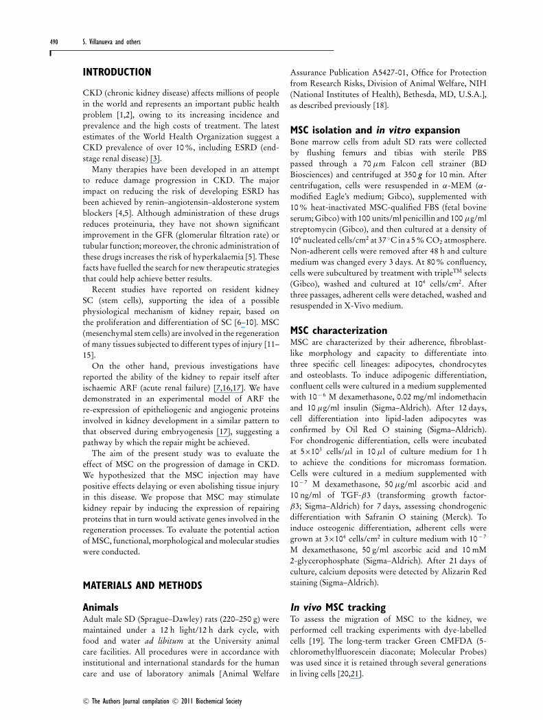
Figure 1 MSC characterization (A), MSC dye tracking in vivo (B), and CD73 + MSC in the kidney (C)(A) MSC were characterized by their capacity to differentiate into three specific cell lineages: chondrocytes, osteoblasts and adipocytes. The chondrogenic differentiationwas confirmed after 7 culture days by Safranin O staining (AA). The osteogenic differentiation was confirmed after 21 culture days by Alizarin Red staining (AC). Theadipogenic differentiation was confirmed after 12 culture days by Oil Red O staining (AE). (AB, AD and AF) The corresponding controls. Scale bar, 200 μm (AA–AD) and50 μm (AE and AF). (B) Microscopic images show kidney sections 24 h after MSC injection incubated with vehicle (upper panel) or with the cell tracker Green CMFDA(lower panel). The nuclei were counterstained with H&E (dark). The right panel (zoom) corresponds to the area enclosed by the segmented square in the merge panel.CM corresponds to kidney injected with culture medium and CM+ MSC corresponds to kidney injected with MSC. Scale bar, 10 μm. (C) CD73, an MSC marker, wasobserved in kidney sections 24 h after MSC injection (GFP panel). DAPI nucleic acid staining is show in blue. The right panels correspond to the Merge and Phasemicroscope techniques. Scale bar, 20 μm.
dithiothreitol, 4 % (w/v) SDS, 0.2 % Bromophenol Blueand 20 % (v/v) glycerol], transferred to nitrocellulosemembranes and blocked as previously described [27].After blocking, membranes were probed with the Ab,washed with TBS-T (Tris-buffered saline containing 1 %Tween 20) and incubated with HRP (horseradishperoxidase)-conjugated secondary Ab for 2 h at roomtemperature. Immunoreactivity was detected usingenhanced chemiluminescence obtained from Pierce. Basalcontrols were obtained from sham kidneys.
Densitometry analyses were performed using theNIH Image program v1.61 (NIH, http://rsb.info.nih.gov/nih-image). α-Tubulin total protein levels were usedto correct variation in sample loading.
Statistical analysisThe Mann–Whitney U test was used with a significancelevel: P < 0.05. Densitometry values are presented asmeans +− S.D. Values are presented in AU (arbitraryunits).
RESULTS
Functional characterization of MSCBone marrow-derived MSC from SD rats with a stablefibroblast-like phenotype were isolated by adherenceseparation. The MSC were able to differentiate intoadipocytes, chondrocytes and osteoblasts (Figure 1).
MSC dye trackingTo assess MSC localization in kidney tissues, we used theCMFDA cell tracker in dye-tracking experiments [19]. At24 h after MSC injection, kidney sections counterstainedwith H&E showed that the presence of GFP (greenfluorescent protein)-positive cells was only observed inanimals receiving Green CMFDA-treated cells (approx.1 %), but not in those treated with vehicle-incubated cells,indicating the migration of injected MSC to the kidney(Figure 1B).
CD73 is a membrane protein involved in theproteolytic activity of extracellular nucleotides and hasbeen described as a positive antigen present in MSC[23]. Immunofluorescence analysis was performed tocorroborate the migration of unlabelled MSC to thekidneys at 24 h after the injection. In agreement withthe previous results, CD73-positive cells were observedin kidney sections at 24 h after the MSC injection(Figure 1C). DAPI nucleic acid stain allows the cellularidentification and evaluation of tissue morphology. TheCD73 reactivity of cells in kidney showed a patternsimilar to what is seen in vitro in cultured MSC (resultsnot shown).
Functional renal damageRenal function was assessed by plasma creatinine levelsin sham animals (Sham) infused with fresh X-Vivomedium, Sham infused with MSC (Sham + MSC), NPX
C© The Authors Journal compilation C© 2011 Biochemical Society
MSC transplantation induces an improvement in CKD 493
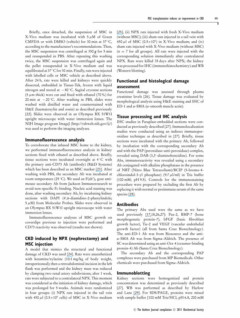
Figure 2 Histological NPX-induced damage in CKD kidneysThe IHC in kidney samples at day 35 post-NPX, injected with fresh X-Vivo medium (NPX) or MSC (NPX+ MSC). Positive staining for macrophages (ED-1) andmiofibroblasts (α-SMA) markers was observed in the interstitium of internal and external medulla in NPX kidneys injected with X-Vivo medium (E and F). A decreasedstaining of these markers was observed in NPX + MSC kidneys (H and I), comparable with kidneys of Sham rats (B and C). The tissue damage was also assessedby H&E staining: NPX rats showed the disappearance of the brush border, epithelium, tubular and glomerular alterations (D). NPX+ MSC showed morphologicalcharacteristics (G) similar to that observed in Sham rats (A). ED-1 and α-SMA markers were detected using peroxidase and developed with DAB (brown reaction).Scale bar, 100 μm. Arrows show the marker localization.
rats infused with fresh X-Vivo medium and NPXinjected with MSC (NPX + MSC). Sham rats injectedwith MSC or X-Vivo had normal plasma creatinine levels(0.3 mg/dl). In NPX animals, creatinine levels increasedto 1.0 +− 0.3 mg/dl, which was significantly higher thanin both Sham groups (P < 0.05). In rats subjectedto NPX + MSC, the creatinine level was reduced to0.4 +− 0.1 mg/dl (P < 0.05 compared with NPX group).Furthermore, creatinine values of the NPX + MSC werenot significantly different from those in Sham animals.
Histological studiesH&E staining of NPX renal sections showed alterationsconsistent with chronic damage: epithelia flattening,dilated tubules and increased fibrotic areas (Figure 2D).NPX treated with a single dose of MSC had normalmorphology (Figure 2G), comparable with that observedin Sham kidneys (Figure 2A and Table 1). We did notfind morphological, functional or histological differencesbetween Sham and Sham + MSC kidneys (results notshown); for this reason, this latter group was excludedin the following IHC analyses.
Damage markers ED-1 (macrophages) and α-SMA(myofibroblasts) were widely distributed in renalparenchyma of NPX animals (Figures 2E and 2F).However, both markers had a minor expression inNPX + MSC animals (Figures 2H and 2I). Figures 2(B)and 2(C) are Sham rats, showing normal IHC distributionof these markers.
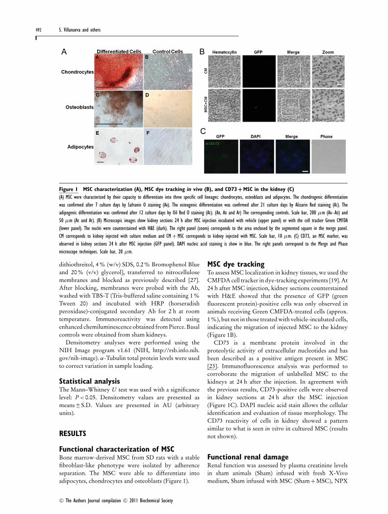
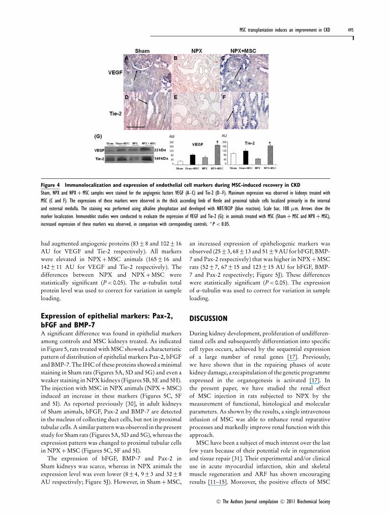
Renal SC detectionThe presence of the renal SC marker Oct-4 was evaluated5 and 35 days after injection. The IHC staining observedin Sham animals was minimal (Figure 3Aa and 3Ad).NPX animals showed a positive stain 5 days after NPX(Figure 3Ab) that was completely inhibited 35 daysafter surgery (Figure 3Ae). NPX animals injected withMSC presented a marked stain at 5 days after injection(Figure 3Ac) that was maintained 35 days after injection(Figure 3Af). Oct-4 expression evaluated by WB 35 daysafter injection is shown in Figure 3(Ag): Sham andNPX animals had reduced Oct-4 expression (15 +− 8and 24 +− 8 AU respectively), which was increased withMSC injection (74 +− 8 and 185 +− 16 AU in Sham + MSCand NPX + MSC respectively). These differences werestatistically significant (P < 0.05).
Additionally, we analysed the co-localization of CD-73 and Oct-4 35 days after injection (Figure 3B). Theco-expression of CD73/Oct-4 was observed in a reducedcell number (0.2 %).
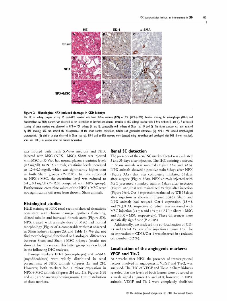
Localization of the angiogenic markers:VEGF and Tie-2At 5 weeks after NPX, the presence of transcriptionalfactors involved in angiogenesis, VEGF and Tie-2, wasanalysed. The IHC of VEGF and Tie-2 in Sham kidneysrevealed that the levels of both factors were observed asa weak signal (Figures 4A and 4D); however, in NPXanimals, VEGF and Tie-2 were completely abolished
C© The Authors Journal compilation C© 2011 Biochemical Society
494 S. Villanueva and others
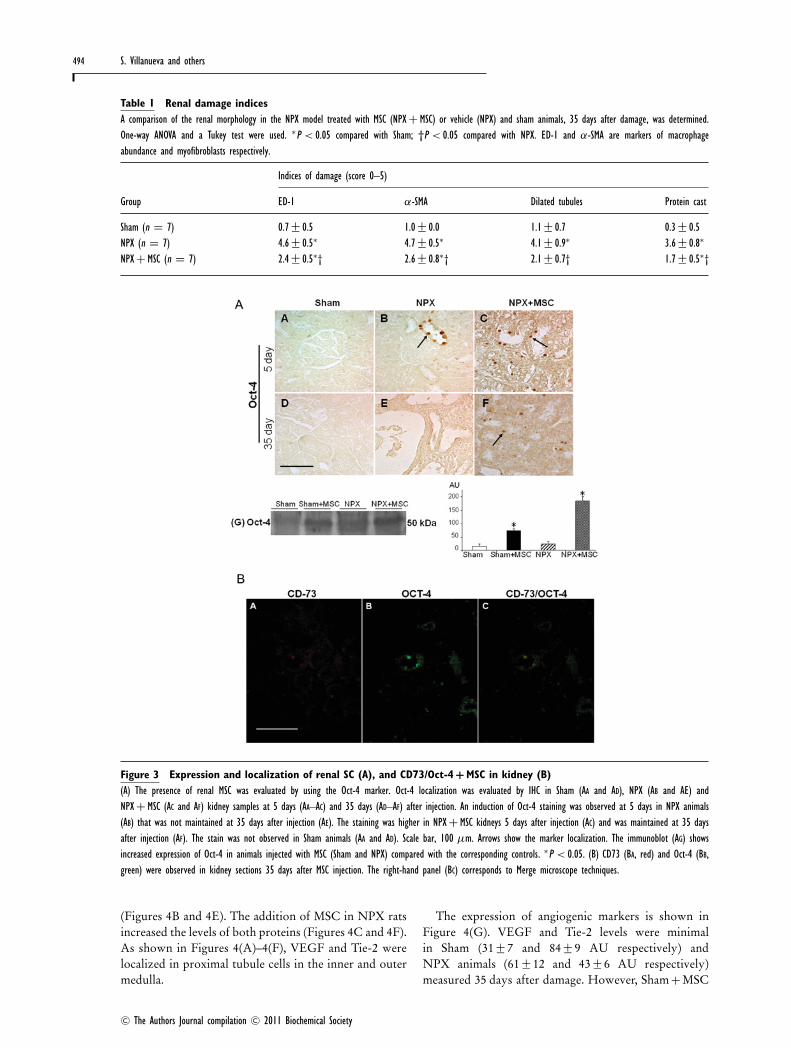
Table 1 Renal damage indicesA comparison of the renal morphology in the NPX model treated with MSC (NPX+ MSC) or vehicle (NPX) and sham animals, 35 days after damage, was determined.One-way ANOVA and a Tukey test were used. ∗P < 0.05 compared with Sham; †P < 0.05 compared with NPX. ED-1 and α-SMA are markers of macrophageabundance and myofibroblasts respectively.
Indices of damage (score 0–5)
Group ED-1 α-SMA Dilated tubules Protein cast
Sham (n = 7) 0.7 +− 0.5 1.0 +− 0.0 1.1 +− 0.7 0.3 +− 0.5NPX (n = 7) 4.6 +− 0.5∗ 4.7 +− 0.5∗ 4.1 +− 0.9∗ 3.6 +− 0.8∗
NPX+ MSC (n = 7) 2.4 +− 0.5∗† 2.6 +− 0.8∗† 2.1 +− 0.7† 1.7+− 0.5∗†
Figure 3 Expression and localization of renal SC (A), and CD73/Oct-4 + MSC in kidney (B)(A) The presence of renal MSC was evaluated by using the Oct-4 marker. Oct-4 localization was evaluated by IHC in Sham (AA and AD), NPX (AB and AE) andNPX+ MSC (AC and AF) kidney samples at 5 days (AA–AC) and 35 days (AD–AF) after injection. An induction of Oct-4 staining was observed at 5 days in NPX animals(AB) that was not maintained at 35 days after injection (AE). The staining was higher in NPX + MSC kidneys 5 days after injection (AC) and was maintained at 35 daysafter injection (AF). The stain was not observed in Sham animals (AA and AD). Scale bar, 100 μm. Arrows show the marker localization. The immunoblot (AG) showsincreased expression of Oct-4 in animals injected with MSC (Sham and NPX) compared with the corresponding controls. ∗P < 0.05. (B) CD73 (BA, red) and Oct-4 (BB,green) were observed in kidney sections 35 days after MSC injection. The right-hand panel (BC) corresponds to Merge microscope techniques.
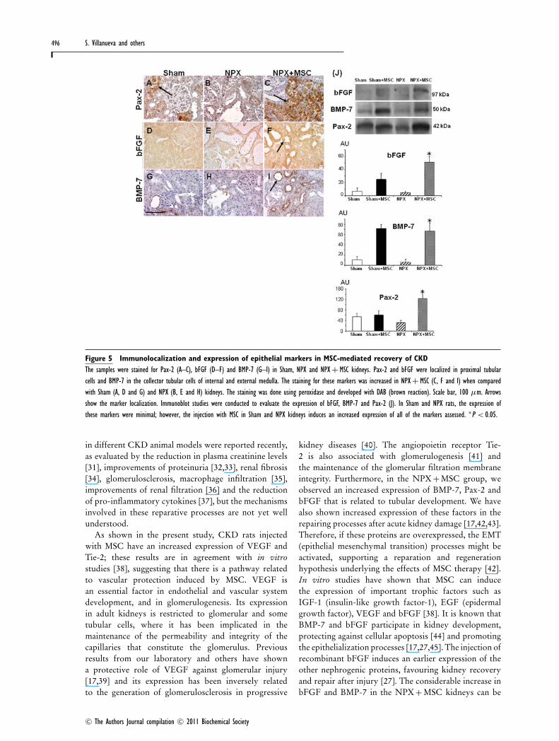
(Figures 4B and 4E). The addition of MSC in NPX ratsincreased the levels of both proteins (Figures 4C and 4F).As shown in Figures 4(A)–4(F), VEGF and Tie-2 werelocalized in proximal tubule cells in the inner and outermedulla.
The expression of angiogenic markers is shown inFigure 4(G). VEGF and Tie-2 levels were minimalin Sham (31 +− 7 and 84 +− 9 AU respectively) andNPX animals (61 +− 12 and 43 +− 6 AU respectively)measured 35 days after damage. However, Sham + MSC
C© The Authors Journal compilation C© 2011 Biochemical Society
MSC transplantation induces an improvement in CKD 495
Figure 4 Immunolocalization and expression of endothelial cell markers during MSC-induced recovery in CKDSham, NPX and NPX + MSC samples were stained for the angiogenic factors VEGF (A–C) and Tie-2 (D–F). Maximum expression was observed in kidneys treated withMSC (C and F). The expressions of these markers were observed in the thick ascending limb of Henle and proximal tubule cells localized primarily in the internaland external medulla. The staining was performed using alkaline phosphatase and developed with NBT/BCIP (blue reaction). Scale bar, 100 μm. Arrows show themarker localization. Immunoblot studies were conducted to evaluate the expression of VEGF and Tie-2 (G): in animals treated with MSC (Sham+ MSC and NPX + MSC),increased expression of these markers was observed, in comparison with corresponding controls. ∗P < 0.05.
had augmented angiogenic proteins (83 +− 8 and 102 +− 16AU for VEGF and Tie-2 respectively). All markerswere elevated in NPX + MSC animals (165 +− 16 and142 +− 11 AU for VEGF and Tie-2 respectively). Thedifferences between NPX and NPX + MSC werestatistically significant (P < 0.05). The α-tubulin totalprotein level was used to correct for variation in sampleloading.
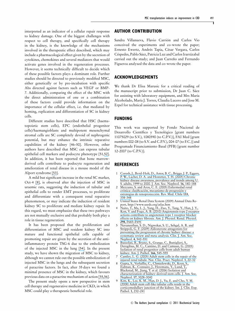
Expression of epithelial markers: Pax-2,bFGF and BMP-7A significant difference was found in epithelial markersamong controls and MSC kidneys treated. As indicatedin Figure 5, rats treated with MSC showed a characteristicpattern of distribution of epithelial markers Pax-2, bFGFand BMP-7. The IHC of these proteins showed a minimalstaining in Sham rats (Figures 5A, 5D and 5G) and even aweaker staining in NPX kidneys (Figures 5B, 5E and 5H).The injection with MSC in NPX animals (NPX + MSC)induced an increase in these markers (Figures 5C, 5Fand 5I). As reported previously [30], in adult kidneysof Sham animals, bFGF, Pax-2 and BMP-7 are detectedin the nucleus of collecting duct cells, but not in proximaltubular cells. A similar pattern was observed in the presentstudy for Sham rats (Figures 5A, 5D and 5G), whereas theexpression pattern was changed to proximal tubular cellsin NPX + MSC (Figures 5C, 5F and 5I).
The expression of bFGF, BMP-7 and Pax-2 inSham kidneys was scarce, whereas in NPX animals theexpression level was even lower (8 +− 4, 9 +− 3 and 32 +− 8AU respectively; Figure 5J). However, in Sham + MSC,
an increased expression of epitheliogenic markers wasobserved (25 +− 3, 68 +− 13 and 51 +− 9 AU for bFGF, BMP-7 and Pax-2 respectively) that was higher in NPX + MSCrats (52 +− 7, 67 +− 15 and 123 +− 15 AU for bFGF, BMP-7 and Pax-2 respectively; Figure 5J). These differenceswere statistically significant (P < 0.05). The expressionof α-tubulin was used to correct for variation in sampleloading.
DISCUSSION
During kidney development, proliferation of undifferen-tiated cells and subsequently differentiation into specificcell types occurs, achieved by the sequential expressionof a large number of renal genes [17]. Previously,we have shown that in the repairing phases of acutekidney damage, a recapitulation of the genetic programmeexpressed in the organogenesis is activated [17]. Inthe present paper, we have studied the renal effectof MSC injection in rats subjected to NPX by themeasurement of functional, histological and molecularparameters. As shown by the results, a single intravenousinfusion of MSC was able to enhance renal reparativeprocesses and markedly improve renal function with thisapproach.
MSC have been a subject of much interest over the lastfew years because of their potential role in regenerationand tissue repair [31]. Their experimental and/or clinicaluse in acute myocardial infarction, skin and skeletalmuscle regeneration and ARF has shown encouragingresults [11–15]. Moreover, the positive effects of MSC
C© The Authors Journal compilation C© 2011 Biochemical Society
496 S. Villanueva and others
Figure 5 Immunolocalization and expression of epithelial markers in MSC-mediated recovery of CKDThe samples were stained for Pax-2 (A–C), bFGF (D–F) and BMP-7 (G–I) in Sham, NPX and NPX+ MSC kidneys. Pax-2 and bFGF were localized in proximal tubularcells and BMP-7 in the collector tubular cells of internal and external medulla. The staining for these markers was increased in NPX+ MSC (C, F and I) when comparedwith Sham (A, D and G) and NPX (B, E and H) kidneys. The staining was done using peroxidase and developed with DAB (brown reaction). Scale bar, 100 μm. Arrowsshow the marker localization. Immunoblot studies were conducted to evaluate the expression of bFGF, BMP-7 and Pax-2 (J). In Sham and NPX rats, the expression ofthese markers were minimal; however, the injection with MSC in Sham and NPX kidneys induces an increased expression of all of the markers assessed. ∗P < 0.05.
in different CKD animal models were reported recently,as evaluated by the reduction in plasma creatinine levels[31], improvements of proteinuria [32,33], renal fibrosis[34], glomerulosclerosis, macrophage infiltration [35],improvements of renal filtration [36] and the reductionof pro-inflammatory cytokines [37], but the mechanismsinvolved in these reparative processes are not yet wellunderstood.
As shown in the present study, CKD rats injectedwith MSC have an increased expression of VEGF andTie-2; these results are in agreement with in vitrostudies [38], suggesting that there is a pathway relatedto vascular protection induced by MSC. VEGF isan essential factor in endothelial and vascular systemdevelopment, and in glomerulogenesis. Its expressionin adult kidneys is restricted to glomerular and sometubular cells, where it has been implicated in themaintenance of the permeability and integrity of thecapillaries that constitute the glomerulus. Previousresults from our laboratory and others have showna protective role of VEGF against glomerular injury[17,39] and its expression has been inversely relatedto the generation of glomerulosclerosis in progressive
kidney diseases [40]. The angiopoietin receptor Tie-2 is also associated with glomerulogenesis [41] andthe maintenance of the glomerular filtration membraneintegrity. Furthermore, in the NPX + MSC group, weobserved an increased expression of BMP-7, Pax-2 andbFGF that is related to tubular development. We havealso shown increased expression of these factors in therepairing processes after acute kidney damage [17,42,43].Therefore, if these proteins are overexpressed, the EMT(epithelial mesenchymal transition) processes might beactivated, supporting a reparation and regenerationhypothesis underlying the effects of MSC therapy [42].In vitro studies have shown that MSC can inducethe expression of important trophic factors such asIGF-1 (insulin-like growth factor-1), EGF (epidermalgrowth factor), VEGF and bFGF [38]. It is known thatBMP-7 and bFGF participate in kidney development,protecting against cellular apoptosis [44] and promotingthe epithelialization processes [17,27,45]. The injection ofrecombinant bFGF induces an earlier expression of theother nephrogenic proteins, favouring kidney recoveryand repair after injury [27]. The considerable increase inbFGF and BMP-7 in the NPX + MSC kidneys can be
C© The Authors Journal compilation C© 2011 Biochemical Society
MSC transplantation induces an improvement in CKD 497
interpreted as an indicator of a cellular repair responseto kidney damage. One of the biggest challenges withrespect to cell therapy, and specifically cell therapyin the kidney, is the knowledge of the mechanismsinvolved in the therapeutic effect described, which mayinclude a pharmacological effect given by the secretion ofcytokines, chemokines and several mediators that wouldactivate genes involved in the regeneration processes.However, it seems technically difficult to decide whichof these possible factors plays a dominant role. Furtherstudies should be directed to previously modified MSC,either genetically or by pre-incubation with specificAbs directed against factors such as VEGF or BMP-7. Additionally, comparing the effect of the MSC withthe direct administration of one or a combinationof these factors could provide information on theimportance of the cellular effect, i.e. that mediated byhoming, replication and differentiation of SC in kidneycells.
Different studies have described that HSC (haema-topoietic stem cells), EPC (endothelial progenitorcells)/haemangioblasts and multipotent mesenchymalstromal cells are SC completely devoid of nephrogenicpotential, but may enhance the intrinsic reparativecapabilities of the kidney [46–50]. However, otherauthors have described that MSC can express tubularepithelial cell markers and podocyte phenotype [51,52].In addition, it has been reported that bone marrow-derived cells contribute to podocyte regeneration andamelioration of renal disease in a mouse model of theAlport syndrome [53].
A mild but significant increase in the renal SC marker,Oct-4 [8], is observed after the injection of MSC inuraemic rats, suggesting the induction of tubular andepithelial cells to render EMT processes, to proliferateand differentiate with a consequent renal reparativephenomenon, or may indicate the induction of residentkidney SC to proliferate and mediate kidney repair. Inthis regard, we must emphasize that these two pathwaysare not mutually exclusive and that probably both play arole in tissue regeneration.
It has been proposed that the proliferation anddifferentiation of MSC and resident kidney SC intomature and functional epithelial cells capable ofpromoting repair are given by the secretion of the anti-inflammatory protein TSG-6 due to the embolizationof the injected MSC in the lung [54]. In the presentstudy, we have shown the migration of MSC to kidney,although we cannot rule out the possible embolization ofinjected MSC in the lungs and the subsequent secretionof paracrine factors. In fact, after 35 days we found aminimal presence of MSC in the kidney, which favoursprevious data on a paracrine mechanism of action [55,56].
The present study opens a new perspective in stemcell therapy and regenerative medicine in CKD, in whichMSC could play a therapeutic beneficial role.
AUTHOR CONTRIBUTION
Sandra Villanueva, Flavio Carrion and Carlos Vioconceived the experiments and co-wrote the paper;Ernesto Ewertz, Andres Tapia, Cesar Vergara, CarlosCespedes, Pablo Saez, Patricia Luz and Carlos Irarrazabalcarried out the study; and Juan Carreno and FernandoFigueroa analysed the data and co-wrote the paper.
ACKNOWLEDGEMENTS
We thank Dr Elisa Marusic for a critical reading ofthe manuscript prior to submission, Dr Juan C. Saezfor assisting with laboratory equipment, and Mrs MariaAlcoholado, Marıa J. Torres, Claudia Lucero and Jose M.Erpel for technical assistance with tissue processing.
FUNDING
This work was supported by Fondo Nacional deDesarrollo Cientıfico y Tecnologico [grant numbers11075029 (to S.V.), 1080590 (to C.P.V.)], FAI Med [grantnumbers 002-08 (to S.V. and C.P.V.), 004-07 (to F.C.)] andProgramade Financiamiento Basal (PFB) [grant number12-2007 (to C.P.V.)].
REFERENCES
1 Coresh, J., Byrd-Holt, D., Astor, B. C., Briggs, J. P., Eggers,P. W., Lacher, D. A. and Hostetter, T. H. (2005) Chronickidney disease awareness, prevalence and trends among U.S. adults, 1999 to 2000. J. Am. Soc. Nephrol. 16, 180–188
2 Mezzano, S. and Aros, C. E. (2005) Enfermedad renalcronica: clasificacion, mecanismo de progresion yestrategias de renoproteccion. Rev. Med. Chile 133,338–348
3 United States Renal Data System (2009) Annual Data Re-port, http://www.usrds.org/atlas.htm
4 Naito, T., Ma, L. J., Yang, H., Zuo, Y., Tang, Y., Han, J. Y.,Kon, V. and Fogo, A. B. (2010) Angiotensin type 2 receptoractions contribute to angiotensin type 1 receptor blockereffects on kidney fibrosis. Am. J. Physiol. Renal. Physiol.298, F683–F691
5 Navaneethan, S. D., Nigwekar, S. U., Sehgal, A. R. andStrippoli, G. F. (2009) Aldosterone antagonists forpreventing the progression of chronic kidney disease: asystematic review and meta-analysis. Clin. J. Am. Soc.Nephrol. 4, 542–551
6 Bussolati, B., Bruno, S., Grange, C., Buttiglieri, S.,Deregibus, M. C., Cantino, D. and Camussi, G. (2005)Isolation of renal progenitor cells from adult humankidney. Am. J. Pathol. 166, 545–555
7 Cantley, L. G. (2005) Adult stem cells in the repair of theinjured renal tubule. Nat. Clin. Pract. Nephrol. 1, 22–32
8 Gupta, S., Verfaillie, C., Chmielewski, D., Kren, S.,Eidman, K., Connaire, J., Heremans, Y., Lund, T.,Blackstad, M., Jiang, Y. et al. (2006) Isolation andcharacterization of kidney-derived stem cells. J. Am. Soc.Nephrol. 17, 3028–3040
9 Kim, K., Lee, K. M., Han, D. J., Yu, E. and Cho, Y. M.(2008) Adult stem cell-like tubular cells reside in thecorticomedullary junction of the kidney. Int. J. Clin. Exp.Pathol. 1, 232–241
C© The Authors Journal compilation C© 2011 Biochemical Society
498 S. Villanueva and others
10 Sagrinati, C., Netti, G. S., Mazzinghi, B., Lazzeri, E.,Liotta, F., Frosali, F., Ronconi, E., Meini, C., Gacci, M.,Squecco, R. et al. (2006) Isolation and characterization ofmultipotent progenitor cells from the Bowman’s capsule ofadult human kidneys. J. Am. Soc. Nephrol. 17, 2443–2456
11 Crop, M. J., Baan, C. C., Korevaar, S. S., Ijzermans, J. N.,Alwayn, I. P., Weimar, W. and Hoogduijn, M.J. (2009)Donor-derived mesenchymal stem cells suppressalloreactivity of kidney transplant patients. Transplantation87, 896–906
12 Poulsom, R., Forbes, S. J., Hodivala-Dilke, K., Ryan, E.,Wyles, S., Navaratnarasah, S., Jeffery, R., Hunt, T., Alison,M., Cook, T. et al. (2001) Bone marrow contributes torenal parenchymal turnover and regeneration. J. Pathol.195, 229–235
13 Quarto, R., Mastrogiacomo, M., Cancedda, R., Kutepov,S. M., Mukhachev, V., Lavroukov, A., Kon, E. andMarcacci, M. (2001) Repair of large bone defects with theuse of autologous bone marrow stromal cells. N. Engl. J.Med. 344, 385–386
14 Semedo, P., Wang, P. M., Andreucci, T. H., Cenedeze, M.A., Teixeira, V. P., Reis, M. A., Pacheco-Silva, A. andCamara, N. O. (2007) Mesenchymal stem cells amelioratetissue damages triggered by renal ischemia and reperfusioninjury. Transplant. Proc. 39, 421–423
15 Sykova, E., Homola, A., Mazanec, R., Lachmann, H.,Konradova, S. L., Kobylka, P., Padr, R., Neuwirth, J.,Komrska, V., Vavra, V. et al. (2006) Autologous bonemarrow transplantation in patients with subacute andchronic spinal cord injury. Cell Transplant. 15, 675–687
16 Morigi, M., Imberti, B., Zoja, C., Corna, D., Tomasoni, S.,Abbate, M., Rottoli, D., Angioletti, S., Benigni, A., Perico,N. et al. (2004) Mesenchymal stem cells are renotropic,helping to repair the kidney and improve function in acuterenal failure. J. Am. Soc. Nephrol. 15, 1794–1804
17 Villanueva, S., Cespedes, C. and Vio, C. P. (2006) Ischemicacute renal failure induces the expression of a wide range ofnephrogenic proteins. Am. J. Physiol. Regul. Integr. Comp.Physiol. 290, R861–R870
18 Villanueva, S., Cespedes, C., Gonzalez, A. A., Roessler, E.and Vio, C. P. (2008) Inhibition of bFGF-receptor type 2increases kidney damage and suppresses nephrogenicprotein expression after ischemic acute renal failure. Am. J.Physiol. Regul. Integr. Comp. Physiol. 294, R819–R828
19 Weir, C., Morel-Kopp, M. C., Gill, A., Tinworth, K., Ladd,L., Hunyor, S. N. and Ward, C. (2008) Mesenchymal stemcells: isolation, characterisation and in vivo fluorescent dyetracking. Heart Lung Circ. 17, 395–403
20 Liebler, J. M., Lutzko, C., Banfalvi, A., Senadheera, D.,Aghamohammadi, N., Crandall, E. D. and Borok, Z.(2008) Retention of human bone marrow-derived cells inmurine lungs following bleomycin-induced lung injury.Am. J. Physiol. Lung Cell. Mol. Physiol. 295, L285–L292
21 Patel, S. A., Meyer, J. R., Greco, S. J., Corcoran, K. E.,Bryan, M. and Rameshwar, P. (2010) Mesenchymal stemcells protect breast cancer cells through regulatory T cells:role of mesenchymal stem cell-derived TGF-β.J. Immunol. 184, 5885–5894
22 Salas, S. P., Giacaman, A., Romero, W., Downey, P.,Aranda, E., Mezzano, D. and Vıo, C. P. (2007) Pregnantrats treated with a serotonin precursor have reduced fetalweight and lower plasma volume and kallikrein levels.Hypertension 50, 773–779
23 Dominici, M., Le Blanc, K., Mueller, I., Slaper-Cortenbach,I., Marini, F., Krause, D., Deans, R., Keating, A., Prockop,Dj. and Horwitz, E. (2006) Minimal criteria for definingmultipotent mesenchymal stromal cells. The InternationalSociety for Cellular Therapy position statement.Cytotherapy 8, 315–317
24 Fleck, C., Appenroth, D., Jonas, P., Koch, M., Kundt, G.,Nizze, H. and Stein, G. (2006) Suitability of 5/6nephrectomy (5/6NX) for the induction of interstitial renalfibrosis in rats – influence of sex, strain, and surgicalprocedure. Exp. Toxicol. Pathol. 57, 195–205
25 Carrion, F., Nova, E., Ruiz, C., Diaz, F., Inostroza, C.,Rojo, D., Monckeberg, G. and Figueroa, F. E. (2010)Autologous mesenchymal stem cell treatment increased Tregulatory cells with no effect on disease activity in twosystemic lupus erythematosus patients. Lupus 19, 317–322
26 Villanueva, S., Cespedes, C., Gonzalez, A. A., Vio, C. P.and Velarde, V. (2007) Effect of ischemic acute renaldamage on the expression of COX-2 and oxidativestress-related elements in rat kidney. Am. J. Physiol. Renal.Physiol. 292, F1364–F1371
27 Villanueva, S., Cespedes, C., Gonzalez, A. and Vio, C. P.(2006) bFGF induces an earlier expression of nephrogenicproteins after ischemic acute renal failure. Am. J. Physiol.Regul. Integr. Comp. Physiol. 291, R1677–R1687
28 Vio, C. P., Cespedes, C., Gallardo, P. and Masferrer, J. L.(1997) Renal identification of cyclooxygenase-2 in a subsetof thick ascending limb cells. Hypertension 3, 687–692
29 Harlow, E. and Lane, D., (1998) Antibodies: A LaboratoryManual, Cold Spring Harbor Laboratory, Plainview, NY
30 Rothenpieler, U. W. and Dressler, G. R. (1993) Pax-2 isrequired for mesenchyme-to-epithelium conversion duringkidney development. Development 119, 711–720
31 Song, J. H. and Humes, H. D. (2009) Renal cell therapyand beyond. Semin. Dial. 22, 603–609
32 Cavaglieri, R. C., Martini, D., Sogayar, M. C. andNoronha, I. L. (2009) Mesenchymal stem cells delivered atthe subcapsule of the kidney ameliorate renal disease in therat remnant kidney model. Transplant. Proc. 41, 947–951
33 Choi, S., Park, M., Kim, J., Hwang, S., Park, S. and Lee, Y.(2009) The role of mesenchymal stem cells in the functionalimprovement of chronic renal failure. Stem Cells Dev. 18,521–529
34 Asanuma, H., Vanderbrink, B. A., Campbell, M. T., Hile,K. L., Zhang, H., Meldrum, D. R. and Meldrum, K. K.(2011) Arterially delivered mesenchymal stem cells preventobstruction-induced renal fibrosis. J. Surg. Res. 168,e51–e59
35 Lee, S. R., Lee, S. H., Moon, J. Y., Park, J. Y., Lee, D., Lim,S. J., Jeong, K. H., Park, J. K., Lee, T. W. and Ihm, C. G.(2010) Repeated administration of bone marrow-derivedmesenchymal stem cells improved the protective effects ona remnant kidney model. Ren. Fail. 32, 840–848
36 Kelley, R., Werdin, E. S., Bruce, A. T., Choudhury, S.,Wallace, S. M., Ilagan, R. M., Cox, B. R., Tatsumi-Ficht, P.,Rivera, E. A., Spencer, T. et al. (2010) Tubular cell-enrichedsubpopulation of primary renal cells improves survival andaugments kidney function in rodent model of chronickidney disease. Am. J. Physiol. Renal. Physiol. 299,F1026–F1039
37 Sangidorj, O., Yang, S. H., Jang, H. R., Lee, J. P., Cha, R.H., Kim, S. M., Lim, C. S. and Kim, Y. S. (2010) Bonemarrow-derived endothelial progenitor cells confer renalprotection in a murine chronic renal failure model. Am. J.Physiol. Renal. Physiol. 299, F325–F335
38 Wakabayashi, K., Nagai, A., Sheikh, A. M., Shiota, Y.,Narantuya, D., Watanabe, T., Masuda, J., Kobayashi, S.,Kim, S. U. and Yamaguchi, S. (2010) Transplantation ofhuman mesenchymal stem cells promotes functionalimprovement and increased expression of neurotrophicfactors in a rat focal cerebral ischemia model. J. Neurosci.Res. 88, 1017–1025
39 Kelly, D. J., Hepper, C., Wu, L. L., Cox, A. J. and Gilbert,R. E. (2003) Vascular endothelial growth factor expressionand glomerular endothelial cell loss in the remnant kidneymodel. Nephrol. Dial. Transplant. 18, 1286–1292
40 Miyamoto, K., Kitamoto, Y., Tokunaga, H., Takeya, M.,Ezaki, T., Imamura, T. and Tomita, K. (2004) Protectiveeffect of vascular endothelial growth factor/vascularpermeability factor 165 and 121 on glomerular endothelialcell injury in the rat. Lab. Invest. 84, 1126–1136
41 Woolf, A. S. (2005) Molecular and genetic analyses of renalcapillary development: Studying the angiopoietin/Tie axis.Kidney Int. 68, 1968–1968
42 Gilbert, S. F. (2000) Reciprocal interactions of developingkidney tissues. In Developmental Biology, 6th edn, p. 462,Sinauer Associates, Sunderland, MA
43 Karavanov, A. A., Karavanova, I., Perantoni, A. andDawid, I. B. (1998) Expression pattern of the rat Lim-1homeobox gene suggest a dual role during kidneydevelopment. Int. J. Dev. Biol. 42, 61–66
C© The Authors Journal compilation C© 2011 Biochemical Society
MSC transplantation induces an improvement in CKD 499
44 Karavanova, I. D., Dove, L. F., Resau, J. H. and Perantoni,A. O. (1996) Conditioned medium from a rat ureteric budcell line in combination with bFGF induces completedifferentiation of isolated metanephric mesenchyme.Development 122, 4159–4167
45 Vrljicak, P., Myburgh, D., Ryan, A. K., van Rooijen, M. A.,Mummery, C. L. and Gupta, I. R. (2004) Smad expressionduring kidney development. Am. J. Physiol. Renal.Physiol. 286, F625–F633
46 Togel, F., Hu, Z., Weiss, K., Isaac, J., Lange, C. andWestenfelder, C. (2005) Administered mesenchymal stemcells protect against ischemic acute renal failure throughdifferentiation-independent mechanisms. Am. J. Physiol.Renal Physiol. 289, F31–F42
47 Bi, B., Schmitt, R., Israilova, M., Nishio, H. and Cantley,L. G. (2007) Stromal cells protect against acute tubularinjury via an endocrine effect. J. Am. Soc. Nephrol. 18,2486–2496
48 Lange, C., Togel, F., Ittrich, H., Clayton, F.,Nolte-Ernsting, C., Zander, A. R. and Westenfelder, C.(2005) Administered mesenchymal stem cells enhancerecovery from ischemia/ reperfusion-induced acute renalfailure in rats. Kidney Int. 68, 1613–1617
49 Togel, F., Cohen, A., Zhang, P., Yang, Y., Hu, Z. andWestenfelder, C. (2009) Autologous and allogeneic marrowstromal cells are safe and effective for the treatment ofacute kidney injury. Stem Cells Dev. 18, 475–485
50 Duffield, J. S., Park, K. M., Hsiao, L. L., Kelley, V. R.,Scadden, D. T., Ichimura, T. and Bonventre, J. V. (2005)Restoration of tubular epithelial cells during repair of thepostischemic kidney occurs independently of bonemarrow-derived stem cells. J. Clin. Invest. 115, 1743–1755
51 Behr, L., Hekmati, M., Fromont, G., Borenstein, N., Noel,L. H., Lelievre-Pegorier, M. and Laborde, K. (2007) Intrarenal arterial injection of autologous mesenchymal stemcells in an ovine model in the postischemic kidney.Nephron Physiol. 107, 65–76
52 Perry, J., Tam, S., Zheng, K., Sado, Y., Dobson, H.,Jefferson, B., Jacobs, R. and Thorner, P. S. (2006) Type IVcollagen induces podocytic features in bone marrowstromal stem cells in vitro. J. Am. Soc. Nephrol. 17,66–76
53 Prodromidi, E. I., Poulsom, R., Jeffery, R., Roufosse,C. A., Pollard, P. J., Pusey, C. D. and Cook, H. T. (2006)Bone marrow-derived cells contribute to podocyteregeneration and amelioration of renal disease in a mousemodel of Alport syndrome. Stem Cells 24, 2448–2455
54 Lee, R. H., Pulin, A. A., Seo, M. J., Kota, D. J., Ylostalo, J.,Larson, B. L., Semprun-Prieto, L., Delafontaine, P. andProckop, D.J. (2009) Intravenous hMSCs improvemyocardial infarction in mice because cells embolized inlung are activated to secrete the anti-inflammatory proteinTSG-6. Cell Stem Cell 5, 54–63
55 Herrera, M. B., Bussolati, B., Bruno, S., Morando, L.,Mauriello-Romanazzi, G., Sanavio, F., Stamenkovic, I.,Biancone, L. and Camussi, G. (2007) Exogenousmesenchymal stem cells localize to the kidney by means ofCD44 following acute tubular injury. Kidney Int. 72,430–431
56 Camussi, G., Deregibus, M. C. and Tetta, C. (2010)Paracrine/endocrine mechanism of stem cells on kidneyrepair: role of microvesicle-mediated transfer of geneticinformation. Curr. Opin. Nephrol. Hypertens. 19,7–12
Received 7 March 2011/4 May 2011; accepted 16 June 2011Published as Immediate Publication 16 June 2011, doi:10.1042/CS20110108
C© The Authors Journal compilation C© 2011 Biochemical Society
Related Documents