Portland State University Portland State University PDXScholar PDXScholar Civil and Environmental Engineering Faculty Publications and Presentations Civil and Environmental Engineering 2020 Menthol in Electronic Cigarettes: A Contributor to Menthol in Electronic Cigarettes: A Contributor to Respiratory Disease? Respiratory Disease? Vijayalekshmi Nair University of California, Riverside Malcolm Tran University of California, Riverside Rachel Z. Behar University of California, Riverside Song Zhai University of California, Riverside Xinping Cui University of California, Riverside See next page for additional authors Follow this and additional works at: https://pdxscholar.library.pdx.edu/cengin_fac Part of the Public Health Commons Let us know how access to this document benefits you. Citation Details Citation Details Nair, V., Tran, M., Behar, R. Z., Zhai, S., Cui, X., Phandthong, R., ... & Volz, D. C. (2020). Menthol in Electronic Cigarettes: A Contributor to Respiratory Disease? This Pre-Print is brought to you for free and open access. It has been accepted for inclusion in Civil and Environmental Engineering Faculty Publications and Presentations by an authorized administrator of PDXScholar. Please contact us if we can make this document more accessible: [email protected].

Welcome message from author
This document is posted to help you gain knowledge. Please leave a comment to let me know what you think about it! Share it to your friends and learn new things together.
Transcript

Portland State University Portland State University
PDXScholar PDXScholar
Civil and Environmental Engineering Faculty Publications and Presentations Civil and Environmental Engineering
2020
Menthol in Electronic Cigarettes: A Contributor to Menthol in Electronic Cigarettes: A Contributor to
Respiratory Disease? Respiratory Disease?
Vijayalekshmi Nair University of California, Riverside
Malcolm Tran University of California, Riverside
Rachel Z. Behar University of California, Riverside
Song Zhai University of California, Riverside
Xinping Cui University of California, Riverside
See next page for additional authors
Follow this and additional works at: https://pdxscholar.library.pdx.edu/cengin_fac
Part of the Public Health Commons
Let us know how access to this document benefits you.
Citation Details Citation Details Nair, V., Tran, M., Behar, R. Z., Zhai, S., Cui, X., Phandthong, R., ... & Volz, D. C. (2020). Menthol in Electronic Cigarettes: A Contributor to Respiratory Disease?
This Pre-Print is brought to you for free and open access. It has been accepted for inclusion in Civil and Environmental Engineering Faculty Publications and Presentations by an authorized administrator of PDXScholar. Please contact us if we can make this document more accessible: [email protected].

Authors Authors Vijayalekshmi Nair, Malcolm Tran, Rachel Z. Behar, Song Zhai, Xinping Cui, Rattapol Phandthong, Yuhuan Wang, Songqin Pan, Wentai Luo, James F. Pankow, David C. Volz, and Prue Talbot
This pre-print is available at PDXScholar: https://pdxscholar.library.pdx.edu/cengin_fac/566

Menthol in Electronic Cigarettes: A Contributor to Respiratory Disease?
Vijayalekshmi Nair1, Malcolm Tran1, Rachel Z. Behar1, Song Zhai2, Xinping Cui2, Rattapol
Phandthong1, Yuhuan Wang1, Songqin Pan3, Wentai Luo4, James F. Pankow4, David C.
Volz5, and Prue Talbot1*
1Department of Molecular, Cell and Systems Biology, University of California, Riverside,
CA 92521, USA
2Department of Statistics, University of California, Riverside, CA 92521, USA
3Proteomics Facility IIGB, University of California, Riverside, CA 92521, USA
4Department of Civil and Environmental Engineering, Portland State University,
Portland, Oregon
5Department of Environmental Sciences, University of California, Riverside, CA 92521,
USA
The first two authors contributed equally to this work
* Lead Contact and Corresponding Author:
Prue Talbot
Telephone: 951-850-7783
Keywords: Menthol, air-liquid interface, reactive oxygen species, inflammation, TRPM8,
respiratory cells
.CC-BY-NC-ND 4.0 International licenseauthor/funder. It is made available under aThe copyright holder for this preprint (which was not peer-reviewed) is the. https://doi.org/10.1101/2020.03.14.988006doi: bioRxiv preprint

SUMMARY
Menthol is widely used in tobacco products. This study compared the effects of menthol
on human bronchial epithelium using submerged cultures, a VITROCELL® cloud chamber that
provides air liquid interface (ALI) exposure without solvents or heating, and a Cultex ALI system
that delivers aerosol equivalent to that inhaled during vaping. In submerged culture, menthol
significantly increased calcium influx and mitochondrial reactive oxygen species (ROS) via the
TRPM8 receptor, responses that were inhibited by a TRPM8 antagonist. VITROCELL® cloud
chamber exposure of BEAS-2B monolayers increased mitochondrial protein oxidation,
expression of the antioxidant enzyme SOD2, activation of NF-κB, and secretion of inflammatory
cytokines (IL-6 and IL-8). Proteomics data collected following ALI exposure of 3D EpiAirway
tissue in the Cultex showed upregulation of NRF-2-mediated oxidative stress, oxidative
phosphorylation, and IL-8 signaling. Across the three platforms, menthol adversely effected
human bronchial epithelium in a manner that could lead to respiratory disease.
.CC-BY-NC-ND 4.0 International licenseauthor/funder. It is made available under aThe copyright holder for this preprint (which was not peer-reviewed) is the. https://doi.org/10.1101/2020.03.14.988006doi: bioRxiv preprint

INTRODUCTION
Flavor chemicals are used in almost all tobacco products, including electronic cigarettes
(ECs) (Behar et al., 2018; Hua et al., 2019; Lisko et al., 2014; Tierney et al., 2016), and
numerous attractive flavors have contributed to the rapid rise in the popularity of ECs
among all age groups in the US (U.S. Department of Health and Human Services, 2016;
Miech et al., 2019; U.S. Department of Health and Human Services, 2016). While most
flavor chemicals in consumer products are “generally regarded as safe” (GRAS), the
Flavor and Extracts Manufacturers Association (FEMA) has cautioned that their GRAS
designation pertains only to ingestion, not inhalation (Hallagan, 2014). Because the data
on flavor chemical ingestion cannot be directly translated to inhalation, the health
consequences of short-and long-term inhalation of flavor chemicals in ECs remain
largely uncharacterized. This problem is compounded by the lack of validated methods
for determining the effects of EC flavor chemicals and their reaction products on the
respiratory system.
Menthol is often used in ECs (Behar et al., 2018; Hua et al., 2019) and is the only
flavor chemical permitted in tobacco cigarettes under the Family Smoking Prevention
and Tobacco Control Act (2009). EC refill fluids and conventional cigarettes sometimes
contain menthol, even when they are sold as non-mentholated (Behar et al., 2018;
Henderson, 2019; Omaiye et al., 2018). Menthol produces a cooling effect upon binding
to the TRPM8 receptor (Transient Receptor Potential Melastatin 8), a cation channel
with selectivity for calcium (Peier et al., 2002). Menthol is used in tobacco products to
impart flavor and to reduce the harshness of inhaled tobacco smoke, making inhalation
of tobacco aerosols easier for novices (DeVito et al., 2019; Willis et al., 2011).
.CC-BY-NC-ND 4.0 International licenseauthor/funder. It is made available under aThe copyright holder for this preprint (which was not peer-reviewed) is the. https://doi.org/10.1101/2020.03.14.988006doi: bioRxiv preprint

Mentholated ECs may facilitate the initiation of smoking, increase nicotine dependence,
and increase progression to conventional cigarette smoking (Food and Drug
Administration, 2011; Nonnemaker et al., 2013; Villanti et al., 2017). Mentholated
tobacco cigarettes also reduce cessation rates when compared to non-mentholated
tobacco cigarettes (Delnevo et al., 2011). Mentholated tobacco cigarettes are widely
distributed among the African American community and adolescent smokers, and are
used more often by women than men (Food and Drug Administration, 2011). In a weight
of evidence analysis on conventional cigarettes, it was concluded that menthol is not
associated with a disease risk to the user (Food and Drug Administration, 2011).
However, this conclusion was based on comparisons of mentholated and non-
mentholated conventional cigarettes, and it may not pertain to ECs, which often have
much higher concentrations of menthol than those in food and other consumer
products, including tobacco cigarettes (Hua et al., 2019; Tierney et al., 2016). As
examples, in mentholated tobacco cigarettes the concentration of menthol was reported
in the range of 0.52- 4.19 mg/cigarette (Ai et al., 2016), and in a second study, the
average concentration was 4.75 mg/cigarette (Paschke et al., 2017). In contrast,
menthol concentration in one EC refill fluid was 85 mg/mL (Behar et al., 2017) and 15
mg/mL in mint flavored JUUL pods (Omaiye et al., 2018), which are popular with high
school-aged users (Barrington-Trimis and Leventhal, 2018).
Existing studies indicate a need for further work on the potential for high menthol
concentrations in ECs to be associated with disease. For example, in submerged 2-
dimensional (2D) cell cultures, EC refill fluids and aerosols had cytotoxic effects on adult
and embryonic cells, and these were often associated with flavor chemical
.CC-BY-NC-ND 4.0 International licenseauthor/funder. It is made available under aThe copyright holder for this preprint (which was not peer-reviewed) is the. https://doi.org/10.1101/2020.03.14.988006doi: bioRxiv preprint

concentrations (Bahl et al., 2012; Behar et al., 2017; Hua et al., 2019). Pure menthol
was cytotoxic to bronchial epithelium at the concentrations found in EC products when
tested in vitro with the MTT assay using 2D submerged cell cultures (Behar et al., 2017;
Hua et al., 2019). Lin et al., (2018) showed that subchronic exposure of mice to
mentholated cigarette smoke induced more inflammation in lungs than smoke from non-
mentholated cigarettes. Recently, serious respiratory illness and death have been
attributed to EC use, and patients requiring hospitalization have been reported to have
“e-cigarette or vaping product use-associated lung injury” (EVALI) (Centers for Disease
Control and Prevention, 2019). The etiology of EVALI is not yet understood, but EC
products with high concentrations of flavor chemicals should be investigated as possible
causative agents.
The purpose of the current study was to understand how menthol, at the
concentrations found in ECs, affects human respiratory epithelium and to compare
responses to menthol across three in vitro platforms. In all protocols, the concentrations
tested produced no effect in the MTT assay (referred to as the MTT NOAEL – no
observed adverse effect level). In the first protocol, BEAS-2B cells from human
bronchial epithelium were exposed to various concentrations of pure menthol using
submerged 2D cultures and endpoints relating to oxidative stress and cell proliferation
were examined. In the second approach, BEAS-2B cells were exposed at the air liquid
interface (ALI) to menthol aerosols using a cloud chamber, which creates aerosol
without heating or solvent (propylene glycol or PG) exposure. This was done to mimic in
vivo exposure, while avoiding the possibility of producing heat-induced reaction
products during aerosolization. Endpoints related to oxidative stress and cytokine
.CC-BY-NC-ND 4.0 International licenseauthor/funder. It is made available under aThe copyright holder for this preprint (which was not peer-reviewed) is the. https://doi.org/10.1101/2020.03.14.988006doi: bioRxiv preprint

signaling were examined. In the third protocol, 3D models of human respiratory
epithelium (EpiAirway tissues) were exposed at the ALI to aerosol created by heating e-
fluid in an EC using a smoking machine. This exposure was similar to actual vaping, as
the heated aerosol contained menthol, PG, and any reaction products that formed
during heating. Proteomics analysis was performed on the EpiAirway tissue exposed to
menthol aerosol. Data were compared across the three in vitro platforms and evaluated
for their potential to contribute to respiratory diseases, such as chronic obstructive
pulmonary disease (COPD), emphysema, and EVALI. To give relevance to our data in
the context of ECs, all menthol concentrations that we tested were within the range
found in EC products (Behar et al., 2017; Hua et al., 2019), and sublethal
concentrations were used in the three in vitro protocols. .
RESULTS
Expression of TRPM8 Receptor
Menthol mediates signal transduction through the TRPM8 receptor, a ligand‑
gated cation channel with moderate to high selectivity for calcium ions (Peier et al.,
2002). The expression of the TRPM8 receptor in human lung epithelial cells and lung
fibroblasts was evaluated using western blotting and immunofluorescence microscopy
(Figures 1A-C). Immunoreactivity of the TRPM8 receptor in BEAS-2B cells was
intermediate between A549 cancer cells and human pulmonary fibroblasts (hPFs)
(Figure 1A). The pattern of fluorescence was punctate and consistent with localization in
the plasma membrane (Figure 1B). BEAS-2B cells treated with secondary antibody
alone (negative control) had no label (Figure 1C).
.CC-BY-NC-ND 4.0 International licenseauthor/funder. It is made available under aThe copyright holder for this preprint (which was not peer-reviewed) is the. https://doi.org/10.1101/2020.03.14.988006doi: bioRxiv preprint

Menthol Fluids and Aerosol Fluids Were Cytotoxic in Submerged Cultures
The cytotoxicity of pure menthol in culture medium (menthol fluid) and menthol
aerosols dissolved in medium (hereafter referred to as aerosol fluid) were examined in
submerged cultures using the MTT assay (Supplementary Figures 1A, B). Test
solutions were considered cytotoxic if absorbance was reduced to < IC70 (reduction of
30% relative to the untreated control) according ISO protocol #10993-5 (ISO-10993-5-
2009). Menthol fluids were cytotoxic in a concentration-dependent manner with the IC70
and IC50 values being 0.26 mg/mL and 0.87 mg/mL, respectively (Supplementary Figure
1A). Menthol concentrations as low as 0.93 mg/mL caused a significant reduction
relative to the control (p <0.01) in the fluid group. Menthol aerosol fluids were likewise
cytotoxic producing an IC70 at 0.369 mg/mL. (Supplementary Figure 1B).
In Submerged Cultures Menthol Induced Calcium Influx in BEAS-2B Cells through
Activation of TRPM8 Receptor
The effect of menthol on calcium influx was measured in BEAS-2B cells using
GCaMP5, a genetically encoded calcium indicator plasmid (Ackerboom et al., 2012).
BEAS-2B cells transfected with GCaMP5 were treated with 0.2 mg/mL of menthol (MTT
NOAEL) and time-lapse video was collected (Figures 1D-G). Intracellular fluorescence
was low prior to treatment (Figure 1D). There was a rapid increase in cytosolic calcium
indicated by increased green fluorescence during the first minute of menthol treatment.
Calcium was initially high in the perinuclear region (Figure 1E) and later became
concentrated in large vesicles that were highly fluorescent (Figure 1F, Supplementary
Video 1). These vesicles bulged from the surface of the cells but were not exocytosed.
Pretreatment of cells with 10 µM BCTC (an antagonist of the TRPM8 receptor) prior to
.CC-BY-NC-ND 4.0 International licenseauthor/funder. It is made available under aThe copyright holder for this preprint (which was not peer-reviewed) is the. https://doi.org/10.1101/2020.03.14.988006doi: bioRxiv preprint

menthol treatment attenuated calcium influx caused by menthol (Figures. 1 H-K). The
time-lapse data were quantified, and significant differences were seen between the
menthol treated group and the group pre-incubated with BCTC prior to menthol
treatment (Figure 1L). These data indicate that menthol caused calcium influx by
activation of the TRPM8 receptor and not non-specific disruption of the cell plasma
membrane.
Menthol Treatment Inhibited Cell Proliferation in Submerged Cultures
Live cell imaging and video bioinformatics software were used to investigate the
effect of menthol fluid and menthol aerosol fluid on cell morphology, proliferation, and
survival (Figure 2). BEAS-2B cells were treated with either 0.02 mg/mL (low) or 0.2
mg/mL (high) concentrations of menthol fluid or menthol aerosol fluid. Treatments were
done either during plating of cells (attaching) (Figures 2 A-J) or after cells had been
plated and attached for 24 h (Figures 2K-T).
The low concentration of both menthol fluid and aerosol fluid did not affect
proliferation of attaching cells (Figures 2D, H, and U). However, when attaching BEAS-
2B cells were treated with the high concentration of menthol fluid during plating, they did
not proliferate (Figure 2F). In contrast, the high concentration (0.2 mg/mL) of menthol
aerosol fluid did not affect proliferation of attaching cells (Figure 2J), probably because
not all menthol transferred to the aerosol fluid.
When attached BEAS-2B cells were treated with the high concentration (0.2
mg/mL) of menthol fluid or menthol aerosol fluid, proliferation was significantly
decreased (Figures 2P,T and V), while the low concentration of aerosol fluid had an
.CC-BY-NC-ND 4.0 International licenseauthor/funder. It is made available under aThe copyright holder for this preprint (which was not peer-reviewed) is the. https://doi.org/10.1101/2020.03.14.988006doi: bioRxiv preprint

effect intermediate between the high concentration and the untreated controls (Figures
2R and V). The low concentration of fluid did not affect attached cells (Figure 2N).
ROS Generation in Submerged Cultures
To investigate effects downstream of menthol-induced calcium elevation, intracellular
ROS was measured in menthol fluid-exposed BEAS-2B cells in submerged culture.
Superoxide (O2•-) generated from mitochondrial oxidative phosphorylation is a major
source of cellular ROS. MitoSOX Red, a fluorescent indicator specific for superoxide,
was used to localize and quantify superoxide in menthol-treated cells. Live cell imaging
results for BEAS-2B cells incubated with MitoSOX showed increased mitochondrial
ROS generation in menthol fluid (0.2 mg/mL, MTT NOAEL) treated cells (Figures. 3A,
B). Menthol induced mitochondrial ROS was decreased when cells were pretreated with
BCTC prior to menthol exposure (Figures. 3C, D)
Oxidative Stress Occurs During ALI Exposure of BEAS-2B Monolayers to
Unheated Menthol Aerosol Generated Using a Cloud Chamber
The preceding studies were done using submerged cultures. A VITROCELL® cloud
chamber was used to determine how monolayers of BEAS-2B cells respond when
exposed to menthol at the air-liquid interface (ALI). The cloud chamber creates an
aerosol without heating, without use of a solvent such as PG, and without introduction of
heat-induced reaction products. Menthol (0.8 mg/mL) aerosol was generated in the
cloud chamber as described in the Transparent Methods. The actual concentration of
menthol in the aerosol in both the VITROCELL® and Cultex experiments was not
directly measured, but 100% transfer was assumed. The MTT assay indicated that
.CC-BY-NC-ND 4.0 International licenseauthor/funder. It is made available under aThe copyright holder for this preprint (which was not peer-reviewed) is the. https://doi.org/10.1101/2020.03.14.988006doi: bioRxiv preprint
cytotoxicity (absorbance < 70% of the control) was not induced by menthol in BEAS-2B
cells using our exposure protocol in the ALI VITROCELL® cloud chamber (Figure 4A).
To visualize mitochondria and oxidation of mitochondrial proteins, we transfected
cells with the MitoTimer plasmid, which is targeted to mitochondria via cytochrome c
(Laker et al., 2014). MitoTimer fluoresces green when mitochondrial protein is not
oxidized. As protein oxidation increases, the fluorescence shifts from green to red. Cells
transfected with the MitoTimer plasmid were exposed to menthol aerosol (0.8 mg/mL) in
the VITROCELL® cloud chamber as described in the Materials and Methods.
Ratiometric analysis of red/green MitoTimer fluorescence revealed a statistically
significant increase of mitochondrial protein oxidation in menthol treated cells (Figures
4B, C). A change in mitochondrial morphology was also observed in treated cells
(Figure 4B). Mitochondria were predominantly networked in control cells (Figure 4B1, 2,
3, 4) and punctate after menthol treatment (Figure 4B: Micrographs 5, 6, 7, and 8)
Cellular ROS levels are regulated by antioxidant systems. The most crucial
antioxidant is manganese superoxide dismutase (MnSOD/SOD-2), which neutralizes
superoxide by converting it into hydrogen peroxide (H2O2) (Holley et al., 2011). Aerosol
generated using 0.8 mg/mL of menthol increased expression of SOD2 in a
concentration dependent manner, as shown in western blots (Figures 4D, F). We next
investigated the effect of TRPM8 silencing on SOD2 expression. Knockdown of TRPM8
using siRNA prior to menthol exposure significantly reversed the effect of menthol
aerosol on SOD2 levels in treated cells (Figures 4 E, G).
Activation of Nuclear Factor Kappa B (NF-κB) is Stimulated by ALI Exposure to
Unheated Menthol Aerosol Generated Using a Cloud Chamber
.CC-BY-NC-ND 4.0 International licenseauthor/funder. It is made available under aThe copyright holder for this preprint (which was not peer-reviewed) is the. https://doi.org/10.1101/2020.03.14.988006doi: bioRxiv preprint

NF-κB is a transcription factor that is activated in response to several stimuli,
including oxidative stress. To evaluate the role of menthol in NF-κB activation, cells
were exposed to menthol aerosol (0.8 mg/mL) in a VITROCELL® cloud chamber as
described in the Transparent Methods. 24 h after menthol aerosol exposure, there was
a significant increase in phospho-NF-κB (active form) expression in the whole cell lysate
when compared to the untreated control (Figure 5A). To assess the translocation of
phospho-NF-κB into the nucleus, cells treated with or without menthol aerosol were
subjected to cell fractionation to separate nuclear and cytoplasmic proteins. There was
a significant increase in phospho-NF-κB in the nuclear fraction of cells exposed to
menthol aerosol (Figure 5B).
Secretion of Inflammatory Cytokines (IL6 and IL8) is Stimulated by ALI Exposure
to Unheated Menthol Aerosol Generated Using a Cloud Chamber
IL-6 and IL-8 are cytokines that are up-regulated in inflamed airways and
airways of asthma patients (Rincon and Irvin, 2012). The effect of menthol aerosol on
secretion of IL6 and IL8 was evaluated following ALI exposure of monolayers of BEAS-
2B cells to menthol aerosol in a VITROCELL® cloud chamber. 24 h after exposure,
conditioned medium was collected from the inserts, and Day 1 cytokine secretion was
analyzed using an ELISA. Fresh medium was added to each insert, and this was
collected and analyzed after an additional 24 h of incubation (Day 2). ALI exposure of
BEAS-2B cells to unheated menthol aerosol caused an elevation of IL6 and IL8
secretion (Figures 5C, D). Menthol increased the secretion of IL6 and IL8 at least two-
fold compared to the control after 24 and 48 h of incubation period.
.CC-BY-NC-ND 4.0 International licenseauthor/funder. It is made available under aThe copyright holder for this preprint (which was not peer-reviewed) is the. https://doi.org/10.1101/2020.03.14.988006doi: bioRxiv preprint

Cytotoxic, TEER, and Proteomic Effects of ALI Exposure of EpiAirway Tissue to
Heated Menthol Aerosol Produced in an EC
Experiments were next performed using 3D EpiAirway tissues to determine if
similar effects on oxidative stress and inflammatory cytokine elevation occurred
following ALI exposure to menthol-containing aerosols created using an EC. In this
protocol, the aerosol was heated and therefore contained, in addition to menthol,
solvent (PG) and any reaction products generated by heating. Endpoints in this protocol
included cytotoxicity (MTT assay), TEER measurements, ELISAs, and proteomics
analysis of cells following exposure.
EpiAirway tissues were exposed at the ALI to 30 puffs of aerosol produced with
an EC at relatively low voltage/power (3V/ 5 watts) then allowed to recover for 24 hours
before evaluation with the TEER (Supplementary Figure 2A) and cytotoxicity assays
(MTT, LDH) (Supplementary Figure 2B, C). Apart from a small decrease in TEER in the
PG control group, tissue integrity was not affected by menthol aerosol treatment when
compared to the clean air control (Supplementary Figure 2A). There was no significant
effect on mitochondrial reductase activity (Supplementary Figure 2B) in the treatment or
PG group. In the LDH assay, there was no effect in the PG control group, and small
decrease in the menthol group (Supplementary Figure 2C), which, although statistically
significant, may not be biologically relevant.
To determine the effect of menthol aerosol exposures on the proteome of
EpiAirway tissue, protein samples were harvested 24 hours after exposure to clean air
(CA), PG vehicle control, or menthol. A mass spectrometry (MS) bottom up proteomics
method with the False Discovery Rate (FDR) controlled at 1% was performed, which
.CC-BY-NC-ND 4.0 International licenseauthor/funder. It is made available under aThe copyright holder for this preprint (which was not peer-reviewed) is the. https://doi.org/10.1101/2020.03.14.988006doi: bioRxiv preprint

identified 4,462 unique proteins in menthol treated cells (Figures 6A). An in-house
statistical method identified 192 significant proteins (35 downregulated and 157
upregulated) in the menthol group and the 22 significant proteins (11 downregulated
and 11 upregulated) in the PG group that had differential abundance relative to clean air
(Figures 6B, D). Our stringent statistical model was developed (Statistics Analysis
Supplemental) to isolate the effect of menthol aerosols from the PG vehicle, resulting in
the unconventional shape of the volcano plots (Figures 6B, C). Despite the efforts to
exclude PG from the analysis, PG still showed an effect on protein expression (Figure
6C, B), which is consistent with recent reports of PG toxicity and respiratory irritation
(Behar et al., 2017; Ghosh et al., 2018)
Protein Pathway Interactome Analysis using DAVID
Menthol and PG aerosol exposure data were analyzed using DAVID to show the
association of pathway clusters. The color-coding in Figure 7 shows pathway clusters
affected by each treatment group (Purple circle: PG, Green circle: Menthol). Menthol
aerosol treated cells expressed proteins related to xenobiotic stress, oxidative stress,
and inflammation among others, including cytoskeletal activity. Mitochondrial pathway
clusters were seen to be affected both by menthol and PG aerosols.
Cell Signaling Pathways Affected by Menthol Aerosol Exposure using IPA
IPA pathway enrichment analysis was used to identify canonical pathways significantly
impacted by menthol aerosol exposure (Figure 8A). A positive z-score (>2) represents
an increase in a cellular process, while a negative z-score (<-2) indicates a decrease.
Enrichment of proteins related to oxidative stress (NRF2 mediated oxidative stress
.CC-BY-NC-ND 4.0 International licenseauthor/funder. It is made available under aThe copyright holder for this preprint (which was not peer-reviewed) is the. https://doi.org/10.1101/2020.03.14.988006doi: bioRxiv preprint

response, EIF2 signaling), inflammatory cytokine signaling (IL8 signaling), metabolic
pathways (oxidative phosphorylation and gluconeogenesis) among other pathways
were found. Top pathways included oxidative phosphorylation (which could increase
oxidative stress) and NRF-2 mediated oxidative stress response. Upregulation of EIF2
signaling was verified using western blotting (Supplemental Figure 3A, B). In addition,
pathways related to cell proliferation regulation (HIPPO signaling, PTEN signaling, and
Cyclins and Cell Cycle Regulation) were downregulated. Chemokine secretion of IL6
and IL8 was investigated using ELISAs and found to be increased significantly in
treatment groups relative to clean air controls (Supplemental Figure 4A, B).
Proteins uploaded into IPA (Ingenuity Pathway Analysis) for the menthol group
were annotated with associations to various cellular processes. 51.02% of the proteins
(N=50) were affiliated with general cellular response, 29% (N=14) with gene expression
regulation, 29% (N=14) with immune response, 27% (N=13) with cancer, and 7.14%
(N=7) with metabolism pathways (Figure 8B).
DISCUSSION
To the best of our knowledge, this is the first study to compare the toxic effects of
MTT NOAEL concentrations of menthol on human respiratory epithelium using
submerged cultures and ALI exposures with and without solvents and with and without
heating the aerosols. In most assays, there was excellent agreement of results across
the three in vitro platforms. At menthol concentrations that did not produce an effect in
the MTT assay, oxidative stress was observed with all three platforms, and cytokine
.CC-BY-NC-ND 4.0 International licenseauthor/funder. It is made available under aThe copyright holder for this preprint (which was not peer-reviewed) is the. https://doi.org/10.1101/2020.03.14.988006doi: bioRxiv preprint

elevation/secretion was found in both ALI exposure protocols. These data show that
screening toxicants using BEAS-2B cells in 2D submerged cultures or in cloud chamber
ALI exposures provides reliable data that could subsequently be confirmed and refined
in the more expensive and labor-intensive 3D ALI EpiAirway model. Our data also
support the use of submerged cultures for assays that are difficult to perform in 3D ALI
exposures, such as monitoring calcium influx through the TRPM8 receptor and live cell
imaging.
Menthol induced cytotoxicity in BEAS-2B cells was concentration-dependent
(submerged culture protocol). Cytotoxicity (MTT assay) was not observed in the
VITROCELL® and Cultex system, probably because exposures were relatively short
compared to 24 hours of continual exposure in submerged cultures. In the live cell
imaging experiment (submerged culture), the ability of attaching cells to better withstand
menthol treatment may be due to removal of cell surface proteins (including TRPM8) by
trypsin during detachment of cells for plating (Zhang et al., 2012). Attached cells likely
regenerated TRPM8 during the 24-h attachment period before treatment and thus were
immediately affected when exposed to menthol. The increased toxicity observed in
menthol aerosol fluids during live cell imaging could be due to reaction products, such
as formaldehyde, acrolein, and acetaldehyde (Kosmider et al., 2014), that formed from
menthol and/or propylene glycol during heating (Behar et al., 2018). In addition,
variations in proliferation of attaching vs attached cells in submerged culture show that
certain cellular responses can vary within exposure protocols and that cell proliferation
is more sensitive to protocol variation than oxidative stress and inflammation. The 3D
EpiAirway (Cultex) data on cell proliferation were inconclusive. Some pathways
.CC-BY-NC-ND 4.0 International licenseauthor/funder. It is made available under aThe copyright holder for this preprint (which was not peer-reviewed) is the. https://doi.org/10.1101/2020.03.14.988006doi: bioRxiv preprint

(downregulation of PTEN signaling, downregulation of HIPPO signaling) suggest an
increase in cell proliferation, while others (upregulation of CHK, downregulation of
cyclins and cell cycle regulation) suggest a decrease (Halder and Johnson, 2011; Jiang
and Liu, 2009; Harvey et al., 2013; Stacey, 2003; Wu et al., 2003; Xiao et al., 2006).
We detected the TRPM8 receptor in BEAS-2B cells, A549 cells, and hPFs with
relatively more expression in the lung cancer cells (A549). Osteosarcoma, pancreatic,
and breast cancer cells also have elevated levels of TRPM8, where it may function in
the development and progression of tumors (Liu et al., 2016; Yee, 2015; Zhao et al.,
2018). In our immunolabeling data, TRPM8 was localized to the plasma membrane,
while another report found it in both the plasma membrane and rough endoplasmic
reticulum (ER) (Sabnis et al., 2008). The differences in labeling may be related to the
use of different antibodies (polyclonal versus monoclonal). In lung cells, TRPM8 is
thought to detect cold temperatures (Bautista et al., 2007), while a less recognized
function may be to respond to inhaled chemicals, such as menthol, and activate
stress/survival responses.
Our data show that the TRPM8 receptor is functional in BEAS-2B cells. In
submerged culture, the initiating event during menthol exposure was a rapid influx of
calcium through the TRPM8 receptor, which was inhibited by BCTC. This observation
agrees with a previous study in which a higher concentration of menthol (2.5 mM vs 1.3
mM in the current study) induced calcium influx into BEAS-2B cells (Sabnis et al.,
2008). Our data showed a rapid increase first in cytosolic calcium (Fig. 1E), suggesting
initial influx through the plasma membrane, followed by increased fluorescence in
vesicles that are likely of ER origin (Fig.1 F). These vesicles moved adjacent to, but did
.CC-BY-NC-ND 4.0 International licenseauthor/funder. It is made available under aThe copyright holder for this preprint (which was not peer-reviewed) is the. https://doi.org/10.1101/2020.03.14.988006doi: bioRxiv preprint

not fuse with, the plasma membrane, suggesting they quickly sequester excess
cytosolic calcium and pump it out near calcium exporters at the cell surface (e.g., Ca2+-
ATPase and Na+/Ca2+ exchanger) (Guerini et al., 2005). It is possible that calcium is
pumped out of the ER via a TRPM8 receptor, which has been reported in the ER
(Sabnis et al., 2008). Because the TRPM8 receptor would have opposite orientations in
the plasma and ER membranes, it is possible TRPM8 in the ER is also activated by
menthol and facilitates removal of excess calcium from the cell.
In submerged cultures, menthol also increased mitochondrial ROS in BEAS-2B
cells, which was likely due to the increase in intracellular calcium. Elevation of cytosolic
calcium can cause a rise in mitochondrial calcium through the mitochondrial uniporter
channel (MCU) (Rizzuto et al., 2000; Samanta et al., 2014), and excess calcium in
mitochondria can enhance ROS generation (Brookes et al., 2004)
In the VITROCELL® cloud chamber, oxidative stress and an inflammatory
response occurred during exposure to relatively low concentrations of menthol (0.8
mg/mL), which did not produce an effect in the cloud chamber MTT assay. The cloud
chamber enabled pure menthol-containing aerosol to be tested without solvents (e.g.,
PG or glycerin) and without heating and reaction-product formation, which distinguishes
this protocol from prior ALI studies with EC flavor chemicals (Azzopardi et al., 2016;
Leigh et al., 2018). BEAS-2B cells exposed to menthol at the ALI showed an increase in
oxidation of mitochondrial proteins and the mitochondrial specific antioxidant enzyme
SOD2, both signs of oxidative stress not previously reported for cells treated with
menthol at the ALI (Muthumalage et al., 2018; Zhao and Xu, 2016; Zhao et al., 2018).
Because these effects were observed in the cloud chamber, they can be attributed to
.CC-BY-NC-ND 4.0 International licenseauthor/funder. It is made available under aThe copyright holder for this preprint (which was not peer-reviewed) is the. https://doi.org/10.1101/2020.03.14.988006doi: bioRxiv preprint

menthol per se and not solvents or heat-generated reaction products. Menthol also
increased the number of punctate mitochondria, a sign of stress that could lead to
mitophagy (Tondera et al., 2009; Zahedi et al., 2018). A similar increase in punctate
mitochondria was observed in BEAS-2B and A549 cells treated with rotenone or
antimycin, and this change was due to calcium influx into the mitochondria and
increased ROS generation (Ahmad et al., 2013). Some of the mitochondrial changes we
observed (increased ROS and oxidation of mitochondrial protein) also occurred
following exposure of neural stem cells to thirdhand cigarette smoke or electronic
cigarette aerosol fluids (Bahl et al., 2016; Zahedi et al., 2018). SOD2, which is located in
the mitochondria, is a major ROS detoxifying enzyme (Holley et al., 2011). The
elevation of SOD2 in BEAS-2B cells exposed to menthol aerosol was inhibited by
siRNA against the TRPM8 receptor, supporting the conclusion that menthol-induced
oxidative stress occurred through activation of this receptor.
In the Cultex protocol, 3D EpiAirway tissue was exposed to aerosol which
contained pure menthol, PG, and reaction products that formed upon heating the refill
fluid in an EC. This aerosol is equivalent to that inhaled by an EC user. Proteins
involved in oxidative stress (e.g., oxidative phosphorylation proteins and NRF-2
mediated oxidative stress) and in inflammatory response (e.g., IL-8 signaling) were
elevated in the Cultex menthol treated group, consistent with data obtained with the
other two exposure protocols. IL-8 signaling, which has been causally linked to acute
inflammation (Harada et al., 1994), was the second most upregulated pathway in our
Cultex exposure data. While the VITROCELL® cloud chamber data clearly show that
menthol by itself can elevate IL8 secretion, the Cultex data further show that PG is also
.CC-BY-NC-ND 4.0 International licenseauthor/funder. It is made available under aThe copyright holder for this preprint (which was not peer-reviewed) is the. https://doi.org/10.1101/2020.03.14.988006doi: bioRxiv preprint

effective, despite efforts to statistically remove its influence from the proteomics
analysis. PG is therefore a concern due to its possible adverse health effects
(Callahan-lyon, 2014; Wieslander et al., 2001) and ubiquitous use in EC products. EC
solvents will be evaluated in more depth in a future study.
In addition to corroborating data obtained with submerged cultures and the cloud
chamber, the proteomics analysis of Cultex data identified other pathways that were
significantly affected in menthol exposed cells. As examples, NGF signaling, which was
increased in the IPA analysis, is involved in activation of NF-κB, a protein that was
detected in the DAVID cluster analysis (Figure 7A, 8). NF-kB is normally present in
inactive form in cells allowing it to become rapidly activated upon exposure to harmful
stimuli (Perkins and Gilmore, 2006). Experiments with tobacco cigarette users and dual
users (EC plus and cigarettes) showed upregulation of NGF signaling, glutathione
transferase, and NRF2 signaling (D’Anna et al., 2015; Ghosh et al., 2018), suggesting
that ECs and conventional cigarettes have similar xenobiotic effects. In addition, Rho
Family GTPase signaling, Rac signaling, and integrin signaling are pathways that affect
the cytoskeleton (Symons, 1996). Their upregulation may have been involved in the
formation of the calcium rich blebs seen in submerged cultures. Blebbing involving
these proteins/pathways has been reported in response to calcium influx in human
embryonic stem cells upon activation of the P2X7 receptor, which causes rapid influx of
calcium (Guan et al., 2016; Weng et al., 2018; Weng and Talbot, 2017).
Our data support the idea that menthol, at concentrations found in EC aerosols,
can disturb cell homeostasis and with chronic exposure may contribute to respiratory
diseases. Elevation of ROS is involved in numerous diseases, including chronic
.CC-BY-NC-ND 4.0 International licenseauthor/funder. It is made available under aThe copyright holder for this preprint (which was not peer-reviewed) is the. https://doi.org/10.1101/2020.03.14.988006doi: bioRxiv preprint

inflammation (Saito et al., 2006; Takeda et al., 1999; Teramoto et al., 1999). One of the
main signaling pathway/transcription factors triggered by oxidative stress is NF-κB
(Perkins and Gilmore, 2006). In humans, the bronchiolar epithelium is an important site
for NF-κB activation and expression of NF-κB dependent inflammatory mediators
(Poynter et al., 2002). NF-κB targets genes that attenuate ROS to promote survival
(Djavaheri-Mergny et al., 2004; Kairisalo et al., 2007) and regulates expression of the
immunomodulatory cytokines. Elevated NF-kB and induced secretion of two
proinflammatory cytokines (IL6 and IL8) are commonly seen in inflammatory pulmonary
diseases (Rincon and Irvin, 2012). Acute and chronic inflammation play roles in the
pathogenesis of many lung disorders, such as asthma, COPD, adult respiratory distress
syndrome, and idiopathic pulmonary fibrosis (Cheng et al., 2007). Although menthol
was not established as the causative agent, chronic inflammation from the use of
tobacco cigarettes has been linked with Acute Respiratory Distress Syndrome (ARDS)
and COPD (Cantin, 2010; Miller et al., 1992; Vaart et al., 2013).
At the high end of the concentration range, menthol is present in some mint
flavored EC refill fluids at 84 mg/mL (Behar et al., 2018), which is well above 1 mg/mL,
which produces a strong cytotoxic effect in the MTT assay, and the 10 mg/mL that was
used for Cultex exposure (Supplemental Fig. 1). Consideration should be given to the
possibility that the high concentrations of flavor chemicals in some EC products
(Omaiye et al., 2019), such as menthol at 84 mg/mL, could kill the respiratory epithelium
resulting in the “burn” characteristics described by some physicians treating EVALI
patients (Butt et al., 2019). The dangers of inhaling high concentrations of menthol are
further supported by a case report in which acute menthol inhalation caused the death
.CC-BY-NC-ND 4.0 International licenseauthor/funder. It is made available under aThe copyright holder for this preprint (which was not peer-reviewed) is the. https://doi.org/10.1101/2020.03.14.988006doi: bioRxiv preprint

of an other-wise healthy factory worker cleaning a peppermint storage vat; after inhaling
menthol fumes for several hours, the 21 year old worker became unconscious, did not
respond to treatment, and died 14 days later (Kumar et al., 2016).
At the time of writing, the Center for Disease Control (CDC) reports 2,807 EVALI
cases and 68 deaths related to EC usage (Centers for Disease Control and Prevention,
2019). While awaiting firm regulations on the use of flavor chemicals in ECs, the FDA
issued a guidance for industry in January 2020 that prohibits the use of flavor
chemicals, excluding only tobacco and menthol flavors (Food and Drug Administration
Center for Tobacco Products, 2020), potentially leaving public health susceptible to
adverse effects from chronic use of menthol at concentrations reported in this study or
from acute harm by products with high concentrations of menthol.
Conclusions
The three in vitro platforms for exposing respiratory epithelium to menthol each
lead to similar conclusions. Concentrations of menthol within the range found in many
EC fluids and aerosols produced rapid calcium influx followed by an increase in
oxidative stress and inflammatory cytokines. These responses were inhibited by BCTC
and siRNA knock-down of the TRPM8 receptor. Taken together, these data provide a
strategy for evaluating the toxicity of inhaled chemicals by first screening in the MTT
assay to identify cytotoxic concentrations and possible modes of action. Authentic
standards can next be tested at the ALI first using cloud chamber exposure to avoid
solvents and reaction products formed by heating, followed by exposure to authentic EC
.CC-BY-NC-ND 4.0 International licenseauthor/funder. It is made available under aThe copyright holder for this preprint (which was not peer-reviewed) is the. https://doi.org/10.1101/2020.03.14.988006doi: bioRxiv preprint

aerosol as done in the Cultex. Using proteomics with ALI exposure systems has the
advantage of both confirming and discovering pathways simultaneously. In future
studies, it will be valuable to show effects similar to those observed with the EpiAirway
protocol in EC users. Validation of the EpiAirway model for translation to in vivo
exposure would be valuable and could replace animal testing, reduce experimental
costs, and accelerate research progress. Data obtained with this approach support the
conclusion that menthol, at concentrations found in EC aerosols, adversely affects
multiple cell types in the respiratory system, which could disrupt tissue homeostasis,
impair cell function, and lead to disease, including some of the recently reported cases
of EVALI.
Limitations of the Study
The effects of menthol were analyzed with exposure to relatively low EC doses.
Increasing the number of puffs or voltage of ECs could increase the toxicity of menthol
aerosols. In another Cultex study, 200 puffs of aerosol delivered over about 30 min
caused differences in cell viability depending on the cell type used (Scheffler et al.,
2015). Therefore, results with ALI exposure will vary depending on the protocol.
Because of the large variability in EC puffing topography (Behar et al., 2015), it would
be useful to develop at least two standard protocols for both the high and low.
METHODS
All methods can be found in the accompanying “Transparent Methods supplemental
file”.
.CC-BY-NC-ND 4.0 International licenseauthor/funder. It is made available under aThe copyright holder for this preprint (which was not peer-reviewed) is the. https://doi.org/10.1101/2020.03.14.988006doi: bioRxiv preprint

SUPPLEMENTAL INFORMATION
Supplemental information can be found at …
ACKNOWLEDGEMENTS
Research reported in this publication was supported by NIDA, NIEHS, and the FDA
Center for Tobacco Products (CTP) grant #s R01DA036493 and R01ES029741. The
content is solely the responsibility of the authors and does not necessarily represent the
official views of the NIH or the Food and Drug Administration. The Orbitrap Fusion mass
spectrometer was purchased with funds from an NIH shared instrumentation grant
(S10OD010669). We thank Lindsey Bustos for her help with the VITROCELL®
exposures as well as Man Wong for his helpful remarks.
AUTHOR CONTRIBUTIONS
Project administration and funding acquisition, P.T and JFP.; Conceptualization, V. N.
and P.T.; Investigation, V. N., M T, R.B, Y.W, R.P., W. L and, proteomics analysis, V.
N., M. T., S. P., Proteomics statistical analysis, S.Z. and X. C.; Writing, all authors.
DECLARATION OF INTERESTS
The authors have no competing interests to declare.
.CC-BY-NC-ND 4.0 International licenseauthor/funder. It is made available under aThe copyright holder for this preprint (which was not peer-reviewed) is the. https://doi.org/10.1101/2020.03.14.988006doi: bioRxiv preprint

References
Akerboom, J., Chen, T.-W., Wardill, T.J., Tian, L., Marvin, J.S., Mutlu, S., Calderon, N.C., Esposti, F., Borghuis, B.G., Sun, X.R., et al. (2012). Optimization of a GCaMP calcium indicator for neural activity imaging. J Neurosci. 2012 Oct 3;32(40):13819-40. DOI: 10.1523/JNEUROSCI.2601- 12.2012 Ahmad, T., Aggarwal, K., Pattnaik, B., Mukherjee, S., Sethi, T., Tiwari, B.K., Kumar, M., Micheal, A., Mabalirajan, U., Ghosh, B., Roy, S.S., Agrawal, A., (2013). Computational classification of mitochondrial shapes reflects stress and redox state. Cell Death Dis. 2013 Jan 17;4: e461. DOI: 10.1038/cddis.2012.213 Ai, J., Taylor, K.M., Lisko, J.G., Tran, H., Watson, C.H., Holman, M.R., (2016). Original investigation menthol content in US marketed cigarettes. Nicotine Tob Res. 2016 Jul;18(7):1575-80. https://doi.org/10.1093/ntr/ntv162 Azzopardi, D., Patel, K., Jaunky, T., Santopietro, S., Camacho, O.M., Mcaughey, J., Gaça, M., (2016). Electronic cigarette aerosol induces significantly less cytotoxicity than tobacco smoke. Toxicol Mech Methods. 2016 Jul;26(6):477-491. DOI: 10.1080/15376516.2016.1217112 Bahl, V., Johnson, K., Phandthong, R., Zahedi, A., Schick, S.F., Talbot, P., (2016). Thirdhand cigarette smoke causes stress-induced mitochondrial hyperfusion and alters the transcriptional profile of stem cells. Toxicol Sci. 2016 Sep;153(1):55-69. DOI: 10.1093/toxsci/kfw102 Bahl, V., Lin, S., Xu, N., Davis, B., Wang, Y., Talbot, P., Oak, R., (2012). Comparison of electronic cigarette refill fluid cytotoxicity using embryonic and adult models. Reprod Toxicol. 2012 Dec;34(4):529-37. DOI: 10.1016/j.reprotox.2012.08.001 Barrington-Trimis, J.L., Leventhal, A., (2018). Adolescents’ use of “pod mod” e-cigarettes - urgent concerns. N Engl J Med. 2018 Sep 20;379(12):1099-1102. DOI: 10.1056/NEJMp1805758 Bautista, D.M., Siemens, J., Glazer, J.M., Tsuruda, P.R., Basbaum, A.I., Stucky, C.L., Jordt, S., Julius, D., (2007). The menthol receptor TRPM8 is the principal detector of environmental cold. Nature. 2007 Jul 12;448(7150):204-8. DOI: 10.1038/nature05910 Behar, R.Z., Hua, M., Talbot, P., (2015). Puffing topography and nicotine intake of electronic cigarette users. PLoS One. 2015 Feb 9;10(2):e0117222. DOI:10.1371/journal.pone.0117222 Behar, R.Z., Luo, W., Mcwhirter, K.J., Pankow, J.F., Talbot, P., (2018). Analytical and toxicological evaluation of flavor chemicals in electronic cigarette refill fluids. Scientific Reports volume 8, Article number: 8288 (2018). DOI: 10.1038/s41598-018-25575-6
.CC-BY-NC-ND 4.0 International licenseauthor/funder. It is made available under aThe copyright holder for this preprint (which was not peer-reviewed) is the. https://doi.org/10.1101/2020.03.14.988006doi: bioRxiv preprint

Behar, R.Z., Wang, Y., Talbot, P., (2017). Comparing the cytotoxicity of electronic cigarette fluids, aerosols and solvents. Tob Control. 2018 May;27(3):325-333. DOI: 10.1136/tobaccocontrol-2016-053472 Brookes, P.S., Yoon, Y., Robotham, J.L., Anders, M.W., Sheu, S., (2004). Calcium, ATP, and ROS: a mitochondrial love-hate triangle. Am J Physiol Cell Physiol. 2004 Oct;287(4):C817-33. DOI: 10.1152/ajpcell.00139.2004 Butt, Y.M., Smith, M.L., Tazelaar, H.D., Vaszar, L.T., Swanson, K.L., Cecchini, M.J., Boland, J.M., Bois, M.C., Boyum, J.H., Froemming, A.T., Khoor, A., Mira-Avendano, I., Patel, A., Larsen, B.T., (2019). Pathology of Vaping-Associated Lung Injury. N Engl J Med 2019; 381:1780-1781. DOI: 10.1056/NEJMc1913069 Callahan-lyon, P., (2014). Electronic cigarettes: human health effects. Tobacco control 23 Suppl 2(Suppl 2): ii36-ii40 · May 2014. http://dx.doi.org/10.1136/tobaccocontrol-2013-051470 Cantin, A.M., (2010). Cellular response to cigarette smoke and oxidants: adapting to survive. Proc Am Thorac Soc. 2010 Nov;7(6):368-75. DOI: 10.1513/pats.201001-014AW Cheng, D., Han, W., Chen, S.M., Taylor, P., Chont, M., Park, G., Sheller, J.R., Polosukhin, V. V, Christman, J.W., Yull, F.E., Blackwell, T.S., Cheng, D., Han, W., Chen, S.M., Sherrill, T.P., Chont, M., Park, G., Sheller, J.R., Polosukhin, V. V, Christman, J.W., Yull, F.E., Blackwell, T.S., (2007). Through the NF- κB pathway 1. J Immunol May 15, 2007, 178 (10) 6504- 6513. DOI: https://doi.org/10.4049/jimmunol.178.10.6504 D’Anna, C., Cigna, D., Costanzo, G., Bruno, A., Ferraro, M., Vincenzo, D., Bianchi, L., Bini, L., Pace, E., (2015). Cigarette smoke alters the proteomic profile of lung fibroblasts. Mol Biosyst. 2015 Jun;11(6):1644-52. DOI: 10.1039/c5mb00188a Delnevo, C.D., Gundersen, D.A., Hrywna, M., Echeverria, S.E., Steinberg, M.B., (2011). Smoking-cessation prevalence among U.S. smokers of menthol versus non-menthol cigarettes. Am J Prev Med. 2011 Oct;41(4):357-65. DOI: 10.1016/j.amepre.2011.06.039 DeVito, E.E., Jensen, K.P., O’Malley, S.S., Gueorguieva, R., Krishnan-Sarin, S., Valentine, G., Jatlow, P.I., Sofuoglu, M., (2019). Modulation of “protective” nicotine perception and use profile by flavorants: preliminary findings in e-cigarettes. Nicotine Tob Res. 2019 Apr 17. pii: ntz057. DOI: 10.1093/ntr/ntz057 Djavaheri-Mergny, M., Javelaud, D., Wietzerbin, J., Besancon, F., (2004). NF- κB activation prevents apoptotic oxidative stress via an increase of both thioredoxin and MnSOD levels in TNFα-treated Ewing sarcoma cells. FEBS Lett. 2004 Nov 11;578(1-2): 111–115. DOI: https://doi.org/10.1016/j.febslet.2004.10.082
.CC-BY-NC-ND 4.0 International licenseauthor/funder. It is made available under aThe copyright holder for this preprint (which was not peer-reviewed) is the. https://doi.org/10.1101/2020.03.14.988006doi: bioRxiv preprint

Ending new nicotine dependencies act of 2019 (ENND Act), S.2519, 116th cong. (2019-2020). Family Smoking Prevention and Tobacco Control Act of 2009, 5 USC §§ 1776-1858 (2009). Food and Drug Administration, (2011). Preliminary scientific evaluation of the possible public health effects of menthol versus nonmenthol cigarettes. Retrieved from https://permanent.access.gpo.gov/gpo39032/Preliminary%20Scientific%20Evaluation%20Menthol%20508%20reduced.pdf Food and Drug Administration, Center for Tobacco Products, (2020). Enforcement Priorities for Electronic Nicotine Delivery Systems (ENDS) and Other Deemed Products on the Market Without Premarket Authorization. Retrieved from: https://www.fda.gov/regulatory-information/search-fda-guidance-documents/enforcement-priorities-electronic-nicotine-delivery-system-ends-and-other-deemed-products-market Ghosh, A., Coakley, R.C., Mascenik, T., Rowell, T.R., Davis, E.S., Rogers, K., Webster, M.J., Dang, H., Herring, L.E., Sassano, M.F., Livraghi-butrico, A., Buren, S.K. Van, Graves, L.M., Herman, M.A., Randell, S.H., Alexis, N.E., Tarran, R., Carolina, N., (2018). Chronic e-cigarette exposure alters the human bronchial epithelial proteome. Am J Respir Crit Care Med. 2018 Jul 1;198(1):67-76. DOI: 10.1164/rccm.201710-2033OC Grady, D., (2019, Nov 8). Lung damage from vaping resembles chemical burns, report says. The New York Times Guan, B.X., Bhanu, B., Talbot, P., Weng, N.J., (2016). Extraction of blebs in human embryonic stem cell videos. IEEE/ACM Trans Comput Biol Bioinform. 2016 Jul-Aug;13(4):678-88. DOI: 10.1109/TCBB.2015.2480091 Guerini, D., Coletto, L., Carafoli, E., (2005) Exporting calcium from cells. Cell Calcium 38: 281-289. Halder, G., Johnson, R.L., (2011). Hippo signaling: growth control and beyond. Development. 2011 Jan;138(1):9-22. DOI: 10.1242/dev.045500 Hallagan, J., (2014). The safety assessment and regulatory authority to use flavors: focus on e-cigarettes. Retrieved from: https://www.femaflavor.org/node/24344 Harada, A., Sekido, N., Akahoshi, T., Wada, T., Mukaida, N., Matsushima, K., (1994). Essential involvement of interleukin-8 (IL-8) in acute inflammation. J Leukoc Biol. 1994 Nov;56(5):559-64. https://doi.org/10.1002/jlb.56.5.559
.CC-BY-NC-ND 4.0 International licenseauthor/funder. It is made available under aThe copyright holder for this preprint (which was not peer-reviewed) is the. https://doi.org/10.1101/2020.03.14.988006doi: bioRxiv preprint

Harvey, K.F., Zhang, X., Thomas, D.M., (2013). The hippo pathway and human cancer. Nat Rev Cancer. 2013 Apr;13(4):246-57. DOI: 10.1038/nrc3458 Henderson, B.J., (2019). Chapter 12 - linking nicotine, menthol, and brain changes. In Neuroscience of Nicotine Mechanisms and Treatment, V. Preedy, ed. (Science Direct), pp. 87-95. Holley, A.K., Bakthavatchalu, V., Velez-roman, J.M., Clair, D.K.S., (2011). Manganese superoxide dismutase: guardian of the powerhouse. Int J Mol Sci. 2011;12(10):7114-62. DOI: 10.3390/ijms12107114 Hua, M., Omaiye, E.E., Luo, W., Mcwhirter, K.J., Pankow, J.F., Talbot, P., (2019). Identification of cytotoxic flavor chemicals in top-selling electronic cigarette refill fluids. Scientific Reports volume 9, Article number: 2782 (2019) .DOI:10.1038/s41598-019-38978-w Jiang, B., Liu, L., (2009). PI3K / PTEN signaling in tumorigenesis and angiogenesis. Adv Cancer Res. 2009;102:19-65. DOI: 10.1016/S0065-230X(09)02002-8 Kairisalo, M., Korhonen, L., Blomgren, K., Lindholm, D., (2007). X-linked inhibitor of apoptosis protein increases mitochondrial antioxidants through NF- κB activation. Biochem Biophys Res Commun. 2007 Dec 7;364(1):138-44. DOI: 10.1016/j.bbrc.2007.09.115 Kosmider, L., Sobczak, A., Fik, M., Knysak, J., Zaciera, M., Kurek, J., Goniewicz, M.L., (2014). Carbonyl compounds in electronic cigarette vapors: effects of nicotine solvent and battery output voltage. Nicotine Tob Res. 2014 Oct;16(10):1319-26. DOI: 10.1093/ntr/ntu078 Kumar, A., Baitha, U., Aggarwal, P., and Jammshed, N. (2016) A fatal case of menthol poisoning. Int Journal of App and Basic Med Res. 6: 137-139. Laker, R.C., Xu, P., Ryall, K.A., Sujkowski, A., Kenwood, B.M., Chain, K.H., Zhang, M., Royal, M.A., Hoehn, K.L., Driscoll, M., Adler, P.N., Wessells, R.J., Saucerman, J.J., Yan, Z., (2014). A novel mitotimer reporter gene for mitochondrial content, structure, stress, and damage in vivo. J Biol Chem. 2014 Apr 25;289(17):12005-15. DOI: 10.1074/jbc.M113.530527 Leigh, N.J., Lawton, R.I., Hershberger, P.A., Goniewicz, M.L., (2018). Flavorings significantly affect inhalation toxicity of aerosol generated from electronic nicotine delivery systems (ENDS). Tobacco Control, 25(Suppl 2), ii81, November 2016. DOI:10.1136/tobaccocontrol-2016-053205 Lin, A.-H., Liu, M., Ko, H., Perng, D., Lee, T., Kou, Y.R., (2018). Menthol cigarette smoke induces more severe lung inflammation than non-menthol cigarette smoke does
.CC-BY-NC-ND 4.0 International licenseauthor/funder. It is made available under aThe copyright holder for this preprint (which was not peer-reviewed) is the. https://doi.org/10.1101/2020.03.14.988006doi: bioRxiv preprint

in mice with subchronic exposure – role of TRPM8. Front Physiol. 2018 Dec 18;9:1817. DOI: 10.3389/fphys.2018.01817 Lisko, J., Stanfill, S., Watson, C., (2014). Analytical methods tobacco products. Anal. Methods 6, 4698–4704. Liu, Z., Wu, H., Wei, Z., Wang, X., Shen, P., (2016). TRPM8�: a potential target for cancer treatment. J Cancer Res Clin Oncol. 2016 Sep;142(9):1871-81. DOI: 10.1007/s00432-015-2112-1 Lloyd, J., O’Malley, P., Miech, R., Bachman, J., Schulenberg, J., (2019). Monitoring the Future National Survey Results on Drug Use, 1975-2015: Overview, Key Findings on Adolescent Drug Use. Retrieved from https://eric.ed.gov/?id=ED578539 Miller, E.J., Cohen, A.B., Nagao, S., Griffith, D.E., Maunder, R.J., Martin, T.R., Wiener-Kronish, J.P., Sticherling, M., Chrisophers, E., Matthay, M.A., (1992). Elevated levels of NAP-1/Interleukin-8 are present in the airspaces of patients with the adult respiratory distress syndrome and are associated with increased mortality. Am Rev Respir Dis. 1992 Aug;146(2):427-32. DOI: 10.1164/ajrccm/146.2.427 Muthumalage, T., Prinz, M., Ansah, K.O., Gerloff, J., Sundar, I.K., Rahman, I., (2018). Inflammatory and oxidative responses induced by exposure to commonly used e-cigarette flavoring chemicals and flavored e-liquids without nicotine. Front Physiol. 2018 Jan 11;8:1130. DOI: 10.3389/fphys.2017.01130 Center for Disease Control and Prevention, (2019). States update number of hospitalized EVALI cases and EVALI deaths. Retrieved from https://www.cdc.gov/media/releases/2019/s1231-evali-cases-update.html Nonnemaker, J., Hersey, J., Homsi, G., Busey, A., Allen, J., Vallone, D., (2013). Initiation with menthol cigarettes and youth smoking uptake. Addiction. 2013 Jan;108(1):171-8. DOI: 10.1111/j.1360-0443.2012.04045.x Omaiye, E.E., Mcwhirter, K.J., Luo, W., Peyton A., Pankow, J.F., Talbot, P. (2019) High concentrations of flavor chemicals are present in electronic cigarette refill fluids. Scientific Reports 9: 2468. https://doi.org/10.1038/s41598-019-39550-2 Omaiye, E.E., Mcwhirter, K.J., Luo, W., Pankow, J.F., Talbot, P., (2018). Toxicity of JUUL fluids and aerosols correlates strongly with nicotine and some flavor chemical concentrations. Chem Res Toxicol. 2019 Jun 17;32(6):1058-1069. DOI: 10.1021/acs.chemrestox.8b00381 Paschke, M., Tkachenko, A., Ackermann, K., Hutzler, C., Henkler, F., Luch, A., (2017). Activation of the cold-receptor TRPM8 by low levels of menthol in tobacco products. Toxicol Lett. 2017 Apr 5;271:50-57. DOI: 10.1016/j.toxlet.2017.02.020
.CC-BY-NC-ND 4.0 International licenseauthor/funder. It is made available under aThe copyright holder for this preprint (which was not peer-reviewed) is the. https://doi.org/10.1101/2020.03.14.988006doi: bioRxiv preprint

Peier, A.M., Moqrich, A., Hergarden, A.C., Reeve, A.J., Andersson, D.A., Story, G.M., Earley, T.J., Dragoni, I., Mcintyre, P., Bevan, S., Patapoutian, A., Diego, S., (2002). A TRP channel that senses cold stimuli and menthol. Cell. 2002 Mar 8;108(5):705-15. DOI:10.1016/s0092-8674(02)00652-9 Perkins, N.D., Gilmore, T.D., (2006). Good cop, bad cop: the different faces of NF- KB. Cell Death Differ. 2006 May;13(5):759-72. DOI: 10.1038/sj.cdd.4401838 Poynter, M.E., Irvin, C.G., Janssen-Heininger, Y.M.W., (2002). Rapid activation of nuclear factor- κB in airway epithelium in a murine model of allergic airway inflammation. Am J Pathol. 2002 Apr;160(4):1325-34. DOI: 10.1016/s0002-9440(10)62559-x Rincon, M., Irvin, C.G., (2012). Role of IL-6 in asthma and other inflammatory pulmonary diseases. Int J Biol Sci. 2012; 8(9): 1281–1290. doi: 10.7150/ijbs.4874 Rizzuto, R., Bernardi, P., Pozzan, T., (2000). Topical review mitochondria as all-round players of the calcium game. J Physiol. 2000 Nov 15;529 Pt 1:37-47. DOI: 10.1111/j.1469-7793.2000.00037.x Sabnis, A.S., Shadid, M., Yost, G.S., Reilly, C.A., (2008). Human lung epithelial cells express a functional cold-sensing TRPM8 variant. Am J Respir Cell Mol Biol. 2008 Oct;39(4):466-74. DOI: 10.1165/rcmb.2007-0440OC Saito, Y., Nishio, K., Ogawa, Y., Kimata, J., Kinumi, T., Yoshida, Y., Noguchi, N., Niki, E., (2006). Turning point in apoptosis/necrosis induced by hydrogen peroxide. Free Radic Res. 2006 Jun;40(6):619-30. DOI: 10.1080/10715760600632552 Samanta, K., Douglas, S., Parekh, A.B., (2014). Mitochondrial calcium uniporter MCU supports cytoplasmic Ca2+ oscillations, store-operated Ca2+ entry and Ca2+ -dependent gene expression in response to receptor stimulation. PLoS ONE (Vol. 9, Issue 7) https://doi.org/10.1371/journal.pone.0101188 Scheffler, S., Dieken, H., Krischenowski, O., Aufderheide, M., (2015). Cytotoxic evaluation of e-liquid aerosol using different lung-derived cell models. Int J Environ Res Public Health. 2015 Oct; 12(10): 12466–12474. DOI: 10.3390/ijerph121012466 Stacey, D.W., (2003). Cyclin D1 serves as a cell cycle regulatory switch in actively proliferating cells. Curr Opin Cell Biol. 2003 Apr;15(2):158-63. DOI: 10.1016/s0955-0674(03)00008-5 Symons, M., (1996). Rho family GTPases�: the cytoskeleton and beyond. Trends Biochem Sci. 1996 May;21(5):178-81. DOI:https://doi.org/10.1016/S0968-0004(96)10022-0
.CC-BY-NC-ND 4.0 International licenseauthor/funder. It is made available under aThe copyright holder for this preprint (which was not peer-reviewed) is the. https://doi.org/10.1101/2020.03.14.988006doi: bioRxiv preprint

Takeda, K., Clausen, B.E., Kaisho, T., Tsujimura, T., Terada, N., Forster, I., Akira, S., (1999). Enhanced Th1 activity and development of chronic enterocolitis in mice devoid of Stat3 in macrophages and neutrophils. Immunity. 1999 Jan;10(1):39-49. DOI: 10.1016/s1074-7613(00)80005-9 Teramoto, S., Tomita, T., Matsui, H., Ohga, E., Matsuse, T., Ouchi, Y., (1999). Hydrogen peroxide-induced apoptosis and necrosis in human lung fibroblasts: protective roles of glutathione. Jpn J Pharmacol. 1999 Jan;79(1):33-40. DOI: 10.1254/jjp.79.33 Tierney, P.A., Karpinski, C.D., Brown, J.E., Luo, W., Pankow, J.F., (2016). Flavour chemicals in electronic cigarette fluids. Tob Control. 2016 Apr;25(e1):e10-5. DOI: 10.1136/tobaccocontrol-2014-052175 Tondera, D., Jourdain, A., Karbowski, M., Mattenberger, Y., Cruz, S. Da, Clerc, P., Raschke, I., Merkwirth, C., Ehses, S., Krause, F., Chan, D.C., Alexander, C., Bauer, C., Youle, R., Langer, T., Martinou, J., (2009). SLP-2 is required for stress-induced mitochondrial hyperfusion. EMBO J. 2009 Jun 3;28(11):1589-600. DOI: 10.1038/emboj.2009.89 U.S. Department of Health and Human Services. (2016). E-Cigarette Use Among Youth and Young Adults. A Report of the Surgeon General. Atlanta, GA: U.S. Department of Health and Human Services, Centers for Disease Control and Prevention, National Center for Chronic Disease Prevention and Health Promotion, Office on Smoking and Health, 2016. Vaart, H.J.D., Murgatroyd, S., Rossiter, H.B., Chen, C., Casaburi, R., Porszasz, J., (2013). Using the power-duration curve to select constant work rates for endurance testing in COPD. American Journal of Respiratory and Critical Care Medicine 2013; 187: A1365 Villanti, A.C., Collins, L.K., Niaura, R.S., Gagosian, S.Y., Abrams, D.B., (2017). Menthol cigarettes and the public health standard: a systematic review. BMC Public Health. 2017 Dec 29;17(1):983. DOI: 10.1186/s12889-017-4987-z Weng, N.J., Cheung, C., Talbot, P., (2018). Dynamic blebbing: A bottleneck to human embryonic stem cell culture that can be overcome by Laminin-Integrin signaling. Stem Cell Res. 2018 Dec; 33:233-246. DOI: 10.1016/j.scr.2018.10.022 Weng, N.J., Talbot, P., (2017). The P2X7 receptor is an upstream regulator of dynamic blebbing and a pluripotency marker in human embryonic stem cells. Stem Cell Res. 2017 Aug; 23:39-49. DOI: 10.1016/j.scr.2017.06.007 Wieslander, G., Norbäck, D., Lindgren, T., (2001). Experimental exposure to propylene glycol mist in aviation emergency training: acute ocular and respiratory effects. Occup Environ Med. 2001 Oct; 58(10): 649–655. DOI: 10.1136/oem.58.10.649
.CC-BY-NC-ND 4.0 International licenseauthor/funder. It is made available under aThe copyright holder for this preprint (which was not peer-reviewed) is the. https://doi.org/10.1101/2020.03.14.988006doi: bioRxiv preprint

Willis, D.N., Liu, B., Ha, M.A., Jordt, S., Morris, J.B., (2011). Menthol attenuates respiratory irritation responses to multiple cigarette smoke irritants. FASEB J. 2011 Dec;25(12):4434-44. DOI: 10.1096/fj.11-188383 Wu, H., Goel, V., Haluska, F.G., (2003). PTEN signaling pathways in melanoma. Oncogene. 2003 May 19;22(20):3113-22. DOI: 10.1038/sj.onc.1206451 Xiao, Z., Xue, J., Sowin, T.J., Zhang, H., (2006). Differential roles of checkpoint kinase 1, checkpoint kinase 2, and mitogen-activated protein kinase-activated protein kinase 2 in mediating DNA damage-induced cell cycle arrest: Implications for cancer therapy. Molecular Cancer Therapeutics 5(8):1935-43. DOI: 10.1158/1535-7163.MCT-06-0077. Yee, N.S., (2015). Roles of TRPM8 ion channels in cancer: proliferation, survival, and invasion. Cancers (Basel). 2015 Oct 23; 7 (4):2134-46. DOI: 10.3390/cancers7040882 Zahedi, A., Phandthong, R., Chaili, A., Remark, G., Talbot, P., (2018). Epithelial-to-mesenchymal transition of A549 lung cancer cells exposed to electronic cigarettes. Lung Cancer. 2018 Aug;122:224-233. DOI: 10.1016/j.lungcan.2018.06.010 Zhang, B., Shan, H., Li, D., Li, Z., Zhu, K., (2012). Different methods of detaching adherent cells significantly affect the detection of TRAIL receptors. Tumori 98, 800–803. DOI: 10.1700/1217.13506 Zhao, W., Xu, H., (2016). High expression of TRPM8 predicts poor prognosis in patients with osteosarcoma. Oncol Lett. 2016 Aug;12(2):1373-1379. DOI: 10.3892/ol.2016.4764 Zhao, Y., Du, L., Du, G., (2018). Menthol. natural small molecule drugs from plants [981-10-8021-6; 981-10-8022-4] pg:289 -294
.CC-BY-NC-ND 4.0 International licenseauthor/funder. It is made available under aThe copyright holder for this preprint (which was not peer-reviewed) is the. https://doi.org/10.1101/2020.03.14.988006doi: bioRxiv preprint

Figures
.CC-BY-NC-ND 4.0 International licenseauthor/funder. It is made available under aThe copyright holder for this preprint (which was not peer-reviewed) is the. https://doi.org/10.1101/2020.03.14.988006doi: bioRxiv preprint

Figure 1. Menthol Induces Calcium Influx via the TRPM8 Receptor in Submerged
Cultures of BEAS-2B Cells
(A) TRPM8 western blot of A549 cells, BEAS-2B cells, and hPFs with β-actin as
the loading control.
(B-C) Immunocytochemical staining of BEAS-2B cells with a human TRPM8
antibody (B), and negative control treated with secondary antibody alone (C).
The nuclei were counterstained using DAPI. This experiment was performed
three times.
(D-K) Time-lapse micrographs of BEAS-2B cells transfected with the GCaMP5
plasmid and treated with 0.2 mg/mL (1.3 mM) of pure menthol (D-G) and TRPM8
inhibitor (BCTC) plus menthol (H-K). This experiment was performed three times.
(L) Graph showing changes in fluorescence intensity in menthol-treated cells with
and without the TRPM8 inhibitor. A two-way ANOVA was performed by
comparing change in green fluorescence versus time, and significant changes in
green fluorescence are indicated by ** and * for p <0.01 and p <0.05,
respectively. Each point is the mean of three experiments ± the standard error of
the mean (SEM).
.CC-BY-NC-ND 4.0 International licenseauthor/funder. It is made available under aThe copyright holder for this preprint (which was not peer-reviewed) is the. https://doi.org/10.1101/2020.03.14.988006doi: bioRxiv preprint

.CC-BY-NC-ND 4.0 International licenseauthor/funder. It is made available under aThe copyright holder for this preprint (which was not peer-reviewed) is the. https://doi.org/10.1101/2020.03.14.988006doi: bioRxiv preprint

Figure 2. Effect of Menthol Fluids and Aerosol Fluids on Proliferation of BEAS-2B
Cells in Submerged Culture.
(A-T) Micrographs of BEAS-2B cells treated with menthol fluid (0.02 mg/mL or
0.2 mg/mL) and menthol aerosol fluid (0.02 mg/mL and 0.2 mg/mL) during plating
(A-J attaching) and 24 h after plating (K-T attached). Cells were imaged live in a
Nikon BioStation CT, and time-lapse images were captured every 2 h for 48 h.
Cells have been segmented with CL-Quant software and colorized to show their
boundaries clearly.
(U and V) Graphs showing confluency of treated cells normalized to untreated
controls versus time in control and treatment groups. Data are plotted as means
of 2 experiments.
.CC-BY-NC-ND 4.0 International licenseauthor/funder. It is made available under aThe copyright holder for this preprint (which was not peer-reviewed) is the. https://doi.org/10.1101/2020.03.14.988006doi: bioRxiv preprint

.CC-BY-NC-ND 4.0 International licenseauthor/funder. It is made available under aThe copyright holder for this preprint (which was not peer-reviewed) is the. https://doi.org/10.1101/2020.03.14.988006doi: bioRxiv preprint

Figure 3. Mitochondrial ROS Generation in Menthol-treated BEAS-2B Cells in
Submerged Culture.
(A) Micrographs of BEAS-2B cells labeled with MitoSOX-Red after no treatment
(control) or treatment with 0.2 mg/mL menthol.
(B) Graph showing average fluorescence intensity per cell in control and
menthol-treated cells. A two tailed t-test was used to compare fluorescent
intensity. In B, each bar is the mean of three experiments ± the standard error of
the mean (SEM). *** = p<0.0001
(C and D) Effects of menthol on BEAS-2B cells after blocking the TRPM8
receptor with BCTC. Cells were labeled with MitoSOX-Red after menthol
treatment (4 h) with and without TRPM8 inhibitor (BCTC). Statistical significance
was determined using a one-way ANOVA and significant changes were isolated
using Dunnett’s posthoc test in which each group was compared to the untreated
control. In D, each bar is the mean of three experiments ± the standard error of
the mean. * = p < 0.05; *** = p<0.00.
.CC-BY-NC-ND 4.0 International licenseauthor/funder. It is made available under aThe copyright holder for this preprint (which was not peer-reviewed) is the. https://doi.org/10.1101/2020.03.14.988006doi: bioRxiv preprint

.CC-BY-NC-ND 4.0 International licenseauthor/funder. It is made available under aThe copyright holder for this preprint (which was not peer-reviewed) is the. https://doi.org/10.1101/2020.03.14.988006doi: bioRxiv preprint

Figure 4. Menthol Exposure at the ALI in a Cloud Chamber Induced an Oxidative
Stress Response and Elevation of an Antioxidant Enzyme.
(A) MTT dose-response curve showing absorbance (percent of control) plotted
as a function of different concentrations (0.15 – 3.125 mg/mL) of menthol aerosol
in ALI exposure. Monolayers of BEAS-2B cells were used in all experiments.
(B) Fluorescent micrographs of BEAS-2B cells transfected with MitoTimer
plasmid. (1, 2) are micrographs of the incubator control, (3, 4) are control aerosol
exposure and (5, 6) are menthol aerosol exposure. (7, 8) are magnified images
showing networked and punctate mitochondria before and after menthol
treatment.
(C) The red/green ratio of the MitoTimer expressing cells is plotted for each
group. Each bar is the mean of three experiments ± the standard error of the
mean. A one-way ANOVA was used to compare means. **** = p< 0.00001
(D) Expression of SOD2 in BEAS-2B cells exposed to menthol aerosol (0.4
mg/mL and 0.8 mg/mL). β-actin was used as the loading control. Inc CN is the
incubator control, and CN is the control exposed to 1% DMSO.
(E) BEAS-2B cells were treated with siRNA against TRPM8 and exposed to
menthol aerosol. Whole cell lysates were then analyzed by western blot for
expression of SOD2. β-actin was used as the loading control
(F and G) Relative expression of SOD2 in western blots D and E respectively.
Bars in F and G are means of three independent experiments and error bars
represent the standard error of the means. A one-way ANOVA with Dunnett’s
.CC-BY-NC-ND 4.0 International licenseauthor/funder. It is made available under aThe copyright holder for this preprint (which was not peer-reviewed) is the. https://doi.org/10.1101/2020.03.14.988006doi: bioRxiv preprint

posthoc test was used to compare means in the knockdown experiment. * = p<
0.05, ** = p<0.01.
.CC-BY-NC-ND 4.0 International licenseauthor/funder. It is made available under aThe copyright holder for this preprint (which was not peer-reviewed) is the. https://doi.org/10.1101/2020.03.14.988006doi: bioRxiv preprint

Figure 5. ALI Exposure to Menthol Aerosol in the Cloud Chamber Stimulated
Activation of NF-kB and Increased Secretion of Immunomodulatory Cytokines in
BEAS-2B cells.
(A and B) Western blot showing expression of phospho-NF-kB in whole cell
extract (A) and in nuclear and cytoplasmic extracts (B) of BEAS-2B cells exposed
.CC-BY-NC-ND 4.0 International licenseauthor/funder. It is made available under aThe copyright holder for this preprint (which was not peer-reviewed) is the. https://doi.org/10.1101/2020.03.14.988006doi: bioRxiv preprint

to menthol aerosol (0.8 mg/mL) in a cloud chamber. β-actin was used as the
loading control.
(C and D) IL-6 levels (Day1 and Day2) and IL-8 (Day1 and Day2) levels in the
culture medium, measured by ELISA. Following menthol exposure in the ALI
chamber, medium was collected after 24 h (Day 1), replaced with fresh medium,
and collected again 24 h later (Day 2). Bars in C and D are the means ± SEM of
three independent experiments. Statistical significance was determined using
one-way ANOVA with Dunnett’s posthoc test. * = p < 0.05; ** = p < 0.01.
.CC-BY-NC-ND 4.0 International licenseauthor/funder. It is made available under aThe copyright holder for this preprint (which was not peer-reviewed) is the. https://doi.org/10.1101/2020.03.14.988006doi: bioRxiv preprint

Figure 6. Identification of Proteins Affected by ALI Exposure of EpiAirway
Tissue to Menthol or PG Aerosol in the Cultex System.
(A) Venn diagram of overlapping proteins identified in each treatment group. Values
above the parentheses indicate all proteins detected after treatment, while values in
parentheses are proteins unique to the CN, PG, and menthol groups.
.CC-BY-NC-ND 4.0 International licenseauthor/funder. It is made available under aThe copyright holder for this preprint (which was not peer-reviewed) is the. https://doi.org/10.1101/2020.03.14.988006doi: bioRxiv preprint

(B) Volcano plot showing proteins significantly changed in the PG group relative to
the clean air controls.
(C) Volcano plot showing proteins significantly changed in the menthol group relative
to the clean air controls.
In B and C, horizontal dashed lines indicate p <0.05. Blue and red dots show down
and up regulated proteins, respectively.
.CC-BY-NC-ND 4.0 International licenseauthor/funder. It is made available under aThe copyright holder for this preprint (which was not peer-reviewed) is the. https://doi.org/10.1101/2020.03.14.988006doi: bioRxiv preprint

Figure 7. DAVID Derived Interactome of Enrichment Clusters in EpiAirway
Tissues Exposed to Menthol or PG at the ALI in the Cultex System
Interaction diagram of proteomics data analyzed with DAVID annotation clustering (p-
value <0.05). Only significant proteins with adjusted p-value <0.05 after statistically
isolating the effect of PG vehicle were considered for menthol. All proteins significant
relative to the Clean Air control (CA) were considered for PG vehicle control.
.CC-BY-NC-ND 4.0 International licenseauthor/funder. It is made available under aThe copyright holder for this preprint (which was not peer-reviewed) is the. https://doi.org/10.1101/2020.03.14.988006doi: bioRxiv preprint

Figure 8. IPA Pathway Analysis and Protein Association Annotations Following
3D Cultex Exposure of EpiAirway at the ALI
(A) Heat map of canonical pathways identified with IPA with table of significantly
affected pathways (Z-Score>=2; Z-Score<=-2) after Cultex exposure to aerosol from a
menthol EC normalized to the Clean Air control (CA).
.CC-BY-NC-ND 4.0 International licenseauthor/funder. It is made available under aThe copyright holder for this preprint (which was not peer-reviewed) is the. https://doi.org/10.1101/2020.03.14.988006doi: bioRxiv preprint

(B) Frequency of proteins associated with disease or function identified by IPA in the
menthol-treated group. Only proteins with adjusted p-values <0.05 after statistical
modeling to isolate the effect of PG vehicle were considered.
.CC-BY-NC-ND 4.0 International licenseauthor/funder. It is made available under aThe copyright holder for this preprint (which was not peer-reviewed) is the. https://doi.org/10.1101/2020.03.14.988006doi: bioRxiv preprint

.CC-BY-NC-ND 4.0 International licenseauthor/funder. It is made available under aThe copyright holder for this preprint (which was not peer-reviewed) is the. https://doi.org/10.1101/2020.03.14.988006doi: bioRxiv preprint

Graphical Abstract. Mechanism of action of menthol on human bronchial
epithelium. Three in vitro platforms were used to study the effect of menthol on
bronchial epithelium. In submerged culture (using BEAS-2B cells), menthol produced
rapid calcium influx followed by an increase in oxidative stress and inflammatory
cytokines. ALI exposure of BEAS-2B cells to unheated menthol in a cloud chamber
caused activation of an inflammatory transcription factor (NF-κB) and oxidative stress.
Proteomics analysis of human EpiAirway tissues exposed at the ALI to heated menthol
EC aerosols identified changes in the expression of proteins involved in oxidative stress
and in an inflammatory response.
.CC-BY-NC-ND 4.0 International licenseauthor/funder. It is made available under aThe copyright holder for this preprint (which was not peer-reviewed) is the. https://doi.org/10.1101/2020.03.14.988006doi: bioRxiv preprint
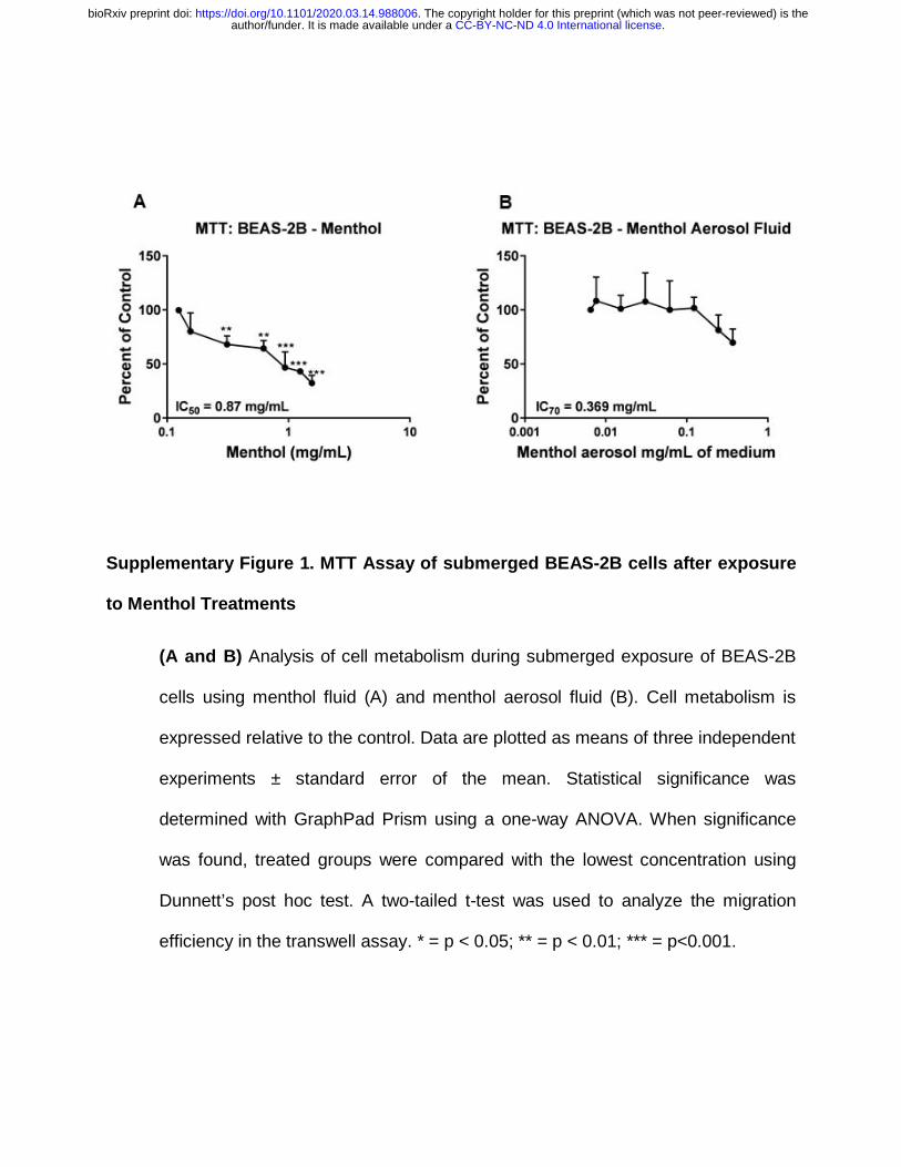
Supplementary Figure 1. MTT Assay of submerged BEAS-2B cells after exposure
to Menthol Treatments
(A and B) Analysis of cell metabolism during submerged exposure of BEAS-2B
cells using menthol fluid (A) and menthol aerosol fluid (B). Cell metabolism is
expressed relative to the control. Data are plotted as means of three independent
experiments ± standard error of the mean. Statistical significance was
determined with GraphPad Prism using a one-way ANOVA. When significance
was found, treated groups were compared with the lowest concentration using
Dunnett’s post hoc test. A two-tailed t-test was used to analyze the migration
efficiency in the transwell assay. * = p < 0.05; ** = p < 0.01; *** = p<0.001.
.CC-BY-NC-ND 4.0 International licenseauthor/funder. It is made available under aThe copyright holder for this preprint (which was not peer-reviewed) is the. https://doi.org/10.1101/2020.03.14.988006doi: bioRxiv preprint

Supplementary Figure 2. Cytotoxicity Assays on 3D EpiAirway Tissues after
exposure to Menthol Aerosols
.CC-BY-NC-ND 4.0 International licenseauthor/funder. It is made available under aThe copyright holder for this preprint (which was not peer-reviewed) is the. https://doi.org/10.1101/2020.03.14.988006doi: bioRxiv preprint

(A) TEER Assay as percent of Clean Air Control.
(B) MTT Assay as percent of Clean Air Control
(C) LDH Assay as percent of Clean Air Control. Statistical significance was
determined with GraphPad Prism using a one-way ANOVA for all assays. When
significance was found, treated groups were compared with the Clean Air Control
using Dunnett’s post hoc test. * = p < 0.05
.CC-BY-NC-ND 4.0 International licenseauthor/funder. It is made available under aThe copyright holder for this preprint (which was not peer-reviewed) is the. https://doi.org/10.1101/2020.03.14.988006doi: bioRxiv preprint

Supplementary Figure 3. Validation of Proteomic Results on 3D EpiAirway Tissue
lysates after exposure in Smoking Machine
(A) Western Blot of eIF2 in duplicate after exposure to menthol (M1 and M2),
propylene glycol (PG1 and PG 2), or Clean Air (CA1 and CA1) in smoking
machine.
(B) Intensity of western blots quantified with ImageJ and plotted with relative
expression to Clean Air control.
.CC-BY-NC-ND 4.0 International licenseauthor/funder. It is made available under aThe copyright holder for this preprint (which was not peer-reviewed) is the. https://doi.org/10.1101/2020.03.14.988006doi: bioRxiv preprint

Supplementary Figure 4. Secretion of cytokines by 3D EpiAirway Tissue into cell
media after a 24hr recovery period
(A) IL-6 secretion in pg/mL.
(B) IL-8 Secretion in pg/mL. Statistical significance was determined with GraphPad
Prism using a one-way ANOVA for all assays. When significance was found,
treated groups were compared with the Clean Air Control using Dunnett’s post
hoc test. * = p < 0.05
.CC-BY-NC-ND 4.0 International licenseauthor/funder. It is made available under aThe copyright holder for this preprint (which was not peer-reviewed) is the. https://doi.org/10.1101/2020.03.14.988006doi: bioRxiv preprint

Supplementary Video 1. Menthol induced calcium flux: Time-lapse video of BEAS-
2B cells transfected with GCaMP5 showing calcium in control and menthol-
treated cells.
(A) 5-minute time course of transfected BEAS-2B cells treated with cell media
(B) 5-minute time course of transfected BEAS-2B cells treated with menthol
.CC-BY-NC-ND 4.0 International licenseauthor/funder. It is made available under aThe copyright holder for this preprint (which was not peer-reviewed) is the. https://doi.org/10.1101/2020.03.14.988006doi: bioRxiv preprint
Related Documents