The PDF of the article you requested follows this cover page. This is an enhanced PDF from The Journal of Bone and Joint Surgery 87:149-165, 2005. doi:10.2106/JBJS.E.00347 J Bone Joint Surg Am. Frank R. Noyes, Sue D. Barber-Westin and Marc Rankin Years Old Meniscal Transplantation in Symptomatic Patients Less Than Fifty This information is current as of September 28, 2005 Reprints and Permissions Permissions] link. and click on the [Reprints and jbjs.org article, or locate the article citation on to use material from this order reprints or request permission Click here to Publisher Information www.jbjs.org 20 Pickering Street, Needham, MA 02492-3157 The Journal of Bone and Joint Surgery on September 28, 2005 www.ejbjs.org Downloaded from

Welcome message from author
This document is posted to help you gain knowledge. Please leave a comment to let me know what you think about it! Share it to your friends and learn new things together.
Transcript

The PDF of the article you requested follows this cover page.
This is an enhanced PDF from The Journal of Bone and Joint Surgery
87:149-165, 2005. doi:10.2106/JBJS.E.00347 J Bone Joint Surg Am.Frank R. Noyes, Sue D. Barber-Westin and Marc Rankin
Years OldMeniscal Transplantation in Symptomatic Patients Less Than Fifty
This information is current as of September 28, 2005
Reprints and Permissions
Permissions] link. and click on the [Reprints andjbjs.orgarticle, or locate the article citation on
to use material from thisorder reprints or request permissionClick here to
Publisher Information
www.jbjs.org20 Pickering Street, Needham, MA 02492-3157The Journal of Bone and Joint Surgery
on September 28, 2005 www.ejbjs.orgDownloaded from
COPYRIGHT © 2005 BY THE JOURNAL OF BONE AND JOINT SURGERY, INCORPORATED
149
Meniscal Transplantation in Symptomatic Patients Less Than Fifty Years OldSurgical TechniqueBy Frank R. Noyes, MD, Sue D. Barber-Westin, BS, and Marc Rankin, MD
Investigation performed at Cincinnati Sportsmedicine and Orthopaedic Center, Cincinnati, Ohio
The original scientific article in which the surgical technique was presented was published in JBJS Vol. 86-A , pp. 1392-1404 , July 2004
INTRODUCTIONMeniscal transplantation remains an evolving area, as investigations of tissue-processing, secondary sterilization, and long-term function continue to be performed to evaluate the overall efficacy of the proce-dure. The primary candidate for the procedure is a young patient who has had a total meniscectomy and has pain in the tibiofemoral com-partment because of early joint arthrosis. There are few treatment op-tions for these patients, and the goal of meniscal transplantation in the short term is to decrease pain, increase knee function, allow pain-free activities of daily living, and delay the progression of tibiofemoral arthrosis. In this report, we describe the preparation of the meniscal transplant and the meticulous surgical technique that is required to achieve an anatomically secure attachment and position in order to provide load-bearing function in the tibiofemoral joint. There are differences between the techniques for medial and lateral meniscal transplantation as a result of the characteristics of the anatomic at-tachment sites.
SURGICAL TECHNIQUESizing and Inspection of Meniscal TransplantsAnteroposterior and lateral radiographs are used to measure the ap-proximate width and length of the meniscal transplant1. The surgeon should have knowledge of the donor-selection criteria and tissue-processing procedures of the tissue bank as these may vary substantially, even among tissue banks that are certified by the American Associa-tion of Tissue Banks and that follow the guidelines of the United States Food and Drug Administration. The implications of different process-
ABSTRACT
BACKGROUND:The purpose of this study was to prospectively evaluate the re-sults of meniscal transplanta-tion in a consecutive series of younger patients treated for pain in the tibiofemoral com-partment following a previous meniscectomy.
METHODS:Forty cryopreserved menisci were implanted into thirty-eight patients. Sixteen knees also had an osteochondral autograft transfer, and nine had a knee ligament reconstruction. The clinical outcome and failure rate of all transplants were evalu-ated at a mean of forty months postoperatively. Meniscal al-lograft characteristics were determined with use of a rating system that combined subjec-tive, clinical, and magnetic reso-nance imaging factors.
continued
on September 28, 2005 www.ejbjs.orgDownloaded from

150
TH E JOUR N AL OF BON E & JOINT SURGER Y · SU R G I C A L TE CH N I QU E S SEPTEMBER 2005 · VOLUME 87-A · SUPPLEMENT 1, PART 2 · JBJS.ORG
ing techniques with regard to graft sterility are important2-4 but beyond the scope of this report.
We advise the surgeon to request that the tissue bank pro-vide, well before the surgery, a photograph of the transplant
that has been selected for each patient. A metric ruler should be placed adjacent to the trans-plant in the photograph to en-sure that the allograft is of adequate size and width. The surgeon should also be aware that certain medial menisci have a hypoplastic anterior horn that is narrow, inserting distal to the medial tibial surface (Type III5), and that these menisci are not acceptable for implantation. Also, if the middle one-third of a medial or lateral meniscus is 8 to 10 mm in width, it is suitable
only for small patients. In addi-tion, if the lateral meniscus has reduced anteroposterior length, less than that calculated on the sagittal radiograph, it is not suit-able for implantation.
The meniscus is thawed, inspected, and prepared prior to the administration of the anes-thesia because it is difficult to detect implant defects through the plastic packaging. The im-plant is also prepared first so that the surgeon can determine the depth and width required for the tibial slot when the cen-
ABSTRACT | continued
RESULTS: Thirty-four (89%) of the thirty-eight patients rated the knee condition as improved. Before surgery, thirty patients (79%) had pain with daily activities, but only four (11%) had such pain at the time of the latest follow-up. While noteworthy pain was present in the tibiofemoral com-partment in all forty knees be-fore surgery, twenty-seven knees (68%) had no pain and thirteen (33%) had only mild compart-ment pain at the time of the lat-est follow-up. Twenty-nine patients (76%) returned to light low-impact sports without prob-lems. Concomitant osteochon-dral autograft transfer and knee ligament reconstruction proce-dures improved knee function and did not increase the rate of complications. Meniscal al-lograft characteristics were nor-mal in seventeen knees (43%), altered in twelve (30%), and failed in eleven (28%).
CONCLUSIONS: The short-term results of menis-cal transplantation are encour-aging in terms of reducing knee pain and increasing function; however, long-term transplant function and any chondroprotec-tive effects remain unknown and require further investigation.
FIG. 1-A
Site of the posterolateral incision for a lateral meniscal transplant.
on September 28, 2005 www.ejbjs.orgDownloaded from
151
TH E JOUR N AL OF BON E & JOINT SURGER Y · SU R G I C A L TE CH N I QU E S SEPTEMBER 2005 · VOLUME 87-A · SUPPLEMENT 1, PART 2 · JBJS.ORG
tral bone-bridge technique is selected.
Technique for Lateral Meniscal TransplantationPreparationThe lateral meniscus, with the anterior and posterior horns re-maining attached centrally to bone, is a better transplant than the medial meniscus. Because the attachment sites and circum-ference tension relationships are not disturbed, an arthroscopi-cally assisted tibial slot method2 of attachment can be performed
with a meticulous inside-out meniscal repair6. The central bone portion of the transplant incorporates the anterior and posterior meniscal attachments and usually measures 8 to 9 mm in width, 35 mm in length, and 10 mm in depth. The posterior 8 to 10 mm of bone that pro-trudes beyond the posterior horn attachment is removed to later produce a buttress against the bone trough in the host knee. Commercially available sizing blocks and channel cut-ters (Stryker Endoscopy, Kala-
mazoo, Michigan, and CryoLife, Kennesaw, Georgia) allow ap-propriate sizing.
Surgical TechniqueThe patient is placed in a supine position on the operating room table, with a tourniquet applied with a leg-holder and the table adjusted to allow 90° of knee flexion. The contralateral lower extremity is placed in a thigh-high elastic stocking and is pad-ded to maintain mild hip flexion to decrease tension on the femo-ral nerve. After examination with
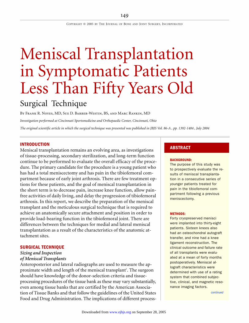
FIG. 1-B FIG. 1-C
Fig. 1-B Incision site in the interval between the posterior edge of the iliotibial band (ITB) and the anterior edge of the biceps tendon. Fig. 1-C The interval between the lateral head of the gastrocnemius and the posterolateral aspect of the capsule is opened bluntly, just proximal to the fibular head, without entering the joint capsule proximally.
on September 28, 2005 www.ejbjs.orgDownloaded from

152
TH E JOUR N AL OF BON E & JOINT SURGER Y · SU R G I C A L TE CH N I QU E S SEPTEMBER 2005 · VOLUME 87-A · SUPPLEMENT 1, PART 2 · JBJS.ORG
the patient under anesthesia, di-agnostic arthroscopy is done to confirm the preoperative diagno-sis and to assess changes in the articular cartilage. An arthro-scopically assisted approach is used in knees that require a cru-ciate ligament reconstruction7. The femoral and tibial tunnels are drilled, and the ligament graft is passed through the tun-nels, with femoral fixation done first, followed by the meniscal transplantation, and then by tibial fixation of the cruciate graft. Fixing the ligament graft at the tibia as the final step al-lows maximum separation of the tibiofemoral joint during meniscal transplantation. It also prevents failure or problems with the ligament fixation or liga-ment graft during the operation.
A limited 3-cm lateral ar-throtomy is made just adjacent to the patellar tendon. Although there are arthroscopic tech-niques for preparation of the tibial slot, we believe that the limited arthrotomy provides su-perior visualization and makes it possible to avoid incision into the patellar tendon. A second, 3-cm posterolateral accessory incision is made, centered just behind the lateral collateral liga-ment (Fig. 1-A)6,8. The interval between the biceps tendon inser-tion and the iliotibial band is identified and incised (Fig. 1-B). The lateral head of the gastro-cnemius is gently dissected with Metzenbaum scissors off of the posterior aspect of the capsule at the joint line (Fig. 1-C). Care is taken at this point because dis-
section that extends too far proximal to the joint line at the posterolateral aspect would enter the joint capsule. If this occurs, a capsular repair is required to maintain joint in-tegrity during the inside-out meniscal repair. The lateral in-ferior genicular artery is also in
close proximity, and it is identi-fied and preserved. The space between the posterolateral as-pect of the capsule and the lat-eral head of the gastrocnemius is further developed bluntly. An appropriately sized popliteal re-tractor (Stryker) is placed di-rectly behind the lateral meniscal
FIG. 2-A
Figs. 2-A through 2-E Tibial slot technique for lateral and medial meniscal transplanta-tion, which is shown here for the lateral meniscus. An arthroscopically assisted tech-nique or a mini-lateral arthrotomy may be used, but we prefer the mini-arthrotomy as it offers superior visualization and allows us to avoid incising the patellar tendon. A de-tailed description of the surgical steps and operative instruments used for the tibial slot
technique is available14. Fig. 2-A A line is established connecting the center of the ante-rior and posterior horn attachments with an electrocautery device. In the mini-open ar-throtomy, a template of the meniscal coronal width is used to verify the medial-to-lateral width of the transplant so that the slot can be moved appropriately to prevent tibial over-hang of the implant.
on September 28, 2005 www.ejbjs.orgDownloaded from

153
TH E JOUR N AL OF BON E & JOINT SURGER Y · SU R G I C A L TE CH N I QU E S SEPTEMBER 2005 · VOLUME 87-A · SUPPLEMENT 1, PART 2 · JBJS.ORG
FIG. 2-B
A burr is used to remove the tibial spine and create a 4-mm straight anterior-to-posterior reference slot along the plane of the tibial slope. This calibrated guide pin sits flush with the articular cartilage.
on September 28, 2005 www.ejbjs.orgDownloaded from

154
TH E JOUR N AL OF BON E & JOINT SURGER Y · SU R G I C A L TE CH N I QU E S SEPTEMBER 2005 · VOLUME 87-A · SUPPLEMENT 1, PART 2 · JBJS.ORG
bed. The tourniquet is inflated only for these two approaches; otherwise, it is not used.
The width of the transplant is determined, and an aluminum foil template of the same width and length as the transplant is cut and is inserted into the lat-eral compartment to determine the lateralmost margin of the bone trough. This sizing step is important to make sure that there is no lateral overhang of
the meniscal body produced by placing the bone trough too far laterally. A rectangular bone trough is prepared at the ante-rior and posterior tibial attach-ment sites of the lateral meniscus to match the dimensions of the prepared lateral meniscal trans-plant. The sequence of steps to prepare the lateral tibial slot is il-lustrated in Figures 2-A through 2-D. The tibial bone slot is 1 to 2 mm wider than the transplant, to facilitate implantation. The anterior and posterior horns of the implant are placed into their normal attachment locations,
adjacent to the anterior cruciate ligament. The allograft is in-serted into the trough (Fig. 2-E), and the bone portion of the graft is seated against the posterior bone buttress to achieve correct anterior-to-posterior placement of the attachment sites. A verti-cal suture in the posterior part of the meniscal body is passed posteriorly to provide tension and facilitate implant placement. The knee is flexed, extended, and rotated to confirm correct al-lograft placement. The posterior suture is tied, and sutures are placed in a vertical fashion into
FIG. 2-C
The drill guide is used with a guide pin that has been marked with a laser to set the depth of a second guide pin. This allows a drill to ream 5 mm less to retain the posterior portion of the tibial slot.
CRITICAL CONCEPTS
INDICATIONS: The indications for a meniscal al-lograft procedure are prior menis-cectomy, an age of fifty years or less, pain in the tibiofemoral com-partment, no radiographic evi-dence of advanced arthrosis, and ≥2 mm of tibiofemoral joint space as seen on 45° weight-bearing posteroanterior radiographs10.
CONTRAINDICATIONS:Contraindications include ad-vanced arthrosis of the knee joint with flattening of the femoral condyle, concavity of the tibial plateau, and osteophytes that prevent anatomic seating of the meniscal allograft11; axial varus malalignment in which a weight-bearing line of <40% of the me-dial-lateral transverse width of the tibial plateau12 is seen on radio-graphs or valgus malalignment in which a weight-bearing line of >60% is seen on radiographs; in-stability of the knee joint or the patient’s refusal to undergo con-comitant knee ligament recon-struction; knee arthrofibrosis; muscular atrophy; and previous joint infection.
continued
on September 28, 2005 www.ejbjs.orgDownloaded from

155
TH E JOUR N AL OF BON E & JOINT SURGER Y · SU R G I C A L TE CH N I QU E S SEPTEMBER 2005 · VOLUME 87-A · SUPPLEMENT 1, PART 2 · JBJS.ORG
FIG. 2-D
An 8-mm drill bit with a collar at the defined depth is used, followed by use of a box cutter to create a rect-angular slot of the desired depth and width.
on September 28, 2005 www.ejbjs.orgDownloaded from

156
TH E JOUR N AL OF BON E & JOINT SURGER Y · SU R G I C A L TE CH N I QU E S SEPTEMBER 2005 · VOLUME 87-A · SUPPLEMENT 1, PART 2 · JBJS.ORG
the anterior one-third of the meniscus, attaching it to the pre-pared meniscal rim under direct visualization.
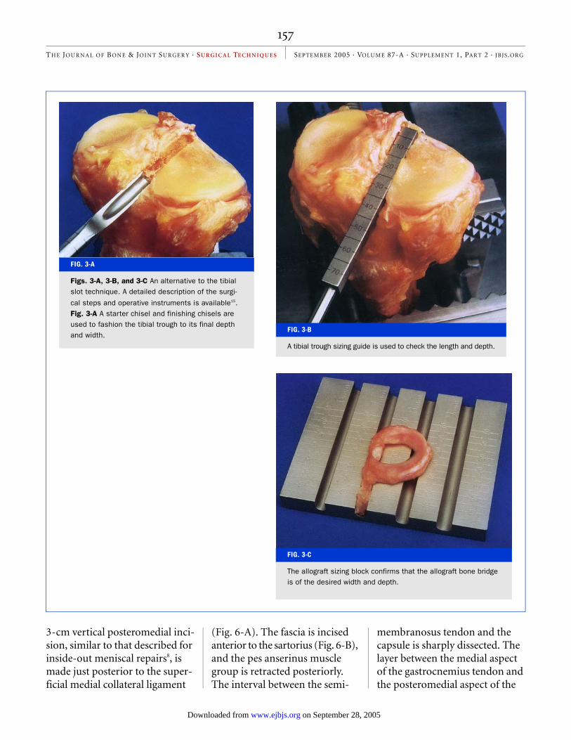
An alternative technique is to use a starter chisel and fin-ishing chisels to fashion the tib-ial trough to its final depth and width (Fig. 3-A). A tibial trough sizing guide is employed to check the length and depth (Fig. 3-B). The allograft sizing block (Fig. 3-C) confirms that the allograft bone bridge is of the correct width and depth.
Two methods are available for fixation of the central bone at-tachment. Two 2-0 nonabsorb-able sutures (Ticron [Davis and Geck, Wayne, New Jersey] or Ethibond [Ethicon, Somerville, New Jersey]) may be placed in the central region, brought through a drill hole, and tied. Our preferred method involves placement of an interference screw (7 × 25 mm),
made of an absorbable composite material, medial and adjacent to the central bone attachment9. The arthrotomy is closed, and the inside-out meniscal repair is performed with multiple verti-cal divergent sutures, which are placed first superiorly to reduce the meniscus (Fig. 4) and then inferiorly in the outer one-third of the implant. Sutures are not placed in the middle and inner thirds of the meniscus to avoid weakening the implant, which has a limited healing capability in these regions (Fig. 5).
Technique for Medial Meniscal TransplantationPreparationThe medial meniscal transplant is inspected to confirm that the size is appropriate and no degen-erative changes are present. The implant is not prepared until it is decided whether the central
bone-bridge technique (which is preferred) or the two-tunnel technique (involving separate anterior and posterior bone attachments and tunnels) is required.
The patient is placed in a supine position on the operating room table, with a tourniquet applied with a leg-holder and the table adjusted to allow 90° of knee flexion. The contralat-eral lower extremity is placed in a thigh-high elastic stocking and is padded to maintain mild hip flexion to decrease tension on the femoral nerve. After examination with the patient under anesthe-sia, diagnostic arthroscopy is done to confirm the preopera-tive diagnosis and assess changes in the articular cartilage.
A 4-cm skin incision is made on the anterior aspect of the tibia adjacent to the tibial tubercle and the patellar tendon. A second,
FIG. 2-E
The lateral meniscal implant with the central bone bridge is ready to be placed into the tibial slot.
on September 28, 2005 www.ejbjs.orgDownloaded from
157
TH E JOUR N AL OF BON E & JOINT SURGER Y · SU R G I C A L TE CH N I QU E S SEPTEMBER 2005 · VOLUME 87-A · SUPPLEMENT 1, PART 2 · JBJS.ORG
FIG. 3-A
Figs. 3-A, 3-B, and 3-C An alternative to the tibial slot technique. A detailed description of the surgi-
cal steps and operative instruments is available15. Fig. 3-A A starter chisel and finishing chisels are used to fashion the tibial trough to its final depth and width.
FIG. 3-B
A tibial trough sizing guide is used to check the length and depth.
FIG. 3-C
The allograft sizing block confirms that the allograft bone bridge is of the desired width and depth.
3-cm vertical posteromedial inci-sion, similar to that described for inside-out meniscal repairs8, is made just posterior to the super-ficial medial collateral ligament
(Fig. 6-A). The fascia is incised anterior to the sartorius (Fig. 6-B), and the pes anserinus muscle group is retracted posteriorly. The interval between the semi-
membranosus tendon and the capsule is sharply dissected. The layer between the medial aspect of the gastrocnemius tendon and the posteromedial aspect of the
on September 28, 2005 www.ejbjs.orgDownloaded from
158
TH E JOUR N AL OF BON E & JOINT SURGER Y · SU R G I C A L TE CH N I QU E S SEPTEMBER 2005 · VOLUME 87-A · SUPPLEMENT 1, PART 2 · JBJS.ORG
capsule is separated with blunt dissection (Fig. 6-C). Great care is taken to identify and avoid in-jury to the infrapatellar branches of the saphenous nerve. The two approaches are performed with the tourniquet inflated to 275 mm Hg and usually require fif-teen minutes; otherwise, the tourniquet is not used.
A medial meniscal trans-plant, with anatomically placed anterior and posterior bone at-
tachments, must be appropri-ately secured to maintain the desired position in the knee joint postoperatively and to provide the circumferential tension re-quired for transplant function. A template of the medial meniscal transplant, made of aluminum foil and measured according to its anterior-posterior and medial-lateral dimensions, is inserted through the limited an-terior arthrotomy incision and is
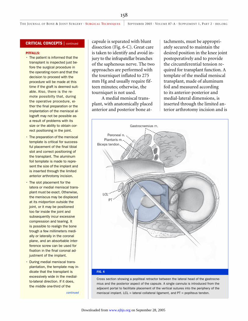
FIG. 4
Cross section showing a popliteal retractor between the lateral head of the gastrocne-mius and the posterior aspect of the capsule. A single cannula is introduced from the adjacent portal to facilitate placement of the vertical sutures into the periphery of the meniscal implant. LCL = lateral collateral ligament, and PT = popliteus tendon.
CRITICAL CONCEPTS | continued
PITFALLS:• The patient is informed that the
transplant is inspected just be-fore the surgical procedure in the operating room and that the decision to proceed with the procedure will be made at this time if the graft is deemed suit-able. Also, there is the re-mote possibilty that, during the operative procedure, ei-ther the final preparation or the implantation of the meniscal al-lograft may not be possible as a result of problems with its size or the ability to obtain cor-rect positioning in the joint.
• The preparation of the meniscal template is critical for success-ful placement of the final tibial slot and correct positioning of the transplant. The aluminum foil template is made to repre-sent the size of the implant and is inserted through the limited anterior arthrotomy incision.
• The slot placement for the latera or medial meniscal trans-plant must be exact. Otherwise, the meniscus may be displaced at its midportion outside the joint, or it may be positioned too far inside the joint and subsequently incur excessive compression and tearing. It is possible to realign the bone trough a few millimeters medi-ally or laterally in the coronal plane, and an absorbable inter-ference screw can be used for fixation in the final coronal ad-justment of the implant.
• During medial meniscal trans-plantation, the template may in-dicate that the transplant is excessively wide in the medial-to-lateral direction. If it does, the middle one-third of the
continued
on September 28, 2005 www.ejbjs.orgDownloaded from

159
TH E JOUR N AL OF BON E & JOINT SURGER Y · SU R G I C A L TE CH N I QU E S SEPTEMBER 2005 · VOLUME 87-A · SUPPLEMENT 1, PART 2 · JBJS.ORG
sized to the medial tibial plateau. This allows the surgeon to mark the position of the central bone trough and to determine whether the meniscal implant will be properly positioned just adja-cent to the tibial attachment of the anterior cruciate ligament, without excessive medial tibial overhang. Next, it is verified that the anterior and posterior at-tachments are located at the ana-tomically correct sites. With the central bone-bridge technique, 4 to 6 mm of the medial tibial eminence is removed. If the im-plant is suitable and there is no medial tibial overhang, then the central bone-bridge technique may be used. If the implant needs to be adjusted to fit to the me-
dial tibial plateau by moving the anterior horn farther laterally, then the two-tunnel technique is selected. Once the technique has been chosen, the meniscal allograft is prepared.
Central Bone-Bridge TechniqueThe central bone-bridge tech-nique for medial meniscal trans-plantation is the same as that described for lateral meniscal transplantation. A reference slot is first created on the tibial plateau in the anteroposterior direction. A guide pin is positioned in the slot, inferiorly on the tibia, and a cannulated drill bit is placed over the pin to drill a tunnel. The final tibial slot is 8 to 9 mm in width and 10 mm in depth. A
FIG. 5
Lateral meniscal graft in place and sutured.
CRITICAL CONCEPTS | continued
transplant would rest outside of the medial tibial plateau in or-der to avoid compromising the attachment of the anterior cru-ciate ligament. The two-tunnel technique is selected to obtain correct anatomic positioning and the desired subsequent cir-cumferential hoop stress.
• The use of multiple vertical di-vergent sutures is required to position the transplant in the anatomically correct manner. There are usually wavy areas in the implant, with loss of circum-ferential tension, that are suc-cessfully removed by correct placement of these sutures.
• We prefer the inside-out me-niscal repair technique, which is considered to be the most precise suturing method.
• We avoid meniscal fixators, with which it is not possible to pro-vide the same secure fit and ex-act placement of the implant.
• The sutures should not be placed in the middle and inner thirds of the meniscus, as this could weaken the implant.
• The suturing of the implant is meticulous, as twelve to fifteen sutures are required both supe-riorly and inferiorly, all placed in a vertical direction. Horizontal sutures have poor holding ability and are therefore not used dur-ing meniscal transplantation.
• Care is taken not to damage the articular cartilage. The technique requires two surgical assis-tants, one dedicated to holding the lower limb to open the me-dial or lateral tibiofemoral com-partment for visualization of the implant and the other seated to retrieve and tie the sutures at the posterior aspect of the joint.
continued
on September 28, 2005 www.ejbjs.orgDownloaded from

160
TH E JOUR N AL OF BON E & JOINT SURGER Y · SU R G I C A L TE CH N I QU E S SEPTEMBER 2005 · VOLUME 87-A · SUPPLEMENT 1, PART 2 · JBJS.ORG
rasp is used to smooth the slot to allow insertion of the central bone bridge of the allograft.
The central bone bridge of the allograft is sized to a width of 7 mm (1 mm less than the di-mension at the tibial site) and a depth of 10 mm9. This allows the
position of the central bone bridge to be adjusted in the ante-rior-posterior direction while the meniscus is positioned to fit in the anatomically correct posi-tion relative to the femoral condyle.
A vertical suture is placed through the junction of the posterior and middle thirds of the meniscus. A single-barrel cannula is used to advance the suture through the capsule at the corresponding attachment site of the meniscus, and the su-ture exits through an accessory incision (Fig. 7). The meniscus
is passed through the arthrot-omy incision into the knee, with tension placed on the su-tures to facilitate proper posi-tioning in the knee joint. Care is taken to align the bone bridge with the recipient tibial slot. The knee is taken through flex-ion and extension and tibial ro-tation to align the implant. Once the appropriate anterior-posterior position of the cen-tral bone bridge is achieved, a guide wire is inserted between the bone bridge and the lateral side of the slot. A tap is inserted over the guide wire to create a
FIG. 6-A
Figs. 6-A, 6-B, and 6-C The accessory posteromedial approach for a medial meniscal al-lograft procedure. Fig. 6-A Site of the posteromedial skin incision.
CRITICAL CONCEPTS | continued
• The suturing of the medial or lateral posterior horn adjacent to the posterior attachment re-quires angulation of the su-ture needle away from the neurovascular structures.
• In order for the meniscal trans-plant to function, it must be placed at the normal anatomic insertion sites. If the posterior horn attachment of the medial or lateral meniscus is placed too far posteriorly, it will not provide proper load-sharing13. Alternatively, a too anterior po-sition of a medial meniscal transplant will produce exces-sive compressive forces and damage the meniscus.
• We disagree with those who have advocated techniques of medial meniscal transplanta-tion in which the posterior bone portion of a medial meniscal implant is not retained and the fibrocartilaginous posterior horn is placed in a posterior tib-ial attachment tunnel. Although such transplants are far easier to prepare and implant surgi-cally, there are inadequate sci-entific data to support the belief that the soft-tissue ends of the meniscal implant (with-out the bone attachment) will heal and provide the circumfer-ential tension in the meniscus that is required for function.
continued
on September 28, 2005 www.ejbjs.orgDownloaded from

161
TH E JOUR N AL OF BON E & JOINT SURGER Y · SU R G I C A L TE CH N I QU E S SEPTEMBER 2005 · VOLUME 87-A · SUPPLEMENT 1, PART 2 · JBJS.ORG
path for an interference screw with the bone bridge held in place manually. An absorbable bone interference screw is in-serted adjacent to the bone bridge.
The joint is again taken through a full range of motion, and the position of the implant is verified. Occasionally, there is an osteophyte on the anterior aspect of the medial tibial pla-teau, and this must be resected to avoid compression of the meniscal implant. The central bone bridge of the implant is fixed with an interference screw
(7 × 25 mm). The meniscus is sutured with vertical divergent sutures (2-0 Ethibond) under direct visualization. The ante-rior arthrotomy is closed, and the inside-out vertical divergent sutures are placed, as described, to sew the meniscus to the me-niscal bed, with removal of any implant undulations and restoration of circumferential meniscal tension. The central bone bridge of the implant provides fixation of the anterior and posterior portions of the implant and healing into the host tibia (Fig. 8).
Two-Tunnel TechniqueIf it is determined that the cen-tral bone-bridge technique is not acceptable, the surgeon must prepare separate anterior and posterior bone portions of the meniscal transplant. Both are se-cured to anatomic attachment sites to provide a functional meniscal implant (Fig. 9). The transplant is prepared with a posterior bone plug, 8 mm in diameter and 12 mm in length, and an anterior bone plug, 12 mm in diameter and 12 mm in length. Two 2-0 nonabsorbable Ethibond sutures are passed ret-
FIG. 6-B FIG. 6-C
Fig. 6-B The incision is shown through the anterior portion of the sartorius fascia. Fig. 6-C The interval is opened between the posterome-dial aspect of the capsule and the gastrocnemius tendon, just proximal to the semimembranosus tendon (arrow). The fascia over the ten-don is excised to its tibial attachment to facilitate retrieval of the meniscal sutures.
on September 28, 2005 www.ejbjs.orgDownloaded from
162
TH E JOUR N AL OF BON E & JOINT SURGER Y · SU R G I C A L TE CH N I QU E S SEPTEMBER 2005 · VOLUME 87-A · SUPPLEMENT 1, PART 2 · JBJS.ORG
rograde through each bone plug, with two additional locking su-tures placed in the meniscus ad-jacent to the bone attachment for subsequent secure fixation of the bone plugs within the tib-ial tunnel.
A guide pin is placed adja-cent to the tibial tubercle and is directed to the anatomic posterior
meniscal attachment. A tibial tun-nel is drilled over the guide wire to a diameter of 8 mm. The bone-tunnel edges are chamfered. A limited notchplasty of the medial femoral condyle is usually re-quired. At least 8 mm of opening adjacent to the posterior cruciate ligament in the femoral notch is needed to pass the posterior os-seous portion of the graft. On oc-casion, a subperiosteal release of the long fibers of the tibial attach-
ment of the medial collateral liga-ment (with later suture-anchor repair) is required to open the medial part of the tibiofemoral joint sufficiently. The meniscal bed is prepared by removing any remaiing meniscal tissue while preserving a 3-mm rim when possible. The meniscal bed is rasped for revascularization of the graft.
A 3-cm anteromedial ar-throtomy is used to pass the pos-
CRITICAL CONCEPTS | continued
AUTHOR UPDATE:In our original study, we used the central bone-bridge technique, which maintains a central bone bridge between the anterior and posterior meniscal attachments, primarily for lateral meniscal transplantation. This is also now our preferred technique for me-dial meniscal transplantation, as described in this article.
Currently, we use a template of the meniscal implant to deter-mine the location of the bone slot. The lateralmost placement of the central bone slot for the medial meniscal implant is lim-ited by the tibial attachment of the anterior cruciate ligament. The anterior horn of the medial meniscus must not be of a Type-III configuration5⎯i.e., it must not insert too far distally on the anterior tibial margin. If assess-ment of the medial meniscal transplant reveals a medial-to-lateral size mismatch, then sep-arate anterior and posterior bone attachments and tunnels are required. The posterior part of the bone-meniscus trans-plant is placed at the normal at-tachment, and the anterior horn is placed in a medial-to-lateral direction to restore correct ten-sioning and position in the joint.
continued
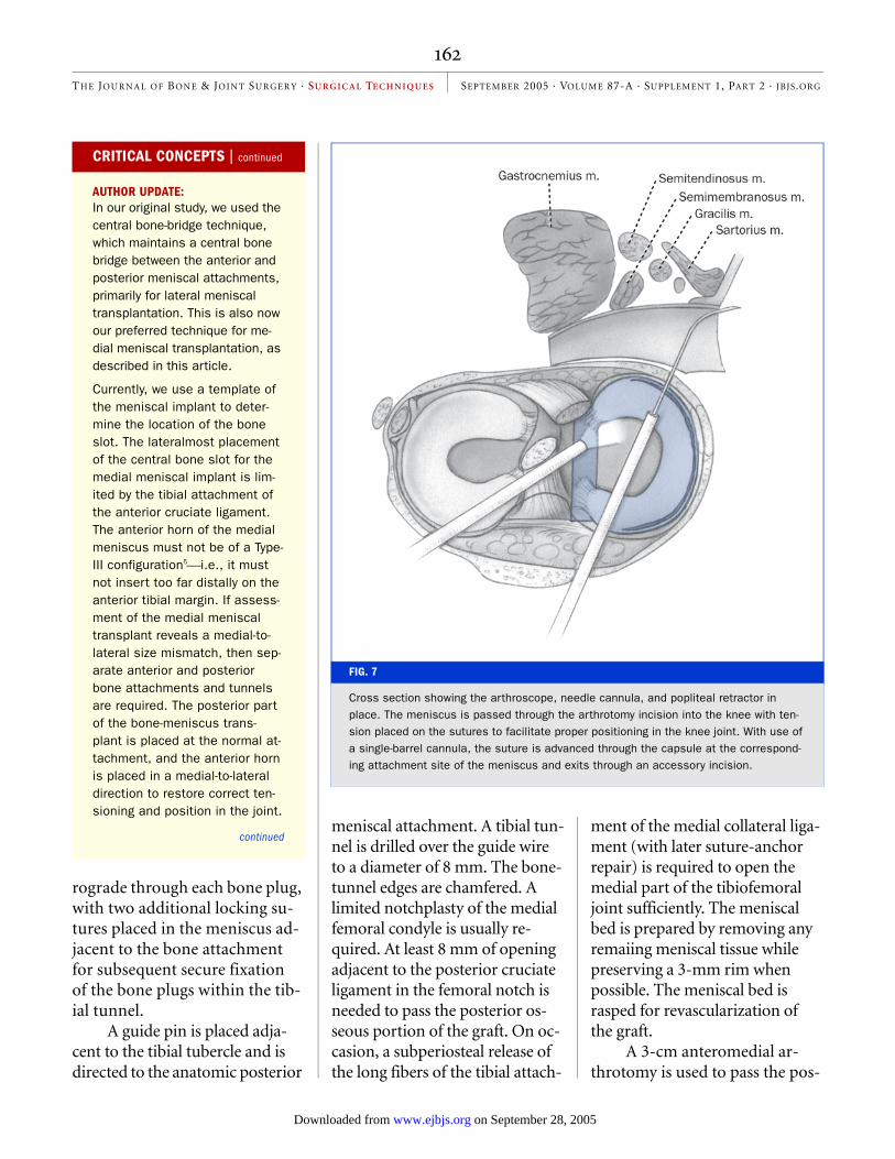
FIG. 7
Cross section showing the arthroscope, needle cannula, and popliteal retractor in place. The meniscus is passed through the arthrotomy incision into the knee with ten-sion placed on the sutures to facilitate proper positioning in the knee joint. With use of a single-barrel cannula, the suture is advanced through the capsule at the correspond-ing attachment site of the meniscus and exits through an accessory incision.
on September 28, 2005 www.ejbjs.orgDownloaded from
163
TH E JOUR N AL OF BON E & JOINT SURGER Y · SU R G I C A L TE CH N I QU E S SEPTEMBER 2005 · VOLUME 87-A · SUPPLEMENT 1, PART 2 · JBJS.ORG
terior bone portion of the graft, with a secondary meniscal body suture passed out through the in-cision for the posteromedial ap-proach. The surgeon is seated with a headlight in place, and the patient’s knee is flexed to 90°. On occasion, there are anterior os-teophytes on the medial tibial plateau that require resection. The posterior attachment guide wire is retrieved, and the sutures
attached to the posterior bone are passed. A second suture is placed into the midportion of the meniscus and is passed inside-out through the incision for the posteromedial approach to guide the meniscus.
The knee is flexed to 20° under a maximum valgus load to pass the posterior bone portions of the graft, with the secondary meniscal body suture held by an
assistant. A nerve hook is used to gently assist the passage of the graft. With use of a headlight and retractors, it is possible to con-firm appropriate passage of the meniscal graft into the medial ti-biofemoral compartment. Care is taken to not advance the poste-rior part of the meniscal body into the tibial tunnel but to only seat the bone portion of the graft in order to avoid shortening of the meniscal graft. The posterior meniscal bone attachment and the midbody sutures are tied over the tibial post to provide tension in the posterior bone at-tachment and the posterior one-third of the meniscus. The knee is flexed and extended to assess meniscal fit and displacement. The optimal location for the an-
CRITICAL CONCEPTS | continued
For tight knees with only a few millimeters of medial joint open-ing, the central bone-bridge tech-nique enables the surgeon to avoid performing a partial de-tachment of the distal part of the medial collateral ligament, which would otherwise be re-quired to gain access to the joint for suturing and to avoid dam-age to the articular cartilage.
There are now newer tech-niques for tissue-processing and advanced donor-screening tests that provide highly safe meniscal transplants with an exceedingly low risk of disease transmission. Advances in tissue-processing and Food and Drug Administra-tion guidelines for tissue banks are important to ensure the safety of allografts.
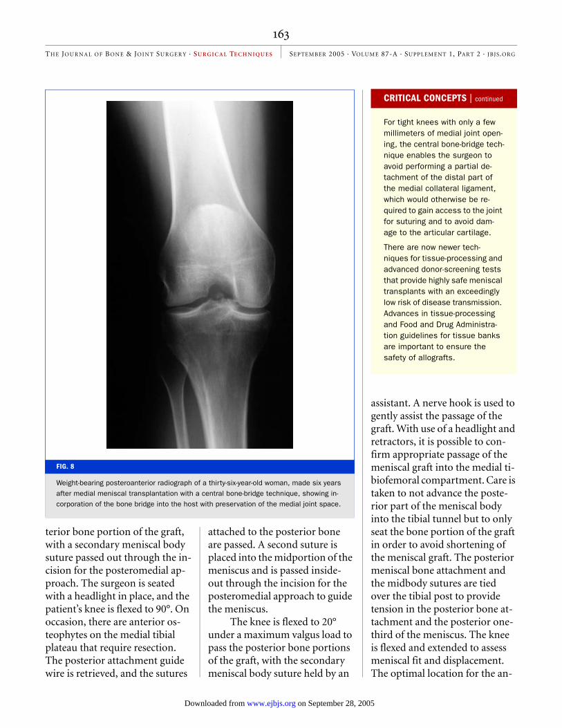
FIG. 8
Weight-bearing posteroanterior radiograph of a thirty-six-year-old woman, made six years after medial meniscal transplantation with a central bone-bridge technique, showing in-corporation of the bone bridge into the host with preservation of the medial joint space.
on September 28, 2005 www.ejbjs.orgDownloaded from

164
TH E JOUR N AL OF BON E & JOINT SURGER Y · SU R G I C A L TE CH N I QU E S SEPTEMBER 2005 · VOLUME 87-A · SUPPLEMENT 1, PART 2 · JBJS.ORG
terior meniscal bone attachment at the anteromedial junction of the tibial plateau is identified, with the medial-to-lateral place-ment in the coronal plane deter-mined with the knee in full extension. A 12-mm rectangular bone attachment is fashioned to correspond to the anterior bone portion of the meniscal graft. A 4-mm bone tunnel is placed at the base of this bone trough, and it exits at the anterior aspect of the tibia just proximal to the pos-terior bone tunnel. The sutures are passed through the bone tun-nel, and the anterior horn is seated. Full knee flexion and ex-tension are again performed to determine proper graft place-ment and fit. Tension is applied to the anterior bone sutures, which are not tied at this point but are used to maintain tension in the graft during the inside-out suture repair. This meticulous seating of the meniscal transplant under circumferential tension with bone attachment of both the anterior and the posterior horn is believed to be crucial for future meniscal weight-bearing position and function.
The anterior arthrotomy is closed, and the arthroscope is in-serted into the anterolateral por-tal for the posterior meniscal repair and into the anteromedial portal for the repairs of the mid-dle and anterior one-thirds, with the single needle cannula in-serted in the other anterior por-tal. The meniscal repair is performed in an inside-out fash-ion, starting with the posterior horn, with use of multiple verti-
FIG. 9
Two-tunnel technique for medial meniscal allograft transplantation. The illustration shows insertion of the transplant, including posteromedial suture placed to facilitate me-niscal reduction. The anterior and posterior bone attachments of the medial meniscal transplant are fixed into separate tibial tunnels.
FIG. 10
Appearance of the final fixation of the medial meniscal transplant in the anterior and posterior tunnels and vertical divergent sutures.
on September 28, 2005 www.ejbjs.orgDownloaded from

165
TH E JOUR N AL OF BON E & JOINT SURGER Y · SU R G I C A L TE CH N I QU E S SEPTEMBER 2005 · VOLUME 87-A · SUPPLEMENT 1, PART 2 · JBJS.ORG
cal divergent sutures of 2-0 non-absorbable Ethibond both superiorly and inferiorly, with constant tensioning of the me-niscus from posterior to anterior to establish circumferential ten-sion. The assistant, seated with a headlight, retrieves the suture needles through the posterome-dial approach. Each suture is placed and tied, bringing the me-niscus directly to the meniscal bed with observation that menis-cal placement, fixation, and ten-sion are correct. The anterior arthrotomy incision is again opened, and the final tensioning and tying of the anterior horn bone sutures are performed with use of the anterior tibial post. Occasionally, additional sutures are required to secure the most anterior one-third of the menis-cus to the capsular attachments, which is done under direct vision (Fig. 10). After final inspection of the graft with knee flexion and extension and tibial rotation, the operative wounds are closed in a routine fashion.
Frank R. Noyes, MDSue D. Barber-Westin, BSMarc Rankin, MDDeaconess Hospital, 311 Straight Street, Cincin-
nati, OH 45219. E-mail address for S.D. Barber-Westin: [email protected]
The authors did not receive grants or outside fund-ing in support of their research or preparation of this manuscript. They did not receive payments or other benefits or a commitment or agreement to provide such benefits from a commercial entity. No commercial entity paid or directed, or agreed to pay or direct, any benefits to any research fund, foundation, educational institution, or other chari-table or nonprofit organization with which the authors are affiliated or associated.
The line drawings in this article are the work of Joanne Haderer Müller of Haderer & Müller ([email protected]).
doi:10.2106/JBJS.E.00347
REFERENCES1. Pollard ME, Kang Q, Berg EE. Radio-graphic sizing for meniscal transplantation. Arthroscopy. 1995;11:684-7.
2. Cole BJ, Carter TR, Rodeo SA. Allograft menisca transplantation: background, tech-niques, and results. J Bone Joint Surg Am. 2002;84:1236-50.
3. Barbour SA, King W. The safe and effective use of allograft tissue—an update. Am J Sports Med. 2003;31:791-7.
4. Vangsness CT Jr, Garcia IA, Mills CR, Kainer MA, Roberts MR, Moore TM. Allograft trans-plantation in the knee: tissue regulation, pro-curement, processing, and sterilization. Am J Sports Med. 2003;31:474-81.
5. Berlet GC, Fowler PJ. The anterior horn of the medical meniscus. An anatomic study of its insertion. Am J Sports Med. 1998;26:540-3.
6. Rubman MH, Noyes FR, Barber-Westin SD.
Technical considerations in the management of complex meniscus tears. Clin Sports Med. 1996;15:511-30.
7. Noyes FR, Barber-Westin SD. A compari-son of results in acute and chronic anterior cruciate ligament ruptures of arthroscopi-cally assisted autogenous patellar tendon reconstruction. Am J Sports Med. 1997;25:460-71.
8. McLaughlin JR, Noyes FR. Arthroscopic meniscus repair: recommended surgical techniques for complex meniscal tears. Tech Orthop. 1993;8:129-36.
9. Farr J, Meneghini RM, Cole BJ. Allograft interference screw fixation in meniscus transplantation. Arthroscopy. 2004;20:322-7.
10. Rosenberg TD, Paulos LE, Parker RD, Coward DB, Scott SM. The forty-five-degree posteroanterior flexion weight-bearing radio-graph of the knee. J Bone Joint Surg Am. 1988;70:1479-83.
11. Noyes FR, Barber-Westin SD, Butler DL, Wilkins RM. The role of allografts in repair and reconstruction of knee joint ligaments and menisci. Instr Course Lect. 1998;47:379-96.
12. Dugdale TW, Noyes FR, Styer D. Preopera-tive planning for high tibial osteotomy. The ef-fect of lateral tibiofemoral separation and tibiofemoral length. Clin Orthop Relat Res. 1992;274:248-64.
13. Alhalki MM, Hull ML, Howell SM. Contact mechanics of the medial tibial plateau after implantation of a medial meniscal allograft. A human cadaveric study. Am J Sports Med. 2000;28:370-6.
14. Farr J, Cole B. Slot instruments for menis-cal transplantation: surgical technique. San Jose, CA: Stryker Endoscopy; 2004.
15. Halbrecht JL. Meniscal reconstruction trough surgical technique. Kennesaw, GA: CryoLife; 2000.
on September 28, 2005 www.ejbjs.orgDownloaded from
Related Documents