Membrane Structure and Function Chapter 8

Membrane Structure and Function Chapter 8 Fluid Mosiac Model.
Dec 30, 2015
Welcome message from author
This document is posted to help you gain knowledge. Please leave a comment to let me know what you think about it! Share it to your friends and learn new things together.
Transcript
Membrane Structure and Function
Chapter 8

Fluid Mosiac Model

Membrane is a fluid mosaic 1. Of lipids, carbohydrates and proteins 2. Held together by hydrophobic
interactions 3. Molecules can drift laterally
A. phospholipids move quickly B. proteins slowly
C. some proteins attached to cytoskeleton and can not move much
Membrane movie

D. Unsaturated hydrocarbon tails enhance fluidity
4. Membranes solidify at critical low temps. A. organisms in colder temps >
concentration of unsaturated phospholipids
B. cholesterol modulates fluidity 1. Less fluid at warmer temps 2. More fluid at lower temps

Membranes: STRUCTURE and FUNCTION 1. Integral proteins
Penetrate the hydrophobic interior of membrane
Unilateral – only partway across membrane
Transmembrane – across membrane

2. Peripheral – attached to surface May be attached to integral proteins,
fibers in ECM (extracellular matrix), or cytoskeleton
Gives animal cells stronger framework

Membrane Proteins

Function of proteins 1. Transport (tunnels) 2. Enzymes 3. Signal transduction 4. Cell-cell recognition. 5.

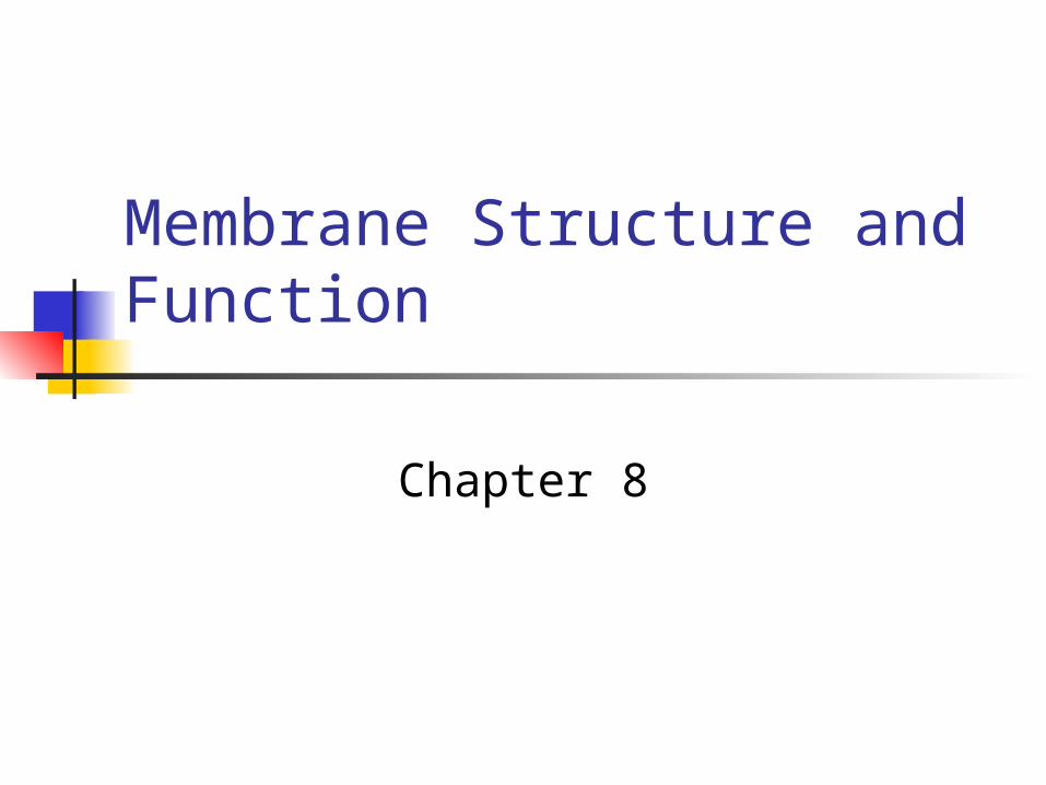
Membranes are bifacial
1. Proteins have distinct orientation2. Carbohydrates restricted to
membranes exterior (oligosaccharide)
1. Glycolipid – attached to phospholipid2. Glycoprotein – attached to protein3. Glcocalyx – all extended
carbohydrates
Bifacial arrangement

Membrane Carbohydrates
1. Function: Cell to cell recognition1. Sorting: animal embryo cells into
tissue and organs2. Rejection of alien cells by immune
system3. Vary from species

Membrane permeability
1. Terms: semi-permeable, selectively permeable, differentially permeable
2. Permeability depends upon:1. phospholipid layer2. Specific integral proteins

Permeability of lipid bilayer
1. Nonpolar molecules:1. Cross with ease2. Smaller molecules cross faster
2. Polar molecules:1. Small, polar uncharged molecules pass
easily ( H2O, CO2)
2. Larger polar uncharged will not (C6H12O6)
3. All ions will not pass easily

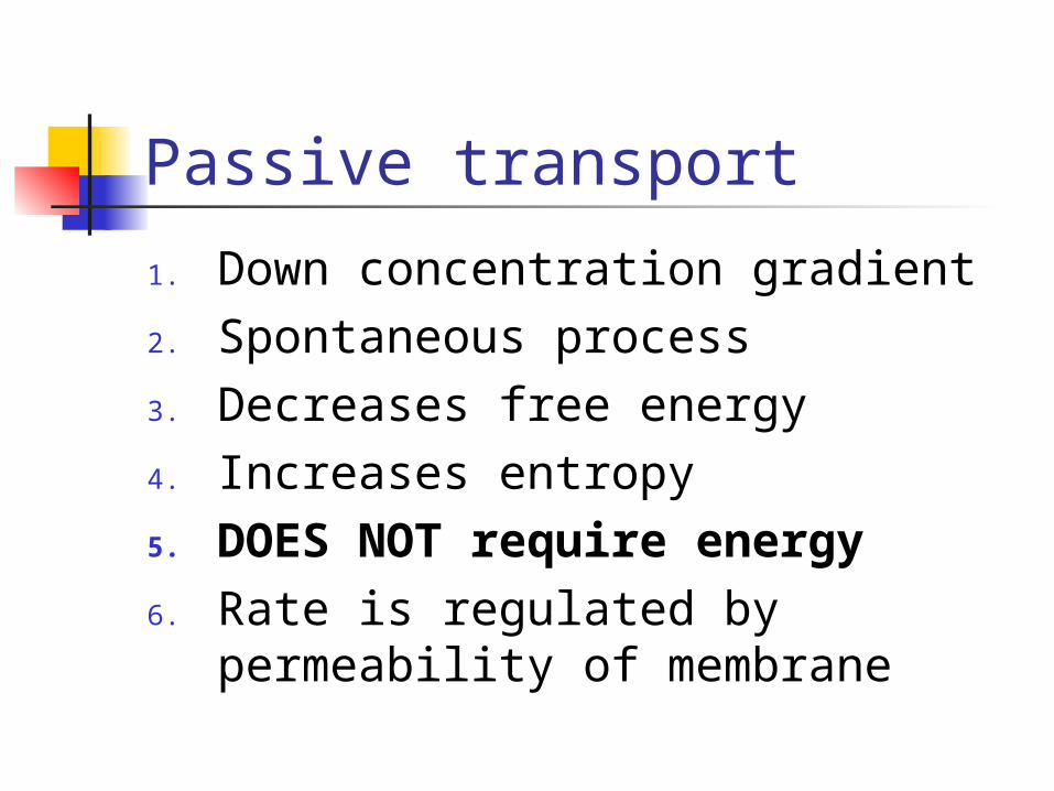
Passive transport1. Does not require energy2. Direction of movement:
1. away from concentration center2. Down the concentration gradient
3. Results from random molecular movement in ALL directions (kinetic energy of molecules)
4. Concentration gradient – change in concentration over a distance in a particular direction

Types of Passive transport1. Diffusion2. Osmosis3. Facilitated diffusion
Passive transport
1. Down concentration gradient2. Spontaneous process3. Decreases free energy4. Increases entropy5. DOES NOT require energy6. Rate is regulated by permeability
of membrane

Diffusion
1. NET movement of a substance down a concentration gradient
2. What direction is this?3. Continues until dynamic
equilibrium is reached (Does movement of molecules stop?)
osmosis

Osmosis1. Diffusion of water across a
selectively permeable membrane2. What direction does water move?3. Osmotic concentration – total
solute concentration of a solution4. Osmotic pressure – measure of a
tendency for a solution to take up water when separated by a membrane
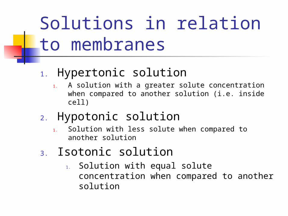
Solutions in relation to membranes
1. Hypertonic solution1. A solution with a greater solute concentration
when compared to another solution (i.e. inside cell)
2. Hypotonic solution1. Solution with less solute when compared to
another solution
3. Isotonic solution1. Solution with equal solute concentration
when compared to another solution

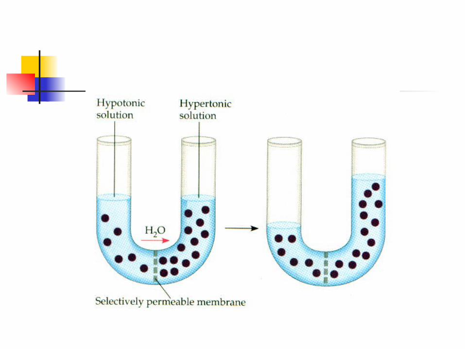


Direction of movement of H2O
1. Down its concentration gradient2. Determined by difference in TOTAL
solute concentration (all types solutes)
3. From hypotonic (hypoosmotic) to hypertonic (hyperosmotic)
4. Hypoosmotic – lower osmotic conc.5. Hyperosmotic – higher osmotic conc.

Osmotic pressure
1. Pure water = zero2. Proportional to its osmotic
concentration1. The greater the solute concentration2. The greater the osmotic pressure

Water balance in animal cells1. In isotonic environments2. No net movement of water3. In hypertonic environments
1. Cell will lose water2. Crenation (shrivel)
4. In hypotonic environments1. Gain water2. lyse

Osmoregulation
1. Contractile vacuoles (protists)1. Pump water out in hypotonic env.
2. Pump out salts 1. Conserve water in hypertonic env.

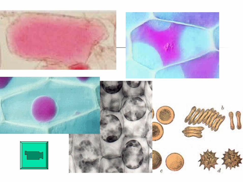
Water balance in cells with cell walls
1. In hypertonic environments1. Plasmolyze-plasma membrane pulls
away from cell wall
2. In hypotonic environments1. Pressure against cell wall equals the
osmotic pressure of cytoplasm2. Dynamic equilibrium established3. turgid

Turgidity
1. Tension found in wall cells2. In hypoosmotic environment3. Ideal state for most plants4. Provides mechanical support for
plants5. Requires cells to be hyperosmotic
to environment

3. In isotonic environments
1. No net movement of water1. Is water moving?
2. Flaccid1. Loss of structural support

Water Potential (Ψ) and Osmosis Define osmosis Water potential (Ψ)
1. The free energy of water2. A consequence of solute
concentration and pressure3. Physical property predicting the
direction of water flow4. Measured in units of pressure
megapascals (MPa)

Movement of water
1. From solution with higher water potential
2. To solution with lower water potential
3. Pure water = 0 MPa 4. Ψ = 0

Change in water potential1. Addition of solutes lowers water
potential (into negative)2. Increased pressure raises water
potential (into positive range3. Bulk flow– movement of water
due to pressure differences4. Faster than movement due to
concentration differences

Effects of pressure and solute concentration
1. Ψ = ΨP + ΨS
2. Example:3. 0.1M solution = ΨS is –0.23
4. In an open container ΨP is 0
5. What is the Ψ?6. Ψ = 0 + (-.23)7. Ψ = -0.23
Movement of water
1. Water would enter solution due to osmotic pressure only
2. Addition of pressure1. Counter affects of osmotic pressure2. Stopping net water movement3. Forcing water from solution into
pure water

Facilitated diffusion
1. Diffusion with help of transport proteins
2. Aids in diffusion of polar molecules and ions

Transport proteinsshare some properties of enzymes
1. Specific2. Can be saturated3. Can be inhibited by molecules
resembling solute4. Do not catalyze reactions

Action of transport proteins
1. Remain in place2. Alternate between two
conformations3. One conformation binds to solute4. Another conformation deposits
solute5. Binding and release of solute may
trigger conformation

Some are selective channels
1. Permeable to specific solutes2. Solutes pass through channels3. Selective channels4. May open in response to
electrical or chemical stimuli1. Na+ and K + ions2. neurotransmitters

Active Transport1. Energy (ATP) requiring process2. Transport protein pumps a molecule
against concentration gradient3. Energetically uphill (+delta G)4. Requires cell to expend energy5. Maintain steep ionic gradients across
membrane1. High affinity for K+ with binding sites
towards ECM

Examples
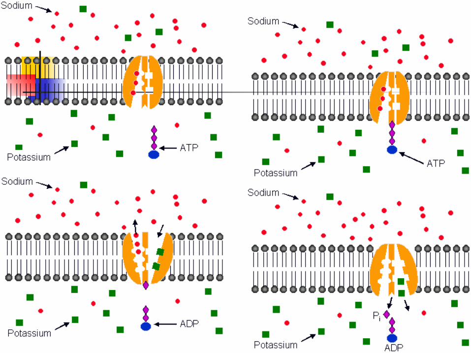
1. Sodium-potassium pump2. Transport protein oscillates
between 2 conformations1. High affinity for Na+ with binding
sites towards cytoplasm

Action
1. ATP phosphorylates transport protein
2. Powers the conformational change3. From Na+ receptive to K+ receptive4. Conformation change translocates
bound solutes5. Na+ K+-pump translocates 3 Na+
out of cell for every 2 K+ into cell

Na+ K+ Pump

Membrane potential
1. Voltage across membranes2. Range: -50 to –200 mv3. Cytoplasm side of cell is negative4. Affects movement of charged
substances across membrane5. Favors diffusion of cations

Two forces drive passive transport of ions
1. Concentration gradient2. Effect of membrane potential

Electrochemical gradient
1. Combined effects of membrane potential and concentration gradient
2. Ions:1. May not always diffuse down their conc.
gradient2. Always diffuse down their electrochemical
gradient
3. Distribution of ions may be different from expected
Cotransport
1. Membrane protein couples the transport of one solute to another
2. Single ATP actively transports one solute
3. indirectly drives the transport of other solutes
4. Against concentration gradients
Involves 2 transport proteins
1. ATP powered pump actively transports one solute (protein)
2. Another transport protein allows solute’s downhill diffusion
1. Solute leaks back across membrane2. As a second solute’s uphill transport
across membrane

Endocytosis
1. Importing macromolecules2. Form vesicles derived from
plasma membrane3. Vesicle forms form a localized
region4. Sinks inward5. Pinches off into cytoplasm

Types of endocytosis1. Phagocytosis
1. Solid particles2. Cell engulfs particle with
pseudopodia3. Vacuole fuses with lysosome
2. Pinocytosis1. Fluid droplets2. Droplets taken into small vesicles3. Not a discriminating process

What is this?

Exocytosis
1. Exporting macromolecules2. Fusion of vesicles with plasma
membrane3. Vesicle budded from ER to Golgi
migrates to plasma membrane4. Used by secretory cells to export
products 1. insulin in pancreas2. neurotansmitters

Path of export vesicle

Endocytosis and exocytosis
Related Documents