Membrane protein sequestering by ionic protein-lipid interactions Geert van den Bogaart 1 , Karsten Meyenberg 2 , H. Jelger Risselada 3 , Hayder Amin 1 , Katrin I. Willig 4 , Barbara E. Hubrich 2 , Markus Dier 1 , Stefan W. Hell 4 , Helmut Grubmüller 3 , Ulf Diederichsen 2 , and Reinhard Jahn 1 1 Department of Neurobiology, Max Planck Institute for Biophysical Chemistry, Am Faßberg 11, 37077, Göttingen, Germany 2 Institute for Organic and Biomolecular Chemistry, Georg-August-University Göttingen, Tammannstraße 2, 37077, Göttingen, Germany 3 Department of Theoretical and Computational Biophysics, Max Planck Institute for Biophysical Chemistry, Am Faßberg 11, 37077, Göttingen, Germany 4 Department of Nanobiophotonics, Max Planck Institute for Biophysical Chemistry, Am Faßberg 11, 37077, Göttingen, Germany Abstract Neuronal exocytosis is catalyzed by the SNARE protein syntaxin-1A 1 . Syntaxin-1A is clustered in the plasma membrane at sites where synaptic vesicles undergo exocytosis 2,3 . However, how syntaxin-1A is sequestered is unknown. Here, we show that syntaxin clustering is mediated by electrostatic interactions with the strongly anionic lipid phosphatidylinositol-4,5-bisphosphate (PIP2). We found with super-resolution STED microscopy on the plasma membrane of PC12 cells that PIP2 is the dominant inner-leaflet lipid in ~73 nm-sized microdomains. This high accumulation of PIP2 was required for syntaxin-1A sequestering, as destruction of PIP2 by the phosphatase synaptojanin-1 reduced syntaxin-1A clustering. Furthermore, co-reconstitution of PIP2 and the C-terminal part of syntaxin-1A in artificial giant unilamellar vesicles resulted in segregation of PIP2 and syntaxin-1A into distinct domains even when cholesterol was absent. Our results demonstrate that electrostatic protein-lipid interactions can result in the formation of microdomains independent of cholesterol or lipid phases. Phosphoinositides are lipids that contain an inositol headgroup conjugated to 1–3 phosphate groups. With ~1% of total lipids in the inner leaflet of the plasma membrane 4 , PIP2 is the most abundant phosphoinositide. Earlier studies identified PIP2 as a second messenger in the phospholipase-C pathway. However, the list of cellular functions of PIP2 is rapidly growing, and PIP2 is also involved in membrane targeting, cytoskeletal attachment, endocytosis and exocytosis 4 . PIP2 interacts with many different proteins, either via unstructured basic residue-rich regions or via more structured domains 4,5 . Correspondence and requests for materials should be addressed to R.J. ([email protected]). Supplementary Information is linked to the online version of the paper at www.nature.com/nature. Author Contributions G.v.d.B and R.J. designed the experiments and wrote the paper. K.M., B.E.H. and U.D. synthesized the peptides. J.H.R. and H.G. performed the simulations. H.A. performed the TIRF and K.I.W. and S.W.H the STED microscopy. M.D. contributed to the protein purification and immunofluorescence. G.v.d.B. performed all other experiments. All authors contributed to the manuscript. The authors declare no competing financial interests. NIH Public Access Author Manuscript Nature. Author manuscript; available in PMC 2012 August 02. Published in final edited form as: Nature. ; 479(7374): 552–555. doi:10.1038/nature10545. NIH-PA Author Manuscript NIH-PA Author Manuscript NIH-PA Author Manuscript

Welcome message from author
This document is posted to help you gain knowledge. Please leave a comment to let me know what you think about it! Share it to your friends and learn new things together.
Transcript

Membrane protein sequestering by ionic protein-lipidinteractions
Geert van den Bogaart1, Karsten Meyenberg2, H. Jelger Risselada3, Hayder Amin1, Katrin I.Willig4, Barbara E. Hubrich2, Markus Dier1, Stefan W. Hell4, Helmut Grubmüller3, UlfDiederichsen2, and Reinhard Jahn1
1Department of Neurobiology, Max Planck Institute for Biophysical Chemistry, Am Faßberg 11,37077, Göttingen, Germany2Institute for Organic and Biomolecular Chemistry, Georg-August-University Göttingen,Tammannstraße 2, 37077, Göttingen, Germany3Department of Theoretical and Computational Biophysics, Max Planck Institute for BiophysicalChemistry, Am Faßberg 11, 37077, Göttingen, Germany4Department of Nanobiophotonics, Max Planck Institute for Biophysical Chemistry, Am Faßberg11, 37077, Göttingen, Germany
AbstractNeuronal exocytosis is catalyzed by the SNARE protein syntaxin-1A1. Syntaxin-1A is clustered inthe plasma membrane at sites where synaptic vesicles undergo exocytosis2,3. However, howsyntaxin-1A is sequestered is unknown. Here, we show that syntaxin clustering is mediated byelectrostatic interactions with the strongly anionic lipid phosphatidylinositol-4,5-bisphosphate(PIP2). We found with super-resolution STED microscopy on the plasma membrane of PC12 cellsthat PIP2 is the dominant inner-leaflet lipid in ~73 nm-sized microdomains. This highaccumulation of PIP2 was required for syntaxin-1A sequestering, as destruction of PIP2 by thephosphatase synaptojanin-1 reduced syntaxin-1A clustering. Furthermore, co-reconstitution ofPIP2 and the C-terminal part of syntaxin-1A in artificial giant unilamellar vesicles resulted insegregation of PIP2 and syntaxin-1A into distinct domains even when cholesterol was absent. Ourresults demonstrate that electrostatic protein-lipid interactions can result in the formation ofmicrodomains independent of cholesterol or lipid phases.
Phosphoinositides are lipids that contain an inositol headgroup conjugated to 1–3 phosphategroups. With ~1% of total lipids in the inner leaflet of the plasma membrane4, PIP2 is themost abundant phosphoinositide. Earlier studies identified PIP2 as a second messenger inthe phospholipase-C pathway. However, the list of cellular functions of PIP2 is rapidlygrowing, and PIP2 is also involved in membrane targeting, cytoskeletal attachment,endocytosis and exocytosis4. PIP2 interacts with many different proteins, either viaunstructured basic residue-rich regions or via more structured domains4,5.
Correspondence and requests for materials should be addressed to R.J. ([email protected]).
Supplementary Information is linked to the online version of the paper at www.nature.com/nature.
Author ContributionsG.v.d.B and R.J. designed the experiments and wrote the paper. K.M., B.E.H. and U.D. synthesized the peptides. J.H.R. and H.G.performed the simulations. H.A. performed the TIRF and K.I.W. and S.W.H the STED microscopy. M.D. contributed to the proteinpurification and immunofluorescence. G.v.d.B. performed all other experiments. All authors contributed to the manuscript.
The authors declare no competing financial interests.
NIH Public AccessAuthor ManuscriptNature. Author manuscript; available in PMC 2012 August 02.
Published in final edited form as:Nature. ; 479(7374): 552–555. doi:10.1038/nature10545.
NIH
-PA Author Manuscript
NIH
-PA Author Manuscript
NIH
-PA Author Manuscript

Neuronal exocytosis requires plasma membrane PIP22,6-8. PIP2 levels at the plasmamembrane determine the rates of vesicle priming, the size of the readily releasable pool, andthe rates of sustained exocytosis in stimulated cells2,6,8. This regulation is probablymediated by interactions of PIP2 with proteins involved in docking and fusion such asrabphilin, CAPS, synaptotagmin, SCAMP2 and Mints6,9. In docking, PIP2 clusters may actas molecular ‘beacons’ that target synaptic vesicles to the fusion sites. Indeed, PIP2 islocally enriched at the sites of docked vesicles and colocalizes with at least 5–10% of themicrodomains of syntaxin-1A (Supp. Fig. 1)2,3,9, the membrane-anchored t-SNARE ofneuronal exocytosis1.
The amount of PIP2 at the sites of membrane fusion in PC12 cells has been estimated at 3–6% PIP2 of surface area (Supp. Fig. 2)9. In these experiments, membrane sheets werespecifically stained for PIP2 with the PH domain of protein lipase C delta fused to GFP9 orcitrine (a YFP analog10; PHPLCδ-citrine, Supp. Fig. 1–2), and the fluorescence from thepunctuated PIP2-microdomains was quantified. However, as explained in reference9, thisapproach underestimates the fraction of PIP2 if the size of the PHPLCδ-microdomains issmaller than the ~200 nm diffraction limited resolution of conventional fluorescencemicroscopy. To obtain a more accurate estimate, we re-analyzed PC12 membrane sheetslabeled with PHPLCδ-citrine or an antibody raised against PIP2 using super resolution STED(stimulation emission depletion) microscopy11 (Fig. 1a–c). These experiments revealed thatthe PIP2 stained clusters are much smaller than anticipated, with an average diameter ofonly 73 ± 42 nm (s.d.). Although this is still a higher estimate since it represents themicrodomain size convoluted with the resolution of the STED microscope (~60 nm), it is ingood agreement with the size of the syntaxin-1A microdomains12. Using this value, we re-calculated the surface density of PIP2 (Supp. Methods). For this calculation, we firstestimated the total amount of PIP2 in a microdomain when sampled with the diffractionlimited resolution of our epi-fluorescence microscope (Supp. Fig. 2; Fig. 1d, black curve).We then calculated the peak concentration when this PIP2 was concentrated into 73 nmmicrodomains (Fig. 1d, red curve). Here, we assumed a Gaussian distribution of PIP2 in themicrodomains. A peak surface density of 82% PIP2 was obtained (Fig. 1d). It needs to bekept in mind that (i) at these high PIP2 concentrations, molecular crowding might hinderbinding of PHPLCδ-citrine, (ii) relatively small errors in microdomain size and resolution ofthe microscopes result in substantial errors, and (iii) PHPLCδ-citrine and antibody bindingmay alter PIP2 localization and is only indicative of PIP2 microdomains. Nevertheless, ourvalues are much higher than any previous estimate and it seems safe to conclude that PIP2 isthe dominant inner-leaflet lipid in the microdomains. The question then arises by whichmolecular mechanism such high concentrations of PIP2 are achieved.
PIP2 has a net negative charge of -3–54 and interacts with polybasic stretches of aminoacids4,5,13,14. Proteins with such stretches can sequester PIP2 even in excess of monovalentanionic lipids, such as MARCKS, spermine and even pentalysine (Lys5)5,14. Similar to theseproteins, syntaxin-1A also possesses a stretch of basic amino acids. These residues areadjacent to the transmembrane domain and are in contact with the head-groups of thephospholipids (Supp. Fig. 3a)15,16. Indeed, it is well established that this conserved stretchwith 5 positive residues (260-KARRKK) interacts with PIP29,15-17. Removal of chargediminishes this interaction (Supp. Fig. 3b–c), but syntaxin-1A remains capable of fusingmembranes even upon removal of all 5 charges9,16. Because PIP2 colocalizes with at least afraction of syntaxin-1A microdomains (Supp. Fig. 1)2, we speculated that their interactionmight drive domain formation similar to various soluble lipid binding proteins5,14. Twoindependent approaches were used to test this hypothesis: (i) reconstitution in giantunilamellar vesicles (GUVs)18, and (ii) hydrolysis of PIP2 in PC12 cells using a membrane-targeted variant of the PIP2 phosphatase synaptojanin-17.
van den Bogaart et al. Page 2
Nature. Author manuscript; available in PMC 2012 August 02.
NIH
-PA Author Manuscript
NIH
-PA Author Manuscript
NIH
-PA Author Manuscript

Murray and Tamm17 showed syntaxin-1A clustered in a non-raft way in neutralcholesterol:phosphatidylcholine (PC) membranes. Here, cholesterol clusters syntaxin-1A bycompeting for solvation by PC. Indeed, a synthetic C-terminal peptide of syntaxin-1A(residues 257–288; 3 mol%; Fig. 2, Supp. Fig. 4a) clustered in domains in > 50% of GUVscomposed of DOPC (1,2-dioleoyl-sn-glycero-3-PC) with 20 mol% cholesterol. This peptidecontained both the polybasic juxtamembrane linker and transmembrane region and was N-terminally labeled with either rhodamine red or Atto647N. Analysis of fluorescence showeda 1.6 ± 0.2 (s.d.; n = 18) fold enrichment of syntaxin-1A257–288 in these clusters, but thislower estimate is limited by the optics. Negatively charged PIP2 or DOPS (1,2-dioleoyl-sn-glycero-3-phosphatidylserine) dispersed these clusters (Fig. 2)17. Thus, while cholesterolcompetition might explain syntaxin-1A clusters that are not enriched in PIP2 (Supp. Fig. 1),they cannot explain the high accumulation of PIP2 at the sites of docked vesicles. However,1.5 mol% (total lipids) PIP2 also clustered syntaxin-1A in 1–10 μm domains in 1–5% of theGUVs (Fig. 2, Supp. Fig. 4b–c). These domains did not depend on cholesterol or DOPS. Inthese domains, PIP2 was 1.9 ± 0.2 (s.d.; n = 13; Supp. Fig. 5) and syntaxin-1A257–288 5.5 ±1.4 (s.d.; n = 27) fold enriched based on fluorescence. Importantly, no domains wereobserved without peptide or when the PIP2 concentration exceeded 5 mol%. Divalentcations can act as bridges between two adjacent lipids and induce aggregation of PIP2 intoclusters19-21, but even 1 mM Ca2+ was not sufficient to compete syntaxin-1A away from themicrodomains. Domains were present with both synthetic dioleoyl-PIP2 and with PIP2extracted from pig brain (Supp. Fig. 4b). Thus, syntaxin-1A can be clustered in themembrane both by cholesterol and PIP2.
These cholesterol- and PIP2-mediated clusters both differ from ‘rafts’. They also differ fromeach other. First, PIP2-domains are always round and only 1–2 per vesicle, whereascholesterol generally (but not always) induces many small domains (Supp. Fig. 4). Second,fluorescence recovery after photobleaching showed that syntaxin-1A remained mobile in thePIP2-domains while syntaxin-1A was essentially immobile in the cholesterol-dependentclusters (Supp. Fig. 6). Syntaxin-1A thus diffuses in the PIP2-domains and forms largecircular domains for minimizing boundary energy21. Third, 6-dodecanoyl-2-dimethylaminonaphthalene (Laurdan)22 showed a high hydration of the PIP2-domains,whereas the cholesterol domains were much denser packed (Supp. Fig. 7). Fourth, phasecontrast microscopy showed a thickening of the cholesterol-dependent clusters, but not ofthe PIP2-domains (Supp. Fig. 8). Thus, even though no saturated lipids are present, thecholesterol-dependent domains show behavior that essentially resembles the Lo phase. Incontrast, the PIP2-domains seem much more disordered and resemble the Ld phase. Ca2+
demixing of polyanionic amphiphiles showed that electrostatic interactions can indeed leadto liquid-like domains21.
The transmembrane helix of syntaxin-1A has been reported to homodimerize. However,introducing the M267A C271A I279A mutations that prevent homodimerization of thesyntaxin-1A peptides23 did not prevent cholesterol or PIP2 mediated clustering (Supp. Fig.9). In contrast, no PIP2-domains were observed when two charges (K264A K265A) fromthe polybasic linker were removed, but cholesterol-dependent clusters were still observed(Supp. Fig. 5). Overexpression of the C-terminal part of syntaxin-1A fused to GFP24 inPC12 cells also showed 4–8-fold loss of clustering of the K264A K265A mutant (Supp. Fig.10–11). These data show that electrostatic interactions between PIP2 and the juxtamembranehelix of syntaxin-1A are sufficient for domain formation.
We then set out to investigate to what extent PIP2 is required for syntaxin-1A clustering inPC12 cells. For this purpose, we expressed a RFP-tagged construct containing thephosphatase domain of synaptojanin-1 fused to a CAAX-box, resulting in its targeting to theplasma membrane7. Synaptojanin-1 is a polyphosphoinositide 5-phosphatase, and the
van den Bogaart et al. Page 3
Nature. Author manuscript; available in PMC 2012 August 02.
NIH
-PA Author Manuscript
NIH
-PA Author Manuscript
NIH
-PA Author Manuscript

expression of the construct completely removed PIP2 from the plasma membrane (Supp.Fig. 12)6,7. Importantly, synaptojanin-1 expression 3.7-fold reduced the punctuatedistribution of endogenous syntaxin-1A (Fig. 3; Supp. Fig. 13). Thus, this provides evidencethat PIP2 is indeed required for at least part of syntaxin-1A microdomain formation.
We performed molecular dynamics simulations to gain insight in the precise conformationof the PIP2-syntaxin-1A microdomains. In these coarse-grained simulations, several atomsare represented by one simulation bead25,26 (Supp. Fig. 14). This allowed for simulations ofrelatively large lipid bilayers of ~2,500 copies of a 4:1 molar ratio of DOPC:DOPS and 40–64 copies of syntaxin-1A257–288 and PIP2. Within 10 μs simulation time, up to 10 copies ofsyntaxin-1A257–288 clustered with PIP2 into microdomains (Supp. Fig. 15). Equal amountsof PIP2 and syntaxin-1A were present in the bulk-phase of those domains, while more PIP2and DOPS associated transiently to the periphery. We used this information to construct adomain with 64 copies of syntaxin-1A (Fig. 4, Supp. Movie 1), which is comparable to thesyntaxin-1A content in the microdomains in PC12 cells12. These domains were stable over 6μs simulation time and contained <10% residual DOPC or DOPS. Together, we concludethat syntaxin-1A and PIP2 can form dynamic, amorphous networks with PIP2 acting as a‘charge-bridge’ and spanning the distance between the various syntaxin-1A molecules (Fig.4c).
In summary, our findings show that electrostatic interactions between the membrane lipidPIP2 and the SNARE syntaxin-1A suffice to induce membrane sequestering andmicrodomain formation without the need for high local PIP2 production or a (complex)‘molecular fence’ restricting PIP2 and protein diffusion27. This does not exclude anadditional role for protein-protein interactions between either transmembrane helices orsoluble domains. In fact, these seem essential for segregation of proteins in similar structureand size, such as syntaxin-1A and syntaxin-4 (both have polybasic regions and clusterseparately)12,24. The mutual enrichment of syntaxin-1A and PIP2 at the fusion sites byelectrostatic interactions has clear advantages. First, accumulation of syntaxin-1A mayfacilitate SNARE interactions and thereby increase the membrane fusion efficiency3,28.Second, the lipid environment modulates the energetic requirements for fusion13,16. Third,both PIP2 and syntaxin-1A function as molecular docking sites and facilitate assembly ofthe complete fusion machinery1,2,6-9. Our findings that electrostatic protein-lipidinteractions are sufficient for membrane sequestering constitute a novel mechanism for theformation of protein microdomains in the membrane that is clearly distinct from the well-established lipid phases18,29.
Methods SummaryPHPLCδ-citrine was expressed in Escherichia coli and purified with his-tag affinitypurification. PC12 cells were maintained and propagated as described3,24. PC12 cells weretransfected using Lipofectamine LTX (Invitrogen). Membrane sheets were prepared byrupturing the cells with probe sonication as described24. Immunostaining24 andmicroscopy11,24 were performed as described. The peptides were synthesized viamicrowave-assisted Fmoc solid phase synthesis. Peptides were mixed with lipids in organicsolvent and GUVs were formed by the drying rehydration procedure. The moleculardynamics simulations were performed with the GROMACS simulation package and theMARTINI coarse-grained model25,26. See Supp. Methods for details.
Supplementary MaterialRefer to Web version on PubMed Central for supplementary material.
van den Bogaart et al. Page 4
Nature. Author manuscript; available in PMC 2012 August 02.
NIH
-PA Author Manuscript
NIH
-PA Author Manuscript
NIH
-PA Author Manuscript

AcknowledgmentsWe thank Matthew Holt, Gertrude Bunt, Fred S. Wouters and Christian Eggeling for advice and Volker Haucke andSeong Joo (Freie Universität Berlin, Germany) for the RFP-synaptojanin-1 construct. G.v.d.B is financed by theHuman Frontier Science Program. This work was supported by the US National Institutes of Health (P01GM072694, to R.J.) and the Deutsche Forschungsgemeinschaft SFB803 (to K.M., J.H.R., U.D., H.G. and R.J.).
References1. Jahn R, Scheller RH. SNAREs-engines for membrane fusion. Nat Rev Mol Cell Biol. 2006; 7:631–
643. [PubMed: 16912714]
2. Aoyagi K, et al. The activation of exocytotic sites by the formation of phosphatidylinositol 4,5-bisphosphate microdomains at syntaxin clusters. J Biol Chem. 2005; 280:17346–17352. [PubMed:15741173]
3. Lang T, et al. SNAREs are concentrated in cholesterol-dependent clusters that define docking andfusion sites for exocytosis. EMBO J. 2001; 20:2202–2213. [PubMed: 11331586]
4. McLaughlin S, Wang J, Gambhir A, Murray D. PIP(2) and proteins: interactions, organization, andinformation flow. Annu Rev Biophys Biomol Struct. 2002; 31:151–175. [PubMed: 11988466]
5. McLaughlin S, Murray D. Plasma membrane phosphoinositide organization by proteinelectrostatics. Nature. 2005; 438:605–611. [PubMed: 16319880]
6. Wen PJ, Osborne SL, Meunier FA. Dynamic control of neuroexocytosis by phosphoinositides inhealth and disease. Prog Lipid Res. 2011; 50:52–61. [PubMed: 20705095]
7. Milosevic I, et al. Plasmalemmal phosphatidylinositol-4,5-bisphosphate level regulates thereleasable vesicle pool size in chromaffin cells. J Neurosci. 2005; 25:2557–2565. [PubMed:15758165]
8. Hay JC, Martin TF. Phosphatidylinositol transfer protein required for ATP-dependent priming ofCa(2+)-activated secretion. Nature. 1993; 366:572–575. [PubMed: 8255295]
9. James DJ, Khodthong C, Kowalchyk JA, Martin TF. Phosphatidylinositol 4,5-bisphosphateregulates SNARE-dependent membrane fusion. J Cell Biol. 2008; 182:355–366. [PubMed:18644890]
10. Griesbeck O, Baird GS, Campbell RE, Zacharias DA, Tsien RY. Reducing the environmentalsensitivity of yellow fluorescent protein. Mechanism and applications. J Biol Chem. 2001;276:29188–29194. [PubMed: 11387331]
11. Hell SW, Wichmann J. Breaking the diffraction resolution limit by stimulated emission:stimulated-emission-depletion fluorescence microscopy. Opt Lett. 1994; 19:780–782. [PubMed:19844443]
12. Sieber JJ, et al. Anatomy and dynamics of a supramolecular membrane protein cluster. Science.2007; 317:1072–1076. [PubMed: 17717182]
13. Williams D, Vicôgne J, Zaitseva I, McLaughlin S, Pessin JE. Evidence that electrostaticinteractions between vesicle-associated membrane protein 2 and acidic phospholipids maymodulate the fusion of transport vesicles with the plasma membrane. Mol Biol Cell. 2009;20:4910–4919. [PubMed: 19812247]
14. Denisov G, Wanaski S, Luan P, Glaser M, McLaughlin S. Binding of basic peptides to membranesproduces lateral domains enriched in the acidic lipids phosphatidylserine and phosphatidylinositol4,5-bisphosphate: an electrostatic model and experimental results. Biophys J. 1998; 74:731–744.[PubMed: 9533686]
15. Kweon DH, Kim CS, Shin YK. The membrane-dipped neuronal SNARE complex: a site-directedspin labeling electron paramagnetic resonance study. Biochemistry. 2002; 41:9264–9268.[PubMed: 12119042]
16. Lam AD, Tryoen-Toth P, Tsai B, Vitale N, Stuenkel EL. SNARE-catalyzed fusion events areregulated by Syntaxin1A-lipid interactions. Mol Biol Cell. 2008; 19:485–497. [PubMed:18003982]
17. Murray DH, Tamm LK. Clustering of syntaxin-1A in model membranes is modulated byphosphatidylinositol 4,5-bisphosphate and cholesterol. Biochemistry. 2009; 48:4617–4625.[PubMed: 19364135]
van den Bogaart et al. Page 5
Nature. Author manuscript; available in PMC 2012 August 02.
NIH
-PA Author Manuscript
NIH
-PA Author Manuscript
NIH
-PA Author Manuscript

18. Bacia K, Schuette CG, Kahya N, Jahn R, Schwille P. SNAREs prefer liquid-disordered over “raft”(liquid-ordered) domains when reconstituted into giant unilamellar vesicles. J Biol Chem. 2004;279:37951–37955. [PubMed: 15226320]
19. Carvalho K, Ramos L, Roy C, Picart C. Giant unilamellar vesicles containingphosphatidylinositol(4,5)bisphosphate: characterization and functionality. Biophys J. 2008;95:4348–4360. [PubMed: 18502807]
20. Levental I, et al. Calcium-dependent lateral organization in phosphatidylinositol 4,5-bisphosphate(PIP2)- and cholesterol-containing monolayers. Biochemistry. 2009; 48:8241–8248. [PubMed:19630438]
21. Christian DA, et al. Spotted vesicles, striped micelles and Janus assemblies induced by ligandbinding. Nat Mater. 2009; 8:843–849. [PubMed: 19734886]
22. Kaiser HJ, et al. Order of lipid phases in model and plasma membranes. Proc Natl Acad Sci USA.2009; 106:16645–16650. [PubMed: 19805351]
23. Laage R, Rohde J, Brosig B, Langosch D. A conserved membrane-spanning amino acid motifdrives homomeric and supports heteromeric assembly of presynaptic SNARE proteins. J BiolChem. 2000; 275:17481–17487. [PubMed: 10764817]
24. Sieber JJ, Willig KI, Heintzmann R, Hell SW, Lang T. The SNARE motif is essential for theformation of syntaxin clusters in the plasma membrane. Biophys J. 2006; 90:2843–2851.[PubMed: 16443657]
25. Marrink SJ, Risselada HJ, Yefimov S, Tieleman DP, de Vries AH. The MARTINI forcefield:coarse grained model for biomolecular simulations. J Phys Chem B. 2007; 111:7812–7824.[PubMed: 17569554]
26. Yesylevskyy S, Schafer LV, Sengupta D, Marrink SJ. Polarizable water model for the coarse-grained Martini force field. PLoS Comp Biol. 2010; 6:e1000810.
27. Fujiwara T, Ritchie K, Murakoshi H, Jacobson K, Kusumi A. Phospholipids undergo hop diffusionin compartmentalized cell membrane. J Cell Biol. 2002; 157:1071–1081. [PubMed: 12058021]
28. van den Bogaart G, Jahn R. Counting the SNAREs needed for membrane fusion. J Mol Cell Biol.2011; 3:204–205. [PubMed: 21525018]
29. Simons K, Ikonen E. Functional rafts in cell membranes. Nature. 1997; 387:569–572. [PubMed:9177342]
van den Bogaart et al. Page 6
Nature. Author manuscript; available in PMC 2012 August 02.
NIH
-PA Author Manuscript
NIH
-PA Author Manuscript
NIH
-PA Author Manuscript

Figure 1.PIP2 is the predominant inner-leaflet lipid in roughly 73 nm-sized microdomains. (a)Confocal and corresponding nanoscale-resolution STED image of a PHPLCδ-citrine stainedmembrane sheet of PC12 cells. Note the increase in resolution. (b) Same as a, but nowimmunostained with a monoclonal PIP2-antibody and a secondary antibody labeled withAlexa Fluor 488. (c) Size distribution of microdomains with PHPLCδ-citrine (blue; n = 433,24 sheets, 2 independent preparations) and PIP2-antibody (pink; n = 2,959, 22 sheets, 2independent preparations). The average diameter (full width at half maximum) was 73 ± 42nm (s.d.) of PHPLCδ-citrine and 87 ± 62 nm (s.d.) for PIP2-antibody. (d) Spatial distributionof PIP2. Black: the PIP2 distribution when sampled at too low (377 nm) diffraction limitedresolution (from Supp. Fig. 2). Red: approximation of the PIP2 distribution in the ~73 nmmicrodomains. PIP2 was accumulated at ~82% of total surface area. See Supp. Methods fordetails.
van den Bogaart et al. Page 7
Nature. Author manuscript; available in PMC 2012 August 02.
NIH
-PA Author Manuscript
NIH
-PA Author Manuscript
NIH
-PA Author Manuscript
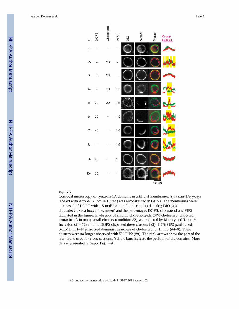
Figure 2.Confocal microscopy of syntaxin-1A domains in artificial membranes. Syntaxin-1A257–288labeled with Atto647N (SxTMH; red) was reconstituted in GUVs. The membranes werecomposed of DOPC with 1.5 mol% of the fluorescent lipid analog DiO (3,3’-dioctadecyloxacarbocyanine; green) and the percentages DOPS, cholesterol and PIP2indicated in the figure. In absence of anionic phospholipids, 20% cholesterol clusteredsyntaxin-1A in many small clusters (condition #2), as predicted by Murray and Tamm17.Inclusion of > 5% anionic DOPS dispersed these clusters (#3). 1.5% PIP2 partitionedSxTMH in 1–10 μm-sized domains regardless of cholesterol or DOPS (#4–8). Theseclusters were no longer observed with 5% PIP2 (#9). The pink arrows show the part of themembrane used for cross-sections. Yellow bars indicate the position of the domains. Moredata is presented in Supp. Fig. 4–9.
van den Bogaart et al. Page 8
Nature. Author manuscript; available in PMC 2012 August 02.
NIH
-PA Author Manuscript
NIH
-PA Author Manuscript
NIH
-PA Author Manuscript

Figure 3.Removal of PIP2 reduces syntaxin-1A clustering in PC12 cells (a) Membrane sheets ofPC12 cells stained with TMA-DPH (1-(4-trimethylammoniumphenyl)-6-phenyl-1,3,5-hexatriene)12,24. Immunostaining with a monoclonal antibody raised against syntaxin-1Aand a secondary antibody labeled with DyLight649 showed that endogenous syntaxin-1Aclustered in microdomains (region b; pink)2,3,9,12,24. Overexpressing the RFP-tagged andmembrane-targeted catalytic region of synaptojanin-1 (residues 498–901; cell outlined inblue)7 reduced this syntaxin-1A clustering 3.7-fold (region c; orange; see Supp. Fig. 13).Synaptojanin-1 is the 5-phosphatase of PIP2 and overexpression of the construct completelyremoves PIP2 from the membrane (Supp. Fig. 12). (b–c) Magnification of the regions ofinterest from a and cross-sections to indicate the clustering.
van den Bogaart et al. Page 9
Nature. Author manuscript; available in PMC 2012 August 02.
NIH
-PA Author Manuscript
NIH
-PA Author Manuscript
NIH
-PA Author Manuscript
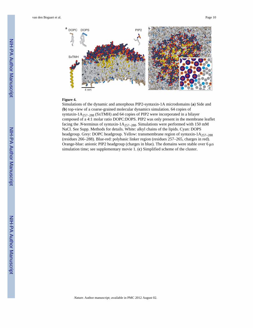
Figure 4.Simulations of the dynamic and amorphous PIP2-syntaxin-1A microdomains (a) Side and(b) top-view of a coarse-grained molecular dynamics simulation. 64 copies ofsyntaxin-1A257–288 (SxTMH) and 64 copies of PIP2 were incorporated in a bilayercomposed of a 4:1 molar ratio DOPC:DOPS. PIP2 was only present in the membrane leafletfacing the N-terminus of syntaxin-1A257–288. Simulations were performed with 150 mMNaCl. See Supp. Methods for details. White: alkyl chains of the lipids. Cyan: DOPSheadgroup. Grey: DOPC headgroup. Yellow: transmembrane region of syntaxin-1A257–288(residues 266–288). Blue-red: polybasic linker region (residues 257–265, charges in red).Orange-blue: anionic PIP2 headgroup (charges in blue). The domains were stable over 6 μssimulation time; see supplementary movie 1. (c) Simplified scheme of the cluster.
van den Bogaart et al. Page 10
Nature. Author manuscript; available in PMC 2012 August 02.
NIH
-PA Author Manuscript
NIH
-PA Author Manuscript
NIH
-PA Author Manuscript
Related Documents