DOI:10.4491/eer.2010.15.3.141 pISSN 1225 -1025 eISSN 2005-968X Environ. Eng. Res. 2010 September,15(3) : 141-147 Research Paper 141 ©Copyright The Korean Society of Environmental Engineers http://www.eer.or.kr Received March 03, 2010 Accepted June 24, 2010 † Corresponding Author E-mail: [email protected] Tel: +82-62-715-3381 Fax: +82-62-715-2434 This is an Open Access article distributed under the terms of the Creative Commons Attribution Non-Commercial License (http://creativecommons. org/licenses/by-nc/3.0/) which permits unrestricted non-commercial use, distribution, and reproduction in any medium, provided the original work is properly cited. Membrane Biofouling of Seawater Reverse Osmosis Initiated by Sporogenic Bacillus Strain Jinwook Lee 1 , Xianghao Ren 2 , Hye-Weon Yu 3 , Sung-Jo Kim 3 , In S. Kim 1,3† 1 Center for Seawater Desalination Plant, Gwangju Institute of Science and Technology, Gwangju 500-712, Korea 2 Key Laboratory of Urban Stormwater System and Water Environment, School of Environment and Energy Engineering, Beijing Univer sity of Civil Engineering and Architecture, Beijing 100-044, China 3 Department of Environmental Science and Engineering, Gwangju Institute of Science and Technology, Gwangju 500-712, Korea Abstract The objective of this study was to assess the biofouling characteristics of the Bacillus biofilm formed on reverse osmosis (RO) mem- branes. For the study, a sporogenic Bacillus sp. was isolated from the seawater intake to a RO process, with two distinct sets of experi- ments performed to grow the Bacillus biofilm on the RO membrane using a lab-scale crossflow membrane test unit. Two operational feds were used, 9 L sterile-filtered seawater and 10 9 Bacillus cells, with flow rates of 1 L/min, and a constant 800 psi-pressure and pH 7.6. From the results, the membrane with more fouling, in which the observed permeate flux decreased to 33% of its initial value, showed about 10 and 100 times greater extracellular polymeric substances and spoOA genes expressions, respectively, than the those of the less fouled membrane (flux declined to 20% of its initial value). Interestingly; however, the number of culturable Bacillus sp. in the more fouled membrane was about 10 times less than that of the less fouled membrane. This indicated that while the number of Bacillus had less relevance with respect to the extent of biofouling, the activation of the genes of interest, which is initiative of biofilm development, had a more positive effect on biofouling than the mass of an individual Bacillus bacterium. Keywords: Bacillus sp., Biofilm, Gene expression, Extracellular polymeric substance, Reverse osmosis membrane, spoOA 1. Introduction Due to the acceleration of global warming and the stress that population growth has placed on the global water supply, the desalination of seawater via reverse osmosis (RO) has shown promise as a technology for overcoming the stress on water re- sources. However, membrane fouling is a common problem, as it causes flux decline, demands frequent cleaning and consumes a large amount of energy, resulting in a shortened lifespan of the system [1]. Among the types of fouling material, severe biofilm formation by variable bacteria, called biofouling, is the most dif- ficult to control. It was reported that 58 of 70 RO membrane and about 70% of the seawater RO membrane installations surveyed in U.S. suffered from biofouling problems [2, 3]. To avoid or re- duce the biofouling on the membrane, a deeper understanding of uncontrollable biofouling is critical. In an attempt to address this issue, a number of studies have focused on measuring the densities of biological matter [4]. About 1 × 10 7 cells per cm 2 of fouled RO membrane surface was observed, with bacteria, such as Pseudomonas, Corynebacteri- um, Bacillus, Arthrobacter, Flavobacter, Mycobacterium, Acineto- bacter and Cytophaga-Flavobacter-Bacteroidetes group found to participate in biofilm development on RO membranes [5, 6]. However, the selection of the most predominant and problem- atic microorganism is not simple since the dominance of bacte- rial species varies from one to facility to another depending on site-specific conditions, such as feed water quality, temperature, location and the processes involved. Accordingly, due to the diversity of predominant bacteria, a model biofilm associated strains or bacterial isolates retrieved from the fouled RO mem- brane are used in biofouling studies. For example, Pseudomonas strains have frequently been studied as primary biofouling mi- croorganisms in the RO process for wastewater treatment [7, 8]. However, the Pseudomonas and RO membranes in these studies have not been employed for seawater treatment. Bacillus sp. strains, or its relatives, have also arisen as prob- lematic bacteria as they are commonly observed in different biofilm community structures on RO/NF membranes used for water treatment [9-12]. The higher hydrophobicity of Bacillus sp. Over that of Pseudomonas enhances the cell adhesion onto RO membranes [13]. Based on our previous studies, the approach using Bacillus

Welcome message from author
This document is posted to help you gain knowledge. Please leave a comment to let me know what you think about it! Share it to your friends and learn new things together.
Transcript
DOI:10.4491/eer.2010.15.3.141pISSN 1225-1025 eISSN 2005-968X
Environ. Eng. Res. 2010 September,15(3) : 141-147
Research Paper
141©Copyright The Korean Society of Environmental Engineers http://www.eer.or.kr
Received March 03, 2010 Accepted June 24, 2010†Corresponding AuthorE-mail: [email protected]: +82-62-715-3381 Fax: +82-62-715-2434
This is an Open Access article distributed under the terms of the Creative Commons Attribution Non-Commercial License (http://creativecommons.org/licenses/by-nc/3.0/) which permits unrestricted non-commercial use, distribution, and reproduction in any medium, provided the original work is properly cited.
Membrane Biofouling of Seawater Reverse Osmosis Initiated by Sporogenic Bacillus Strain
Jinwook Lee1, Xianghao Ren2, Hye-Weon Yu3, Sung-Jo Kim3, In S. Kim1,3†
1Center for Seawater Desalination Plant, Gwangju Institute of Science and Technology, Gwangju 500-712, Korea2Key Laboratory of Urban Stormwater System and Water Environment, School of Environment and Energy Engineering, Beijing Univer sity of Civil Engineering and Architecture, Beijing 100-044, China 3Department of Environmental Science and Engineering, Gwangju Institute of Science and Technology, Gwangju 500-712, Korea
Abstract
The objective of this study was to assess the biofouling characteristics of the Bacillus biofilm formed on reverse osmosis (RO) mem-branes. For the study, a sporogenic Bacillus sp. was isolated from the seawater intake to a RO process, with two distinct sets of experi-ments performed to grow the Bacillus biofilm on the RO membrane using a lab-scale crossflow membrane test unit. Two operational feds were used, 9 L sterile-filtered seawater and 109 Bacillus cells, with flow rates of 1 L/min, and a constant 800 psi-pressure and pH 7.6. From the results, the membrane with more fouling, in which the observed permeate flux decreased to 33% of its initial value, showed about 10 and 100 times greater extracellular polymeric substances and spoOA genes expressions, respectively, than the those of the less fouled membrane (flux declined to 20% of its initial value). Interestingly; however, the number of culturable Bacillus sp. in the more fouled membrane was about 10 times less than that of the less fouled membrane. This indicated that while the number of Bacillus had less relevance with respect to the extent of biofouling, the activation of the genes of interest, which is initiative of biofilm development, had a more positive effect on biofouling than the mass of an individual Bacillus bacterium.
Keywords: Bacillus sp., Biofilm, Gene expression, Extracellular polymeric substance, Reverse osmosis membrane, spoOA
1. Introduction
Due to the acceleration of global warming and the stress that population growth has placed on the global water supply, the desalination of seawater via reverse osmosis (RO) has shown promise as a technology for overcoming the stress on water re-sources. However, membrane fouling is a common problem, as it causes flux decline, demands frequent cleaning and consumes a large amount of energy, resulting in a shortened lifespan of the system [1]. Among the types of fouling material, severe biofilm formation by variable bacteria, called biofouling, is the most dif-ficult to control. It was reported that 58 of 70 RO membrane and about 70% of the seawater RO membrane installations surveyed in U.S. suffered from biofouling problems [2, 3]. To avoid or re-duce the biofouling on the membrane, a deeper understanding of uncontrollable biofouling is critical.
In an attempt to address this issue, a number of studies have focused on measuring the densities of biological matter [4]. About 1 × 10⁷ cells per cm2 of fouled RO membrane surface was observed, with bacteria, such as Pseudomonas, Corynebacteri-um, Bacillus, Arthrobacter, Flavobacter, Mycobacterium, Acineto-
bacter and Cytophaga-Flavobacter-Bacteroidetes group found to participate in biofilm development on RO membranes [5, 6]. However, the selection of the most predominant and problem-atic microorganism is not simple since the dominance of bacte-rial species varies from one to facility to another depending on site-specific conditions, such as feed water quality, temperature, location and the processes involved. Accordingly, due to the diversity of predominant bacteria, a model biofilm associated strains or bacterial isolates retrieved from the fouled RO mem-brane are used in biofouling studies. For example, Pseudomonas strains have frequently been studied as primary biofouling mi-croorganisms in the RO process for wastewater treatment [7, 8]. However, the Pseudomonas and RO membranes in these studies have not been employed for seawater treatment.
Bacillus sp. strains, or its relatives, have also arisen as prob-lematic bacteria as they are commonly observed in different biofilm community structures on RO/NF membranes used for water treatment [9-12]. The higher hydrophobicity of Bacillus sp. Over that of Pseudomonas enhances the cell adhesion onto RO membranes [13].
Based on our previous studies, the approach using Bacillus
Jinwook Lee, Xianghao Ren, Hye-Weon Yu, Sung-Jo Kim, In S. Kim
142DOI:10.4491/eer.2010.15.3.141
biofilm formation in a crossflow membrane test unit for seawa-ter RO, as well as the spoOA gene expression assay of Bacillus biofilm, have rarely been reported. The spoOA gene is known as a sporulation transcription factor and its production is required by Bacillus cells to enter the biofilm developmental process [14]. Thus, the measurement of spoOA gene expression is assumed to be a biofouling indicator. The objective of this study was to di-agnose the seawater RO biofouling induced by a Bacillus strain. When Bacillus adhesion and growth were allowed to occur in two different RO systems with feed seawater, the relevance be-tween the number of Bacillus and the extent of biofouling was assessed. For biofouling measurements, not only typical bio-mass accumulation parameters, but a specific gene expression assay was performed.
2. Materials and Methods
2.1. Characteristics of Seawater Sample
The seawater was sampled from what will be the intake to a seawater reverse osmosis (SWRO) desalination plant (longitude 35°13’32N; latitude 129°14’37N) due to be constructed in 2011 in Korea through the seaHERO R&D program supported by the Ministry of Land, Transport and Maritime Affairs [15]. Water was taken from 7 meters below sea level. The seawater quality data, regularly acquired in 2008 and 2009, are presented in Table 1, and were observed to be fairly consistent. It should be noted that the cell densities in the collected samples were not relatively high compared to those observed in other studies; for example,
bacterial cells in the northern Gulf of Mexico ranged from 0.4 × 10⁶ cells/mL offshore to 1.0 × 10⁶ cells/mL inshore [16].
An untreated seawater sample was used for the isolation of bacteria, including sporogenous Bacillus strains, with sterile mi-cro filtered seawater used as the feed solution for the formation of the biofilm in a lab-scale crossflow membrane test unit.
2.2. Isolation of Bacillus sp. from the Seawater Sample and Sequencing
For the isolation of the bacteria from the seawater sample, the following membrane filtration technique was used. After fil-tering through a 0.45 µm pore-size cellulose acetate membrane, the cells remaining on the filter were transferred to a marine agar plate (Difco 2216) and incubated at 37°C until individual colo-nies were grown. Each colony was then genetically identified by sequencing of the amplified 16S rRNA gene. For amplification, a universal bacterial primer set, forward primer 9F (5’-GAG TTT GAT CCT GGC TCA G-3’) and reverse primer 1512R (5’-ACG GHT ACC TTG TTA CGA CTT-3’) was used [12]. In total, 37 strains were purified, with 7 of these isolates deemed to be close to Bacillus sp., as their sequence homologies were more than 99%; Ten se-quence sources of the representative bacteria belonging to the phylum-based bacterial group were then registered in Genbank (Genbank accession no. FJ869031-FJ869040; Table 2). Finally, the Bacillus sp. Sea-3 (FJ869033) culture, due to its specificity with respect to color and colony morphology, was used to investigate the biofilm formation on an RO membrane.
2.3. Operation of RO Membrane Module and Biofilm Formation on RO Membrane
Two distinct sets of experiments were performed, based on fouling protocols, using a lab-scale crossflow membrane test unit (Sepa CF II; GE Osmonics, Minnetonka, MN, USA). The fouling protocols can be roughly summarized into two main categories. One being a disinfecting RO test unit before applying the fouling protocol (Case I), with the other being an omitting disinfection of an RO test unit (Case II). For the Case I experi-ment, the lab-scale crossflow membrane test unit was disin-
Table 1. Composition of the seawater feed used in this study
Water quality parameter (unit) Average measurement (stdev)
Total count (cells/mL) 4.12E+5 ~ 8.35E+5
pH 7.66 (0.28)
Turbidity (NTU) 0.51 (0.17)
TSS (mg/L) 19.94 (2.09)
TDS (mg/L) 36,642.60 (2,456.10)
TOC (mg/L) 2.42 (0.89)
Na+ (mg/L) 11,070.20 (1,497.63)
K+ (mg/L) 434.80 (35.31)
Mg2+ (mg/L) 1,346.80 (134.39)
Ca2+ (mg/L) 569.94 (114.65)
NO3- (mg/L) 0.01 (0.02)
NH4+ (mg/L) 0.01 (0.03)
HCO3- (mg/L) 2,426.80 (32.09)
Cl- (mg/L) 18,876.80 (1,541.59)
SO42- (mg/L) 2,740.80 (463.66)
B (mg/L) 4.23 (0.33)
Si (mg/L) 0.22 (0.11)
Fe (mg/L) 0.08 (0.05)
Zn (mg/L) 0.02 (0.03)
Sr (mg/L) 6.75 (0.07)
Table 2. NCBI accession numbers of the bacteria isolated from seawater
Microorganism(based on 16S ribosomal RNA gene)
Sequence length
(bp)
NCBIaccession number
Roseobacter sp. Sea-1 1198 FJ869031
Tenacibaculum discolor strain Sea-2 1235 FJ869032
Bacillus sp. Sea-3 1264 FJ869033
Bacillus sp. Sea-4 1287 FJ869034
Pseudomonas marincola strain Sea-5 1213 FJ869035
Bacillus oshimensis strain Sea-7 1243 FJ869036
Bacillus horikoshii strain Sea-9 793 FJ869037
Burkholderia sp. Sea-8 784 FJ869038
Bacillus lehensis strain Sea-10 784 FJ869039
Microbacterium sp. Sea-11 750 FJ869040
NCBI: National Center for Biotechnology Information.
Membrane Biofouling of Seawater Reverse Osmosis Initiated by Sporogenic Bacillus Strain
143 http://www.eer.or.kr
fected with 0.5% sodium hypochlorite, 5 mM ethylene diamine tetra acetate (EDTA), 2 mM sodium dodecyl sulfate (SDS), 95% ethanol and deionized (DI) water [17] prior to the addition of the Bacillus foulants into sterilized seawater feed. In the second set of experiments (Case II), the test unit was cleaned with DI water only, with biofilm formation then carried out in the same man-ner as in Case I.
A flat sheet polyacrylamide RO membrane, having an effec-tive area of 140 cm2, with a spacer, was placed in the test unit (part no. 1142819, GE Osmonics). The experiment was per-formed in a 20 L feed tank with a 9 L feedwater volume. The feed-water was composed of micro filtered, sterilized seawater dosed with109 cells of a Bacillus sp. Sea-3 isolate. During the 200-250 hours operation period, at a pressure of 800 psi and crossflow velocity of 1 L/min at 20°C, both the permeate and retentate were recirculated to the feed tank.
2.4. Autopsy of Biofouled RO Membranes and SEM Ob-servation for the Surface of RO Membrane
The RO membrane was retrieved from the test unit and cut into 10 cm × 1 cm sized-pieces. Each piece was then placed in a 15 mL conical tube containing 10 mL of distilled sterile water. To analyze the microbiological and organic compounds based on criteria, such as dry weight, organic matter, protein and carbo-hydrate contents, each rectangular piece of RO membrane was sonicated for 10-15 minutes using a bath-type sonicator, as the biofilm microorganisms adhering to the surface of membranes were not sufficiently detached by scraping and vigorous stirring only.
For scanning electron microscope (SEM) observation, the fouled regions of the RO membranes were carefully cut into very small pieces of less than 1 cm × 1 cm. The SEM observation re-quired pretreatment of the membrane sections. The membrane samples were fixed with a 2% glutaraldehyde solution for 1 hour at 4°C, dried for 5 minutes at 35°C, and subsequently serially de-hydrated for 3 minutes in 50, 80 and 96% EtOH solutions. After placing in a drying chamber overnight, the membrane samples were then coated with Pt for 35 s using ion sputtering (E-1030; Hitachi, Tokyo, Japan) prior to the SEM (S-4700; Hitachi) obser-vation.
Note: the entire process of membrane autopsy and sam-pling was undertaken under aseptic conditions. All solutions were prepared with distilled water, autoclaved, and then filtered through a cellulose acetate membrane with a pore size of 0.2 µm. All storage receptacles and filtration membranes were sterile and disposable.
2.5. Measurements of Total Bacteria Count, Bacillus sp. Count and Viability
Direct measurements of total count and viability were car-ried out based on fluorescent staining. For the total count, 1 mg/mL 4‘-6-diamidino-2-phenylindole (DAPI) was applied to the biofilm extract sample for DNA staining, with the staining solution transferred to a 0.2 μm-pore sized black polycarbon-ate membrane for confocal laser scanning microscopy (CLSM) observation. Ten fluorescent images were randomly captured using a LSM5 (Zeiss, Oberkochen, Germany) inverted CLSM, linked to a PC, with LSM software (PASCAL, Oberkochen, Ger-many) used to control all the system components. A mercury lamp was used as the light source, and imaging was achieved us-
ing a C-Apochromat 40× objective. A quantitative analysis of the fluorescent signal on the image captured from a LSM confocal microscope was then conducted using image analyzer software (isolution/Lite, iMTechnology, Korea).
To determine the viability, a LIVE/DEAD BacLight staining kit (L-7012; Molecular Probes, USA) was used to enumerate the viable bacteria from the biofilm cells. The staining procedure followed the instructions provided by the manufacturer; appli-cation of the two different dyes resulted in green fluorescence, indicating viable cells, and red fluorescence for dead cells, due to SYTO 9 and PI, respectively. Finally, the viability (% of viable cells) was obtained using a flow cytometer (Cell Lab Quanta SC; Beckman Coulter, USA).
Spread plate counting was used to enumerate the viable Bacillus sp. Sea-3 cells from the biofilm extract. Here, 10 µL of the biofilm extract was spread on marine agar plates, with the plates incubated at 37°C overnight. Bacillus sp. Sea-3 was easily discriminated from the other species since it had orange-col-ored colonies. Twelve other colonies, with different colors and morphologies were identified using polymerase chain reaction (PCR)-amplified 16S rRNA genes; PCR-based 16S rRNA gene amplification and sequencing were undertaken in the same manner outlined above. All determinations were performed at least in triplicate.
2.6. Measurements of EPS Proteins and EPS Hexose
The biofilms extracted from the 10 cm × 1 cm rectangular RO membrane pieces into 10 mL sterile water were stored in 15 mL conical tubes. Several of the conical tubes were then used to de-termine the dry weight, organic matter content and EPS hexose content [4]. Here, the EPS protein concentration was measured using a Micro BCA protein assay kit (Cat no. 23235; Thermo Sci-entific Pierce, USA), with EPS extract prepared from the biofilm extract [18].
2.7. Measurement of spoOA Gene Expressions by RT-PCR
DNA and RNA extractions from the biofilm cells were car-ried out, with the same volume of biofilm cells being used in each extraction. Genomic DNA was extracted using a phenol/chloroform extraction and ethanol precipitation method. The extracted DNA was then quantified using a spectrophotometer (Ultraspec 3100pro, Amersham Pharmacia Biotech, Sweden) at 260 nm. For RNA extraction, bead beating (0.1 mm zirconia/silica beads, Biospec Products, USA) was applied with 10% SDS for lysis of the biofilm cells. The cells were then lysed in a bead-beater for 60 seconds at 4,600 rpm, then centrifuged for 3 min-utes at 12,000 rpm and 4°C. The supernatant was collected in a new sterile 2 mL tube, with RNAiso plus (Cat no. 9108; TaKaRa, Japan) then applied to isolate RNA according to the manufactur-er’s protocols. Subsequently, reverse transcription (AccuPower RT PreMix; K-2041, Bioneer, Korea) was performed using 10 µL of the RNA extract to make cDNA; this cDNA was further used for a real-time reverse transcriptase (RT)-PCR assay in order to quantify the amount of the transcribed spoOA gene.
RT-PCR was conducted and then analyzed using a Rotor-Gene 6000 system (Corbett Research, USA), with SYBR Green, a fluorescent double-stranded DNA (dsDNA)-intercalating dye. Two primers, forward: 5’- GGN RTN CCN GCN CAY ATH AAR GGN TA -3’) and reverse: 5’- CAT NGC DAT RAA YTC NSW RTT NGT NGG YTT -3’), were used to amplify the results by about
Jinwook Lee, Xianghao Ren, Hye-Weon Yu, Sung-Jo Kim, In S. Kim
144DOI:10.4491/eer.2010.15.3.141
300 bps against the spoOA gene of Bacillus sp. Sea-3[19]. For RT-PCR, individual 20-µL reaction mixtures contained 10 pmole of each primer, 2 µL of the template DNA and 10 µL of the SYBR Green PCR master mix (Cat. no. QT605-5100, SeniMixPlus SYBR; Quantace, Finchley, UK). The thermal cycling reactions con-sisted of an initial denaturation/enzyme activation step (94°C, 15 minutes) followed by 40 cycles of denaturation (94°C, 30 sec-onds), primer annealing (55°C, 30 seconds) and extension (72°C, 30 seconds); for each cycle, the fluorescence was recorded for the final 5 seconds at 72°C.
To determine the specificities of the amplified products af-fected by nonspecific amplification and/or primer dimmer formation, a melting-point analysis was carried out after com-pletion of the amplification; slow heating at 1°C per step from 72-95°C was performed, while continuously acquiring fluores-cence data during the 5 s hold per step. A standard curve for the RT-PCR assay was created by serially diluting the standard plas-mids (about 300 bps-spoOA gene fragment of Bacillus sp. MOLA 87 [Accession no. AM990861]), ranging from 8.8 × 10² to 8.8 × 10⁷ copies. By comparing the threshold cycle (C
T) values, the stan-
dard curve was then used to calculate the number of both the spoOA genes expressed during biofilm formation and the spoOA DNA gene in the biofilm cells.
3. Results
3.1. Permeate Flux Performance
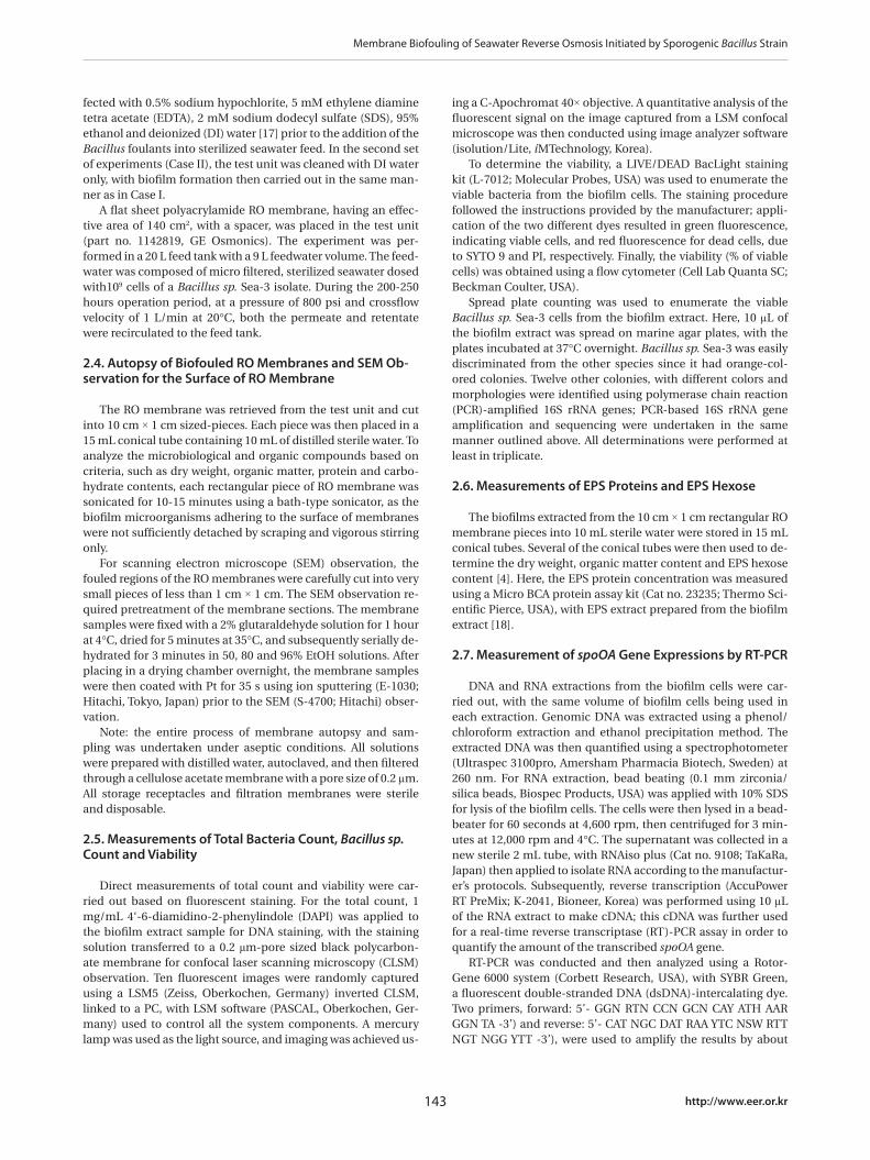
The decline in the permeate flux was determined for both Cases I and II experiments. For Case I, the membrane test unit was cleaned with various detergents and chemical disinfectants before adding the Bacillus sp. as the biofoulant; whereas, for Case II, the same test unit was only circulated with distilled wa-ter. The different cleaning methods might not have a significant influence on the development of a biofilm on the membrane surface. However, our focus in this study was on obtaining two comparable biofilms in terms of biofouling characteristics than on the experimental conditions. During crossflow filtration, the permeate flux in both cases gradually decreased, and after 272 and 208 hours, the fluxes declined by 20 and 33% in Cases I and II, respectively (Fig. 1). A minor permeate flux decline was ob-
served in the control experiment, which contained no Bacillus strain. Note: at the end of the Case II period, a relatively rapid decline of the flux rate was observed compared to Case I.
0 50 100 150 200 250
Perm
eate
Flu
x (%
)
40
60
80
100
120
Case II
Case I
Control
Operation time (hrs)
Fig. 1. Decline of the permeate fluxes in Cases I and II due to bio-film formation on the reverse osmosis membranes.
a
b
c
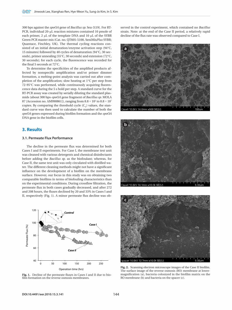
Fig. 2. Scanning electron microscope images of the Case II biofilm. The surface image of the reverse osmosis (RO) membrane at lower-magnification (a), bacteria colonized in the biofilm matrix on the RO membrane (b) and bacteria on the spacer (c).
Membrane Biofouling of Seawater Reverse Osmosis Initiated by Sporogenic Bacillus Strain
145 http://www.eer.or.kr
3.2. Bacillus Biofilm Autopsy Formed on RO Membrane
Fig. 2 presents the SEM images of the membrane surface morphology. The fouling of the membrane in Case II initially ap-pears as in a web-like form at lower magnification (a), although a closer observation of the membrane surface revealed a large amount of bacteria embedded or grown beneath the inorganic aggregates (b). Bacteria attached to the spacer surface used in the assembly of the test unit were also observed (c). However, it was hard to capture the images of bacteria present in the biofilm for Case I (data not shown). The energy-dispersive X-ray (EDX) spectra of the biofilms in Cases I and II indicated the presence of Na (1.19%), Si (2.60%), S (0.91%), C (47.46%) and O (47.83%). The composition found from the EDX analysis was relatively constant, with less than 1% fluctuations.
3.3. Measurements of Total Count, Viability, Dry Weight, Organic Matters, EPS Protein and EPS Hexose
The direct fluorescence microscopic counts for enumeration of the total count included both live and dead cells. Based on the results shown in Table 3, the difference in the total counts between the two biofilms was not significant (p > 0.05). The vi-abilities of all the bacteria in the biofilm extracts from Cases I and II were determined using flow cytometry; the percentages of viable cells were about 70% in both biofilms, but there was no statistical difference (p > 0.05). From the results of the total counts and viabilities, it was determined that both the Cases I and II biofilms had similar amounts of biomass.
In both cases, dry weights of 12-13 mg per 140 cm2 of the RO membranes were measured, with the dry masses mainly com-posed of organic matter, at 11-12 mg per 140 cm2 (90.5-90.6%). EPS protein and EPS hexose measurements were then carried out by extracting the EPS from each biofilm. Many of the bacte-rial EPS were found to possess 1, 3- or 1, 4- β-linked hexose resi-dues [20] and the most common monosaccharides in the EPS were hexose D-glucose, D-galactose and D-mannose, with pen-tose, hexosamine and glucuronic acids, as well as hexose also found in the EPS from the biofilms. The phenol-sulfuric acid method was used to quantify the carbohydrates, which mainly estimates the concentration of hexose sugars because different sugars have different absorbencies. Thus, the term ‘EPS hexose’
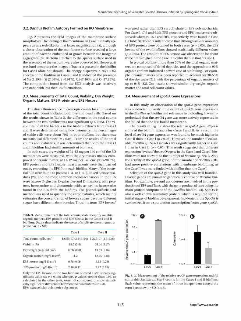
Table 3. Measurements of the total counts, viabilities, dry weights, organic matters, EPS protein and EPS hexose in the Cases I and II biofilms. Data values indicate the mean of triplicate measurements (error bar, 1 × SD)
Case I Case II
Total count (cells/cm2) 1.65E+07 (2.34E+06) 1.32E+07 (2.31E+6)
Viability (%) 69.5 (5.8) 68.04 (3.67)
Dry weight (mg/140 cm2) 12.37 (0.81) 13.53 (1.46)
Organic matter (mg/140 cm2) 11.2 12.25 (1.48)
EPS hexose (mg/140 cm2) 0.78 (0.09) 8.13 (0.73)
EPS protein (mg/140 cm2) 2.16 (0.11) 2.27 (0.18)
Only the EPS hexose in the two biofilms showed a statistically sig-nificant value (at p < 0.05); whereas, p values greater than 0.05, as calculated in the other tests, were not considered to show statisti-cally significant differences between the two biofilms (n = 3).EPS: extracellular polymeric substances.
Fig. 3. (a) Measurement of the relative spoOA gene expression and (b) culturable Bacillus sp. Sea-3 counts for the Cases I and II biofilms. Each value represents the mean of three independent assays; the error bars show 1 × SD (n = 3).
Rela
tive
sp
o0A
gen
e ex
pre
ssio
n
0.01
0.1
1
10
100
Case I Case II
Col
ony/
cm2
1e+3
1e+4
1e+5
Case I Case II
was used rather than EPS carbohydrate or EPS polysaccharide. For Case I, 17.5 and 6.3% EPS proteins and EPS hexose were ob-served; whereas, 16.7 and 60%, respectively, were found in Case II (Table 3). These results showed that although similar amounts of EPS protein were obtained in both cases (p > 0.05), the EPS hexose of the two biofilms showed statistically different values (p < 0.05). The amount of EPS hexose was observed to be about three times higher in the Case II biofilm than in that of Case I.
In typical biofilms, more than 50% of the total organic mat-ters are composed of dried deposits, and the approximate 90% organic content indicated a severe case of biofouling. For exam-ple, organic matters have been reported to account for 30-55% of the dry mass [21], with the percentage of organic matters of up to 94% [22]. Our results showed similar dry weight, organic matter and total cell count values.
3.4. Measurement of spoOA Gene Expressions
In this study, an observation of the spoOA gene expression was conducted to verify if the extent of spoOA gene expression in the Bacillus sp. biofilm had relevance to biofouling. It was hy-pothesized that the spoOA gene was more actively expressed in the fouled than the less fouled membrane.
The results in Fig. 3a show the relative spoOA gene expres-sions of the biofilm extracts for Cases I and II. As a result, the level of spoOA gene expression was found to be much higher in Case II than in Case I (p < 0.05). However, the number of cultur-able Bacillus sp. Sea-3 isolates was significantly higher in Case I than in Case II (p < 0.05). This result suggested that different expression levels of the spoOA gene in the Case I and Case II bio-films were not relevant to the number of Bacillus sp. Sea-3. Also, the activity of the spoOA gene, not the number of Bacillus cells, had more positive correlations with membrane biofouling, as the Case II was more fouled with biofilm than the Case I.
Selection of the spoOA gene in this study was well founded. Diverse genes are known to genetically control of Bacillus bio-films. For example, yve and eps operons are involved in the pro-duction of EPS and TasA, with the gene product of tasA being the main protein component of the Bacillus biofilm [23]. SpoOA is also a well known regulatory protein, which is required for the initial stages of biofilm development. Incidentally, the SpoOA is synthesized from a sporulation transcription factor gene, spoOA.
a b
Jinwook Lee, Xianghao Ren, Hye-Weon Yu, Sung-Jo Kim, In S. Kim
146DOI:10.4491/eer.2010.15.3.141
The expression of this gene is required for both sporulation and biofilm construction. It is also known that spoOA gene expres-sion is involved in cell-cell communication, because a mutant of the Bacillus strain defective in the synthesis of SpoOA protein could not form multi cellular structures in biofilms. Secondly, SpoOA negatively controls abrB activity, which has a role in sup-pressing the relevant genes involved in intercellular adhesion [14]. Third, the synthesis of TasA protein requires the transcrip-tion factor SpoOA.
In terms of the relationship between the bacterial mass of the biofilm and biofouling, the mass of an individual bacterium did not give rise to significant biofouling. For this reason, it has proved quite difficult to control biofouling by reducing the num-ber of microorganisms in water due to the unclear link between microbial communities and the membrane [24]. However, the activation of genes of interest, which indicate a functional in-clination to form the biofilm on an RO membrane, had a much more significant effect on biofouling. Studies to determine the gene expression patterns in bacterial biofilms have recently in-creased. For instance, biofilm genes essential in the initial step of adhesion may not be activated in biofilms formed later and in those with more complex structures [25]; also, EPS synthesis-related genes increase during biofilm formation; whereas, pili or flagella-related genes decrease [17].
3.5. Bacillus and Other Culturable Bacteria in Case II Biofilm
When each biofilm extract was cultured on marine agar, a yellow colony, revealing Bacillus sp. Sea-3, was only grown from in the Case I biofilm. However, other colonies were grown with a yellow-colored colony in the Case II biofilm. Identification of other bacteria was performed using 16S rRNA gene sequencing
and based on a comparison with the GenBank database using a basic local alignment search tool (BLAST) search. The isolates screened in this study had similarities ranging from 99 to 100%.
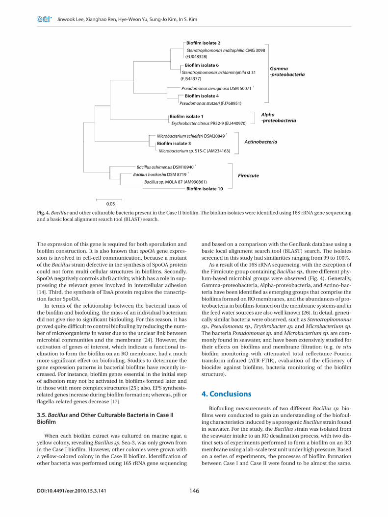
As a result of the 16S rRNA sequencing, with the exception of the Firmicute group containing Bacillus sp., three different phy-lum-based microbial groups were observed (Fig. 4). Generally, Gamma-proteobacteria, Alpha-proteobacteria, and Actino-bac-teria have been identified as emerging groups that comprise the biofilms formed on RO membranes, and the abundances of pro-teobacteria in biofilms formed on the membrane systems and in the feed water sources are also well known [26]. In detail, geneti-cally similar bacteria were observed, such as Stenotrophomonas sp., Pseudomonas sp., Erythrobacter sp. and Microbacterium sp. The bacteria Pseudomonas sp. and Microbacterium sp. are com-monly found in seawater, and have been extensively studied for their effects on biofilms and membrane filtration (e.g. in situ biofilm monitoring with attenuated total reflectance-Fourier transform infrared (ATR-FTIR), evaluation of the efficiency of biocides against biofilms, bacteria monitoring of the biofilm structure).
4. Conclusions
Biofouling measurements of two different Bacillus sp. bio-films were conducted to gain an understanding of the biofoul-ing characteristics induced by a sporogenic Bacillus strain found in seawater. For the study, the Bacillus strain was isolated from the seawater intake to an RO desalination process, with two dis-tinct sets of experiments performed to form a biofilm on an RO membrane using a lab-scale test unit under high pressure. Based on a series of experiments, the processes of biofilm formation between Case I and Case II were found to be almost the same.
Biofilm isolate 2
Stenotrophomonas maltophilia CMG 3098 (EU048328)
Biofilm isolate 6
Stenotrophomonas acidaminiphila st 31 (FJ544377)
Pseudomonas aeruginosa DSM 50071 T
Biofilm isolate 4
Pseudomonas stutzeri (FJ768951)
Biofilm isolate 1
Erythrobacter citreus PR52-9 (EU440970)
Microbacterium schleiferi DSM20849 T
Biofilm isolate 3
Microbacterium sp. S15-C (AM234163)
Bacillus oshimensis DSM18940 T
Bacillus horikoshii DSM 8719 T
Bacillus sp. MOLA 87 (AM990861)
Biofilm isolate 10
0.05
Actinobacteria
Firmicute
Alpha-proteobacteria
Gamma-proteobacteria
Fig. 4. Bacillus and other culturable bacteria present in the Case II biofilm. The biofilm isolates were identified using 16S rRNA gene sequencing and a basic local alignment search tool (BLAST) search.
Membrane Biofouling of Seawater Reverse Osmosis Initiated by Sporogenic Bacillus Strain
147 http://www.eer.or.kr
However, in Case I the membrane test unit was disinfected with various detergents and chemicals; whereas, the same test unit was only circulated with distilled water in Case II.
In Case II, more biofilm fouling was observed than in Case I due to the higher amount of EPS hexose and larger decline of the permeate flux in Case II. From the results, no significant dif-ferences were observed in the total counts, dry weights, total or-ganic matters and amounts of EPS protein between the two bio-films. However, significant differences in the EPS hexose, spoOA gene expression and number of culturable Bacillus sp. Sea-3 were observed. Higher spoOA gene expression and less number of Bacillus sp. Sea-3 were observed in the more fouled Case II biofilm. This implies that the activity of the spoOA gene was not correlated with the number of Bacillus cells, but was well cor-related with membrane biofouling.
It was noted in this study that the extent of spoOA gene ex-pression, as a biofouling indicator, was limited to the biofilm mainly induced by sporogenic Bacillus strains, since the spoOA gene was only found in Bacillus. In particular; therefore, the genes of interest in the SWRO system can be applied as targets in the diagnosis of biofouling. Finally, the bacterial species or strains showing a significant expression pattern of a particular gene are suggested as target microorganisms for the develop-ment of a monitoring system for RO processes.
Acknowledgements
This research was supported by a grant (07SeaHeroA01-01) from the Plant Technology Advancement Program, funded by the Ministry of Land, Transport and Maritime Affairs of the Ko-rean Government.
References
1. Al-Ahmad M, Abdul Aleem FA, Mutiri A, Ubaisy A. Biofuoling in RO membrane systems Part 1: Fundamentals and control. Desalination 2000;132:173-179.
2. Paul DH. Reverse osmosis: scaling, fouling and chemical at-tack. Desal. Water Reuse 1991;1:8-11.
3. Khedr MG. Membrane fouling problems in reverse osmosis desalination applications. Desal. Water Reuse 2000;10:8-17.
4. Jang N, Shon H, Ren X, Vigneswaran S, Kim IS. Characteris-tics of bio-foulants in the membrane bioreactor. Desalina-tion 2006;200:201-202.
5. Ridgway HF, Kelly A, Justice C, Olson BH. Microbial foul-ing of reverse-osmosis membranes used in advanced wastewater treatment technology: Chemical, bacteriologi-cal, and ultrastructural analyses. Appl. Environ. Microbiol. 1983;45:1066-1084.
6. Baker JS, Dudley LY. Biofouling in membrane systems - a re-view. Desalination 1998;118:81-90.
7. Sadr Ghayeni SB, Beatson PJ, Schneider RP, Fane AG. Adhe-sion of waste water bacteria to reverse osmosis membranes. J. Membr. Sci. 1998;138:29-42.
8. Flemming HC, Schaule G. Biofouling on membranes - A mi-crobiological approach. Desalination 1988;70:95-119.
9. Bereschenko LA, Heilig GHJ, Nederlof MM, Van Loosdrecht MCM, Stams AJM, Euverink GJW. Molecular characterization
of the bacterial communities in the different compartments of a full-scale reverse-osmosis water purification plant. Appl. Environ. Microbiol. 2008;74:5297-5304.
10. Chen CL, Liu WT, Chong ML, et al. Community structure of microbial biofilms associated with membrane-based water purification processes as revealed using a polyphasic ap-proach. Appl. Microbiol. Biotechnol. 2004;63:466-473.
11. Ivnitsky H, Katz I, Minz D, et al. Bacterial community com-position and structure of biofilms developing on nanofiltra-tion membranes applied to wastewater treatment. Water Res. 2007;41:3924-3935.
12. Lee J, Kim SM, Jung JY, Oh BS, Kim IS, Hong SK. Analysis of total bacteria, enteric members of γ-proteobacteria and microbial communities in seawater as indirect indicators of quantifying biofouling. Environ. Eng. Res. 2009;14:19-25.
13. Jung JY, Lee J, Kim S, Kim IS. Characteristics on the adhe-sion and the high pressure resistance of bacteria in biofilm of SWRO process. J. of KSEE 2009;31:51-57.
14. Hamon MA, Lazazzera BA. The sporulation transcription factor Spo0A is required for biofilm development in Bacillus subtilis. Mol. Microbiol. 2001;42:1199-1209.
15. Kim S, Cho D, Lee MS, Oh BS, Kim JH, Kim IS. SEAHERO R&D program and key strategies for the scale-up of a seawater re-verse osmosis (SWRO) system. Desalination 2009;238:1-9.
16. Jochem FJ. Morphology and DNA content of bacterioplank-ton in the northern Gulf of Mexico: Analysis by epifluores-cence microscopy and flow cytometry. Aquat. Microb. Ecol. 2001;25:179-194.
17. Herzberg M, Elimelech M. Physiology and genetic traits of reverse osmosis membrane biofilms: A case study with Pseu-domonas aeruginosa. ISME Journal 2008;2:180-194.
18. Bin Z, Baosheng S, Min J, Taishi G, Zhenghong G. Extrac-tion and analysis of extracellular polymeric substances in membrane fouling in submerged MBR. Desalination 2008;227:286-294.
19. Brill JA, Wiegel J. Differentiation between spore-forming and asporogenic bacteria using a PCR and Southern hybrid-ization based method. J. Microbiol. Methods 1997;31:29-36.
20. Sutherland IW. Biofilm exopolysaccharides: a strong and sticky framework. Microbiology 2001;147:3-9.
21. Schneider RP, Ferreira LM, Binder P, et al. Dynamics of or-ganic carbon and of bacterial populations in a conventional pretreatment train of a reverse osmosis unit experiencing severe biofouling. J. Membr. Sci. 2005;266:18-29.
22. Hu JY, Song LF, Ong SL, Phua ET, Ng WJ. Biofiltration pre-treatment for reverse osmosis (RO) membrane in a water reclamation system. Chemosphere 2005;59:127-133.
23. Branda SS, Chu F, Kearns DB, Losick R, Kolter R. A major protein component of the Bacillus subtilis biofilm matrix. Mol. Microbiol. 2006;59:1229-1238.
24. Flemming HC. Reverse osmosis membrane biofouling. Exp. Therm Fluid Sci. 1997;14:382-391.
25. Beloin C, Ghigo JM. Finding gene-expression patterns in bacterial biofilms. Trends Microbiol. 2005;13:16-19.
26. Lawrence JR, Swerhone GDW, Leppard GG, et al. Scanning transmission X-ray, laser scanning, and transmission elec-tron microscopy mapping of the exopolymeric matrix of microbial biofilms. Appl. Environ. Microbiol. 2003;69:5543-5554.
Related Documents


