Melatonin induces transcriptional regulation of Bim by FoxO3a in HepG2 cells S Carbajo-Pescador 1 , C Steinmetz 2 , A Kashyap 2 , S Lorenz 2 , J L Mauriz 1 , M Heise 3 , P R Galle 2 , J Gonza ´ lez-Gallego 1 and S Strand * ,2 1 Centro de Investigacio ´ n Biome ´ dica en Red de Enfermedades Hepa ´ ticas y Digestivas (CIBERehd) and Institute of Biomedicine, University of Leo ´ n, Leo ´ n, Spain; 2 Department of Internal Medicine, University Medical Center, Johannes Gutenberg University, Mainz, Germany and 3 Department of Transplantation and Hepatobiliary Surgery, University Medical Center, Johannes Gutenberg University, Mainz, Germany Background: Melatonin induces apoptosis in many different cancer cell lines, including hepatocellular carcinoma cells. However, the responsible pathways have not been clearly elucidated. A member of the forkhead transcription factors’ family, FoxO3a, has been implicated in the expression of the proapoptotic protein Bim (a Bcl-2-interacting mediator of cell death). In this study, we used human HepG2 liver cancer cells as an in vitro model to investigate whether melatonin treatment induces Bim through regulation by the transcription factor FoxO3a. Methods: Cytotoxicity of melatonin was compared in HepG2 hepatoblastoma cells and primary human hepatocytes. Proapoptotic Bim expression was analysed by reverse transcriptase–polymerase chain reaction and western blot. Reporter gene assays and chromatin immunoprecipitation assays were performed to analyse whether FoxO3a transactivates the Bim promoter. Small interfering RNA (siRNA) was used to study the role of FoxO3a in Bim expression. Immunofluorescence was performed to analyse FoxO3a localisation in HepG2 cells. Results: Melatonin treatment induces apoptosis in HepG2 cells, but not in primary human hepatocytes. The proapoptotic effect was mediated by increased expression of the BH3-only protein Bim. During melatonin treatment, we observed increased transcriptional activity of the forkhead-responsive element and could demonstrate that FoxO3a binds to a specific sequence within the Bim promoter. Furthermore, melatonin reduced phosphorylation of FoxO3a at Thr 32 and Ser 253 , and induced its increased nuclear localisation. Moreover, silencing experiments with FoxO3a siRNA prevented Bim upregulation. Conclusion: This study shows that melatonin can induce apoptosis in HepG2 hepatocarcinoma cells through the upregulation of proapoptotic Bim mediated by nuclear translocation and activation of the transcription factor FoxO3a. Each year, hepatocellular carcinoma (HCC) is diagnosed in more than half a million people worldwide, being the most common malignant hepatobiliar disease (El-Serag, 2011; Jemal et al, 2011). Viral hepatitis B and C, as well as alcohol abuse, are the main risk factors for its development (Cornella et al, 2011). Dysregulation of cellular proliferation and apoptosis are frequent events related with malignant phenotype and poor responsiveness of HCC towards chemotherapy (Mu ¨ller et al, 1997). For this reason, advances in understanding these processes are needed for developing effective pharmacological therapies for HCC. Melatonin, the hormone of the pineal gland, controls circadian rhythms, and it has been reported to exert additional functions in other organs. A large number of studies have demonstrated the protective role of melatonin in different pathophysiological situations in the liver, showing antioxidant and antiapoptotic proprieties (Pan et al, 2006; Subramanian et al, 2007; Thong-Ngam et al, 2007; Tahan et al, 2009). On the other hand, in vitro studies with different cancer cell lines have provided evidence for melatonin induction of apoptosis in tumour cells (Hill and Blask, 1988; Farriol et al, 2000; Futagami et al, 2001; Cini et al, 2005; 442 www.bjcancer.com | DOI:10.1038/bjc.2012.563 *Correspondence: Dr S Strand; E-mail: [email protected] Received 23 July 2012; revised 13 November 2012; accepted 17 November 2012; published online 20 December 2012 & 2013 Cancer Research UK. All rights reserved 0007 – 0920/13 FULL PAPER Keywords: FoxO3a; Bim; hepatocellular carcinoma; melatonin British Journal of Cancer (2013) 108, 442–449 | doi: 10.1038/bjc.2012.563

Welcome message from author
This document is posted to help you gain knowledge. Please leave a comment to let me know what you think about it! Share it to your friends and learn new things together.
Transcript
Melatonin induces transcriptional regulationof Bim by FoxO3a in HepG2 cellsS Carbajo-Pescador1, C Steinmetz2, A Kashyap2, S Lorenz2, J L Mauriz1, M Heise3, P R Galle2,J Gonzalez-Gallego1 and S Strand*,2
1Centro de Investigacion Biomedica en Red de Enfermedades Hepaticas y Digestivas (CIBERehd) and Institute of Biomedicine,University of Leon, Leon, Spain; 2Department of Internal Medicine, University Medical Center, Johannes Gutenberg University,Mainz, Germany and 3Department of Transplantation and Hepatobiliary Surgery, University Medical Center, Johannes GutenbergUniversity, Mainz, Germany
Background: Melatonin induces apoptosis in many different cancer cell lines, including hepatocellular carcinoma cells. However,the responsible pathways have not been clearly elucidated. A member of the forkhead transcription factors’ family, FoxO3a, hasbeen implicated in the expression of the proapoptotic protein Bim (a Bcl-2-interacting mediator of cell death). In this study, weused human HepG2 liver cancer cells as an in vitro model to investigate whether melatonin treatment induces Bim throughregulation by the transcription factor FoxO3a.
Methods: Cytotoxicity of melatonin was compared in HepG2 hepatoblastoma cells and primary human hepatocytes.Proapoptotic Bim expression was analysed by reverse transcriptase–polymerase chain reaction and western blot. Reporter geneassays and chromatin immunoprecipitation assays were performed to analyse whether FoxO3a transactivates the Bim promoter.Small interfering RNA (siRNA) was used to study the role of FoxO3a in Bim expression. Immunofluorescence was performed toanalyse FoxO3a localisation in HepG2 cells.
Results: Melatonin treatment induces apoptosis in HepG2 cells, but not in primary human hepatocytes. The proapoptotic effectwas mediated by increased expression of the BH3-only protein Bim. During melatonin treatment, we observed increasedtranscriptional activity of the forkhead-responsive element and could demonstrate that FoxO3a binds to a specific sequencewithin the Bim promoter. Furthermore, melatonin reduced phosphorylation of FoxO3a at Thr32 and Ser253, and induced itsincreased nuclear localisation. Moreover, silencing experiments with FoxO3a siRNA prevented Bim upregulation.
Conclusion: This study shows that melatonin can induce apoptosis in HepG2 hepatocarcinoma cells through the upregulation ofproapoptotic Bim mediated by nuclear translocation and activation of the transcription factor FoxO3a.
Each year, hepatocellular carcinoma (HCC) is diagnosed in morethan half a million people worldwide, being the most commonmalignant hepatobiliar disease (El-Serag, 2011; Jemal et al, 2011).Viral hepatitis B and C, as well as alcohol abuse, are the main riskfactors for its development (Cornella et al, 2011). Dysregulation ofcellular proliferation and apoptosis are frequent events related withmalignant phenotype and poor responsiveness of HCC towardschemotherapy (Muller et al, 1997). For this reason, advances inunderstanding these processes are needed for developing effectivepharmacological therapies for HCC.
Melatonin, the hormone of the pineal gland, controls circadianrhythms, and it has been reported to exert additional functions inother organs. A large number of studies have demonstrated theprotective role of melatonin in different pathophysiologicalsituations in the liver, showing antioxidant and antiapoptoticproprieties (Pan et al, 2006; Subramanian et al, 2007; Thong-Ngamet al, 2007; Tahan et al, 2009). On the other hand, in vitro studieswith different cancer cell lines have provided evidence formelatonin induction of apoptosis in tumour cells (Hill and Blask,1988; Farriol et al, 2000; Futagami et al, 2001; Cini et al, 2005;
442 www.bjcancer.com | DOI:10.1038/bjc.2012.563
*Correspondence: Dr S Strand; E-mail: [email protected]
Received 23 July 2012; revised 13 November 2012; accepted 17 November 2012; published online 20 December 2012
& 2013 Cancer Research UK. All rights reserved 0007 – 0920/13
FULL PAPER
Keywords: FoxO3a; Bim; hepatocellular carcinoma; melatonin
British Journal of Cancer (2013) 108, 442–449 | doi: 10.1038/bjc.2012.563
Garcia-Santos et al, 2006; Garcia-Navarro et al, 2007; Cabrera et al,2010; Chiu et al, 2010; Gonzalez et al, 2010). We have recentlyreported that melatonin administration induces cell cycle arrestand apoptosis in hepatocarcinoma HepG2 cells through MT1melatonin receptor by modulation of cAMP basal levels and ERKkinase activation (Carbajo-Pescador et al, 2009, 2011). Further-more, melatonin-induced apoptosis was related with enhancedcaspase-3 and caspase-9 activity, cytosolic cytochrome c releaseand upregulation of the proapoptotic protein Bax (Martin-Renedoet al, 2008). Nevertheless, the molecular pathways that underliemelatonin-induced apoptosis in human HCC are not fullyelucidated.
The FoxO subfamily of forkhead transcription factors (FoxO1/FKHR, FoxO3/FKHRL1 and FoxO4/AFX identified in mammals)plays an important role in tumour suppression by upregulatingtarget genes involved in cell cycle arrest and apoptosis. Interestingly,low levels of FoxO3 have been reported to confer chemotherapyresistance in human cancers, being significantly associated withpoor prognosis in cancer patients (Jin et al, 2004; Fei et al, 2009; Suet al, 2011). Moreover, enhanced activity and expression of activeforms of FoxO transcription factors is required for tumourchemosensitisation (Sunters et al, 2003; Paroni et al, 2011). FoxOproteins are activated in response to a wide range of externalstimuli. Regulation of its activity depends mainly on changes in thesubcellular localisation, achieved via post-translational modifica-tions, including phosphorylation, acetylation and ubiquitination(Calnan and Brunet, 2008). Several genetic and biochemical studiesindicate that the FoxO family is a key downstream target of thePI3K-Akt pathway in development and longevity (Lin et al, 1997;Brunet et al, 1999). Thus, phosphorylation of FoxO factors inspecific serine and/or threonine sites modulates their subcellularlocalisation (Rena et al, 2002; Barthel et al, 2005; Anton et al, 2007).Once placed in the nucleus, they play tumour suppressor rolesthrough enhanced transcription of proapoptotic genes, such asBCL6, a Bcl-2-interacting mediator of cell death (Bim), and Fasligand (Dijkers et al, 2000; Yang et al, 2006).
Bim is a proapoptotic member of the Bcl-2 family, and is one ofthe main downstream targets of FoxO3a. After transcription, BimmRNA undergoes an alternate splicing, giving three isoforms(BimS, BimL and BimEL) with different length (Ewings et al, 2007).Interestingly, there are in vivo and in vitro evidence demonstratingan essential role of Bim proteins in Bax activation (Ren et al, 2010).Based on this information, we focused this study on the FoxO3aregulation of Bim expression after treatment with pharmacologicalconcentrations of melatonin, in an attempt to gain furthermechanistic insights on the molecular pathways leading tomelatonin-induced apoptosis in HepG2 liver cancer cells.
MATERIALS AND METHODS
Cell culture. HepG2 cells were obtained from the American TypeCulture Collection (Manassas, VA, USA) and grown in Dulbecco’smodified Eagle’s medium (DMEM). Primary human hepatocyteswere isolated from healthy liver tissue of patients undergoingpartial hepatectomy by two-step collagenase perfusion. Cells wereseeded on collagen-coated culture dishes in Williams mediumsupplemented with 10% fetal bovine serum, 15 mmol l� 1 HEPES(pH 7.4), 2 mmol l� 1 glutamine, 100 U ml� 1 penicillin and100 mg ml� 1 streptomycin. LY294002 was from Tocris (Bristol,UK). Melatonin and epidermal growth factor (EGF) were obtainedfrom Sigma-Aldrich (St Louis, MO, USA).
Viability assays. HepG2 cells or primary human hepatocyte wereseeded in 96-well plates. Melatonin dissolved in dimethylsulphoxide (DMSO) was added to the cells at the concentrationsas indicated in the figures. Apoptosis was induced in HepG2 cells
with 200 ng ml� 1 of the monoclonal antibody (Ab) to human(APO-1/Fas) anti-APO-1, kindly provided by Peter H Krammer.Cell viability was determined using the CellTiter-Glo (Promega,Fitchburg, WI, USA) and 3-(4,5-Dimethythiazol-2-yl)-2,5-diphe-nyl tetrazolium bromide (MTT) assays (Sigma-Aldrich). CellTiter-Glo assay was performed according to the manufacturer’sinstructions (Promega). Luminescence was determined in aSaphire luminometer (Tecan Austria, Grodig, Austria). The MTTassay was carried out as described by Denizot and Lang (1986).Briefly, after exposure of cells to melatonin, culture media werechanged by serum-free culture media. 3-(4,5-Dimethythiazol-2-yl)-2,5-diphenyl tetrazolium bromide dissolved in phosphate-bufferedsaline (PBS) was added to each well for 3 h. After this interval, theculture media containing MTT were discarded and DMSO wasadded to each well, dissolving the precipitate. The optical densitieswere measured at 560 nm spectral wavelength using microtitreplate reader (Synergy HT Multi-Mode Microplate Reader; Bio-TekInstruments Inc., Winooski, VT, USA).
Transfection and luciferase reporter assay. Transient transfec-tion of HepG2 human hepatocytes was performed using theTransFectin reagent (Bio-Rad, Hercules, CA, USA). Constructsused were the FHRE-Luciferase reporter (Addgene plasmid 1789kindly provided by M Greenberg’s lab) (Tran et al, 2002) and theFoxO3a expression construct (Addgene plasmid 8355 kindlyprovided by A Brunet’s lab) (Brunet et al, 1999). Inducibleactivation of FoxO3a was performed through transfection of theHA-FoxO3a-WT-ER plasmid. The HA-FoxO3a-WT-ER fusionprotein is constitutively expressed but is inhibited unless exposedto a modified ligand for the oestrogen receptor (ER), 4-hydroxy-tamoxifen (4-OHT). HepG2 cells were transfected using theTransFectin reagent (Bio-Rad, Munich, Germany) with 1 mg ofHA-FoxO3a-WT-ER plasmid (Tran et al, 2002). Activation of theaccumulated FoxO3a protein was induced by treatment with theER ligand 4-OHT 1 h before melatonin treatment. The luciferasereporter activity was measured using a commercially availableluciferase assay system (Promega). Transfection efficiency wasnormalised by b-galactosidase activity.
Western blot analysis. After treatments, cultured cells werewashed two times with ice-cold PBS and lysed by adding ice-coldRIPA buffer containing 50 mM Tris-HCl (pH 7.4), 150 mM NaCl,2 mM EDTA, 0.1% Triton X-100, 10% sodium deoxycholate, 10%SDS, 1 mM NaF and protease cocktail inhibitor (Roche, Basel,Switzerland), and scraped off the plate. The extracts weretransferred to a microfuge tube and centrifuged for 10 min at15 000 g. Equal amounts of the supernatant protein (20 mg) wereseparately subjected to SDS–PAGE and transferred to a PVDFmembrane (Bio-Rad). Primary Abs were diluted in blockingsolution and incubated overnight at 4 1C with polyclonal Ab toBim (rabbit, 1 : 1000 dilution; eBioscience, San Diego, CA, USA),phospho-FoxO3a Thr32 and FoxO3a Ser253(rabbit, 1:1000 dilution;Cell Signalling Technology, Beverly, MA, USA) and FoxO3a(rabbit, 1:1000 dilution; Abcam, Cambridge, UK). Equal loading ofprotein was demonstrated by probing the membranes with a rabbitanti-b-actin polyclonal Ab (1:2000 dilution; Sigma), anti-lamin B1(H-90) (1:2000 dilution; Santa Cruz Biotechnology, Santa Cruz,CA, USA) or anti-tubulin (Sigma-Aldrich). After washing withPBST, membranes where incubated with phosphatase-conjugatedanti-rabbit secondary Ab from Sigma-Aldrich diluted in blockingsolution and incubated for 1 h at room temperature. The proteinswere detected and visualised by chemiluminescence using theCDP star detection system (Tropix Applied Biosystems,Carlsbad, CA, USA). Densitometry analysis of specific bands wasperformed by the Scion Image software (Scion Corporation,Frederick, MD, USA).
Melatonin and FoxO3a BRITISH JOURNAL OF CANCER
www.bjcancer.com | DOI:10.1038/bjc.2012.563 443
Real-time reverse transcriptase–polymerase chain reaction. Forreal-time reverse transcriptase–polymerase chain reaction (RT–PCR), confluent HepG2 cells growing in complete media werereplated in six-well culture plates, at a density of 150 000 cell perwell in a total volume of 2 ml of complete medium. Aftertreatment, total RNA was obtained by using a Trizol reagent(Applied Biosystems, Carlsbad, CA, USA) and quantified byspectrophotometry (Nanodrop 1000; Thermo Scientific, Waltham,MA, USA). The iScript cDNA Synthesis Kit (Bio-Rad) was used toreverse transcribe RNA into cDNA. Real-time PCR was performedusing the iCycler Absolute QPCR SYBR Green Mix (ABgene,Waltham, MA, USA). Bim mRNA levels were normalised to RNApolymerase II (RpII) using the 2�DCt method based on thethreshold cycle (CT) value. Primer sequences were as follows: Bim,50-AACCACTATCTCAGTGCAAT-30 and 50-GGTCTTCGGCTGCTTGGTAA-30; RPII, 50-GCACCACGTCCAATGACAT-30 and50-GTGCGGCTGCTTCCATAA-30.
Small interfering RNA transfection. HepG2 cells (0.5� 106 cellsper ml) were seeded in DMEM medium without antibioticsovernight. After washing the cells with PBS, 1 ml of media withoutantibiotics were added. Thereafter, 200 ml of Lipofectamine 2000complex was added into each plate. The cells were transfected withFoxO3a small interfering (siRNA) (FKHRL1 siRNA sc-37887) andBim siRNA (sc-29802) (Santa Cruz Biotechnology) for 48 haccording to the manufacturer’s instructions. A non-targetingsiRNA-A sc-37007 was used as a negative control. At 48 h aftertransfection, medium was replaced for complete DMEM and cellswere treated with or without melatonin.
Chromatin immunoprecipitation assays. Chromatin-immuno-precipitation (ChIP) assays were performed using chromatinimmunoprecipitation kit (Upstate Cell Signaling, Lake Placid,NY, USA) according to the manufacturer’s instructions. Samplestreated with the 1000mM melatonin concentration were immuno-precipitated with anti-FoxO3a Ab (Abcam) or rabbit IgG (Sigma).Polymerase chain reaction was performed using primers specificfor the Bim promoter: forward, 5’-CCTTCGCGAGGACCAACC-CAGTC-3’ and reverse, 5’-CCGCTCCTACGCCCAATCACTGC-3’.
Immunofluorescence. HepG2 cells were seeded in eight-wellchamber slides and then treated with melatonin as indicated.After treatment, they were fixed in 4% paraformaldehyde andstained with Abs to FoxO3a (rabbit, 1:100 dilution; Abcam). AlexaFluor 488-labelled anti-rabbit Ab (Invitrogen, Carlsbad, CA, USA)was used as a secondary Ab. Counterstaining of nuclei wasperformed with Hoechst 33342 (Invitrogen). The cells were imageddirectly in the chambers using a Zeiss LSM 710 NLO confocal laserscanning microscope and image analysis was performed using theZeiss Zen-2009 software (Carl Zeiss Microimaging GmbH, Jena,Germany).
Statistical analysis. Results are expressed as mean values±s.e.m.of the indicated number of experiments. A t-test was used todetermine differences between pairs of treatments, as indicated inResults. One-way ANOVA followed by Student–Newmann–Keulspost hoc test was used to determine differences between the meanvalues of the different treated groups. Po0.05 was consideredsignificant. Values were analysed using the statistical packageStatistica 10.0 (Statsoft Inc., Tulsa, OK, USA).
RESULTS
Effect of melatonin treatment on cell viability. Most types ofantitumour therapy result in a certain amount of damage tohealthy tissues with associated side effects. Previously, we haveshown that melatonin has oncostatic effects in HepG2 liver cancer
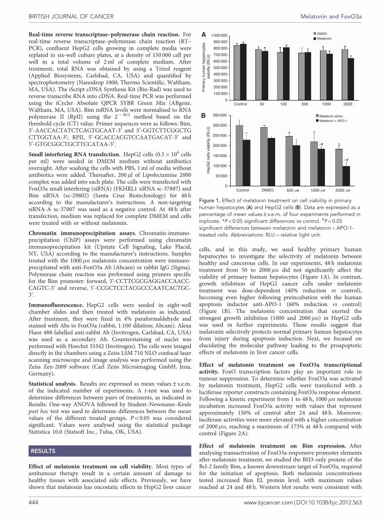
cells, and in this study, we used healthy primary humanhepatocytes to investigate the selectivity of melatonin betweenhealthy and cancerous cells. In our experiments, 48 h melatonintreatment from 50 to 2000 mM did not significantly affect theviability of primary human hepatocytes (Figure 1A). In contrast,growth inhibition of HepG2 cancer cells under melatonintreatment was dose-dependent (40% reduction vs control),becoming even higher following preincubation with the humanapoptosis inductor anti-APO-1 (60% reduction vs control)(Figure 1B). The melatonin concentration that exerted thestrongest growth inhibition (1000 and 2000mM) in HepG2 cellswas used in further experiments. These results suggest thatmelatonin selectively protects normal primary human hepatocytesfrom injury during apoptosis induction. Next, we focused onelucidating the molecular pathway leading to the proapoptoticeffects of melatonin in liver cancer cells.
Effect of melatonin treatment on FoxO3a transcriptionalactivity. FoxO transcription factors play an important role intumour suppression. To determine whether FoxO3a was activatedby melatonin treatment, HepG2 cells were transfected with aluciferase reporter constructs containing FoxO3a response element.Following a kinetic experiment from 1 to 48 h, 1000 mM melatoninincubation increased FoxO3a activity with values that representapproximately 150% of control after 24 and 48 h. Moreover,luciferase activities were more elevated with a higher concentrationof 2000 mM, reaching a maximum of 173% at 48 h compared withcontrol (Figure 2A).
Effect of melatonin treatment on Bim expression. Afteranalysing transactivation of FoxO3a-responsive promoter elementsafter melatonin treatment, we studied the BH3-only protein of theBcl-2 family Bim, a known downstream target of FoxO3a, requiredfor the initiation of apoptosis. Both melatonin concentrationstested increased Bim EL protein level, with maximum valuesreached at 24 and 48 h. Western blot results were consistent with
1000 000
900 000
DMSO
Melatonin
Melatonin aloneMelatonin + APO-1
*
**#
*#
800 000
700 000
600 000
500 000
400 000
300 000
Prim
ary
hum
an h
epat
ocyt
es v
iabi
lity
(RLU
)H
epG
2 ce
lls v
iabi
lity
(RLU
)
200 000
100 000
0Control
Control DMSO
50 100 500 1000 2000
500 �M 1000 �M 2000 �M0
50 000
100 000
150 000
200 000
250 000
300 000
350 000
Figure 1. Effect of melatonin treatment on cell viability in primaryhuman hepatocytes (A) and HepG2 cells (B). Data are expressed as apercentage of mean values±s.e.m. of four experiments performed intriplicate. *Po0.05 significant differences vs control. #Po0.05significant differences between melatonin and melatoninþAPO-1-treated cells. Abbreviations: RLU¼ relative light unit.
BRITISH JOURNAL OF CANCER Melatonin and FoxO3a
444 www.bjcancer.com | DOI:10.1038/bjc.2012.563
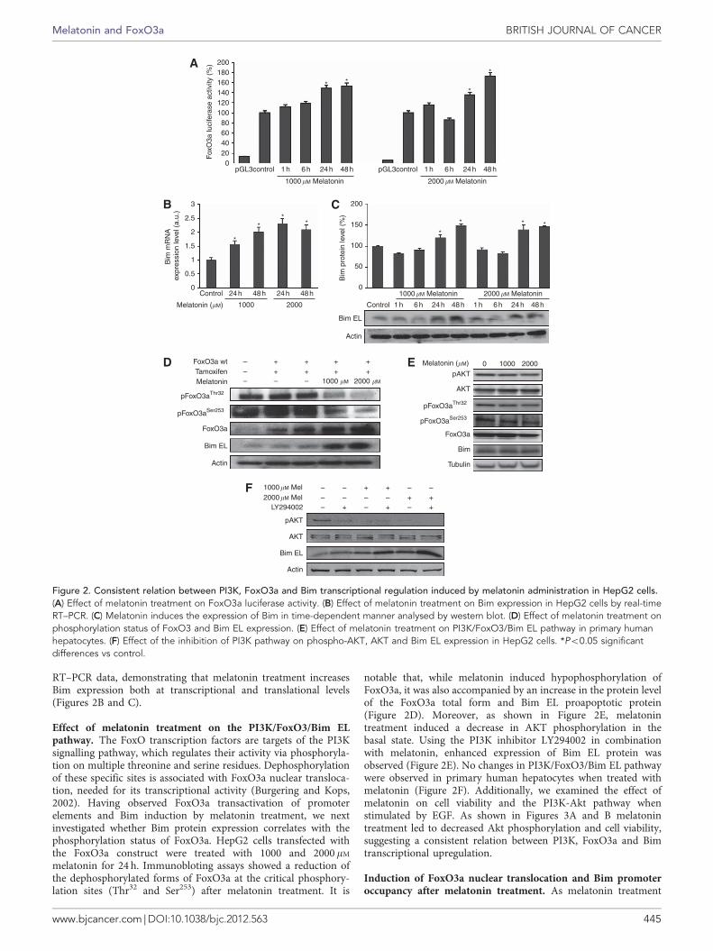
RT–PCR data, demonstrating that melatonin treatment increasesBim expression both at transcriptional and translational levels(Figures 2B and C).
Effect of melatonin treatment on the PI3K/FoxO3/Bim ELpathway. The FoxO transcription factors are targets of the PI3Ksignalling pathway, which regulates their activity via phosphoryla-tion on multiple threonine and serine residues. Dephosphorylationof these specific sites is associated with FoxO3a nuclear transloca-tion, needed for its transcriptional activity (Burgering and Kops,2002). Having observed FoxO3a transactivation of promoterelements and Bim induction by melatonin treatment, we nextinvestigated whether Bim protein expression correlates with thephosphorylation status of FoxO3a. HepG2 cells transfected withthe FoxO3a construct were treated with 1000 and 2000 mM
melatonin for 24 h. Immunobloting assays showed a reduction ofthe dephosphorylated forms of FoxO3a at the critical phosphory-lation sites (Thr32 and Ser253) after melatonin treatment. It is
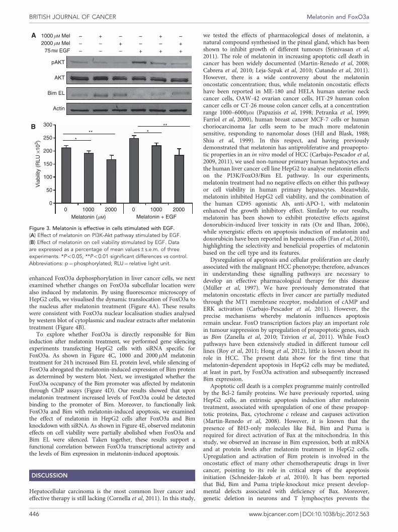
notable that, while melatonin induced hypophosphorylation ofFoxO3a, it was also accompanied by an increase in the protein levelof the FoxO3a total form and Bim EL proapoptotic protein(Figure 2D). Moreover, as shown in Figure 2E, melatonintreatment induced a decrease in AKT phosphorylation in thebasal state. Using the PI3K inhibitor LY294002 in combinationwith melatonin, enhanced expression of Bim EL protein wasobserved (Figure 2E). No changes in PI3K/FoxO3/Bim EL pathwaywere observed in primary human hepatocytes when treated withmelatonin (Figure 2F). Additionally, we examined the effect ofmelatonin on cell viability and the PI3K-Akt pathway whenstimulated by EGF. As shown in Figures 3A and B melatonintreatment led to decreased Akt phosphorylation and cell viability,suggesting a consistent relation between PI3K, FoxO3a and Bimtranscriptional upregulation.
Induction of FoxO3a nuclear translocation and Bim promoteroccupancy after melatonin treatment. As melatonin treatment
200
200
180160140
Fox
O3a
luci
fera
se a
ctiv
ity (
%)
Bim
mR
NA
exp
ress
ion
leve
l (a.
u.)
120100806040200
0
50
100
150*
*
*
***
* *
Bim
pro
tein
leve
l (%
)
pGL3control 1 h 6 h 24 h 48 h
1000�M Melatonin
1000�M Melatonin
pGL3control 1 h
1 h
6 h
6 h
24 h
24 h
48 h
48 h 1 h 6 h 24 h 48 h
2000�M Melatonin
2000�M Melatonin0
Control
Control
FoxO3a wt –––
+ ++ +
1000 �M 2000 �M
1000�M Mel2000�M Mel
LY294002
+ +++
– –
––
–– –
––
–
–+
+ +
++ +
+–
TamoxifenMelatonin
pFoxO3aThr32
AKT
pAKT
pFoxO3aSer253
FoxO3a
Bim EL
Bim EL
Bim EL
Actin
Actin
Actin
–
Melatonin (�M) 0 1000 2000
pFoxO3aThr32
AKT
pAKT
pFoxO3aSer253
FoxO3a
Bim
Tubulin
Melatonin (�M)
0.5
1
1.5
2*
**
*2.5
3
24 h
1000 2000
24 h48 h 48 h
Figure 2. Consistent relation between PI3K, FoxO3a and Bim transcriptional regulation induced by melatonin administration in HepG2 cells.(A) Effect of melatonin treatment on FoxO3a luciferase activity. (B) Effect of melatonin treatment on Bim expression in HepG2 cells by real-timeRT–PCR. (C) Melatonin induces the expression of Bim in time-dependent manner analysed by western blot. (D) Effect of melatonin treatment onphosphorylation status of FoxO3 and Bim EL expression. (E) Effect of melatonin treatment on PI3K/FoxO3/Bim EL pathway in primary humanhepatocytes. (F) Effect of the inhibition of PI3K pathway on phospho-AKT, AKT and Bim EL expression in HepG2 cells. *Po0.05 significantdifferences vs control.
Melatonin and FoxO3a BRITISH JOURNAL OF CANCER
www.bjcancer.com | DOI:10.1038/bjc.2012.563 445
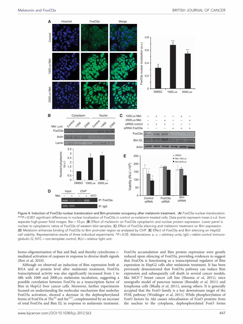
enhanced FoxO3a dephosphorylation in liver cancer cells, we nextexamined whether changes on FoxO3a subcellular location werealso induced by melatonin. By using fluorescence microscopy ofHepG2 cells, we visualised the dynamic translocation of FoxO3a tothe nucleus after melatonin treatment (Figure 4A). These resultswere consistent with FoxO3a nuclear localisation studies analysedby western blot of cytoplasmic and nuclear extracts after melatonintreatment (Figure 4B).
To explore whether FoxO3a is directly responsible for Biminduction after melatonin treatment, we performed gene silencingexperiments transfecting HepG2 cells with siRNA specific forFoxO3a. As shown in Figure 4C, 1000 and 2000mM melatonintreatment for 24 h increased Bim EL protein level, while silencing ofFoxO3a abrogated the melatonin-induced expression of Bim proteinas determined by western blot. Next, we investigated whether theFoxO3a occupancy of the Bim promoter was affected by melatoninthrough ChIP assays (Figure 4D). Our results showed that uponmelatonin treatment increased levels of FoxO3a could be detectedbinding to the promoter of Bim. Moreover, to functionally linkFoxO3a and Bim with melatonin-induced apoptosis, we examinedthe effect of melatonin in HepG2 cells after FoxO3a and Bimknockdown with siRNA. As shown in Figure 4E, observed melatonineffects on cell viability were partially abolished when FoxO3a andBim EL were silenced. Taken together, these results support afunctional correlation between FoxO3a transcriptional activity andthe levels of Bim expression in melatonin-induced apoptosis.
DISCUSSION
Hepatocellular carcinoma is the most common liver cancer andeffective therapy is still lacking (Cornella et al, 2011). In this study,
we tested the effects of pharmacological doses of melatonin, anatural compound synthesised in the pineal gland, which has beenshown to inhibit growth of different tumours (Srinivasan et al,2011). The role of melatonin in increasing apoptotic cell death incancer has been widely documented (Martin-Renedo et al, 2008;Cabrera et al, 2010; Leja-Szpak et al, 2010; Cutando et al, 2011).However, there is a wide controversy about the melatoninoncostatic concentration; thus, while melatonin oncostatic effectshave been reported in ME-180 and HELA human uterine neckcancer cells, OAW-42 ovarian cancer cells, HT-29 human coloncancer cells or CT-26 mouse colon cancer cells, at a concentrationrange 1000–6000mM (Papazisis et al, 1998; Petranka et al, 1999;Farriol et al, 2000), human breast cancer MCF-7 cells or humanchoriocarcinoma Jar cells seem to be much more melatoninsensitive, responding to nanomolar doses (Hill and Blask, 1988;Shiu et al, 1999). In this respect, and having previouslydemonstrated that melatonin has antiproliferative and proapopto-tic properties in an in vitro model of HCC (Carbajo-Pescador et al,2009, 2011), we used non-tumour primary human hepatocytes andthe human liver cancer cell line HepG2 to analyse melatonin effectson the PI3K/FoxO3/Bim EL pathway. In our experiments,melatonin treatment had no negative effects on either this pathwayor cell viability in human primary hepatocytes. Meanwhile,melatonin inhibited HepG2 cell viability, and the combination ofthe human CD95 agonistic Ab, anti-APO-1, with melatoninenhanced the growth inhibitory effect. Similarly to our results,melatonin has been shown to exhibit protective effects againstdoxorubicin-induced liver toxicity in rats (Oz and Ilhan, 2006),while synergistic effects on apoptosis induction of melatonin anddoxorubicin have been reported in hepatoma cells (Fan et al, 2010),highlighting the selectivity and beneficial properties of melatoninbased on the cell type and its features.
Dysregulation of apoptosis and cellular proliferation are clearlyassociated with the malignant HCC phenotype; therefore, advancesin understanding these signalling pathways are necessary todevelop an effective pharmacological therapy for this disease(Muller et al, 1997). We have previously demonstrated thatmelatonin oncostatic effects in liver cancer are partially mediatedthrough the MT1 membrane receptor, modulation of cAMP andERK activation (Carbajo-Pescador et al, 2011). However, theprecise mechanisms whereby melatonin influences apoptosisremain unclear. FoxO transcription factors play an important rolein tumour suppression by upregulation of proapoptotic genes, suchas Bim (Zanella et al, 2010; Tzivion et al, 2011). While FoxOpathways have been extensively studied in different tumour celllines (Roy et al, 2011; Hong et al, 2012), little is known about itsrole in HCC. The present data show for the first time thatmelatonin-dependent apoptosis in HepG2 cells may be mediated,at least in part, by FoxO3a activation and subsequently increasedBim expression.
Apoptotic cell death is a complex programme mainly controlledby the Bcl-2 family proteins. We have previously reported, usingHepG2 cells, an extrinsic apoptosis induction after melatonintreatment, associated with upregulation of one of these proapop-totic proteins, Bax, cytochrome c release and caspases activation(Martin-Renedo et al, 2008). However, it is known that thepresence of BH3-only molecules like Bid, Bim and Puma isrequired for direct activation of Bax at the mitochondria. In thisstudy, we observed an increase in Bim expression, both at mRNAand at protein levels after melatonin treatment in HepG2 cells.Upregulation and activation of Bim protein is involved in theoncostatic effect of many other chemotherapeutic drugs in livercancer, pointing to its role in critical steps of the apoptosisinitiation (Schneider-Jakob et al, 2010). It has been reportedthat Bid, Bim and Puma triple-knockout mice present develop-mental defects associated with deficiency of Bax. Moreover,genetic deletion in neurons and T lymphocytes prevents the
1000�M Mel ––– –
––
–––
––+
++ +
+++
2000�M Mel75 nM EGF
pAKT
AKT
Bim EL
Actin
300
250
200
150
1000 2000Melatonin (�M) Melatonin + EGF
****
**
100
50
Via
bilit
y (R
LU ×
103 )
00 1000 20000
Figure 3. Melatonin is effective in cells stimulated with EGF.(A) Effect of melatonin on PI3K-Akt pathway stimulated by EGF.(B) Effect of melatonin on cell viability stimulated by EGF. Dataare expressed as a percentage of mean values±s.e.m. of threeexperiments. *Po0.05, **Po0.01 significant differences vs control.Abbreviations: p¼phosphorylated; RLU¼ relative light unit.
BRITISH JOURNAL OF CANCER Melatonin and FoxO3a
446 www.bjcancer.com | DOI:10.1038/bjc.2012.563
homo-oligomerisation of Bax and Bad, and thereby cytochrome c-mediated activation of caspases in response to diverse death signals(Ren et al, 2010).
Although we observed an induction of Bim expression both atRNA and at protein level after melatonin treatment, FoxO3atranscriptional activity was also significantly increased from 1 to48h with 1000 and 2000mM melatonin incubation, suggesting apossible correlation between FoxO3a as a transcription factor ofBim in HepG2 liver cancer cells. Moreover, further experimentsfocused on understanding the molecular mechanism that underlayFoxO3a activation, showed a decrease in the dephosphorylatedforms of FoxO3a at Thr32 and Ser253, complemented by an increaseof total FoxO3a and Bim EL in response to melatonin treatment.
FoxO3a accumulation and Bim protein expression were greatlyreduced upon silencing of FoxO3a, providing evidences to suggestthat FoxO3a is functioning as a transcriptional regulator of Bimexpression in HepG2 cells after melatonin treatment. It has beenpreviously demonstrated that FoxO3a pathway can induce Bimexpression and subsequently cell death in several cancer models,like MCF-7 breast cancer cell line (Sharma et al, 2011), micexenografts model of pancreas tumour (Boreddy et al, 2011) andlymphoma cells (Bhalla et al, 2011), among others. It is generallyaccepted that the FoxO family is a key downstream target of thePI3K pathway (Weidinger et al, 2011). While phosphorylation ofFoxO factors by Akt causes relocalisation of FoxO proteins fromthe nucleus to the cytoplasm, dephosphorylated FoxO forms
Hoechst
Con
trol
1000
� M M
el.
2000
�M M
el.
FoxO3a Merge
0.8
0.6
0.4
0.2
0DMSO 1000�M 2000�M
***
***
Cytoplasm
Mel. (�M)
FoxO3a
Actin
Lamin
Contro
l
1000
2000
Contro
l
1000
2000
Nuclei
0
Input
Mel. – –+ + NTC
IP: IgG
ChIP
* *
0.5
1
1.5
2
Nuc
lear
/cyt
opla
smic
(a.u
.)
2.5
3.5
3
DMSO 1000�M 2000�M
1000�M Mel. – –
––
– – ––– –
– –
––+ +
+
+++++
++2000�M Mel.siRNA control
siRNA FoxO3a
FoxO3a
Bim EL
Actin
DMSO
Mel 1000�M
Mel 2000�M
*
*
200
250
0
50
100
150
Via
bilit
y (R
LU ×
103 )
ControlsiRNA
BimsiRNA
Fox
O3a
nuc
lear
loca
lisat
ion
(a.u
.)
IP: FoxO3aFoxO3asiRNA
Figure 4. Induction of FoxO3a nuclear translocation and Bim promoter occupancy after melatonin treatment. (A) FoxO3a nuclear translocation.***Po0.001 significant differences in nuclear localisation of FoxO3a in control vs melatonin-treated cells. Data points represent mean±s.d. fromseparate high-power field images. Bar¼10mM. (B) Effect of melatonin on FoxO3a cytoplasmic and nuclear protein expression. Lower panel isnuclear to cytoplasmic ratios of FoxO3a of western blot samples. (C) Effect of FoxO3a silencing and melatonin treatment on Bim expression.(D) Melatonin enhances binding of FoxO3a to Bim promoter region as analysed by ChIP. (E) Effect of FoxO3a and Bim silencing on HepG2cell viability. Representative results of three individual experiments. *Po0.05. Abbreviations: a. u.¼ arbitrary units; IgG¼ rabbit control immuno-globulin G; NTC¼non-template control; RLU¼ relative light unit.
Melatonin and FoxO3a BRITISH JOURNAL OF CANCER
www.bjcancer.com | DOI:10.1038/bjc.2012.563 447
activate target genes (Hong et al, 2012). Therefore, our workprovides clear evidence of melatonin-induced activation of FoxO3in cells, promoting changes in its phosphorylation status. More-over, translocation of FoxO3a to the nucleus was also confirmed byfluorescence microscopy experiments as well as by FoxO3a westernblot in nuclear and cytoplasmic extracts. In this study, melatonincaused an inhibition of AKT phosphorylation even after EGFstimulation, and the PI3K inhibitor LY294002, combined withmelatonin, resulted in a synergic effect enhancing Bim proteinexpression. Our knowledge of the mechanisms by which melatonininduces apoptosis in human HepG2 hepatoma cells are limited;however, the FoxO3a/Bim pathway has been shown to participatein apoptotic processes in response to other chemotherapeuticagents like cisplatin (Fernandez de Mattos et al, 2008; Yuan et al,2011). Moreover, resveratrol, another antioxidant molecule, hasbeen reported to behave as melatonin, exerting an oncostatic andproapoptotic activity in different tumour cells, including HepG2(Hsieh et al, 2005; Notas et al, 2006). Although little is knownabout the resveratrol effect on the FoxO3a pathway, several groupshave reported FoxO3a dephosphorylation, nuclear translocationand Bim induction after resveratrol treatment in in vitro cancermodels (Chen et al, 2010; Roy et al, 2011), helping us to supportour hypothesis.
Our study provides important information regarding themechanisms by which melatonin regulates apoptosis through theactivation of FoxO transcription factors. Taken together, all theseresults demonstrate that Bim plays a significant role in melatonin-induced apoptosis in HepG2 liver cancer cells, most likely throughthe activity of FoxO3a. Thereby, while this work could represent asignificant advance for the understanding of the melatoninoncostatic pathway in vitro, further in vivo experiments arerequired to bridge the gap between clinical applications and toinvestigate whether this indol could be safely used as a therapeuticdrug in HCC treatment, perhaps as an adjuvant.
ACKNOWLEDGEMENTS
Sara Carbajo-Pescador is granted by the Consejerıa de Educacion(Junta de Castilla y Leon, Spain) and Fondo Social Europeo.CIBERehd is funded by Instituto de Salud Carlos III. This workhas been partially supported by Junta de Castilla y Leon (ref.LE117A11-2), and Fundacion Investigacion Sanitaria en Leon andthe Forschungszentrum Immunologie (FZI), Mainz.
CONFLICT OF INTEREST
The authors declare no conflict of interest.
REFERENCES
Anton S, Melville L, Rena G (2007) Epigallocatechin gallate (EGCG) mimicsinsulin action on the transcription factor FOXO1a and elicits cellularresponses in the presence and absence of insulin. Cell Signal 19: 378–383.
Barthel A, Schmoll D, Unterman TG (2005) FoxO proteins in insulin actionand metabolism. Trends Endocrinol Metab 16: 183–189.
Bhalla S, Evens AM, Dai B, Prachand S, Gordon LI, Gartenhaus RB (2011)The novel anti-MEK small molecule AZD6244 induces BIM-dependentand AKT-independent apoptosis in diffuse large B-cell lymphoma. Blood118: 1052–1061.
Boreddy SR, Pramanik KC, Srivastava SK (2011) Pancreatic tumoursuppression by benzyl isothiocyanate is associated with inhibition of PI3K/AKT/FOXO pathway. Clin Cancer Res 17: 1784–1795.
Brunet A, Bonni A, Zigmond MJ, Lin MZ, Juo P, Hu LS, Anderson MJ, ArdenKC, Blenis J, Greenberg ME (1999) Akt promotes cell survival by
phosphorylating and inhibiting a forkhead transcription factor. Cell 96:857–868.
Burgering BM, Kops GJ (2002) Cell cycle and death control: long liveforkheads. Trends Biochem Sci 27: 352–360.
Cabrera J, Negrin G, Estevez F, Loro J, Reiter RJ, Quintana J (2010) Melatonindecreases cell proliferation and induces melanogenesis in humanmelanoma SK-MEL-1 cells. J Pineal Res 49: 45–54.
Calnan DR, Brunet A (2008) The FoxO code. Oncogene 27: 2276–2288.Carbajo-Pescador S, Garcia-Palomo A, Martin-Renedo J, Piva M, Gonzalez-
Gallego J, Mauriz JL (2011) Melatonin modulation of intracellularsignaling pathways in hepatocarcinoma HepG2 cell line: role of the MT1receptor. J Pineal Res 51: 463–471.
Carbajo-Pescador S, Martin-Renedo J, Garcia-Palomo A, Tunon MJ, MaurizJL, Gonzalez-Gallego J (2009) Changes in the expression of melatoninreceptors induced by melatonin treatment in hepatocarcinoma HepG2cells. J Pineal Res 47: 330–338.
Chen Q, Ganapathy S, Singh KP, Shankar S, Srivastava RK (2010) Resveratrolinduces growth arrest and apoptosis through activation of FOXOtranscription factors in prostate cancer cells. PLoS One 5: e15288.
Chiu CC, Chen JY, Lin KL, Huang CJ, Lee JC, Chen BH, Chen WY, Lo YH,Chen YL, Tseng CH, Chen YL, Lin SR (2010) p38 MAPK and NF-kappaBpathways are involved in naphtho[1,2-b] furan-4,5-dione induced anti-proliferation and apoptosis of human hepatoma cells. Cancer Lett 295: 92–99.
Cini G, Neri B, Pacini A, Cesati V, Sassoli C, Quattrone S, D’Apolito M, FazioA, Scapagnini G, Provenzani A, Quattrone A (2005) Antiproliferativeactivity of melatonin by transcriptional inhibition of cyclin D1 expression:a molecular basis for melatonin-induced oncostatic effects. J Pineal Res 39:12–20.
Cornella H, Alsinet C, Villanueva A (2011) Molecular Pathogenesis ofHepatocellular Carcinoma. Alcohol Clin Exp Res 35: 821–825.
Cutando A, Aneiros-Fernandez J, Aneiros-Cachaza J, Arias-Santiago S (2011)Melatonin and cancer: current knowledge and its application to oral cavitytumours. J Oral Pathol Med 40: 593–597.
Denizot F, Lang R (1986) Rapid colorimetric assay for cell growth andsurvival. Modifications to the tetrazolium dye procedure giving improvedsensitivity and reliability. J Immunol Methods 89: 271–277.
Dijkers PF, Medema RH, Lammers JW, Koenderman L, Coffer PJ (2000)Expression of the pro-apoptotic Bcl-2 family member Bim is regulated bythe forkhead transcription factor FKHR-L1. Curr Biol 10: 1201–1204.
El-Serag HB (2011) Hepatocellular carcinoma. N Engl J Med 365: 1118–1127.Ewings KE, Wiggins CM, Cook SJ (2007) Bim and the pro-survival Bcl-2
proteins: opposites attract, ERK repels. Cell Cycle 6: 2236–2240.Fan LL, Sun GP, Wei W, Wang ZG, Ge L, Fu WZ, Wang H (2010) Melatonin
and doxorubicin synergistically induce cell apoptosis in human hepatomacell lines. World J Gastroenterol 16: 1473–1481.
Farriol M, Venereo Y, Orta X, Castellanos JM, Segovia-Silvestre T (2000)In vitro effects of melatonin on cell proliferation in a colonadenocarcinoma line. J Appl Toxicol 20: 21–24.
Fei M, Zhao Y, Wang Y, Lu M, Cheng C, Huang X, Zhang D, Lu J, He S, ShenA (2009) Low expression of Foxo3a is associated with poor prognosisin ovarian cancer patients. Cancer Invest 27: 52–59.
Fernandez de Mattos S, Villalonga P, Clardy J, Lam EW (2008) FOXO3amediates the cytotoxic effects of cisplatin in colon cancer cells. Mol CancerTher 7: 3237–3246.
Futagami M, Sato S, Sakamoto T, Yokoyama Y, Saito Y (2001) Effects ofmelatonin on the proliferation and cis-diamminedichloroplatinum(CDDP) sensitivity of cultured human ovarian cancer cells. Gynecol Oncol82: 544–549.
Garcia-Navarro A, Gonzalez-Puga C, Escames G, Lopez LC, Lopez A, Lopez-Cantarero M, Camacho E, Espinosa A, Gallo MA, Acuna-Castroviejo D(2007) Cellular mechanisms involved in the melatonin inhibitionof HT-29 human colon cancer cell proliferation in culture. J Pineal Res 43:195–205.
Garcia-Santos G, Antolin I, Herrera F, Martin V, Rodriguez-Blanco J, del PilarCarrera M, Rodriguez C (2006) Melatonin induces apoptosis in humanneuroblastoma cancer cells. J Pineal Res 41: 130–135.
Gonzalez A, Del Castillo-Vaquero A, Miro-Moran A, Tapia JA, Salido GM(2010) Melatonin reduces pancreatic tumour cell viability by alteringmitochondrial physiology. J Pineal Res 50: 250–260.
Hill SM, Blask DE (1988) Effects of the pineal hormone melatonin on theproliferation and morphological characteristics of human breast cancercells (MCF-7) in culture. Cancer Res 48: 6121–6126.
BRITISH JOURNAL OF CANCER Melatonin and FoxO3a
448 www.bjcancer.com | DOI:10.1038/bjc.2012.563
Hong ZY, Lee HJ, Shin DY, Kim SK, Seo M, Lee EJ (2012) Inhibition of Akt/FOXO3a signaling by constitutively active FOXO3a suppresses growth offollicular thyroid cancer cell lines. Cancer Lett 314: 34–40.
Hsieh TC, Wang Z, Hamby CV, Wu JM (2005) Inhibition of melanoma cellproliferation by resveratrol is correlated with upregulation of quinonereductase 2 and p53. Biochem Biophys Res Commun 334: 223–230.
Jemal A, Bray F, Center MM, Ferlay J, Ward E, Forman D (2011) Globalcancer statistics. CA Cancer J Clin 61: 69–90.
Jin GS, Kondo E, Miyake T, Shibata M, Takashima T, Liu YX, Hayashi K,Akagi T, Yoshino T (2004) Expression and intracellular localization ofFKHRL1 in mammary gland neoplasms. Acta Med Okayama 58:197–205.
Leja-Szpak A, Jaworek J, Pierzchalski P, Reiter RJ (2010) Melatonin inducespro-apoptotic signaling pathway in human pancreatic carcinoma cells(PANC-1). J Pineal Res 49: 248–255.
Lin K, Dorman JB, Rodan A, Kenyon C (1997) Daf-16: An HNF-3/forkheadfamily member that can function to double the life-span of Caenorhabditiselegans. Science 278: 1319–1322.
Martin-Renedo J, Mauriz JL, Jorquera F, Ruiz-Andres O, Gonzalez P,Gonzalez-Gallego J (2008) Melatonin induces cell cycle arrest andapoptosis in hepatocarcinoma HepG2 cell line. J Pineal Res 45:532–540.
Muller M, Strand S, Hug H, Heinemann EM, Walczak H, Hofmann WJ,Stremmel W, Krammer PH, Galle PR (1997) Drug-induced apoptosis inhepatoma cells is mediated by the CD95 (APO-1/Fas) receptor/ligandsystem and involves activation of wild-type p53. J Clin Invest 99: 403–413.
Notas G, Nifli AP, Kampa M, Vercauteren J, Kouroumalis E, Castanas E(2006) Resveratrol exerts its antiproliferative effect on HepG2hepatocellular carcinoma cells, by inducing cell cycle arrest, and NOSactivation. Biochim Biophys Acta 1760: 1657–1666.
Oz E, Ilhan MN (2006) Effects of melatonin in reducing the toxic effects ofdoxorubicin. Mol Cell Biochem 286: 11–15.
Pan M, Song YL, Xu JM, Gan HZ (2006) Melatonin ameliorates nonalcoholicfatty liver induced by high-fat diet in rats. J Pineal Res 41: 79–84.
Papazisis KT, Kouretas D, Geromichalos GD, Sivridis E, Tsekreli OK,Dimitriadis KA, Kortsaris AH (1998) Effects of melatonin on proliferationof cancer cell lines. J Pineal Res 25: 211–218.
Paroni G, Fratelli M, Gardini G, Bassano C, Flora M, Zanetti A, Guarnaccia V,Ubezio P, Centritto F, Terao M, Garattini E (2011) Synergistic antitumoractivity of lapatinib and retinoids on a novel subtype of breast cancer withcoamplification of ERBB2 and RARA. Oncogene.
Petranka J, Baldwin W, Biermann J, Jayadev S, Barrett JC, Murphy E (1999)The oncostatic action of melatonin in an ovarian carcinoma cell line.J Pineal Res 26: 129–136.
Ren D, Tu HC, Kim H, Wang GX, Bean GR, Takeuchi O, Jeffers JR, ZambettiGP, Hsieh JJ, Cheng EH (2010) BID, BIM, and PUMA are essential foractivation of the BAX- and BAK-dependent cell death program. Science330: 1390–1393.
Rena G, Woods YL, Prescott AR, Peggie M, Unterman TG, Williams MR,Cohen P (2002) Two novel phosphorylation sites on FKHR that arecritical for its nuclear exclusion. EMBO J 21: 2263–2271.
Roy SK, Chen Q, Fu J, Shankar S, Srivastava RK (2011) Resveratrolinhibits growth of orthotopic pancreatic tumours through activation ofFOXO transcription factors. PLoS One 6: e25166.
Schneider-Jakob S, Corazza N, Badmann A, Sidler D, Stuber-Roos R, KeoghA, Frese S, Tschan M, Brunner T (2010) Synergistic induction of cell death
in liver tumour cells by TRAIL and chemotherapeutic drugs via the BH3-only proteins Bim and Bid. Cell Death Dis 1: e86.
Sharma G, Kar S, Palit S, Das PK (2011) 18Beta-glycyrrhetinic acid inducesapoptosis through modulation of Akt/FOXO3a/Bim pathway in humanbreast cancer MCF-7 cells. J Cell Physiol 227: 1923–1931.
Shiu SY, Li L, Xu JN, Pang CS, Wong JT, Pang SF (1999) Melatonin-inducedinhibition of proliferation and G1/S cell cycle transition delay of humanchoriocarcinoma JAr cells: possible involvement of MT2 (MEL1B)receptor. J Pineal Res 27: 183–192.
Srinivasan V, Pandi-Perumal SR, Brzezinski A, Bhatnagar KP, Cardinali DP(2011) Melatonin, immune function and cancer. Recent Pat Endocr MetabImmune Drug Discov 5: 109–123.
Su JL, Cheng X, Yamaguchi H, Chang YW, Hou CF, Lee DF, Ko HW, HuaKT, Wang YN, Hsiao M, Chen PB, Hsu JM, Bast Jr RC, Hortobagyi GN,Hung MC (2011) FOXO3a-dependent mechanism of E1A-inducedchemosensitization. Cancer Res 71: 6878–6887.
Subramanian P, Mirunalini S, Pandi-Perumal SR, Trakht I, Cardinali DP(2007) Melatonin treatment improves the antioxidant status and decreaseslipid content in brain and liver of rats. Eur J Pharmacol 571: 116–119.
Sunters A, Fernandez de Mattos S, Stahl M, Brosens JJ, Zoumpoulidou G,Saunders CA, Coffer PJ, Medema RH, Coombes RC, Lam EW (2003)FoxO3a transcriptional regulation of Bim controls apoptosis in paclitaxel-treated breast cancer cell lines. J Biol Chem 278: 49795–49805.
Tahan V, Atug O, Akin H, Eren F, Tahan G, Tarcin O, Uzun H, Ozdogan O,Tarcin O, Imeryuz N, Ozguner F, Celikel C, Avsar E, Tozun N (2009)Melatonin ameliorates methionine- and choline-deficient diet-inducednonalcoholic steatohepatitis in rats. J Pineal Res 46: 401–407.
Thong-Ngam D, Samuhasaneeto S, Kulaputana O, Klaikeaw N (2007) N-acetylcysteine attenuates oxidative stress and liver pathology in rats withnon-alcoholic steatohepatitis. World J Gastroenterol 13: 5127–5132.
Tran H, Brunet A, Grenier JM, Datta SR, Fornace Jr AJ, DiStefano PS, ChiangLW, Greenberg ME (2002) DNA repair pathway stimulated by theforkhead transcription factor FOXO3a through the Gadd45 protein.Science 296: 530–534.
Tzivion G, Dobson M, Ramakrishnan G (2011) FoxO transcription factors:regulation by AKT and 14-3-3 proteins. Biochim Biophys Acta 1813:1938–1945.
Weidinger C, Krause K, Mueller K, Klagge A, Fuhrer D (2011) FOXO3 isinhibited by oncogenic PI3K/Akt signaling but can be reactivated by theNSAID sulindac sulfide. J Clin Endocrinol Metab 96: E1361–E1371.
Yang JY, Xia W, Hu MC (2006) Ionizing radiation activates expression ofFOXO3a, Fas ligand, and Bim, and induces cell apoptosis. Int J Oncol 29:643–648.
Yuan Z, Wang F, Zhao Z, Zhao X, Qiu J, Nie C, Wei Y (2011) BIM-mediatedAKT phosphorylation is a key modulator of arsenic trioxide-inducedapoptosis in cisplatin-sensitive and -resistant ovarian cancer cellsPLoS One 6: e20586.
Zanella F, Link W, Carnero A (2010) Understanding FOXO, new views on oldtranscription factors. Curr Cancer Drug Targets 10: 135–146.
This work is published under the standard license to publish agree-ment. After 12 months the work will become freely available andthe license terms will switch to a Creative Commons Attribution-NonCommercial-Share Alike 3.0 Unported License.
Melatonin and FoxO3a BRITISH JOURNAL OF CANCER
www.bjcancer.com | DOI:10.1038/bjc.2012.563 449
Related Documents