Melanoblasts’ Proper Location and Timed Differentiation Depend on Notch/RBP-J Signaling in Postnatal Hair Follicles Genevie `ve Aubin-Houzelstein 1,2 , Johanna Djian-Zaouche 1,2 , Florence Bernex 1 , Ste ´phanie Gadin 1 , Ve ´ronique Delmas 3 , Lionel Larue 3 and Jean-Jacques Panthier 1,2 The Notch/RBP-J pathway is involved in a variety of developmental processes and in tissue homeostasis. In the melanocyte lineage, it has been shown that Notch signaling acts through Hes1 to maintain the melanocyte stem cell population in the hair follicle. This study was designed to determine whether Notch signaling is implicated in other steps of melanocyte-lineage postnatal development. For this purpose, we developed mice in which the RBP-J gene was conditionally ablated in the melanocyte lineage and used the Dct-lacZ reporter transgene to track melanocytes and their precursors in individual hair follicles. We determine that Notch/RBP-J-deficient melanoblasts are in reduced number within the hair follicle and gather within its lower permanent part. Moreover, our results show that Notch signaling is necessary to prevent differentiation of melanocyte stem cells and of melanoblasts before they reach the hair bulb. Finally, our data show that Notch signaling is involved in proper location of melanoblasts in the outer root sheath and of melanocytes in the hair matrix. These findings reveal previously unrecognized roles for Notch signaling in the melanocyte lineage. Journal of Investigative Dermatology advance online publication, 8 May 2008; doi:10.1038/jid.2008.120 INTRODUCTION In the mouse skin, most melanocytes are found in the hair follicles (HFs) (Slominski and Paus, 1993). During develop- ment, melanocyte precursor cells emerge from the neural crest, migrate, and proliferate in the mesenchyme, colonize first the epidermis, then the forming HFs. There, they segregate to form two subpopulations: terminally differen- tiated, pigmented melanocytes, found in the hair matrix, responsible for the coloring of the hair shaft; and melanocyte stem cells (MSCs), found in an expanded region of the outer root sheath (ORS), the bulge, which corresponds to the lower permanent part (LPP) of the HF (Nishimura et al., 2002). Throughout postnatal life, hair growth occurs in cycles. A finite period of hair fiber production (anagen) is followed by a regression phase (catagen) and a resting period (telogen). Melanogenesis is strictly coupled to the anagen stage of the hair cycle. During the first anagen, hair-matrix melanocytes differentiate directly from the embryonic melanoblasts bypassing MSCs (Mak et al., 2006). They die by apoptosis during catagen. During the following anagens, the melano- cyte population is renewed after stimulation of the MSCs. MSCs give rise to a transit-amplifying cell population; these committed and undifferentiated transit-amplifying cells are called melanoblasts (MBs) in the rest of this paper. The MBs exit the bulge and migrate downwards along the ORS to populate the hair matrix (Botchkareva et al., 2001; Nishimura et al., 2002). In the bulge, MSCs express a few known melanocyte markers, namely the dopachrome tautomerase (DCT) protein and the paired box transcription factor 3 PAX3 (Nishimura et al., 2002; Lang et al., 2005); they are negative for the proliferation marker Ki67 (Botchkareva et al., 2001; Osawa et al., 2005), suggesting that they are quiescent. Stem cell-derived MBs in the ORS and pigment-producing melanocytes in the hair matrix express numerous additional markers, including the receptor tyrosine kinase KIT, the basic helix–loop–helix zipper (bHLH-Zip) microphthalmia-asso- ciated transcription factor (MITF), the high-mobility group transcription factor SOX10 and proteins involved in melanin biosynthesis (Botchkareva et al., 2001; Lang et al., 2005; Osawa et al., 2005). The Notch pathway has pleiotropic actions during normal development and tissue homeostasis, and its dysfunction contributes to many cancers (Bolos et al., 2007; Hurlbut et al., 2007; Roy et al., 2007). The Notch pathway regu- lates the developmental selection made by individual cells & 2008 The Society for Investigative Dermatology www.jidonline.org 1 ORIGINAL ARTICLE Received 18 January 2008; revised 3 March 2008; accepted 19 March 2008 1 INRA, UMR955 Ge ´ne ´tique Mole ´culaire et Cellulaire; Ecole Nationale Ve ´te ´rinaire d’Alfort, Maisons-Alfort, France; 2 Institut Pasteur, Unite ´ de Ge ´ne ´tique Fonctionnelle de la Souris; CNRS, URA 2578, De ´partement de Biologie du De ´veloppement; USC INRA, Paris, France and 3 CNRS, UMR146 Ge ´ne ´tique du De ´veloppement des Me ´lanocytes; Institut Curie, Ba ˆt. 110, Orsay, France Correspondence: Dr Genevie `ve Aubin-Houzelstein and Dr Jean-Jacques Panthier, Unite ´ de Ge ´ne ´tique Fonctionnelle de la Souris, Institut Pasteur, 25 rue du Docteur Roux, Paris F-75724, France. E-mails: [email protected] and [email protected] Abbreviations: b-gal, b-galactosidase; DCT, dopachrome tautomerase; E, embryonic day; HB, hair bulb; HF, hair follicle; KO, knockout; LPP, lower permanent part; MB, melanoblast; MSC, melanocyte stem cell; ORS, outer root sheath; P, postanatal day; TYRP1, Tyrosine-Related Protein 1; UPP, upper permanent part; UTP, upper transitory part

Welcome message from author
This document is posted to help you gain knowledge. Please leave a comment to let me know what you think about it! Share it to your friends and learn new things together.
Transcript
Melanoblasts’ Proper Location and TimedDifferentiation Depend on Notch/RBP-J Signalingin Postnatal Hair FolliclesGenevieve Aubin-Houzelstein1,2, Johanna Djian-Zaouche1,2, Florence Bernex1, Stephanie Gadin1,Veronique Delmas3, Lionel Larue3 and Jean-Jacques Panthier1,2
The Notch/RBP-J pathway is involved in a variety of developmental processes and in tissue homeostasis. In themelanocyte lineage, it has been shown that Notch signaling acts through Hes1 to maintain the melanocyte stemcell population in the hair follicle. This study was designed to determine whether Notch signaling is implicatedin other steps of melanocyte-lineage postnatal development. For this purpose, we developed mice in which theRBP-J gene was conditionally ablated in the melanocyte lineage and used the Dct-lacZ reporter transgene totrack melanocytes and their precursors in individual hair follicles. We determine that Notch/RBP-J-deficientmelanoblasts are in reduced number within the hair follicle and gather within its lower permanent part.Moreover, our results show that Notch signaling is necessary to prevent differentiation of melanocyte stem cellsand of melanoblasts before they reach the hair bulb. Finally, our data show that Notch signaling is involved inproper location of melanoblasts in the outer root sheath and of melanocytes in the hair matrix. These findingsreveal previously unrecognized roles for Notch signaling in the melanocyte lineage.
Journal of Investigative Dermatology advance online publication, 8 May 2008; doi:10.1038/jid.2008.120
INTRODUCTIONIn the mouse skin, most melanocytes are found in the hairfollicles (HFs) (Slominski and Paus, 1993). During develop-ment, melanocyte precursor cells emerge from the neuralcrest, migrate, and proliferate in the mesenchyme, colonizefirst the epidermis, then the forming HFs. There, theysegregate to form two subpopulations: terminally differen-tiated, pigmented melanocytes, found in the hair matrix,responsible for the coloring of the hair shaft; and melanocytestem cells (MSCs), found in an expanded region of the outerroot sheath (ORS), the bulge, which corresponds to the lowerpermanent part (LPP) of the HF (Nishimura et al., 2002).Throughout postnatal life, hair growth occurs in cycles. Afinite period of hair fiber production (anagen) is followed by aregression phase (catagen) and a resting period (telogen).
Melanogenesis is strictly coupled to the anagen stage of thehair cycle. During the first anagen, hair-matrix melanocytesdifferentiate directly from the embryonic melanoblastsbypassing MSCs (Mak et al., 2006). They die by apoptosisduring catagen. During the following anagens, the melano-cyte population is renewed after stimulation of the MSCs.MSCs give rise to a transit-amplifying cell population; thesecommitted and undifferentiated transit-amplifying cells arecalled melanoblasts (MBs) in the rest of this paper. The MBsexit the bulge and migrate downwards along the ORS topopulate the hair matrix (Botchkareva et al., 2001; Nishimuraet al., 2002). In the bulge, MSCs express a few knownmelanocyte markers, namely the dopachrome tautomerase(DCT) protein and the paired box transcription factor 3 PAX3(Nishimura et al., 2002; Lang et al., 2005); they are negativefor the proliferation marker Ki67 (Botchkareva et al., 2001;Osawa et al., 2005), suggesting that they are quiescent. Stemcell-derived MBs in the ORS and pigment-producingmelanocytes in the hair matrix express numerous additionalmarkers, including the receptor tyrosine kinase KIT, the basichelix–loop–helix zipper (bHLH-Zip) microphthalmia-asso-ciated transcription factor (MITF), the high-mobility grouptranscription factor SOX10 and proteins involved in melaninbiosynthesis (Botchkareva et al., 2001; Lang et al., 2005;Osawa et al., 2005).
The Notch pathway has pleiotropic actions during normaldevelopment and tissue homeostasis, and its dysfunctioncontributes to many cancers (Bolos et al., 2007; Hurlbutet al., 2007; Roy et al., 2007). The Notch pathway regu-lates the developmental selection made by individual cells
& 2008 The Society for Investigative Dermatology www.jidonline.org 1
ORIGINAL ARTICLE
Received 18 January 2008; revised 3 March 2008; accepted 19 March 2008
1INRA, UMR955 Genetique Moleculaire et Cellulaire; Ecole NationaleVeterinaire d’Alfort, Maisons-Alfort, France; 2Institut Pasteur, Unite deGenetique Fonctionnelle de la Souris; CNRS, URA 2578, Departement deBiologie du Developpement; USC INRA, Paris, France and 3CNRS, UMR146Genetique du Developpement des Melanocytes; Institut Curie, Bat. 110,Orsay, France
Correspondence: Dr Genevieve Aubin-Houzelstein and Dr Jean-JacquesPanthier, Unite de Genetique Fonctionnelle de la Souris, Institut Pasteur,25 rue du Docteur Roux, Paris F-75724, France.E-mails: [email protected] and [email protected]
Abbreviations: b-gal, b-galactosidase; DCT, dopachrome tautomerase;E, embryonic day; HB, hair bulb; HF, hair follicle; KO, knockout; LPP, lowerpermanent part; MB, melanoblast; MSC, melanocyte stem cell; ORS, outerroot sheath; P, postanatal day; TYRP1, Tyrosine-Related Protein 1; UPP, upperpermanent part; UTP, upper transitory part

according to the state of the adjacent cells (Artavanis-Tsakonaset al., 1995, 1999). It is also frequently involved in themaintenance of populations of undifferentiated committedprogenitors (Yoon and Gaiano, 2005). In addition, Notchsignals are necessary for proliferation, migration, and rescuefrom apoptosis in various cell systems (Chiba, 2006). In mouse,four Notch genes (Notch1 through Notch4) and five Notchligands encoding genes (Delta-like1, Delta-like3, Delta-like4,Jagged1, and Jagged2) have been identified. As Notchreceptors and their ligands are transmembrane proteins,Notch signaling requires direct cell–cell contact. On ligandbinding, the Notch receptor is proteolytically cleaved,releasing its intracellular domain, NotchIC, from the mem-brane. NotchIC is then translocated to the nucleus, where itinteracts with the recombination signal binding protein-J(RBP-J) within a multiproteic complex. The complex activatesvarious target genes, such as members of the hairy andenhancer of split protein family. Importantly, all four mouseNotch-family members interact physically with RBP-J(Kato et al., 1996).
In the melanocyte lineage, Notch signaling acts throughHes1 (hairy/enhancer of split 1) and plays a role in themaintenance of MSCs and MBs (Moriyama et al., 2006;Pinnix and Herlyn, 2007; Schouwey et al., 2007; Kumanoet al., 2008). Conditional ablation of Rbp-J in the melanocytelineage produces a diluted coat color at birth and accelerateshair graying after the first hair moult (Moriyama et al., 2006).A similar phenotype is observed when Notch1 and/or Notch2are ablated in the melanocyte lineage; the severity of the hair-graying phenotype depends on the number of inactivatedNotch alleles (Schouwey et al., 2007).
This study was designed to determine whether Notchsignaling is required for other steps of melanocyte lineagepostnatal development and biology than MSCs maintenance.For this purpose, we re-investigated the phenotype ofconditional Rbp-J knockout (KO) mice using the Tg(Dct-lacZ)reporter transgene (MacKenzie et al., 1997). We observedthat the number and distribution of cells of the melanocytelineage are impaired in the HFs of conditional Rbp-J KOmice. Moreover, Notch/RBP-J-deficient MBs and melano-cytes are found in ectopic locations relative to the outer rootsheath and hair matrix, respectively. Finally, Notch/RBP-J-deficient MSCs and MBs undergo precocious differentiationinto fully mature, pigmented melanocytes in the LPP,whereas MBs are in reduced number and remain undiffer-entiated in the hair matrix. Thus, Notch/RBP-J signaling playsa role in maintaining MSCs and their MB progeny in anundifferentiated state.
RESULTSCoat color dilution in Notch/RBP-J-deficient mice
To investigate the roles of the Notch pathway in melanocytelineage postnatal development, we conditionally ablatedNotch signaling in the melanocyte lineage of mice. For thispurpose, we used the same conditional mutant as used byMoriyama et al. (2006). This mutant carries two floxed Rbp-Jf
alleles (Han et al., 2002) and the Tg(Tyr-Cre)1Lru transgene(Delmas et al., 2003), and is referred to as cRBP-J KO for
conditional RBP-J KO in the rest of this paper. In cRBP-J KOmice, Cre recombinase is active from embryonic day 11.5(E11.5) onwards (Delmas et al., 2003). To follow themelanocyte lineage in cRBP-J KO mice, we took advantageof the Tg(Dct-lacZ) reporter transgene that allows identifica-tion of MBs and melanocytes by X-Gal staining (MacKenzieet al., 1997). This reporter can be used to monitor cells of themelanocyte lineage in cRBP-J KO mice, since Dct-promoteractivity is not decreased when Notch signaling is blocked(Kumano et al., 2008). We studied the coat-color phenotypeof cRBP-J KO; Tg(Dct-lacZ) and control mice. At postnatalday 8 (P8), cRBP-J KO; Tg(Dct-lacZ) mice had a diluted coatcolor (Figure 1a). At P30, coat color was further diluted(Figure 1b) and turned white within a few months (data notshown). The penetrance of the conditional RBP-J mutationwas complete, independent of genetic background. This age-dependent coat-color whitening was identical to that ofcRBP-J KO mice that do not carry the Dct-lacZ transgene(Moriyama et al., 2006, and our own observations).
The number of melanoblasts is reduced in cRBP-J KO;Tg(Dct-lacZ) postnatal HFs
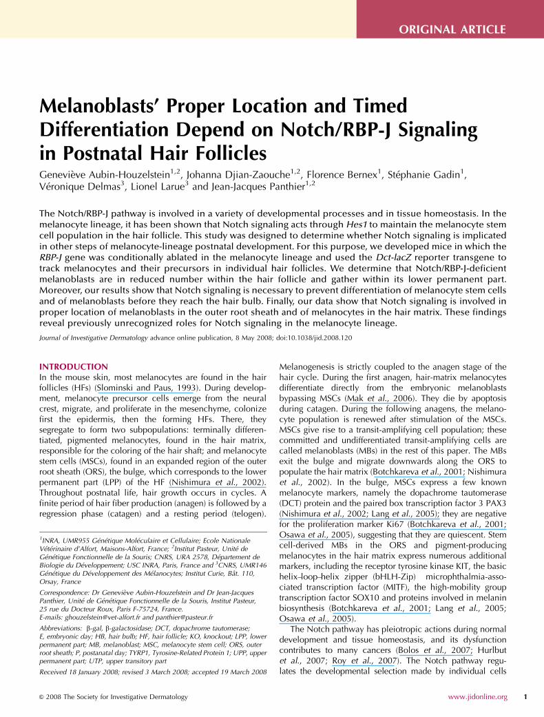
We isolated HFs from the skin of cRBP-J KO; Tg(Dct-lacZ)and Tg(Dct-lacZ) control mice and counted b-galactosidase(b-gal)-positive cells within the HFs, excluding the hair matrixwhere they were not countable. Indeed, in the hair matrix,the density of b-gal-positive cells was very high and the X-Galprecipitate leaked so that the boundaries of individual b-gal-positive cells were not visible. No X-Gal staining could bedetected in HFs from the skin of cRBP-J KO and Rbp-Jf/f micelacking the Dct-lacZ transgene, indicating that the lacZreporter under the control of the Dct promoter is a robust andreliable marker of melanoblasts within the HF (Figure S1). Weanalyzed HFs at both P8 and P30 that corresponded to theanagen of the first and second hair cycles, respectively. In P8control HFs (Figure 1c), the average number of b-gal-positivecells per HF was 5.6±2.5 (n¼ 47 HFs). In P8 cRBP-J KO;Tg(Dct-lacZ) HFs (Figure 1d), the count was 1.8±1.6(n¼124 HFs). The number of b-gal-positive cells wassignificantly reduced in cRBP-J KO; Tg(Dct-lacZ) HFs ascompared with that in Tg(Dct-lacZ) controls (Mann–WhitneyU-test, Po10�4). In P30 control HFs (Figure 1e), the averagenumber of b-gal-positive cells per HF was 6.2±2.5 (n¼84HFs). In P30 cRBP-J KO; Tg(Dct-lacZ) HFs (Figure 1f), thecount was 1.2±1.3 (n¼162). The number of b-gal-positivecells was significantly reduced in cRBP-J KO; Tg(Dct-lacZ)HFs as compared with that in Tg(Dct-lacZ) controls (Man-n–Whitney U-test, Po10�4). At P8 and P30, in both controlsand mutants the number of b-gal-positive cells was notnoticeably different between HFs producing guard, auchene,awl, or zigzag hair shafts.
We compared the mean number of b-gal-positive cells perHF at P8 and P30. In Tg(Dct-lacZ) control HFs, there wasno difference in the mean number of b-gal-positive betweenP8 and P30 (Mann–Whitney U-test). By contrast, in cRBP-JKO; Tg(Dct-lacZ) HFs the mean number of b-gal-positivecells was lower at P30 than at P8 (Mann–Whitney U-test,Po10�3).
2 Journal of Investigative Dermatology
G Aubin-Houzelstein et al.Notch Signaling in Melanocyte

Altogether, these results show that the number of MBs isreduced in the HFs of cRBP-J KO mice at both P8 and P30 ascompared with that in control mice. Moreover, whereas thenumber of MBs remains constant between the first andsecond hair cycles in the HFs of control mice, it dwindlessignificantly in the HFs of cRBP-J KO mice.
Absence of Notch signaling alters distribution of melanoblastsin postnatal HFs
We examined whether the reduced number of b-gal-positivecells in cRBP-J KO; Tg(Dct-lacZ) HFs was homogenous orcould be attributed to a specific region of the HF. To this end,we split each HF in four parts corresponding to anatomicallydistinct regions of the follicle: (i) the upper permanentpart (UPP), from the epidermis to the bulge; (ii) the LPP, thebulge where the MSCs reside; (iii) the upper transitory part(UTP), from the LPP to the bulb; and (iv) the hair bulb (HB)(Figure 1c). The LPP or bulge is located at the insertion of thearrector pili muscle (Muller-Rover et al., 2001). It extendsfrom the lower part of the insertion site of the arrector pilimuscle to the opening of the sebaceous gland duct
(Paus et al., 1999; Tiede et al., 2007). In HFs where thesemarks were visible, the lower part of the insertion of thearrector pili muscle was situated at the junction of thedermis and the subcutis, and the LPP covered 120 mm onaverage. Therefore, in HFs where the arrector pili and/or theopening of the sebaceous gland were not visible, the lowerboundary for the LPP was chosen at the junction of thedermis and the subcutis, and its upper boundary 120mmabove. We counted the numbers of b-gal-positive cells in thethree upper parts for each HF; b-gal-positive cells withinthe HB were not countable. We compared the mean numbersof b-gal-positive cells in each part between Tg(Dct-lacZ)control and cRBP-J KO; Tg(Dct-lacZ) HFs (Figure 1g and h).At P8, the means of b-gal-positive cells were significantlylower in cRBP-J KO; Tg(Dct-lacZ) UPP, LPP, and UTP thanin controls (Mann–Whitney tests, all Po10�3) (Figure 1g).At P30, the mean numbers of b-gal-positive cells in all parts,except UPP, were significantly lower in cRBP-J KO; Tg(Dct-lacZ) than in controls (Mann–Whitney tests, Po10�3,except for the UPP where P-value was not significant)(Figure 1h).
LPP
UTP
UPP
63
12
25
18
52
30
UTP
UPP
LPP *
***
***
***
13
47
40
***
***
***
NS 5
56
39
P8 controls(N=47)
P8 cRBP-J KO(N=124)
100
80
60
40
20
0P30 controls
(N=84)P30 cRBP-J KO
(N=162)
100
80
60
40
20
0
6.2
1.2
***P8 P30
***
******
2.4
0.6
***
3.5
0.5
***
0.20.3
NS
0
2
4
6
Total UPP LPP UTP Total UPP LPP UTP
ControlscRBP-J KO
5.6
0.61.4
3.6
1.8 0.3 0.90.5
ControlscRBP-J KO
***
* **
UPP
LPP
UTP
HB
*****
** *
P8 Tg(Dct-lacZ) P30 Tg(Dct-lacZ)
P8 control P8 cRBP-J KO P30 control P30 cRBP-J KO
0
2
4
6
Num
ber
of β
-gal
+ce
lls
Num
ber
of β
-gal
+ce
lls
Per
cent
age
of β
-gal
+ce
lls
Per
cent
age
of β
-gal
+ce
lls
Figure 1. Coat-color phenotype and distribution of b-gal-positive cells in cRBP-J KO; Tg(Dct-lacZ) and control HFs at P8 and P30. (a) Coat-color phenotype of
two 8-day-old (P8) cRBP-J KO; Tg(Dct-lacZ) pups (middle) and two Tg(Dct-lacZ) control littermates (sides). (b) Thirty-day-old (P30) cRBP-J KO; Tg(Dct-lacZ)
mouse (top) and Tg(Dct-lacZ) control littermate (bottom). (c–f) HFs were dissected after X-Gal staining of the skin; asterisks indicate the position of the LPP.
Bars¼ 200 mm. (c) HFs from a P8 Tg(Dct-lacZ) control mouse; parentheses show the location for the UPP, LPP, UTP, and HB on the HF situated on the left.
(d) HFs from a P8 cRBP-J KO; Tg(Dct-lacZ) mouse; the arrow points at a dendritic b-gal-positive cell. (e) HFs from a P30 Tg(Dct-lacZ) control mouse; the hair
clubs from the first hair cycle are still present, allowing location of LPP (asterisks). (f) HFs from a P30 cRBP-J KO; Tg(Dct-lacZ) mouse; the arrow points at a
dendritic and pigmented b-gal-positive cell. (g–h) Means of the number of b-gal-positive cells in the Tg(Dct-lacZ) control (black histograms) and cRBP-J KO;
Tg(Dct-lacZ) (white histograms) HFs, UPP, LPP, and UTP at P8 (g) and P30 (h). The total corresponds to the sum of b-gal-positive cells mean numbers in the UPP,
LPP, and UTP. Means for the considered region are given rounded to the nearest tenth either on or above each histogram. Bars represent SEM. Data were
analyzed using Mann–Whitney tests: ***Po10�3; NS, non-significant. (i, j) Distribution patterns of b-gal-positive cells in HFs according to their location (black,
UTP; white, LPP; gray, UPP) at P8 (i) and P30 (j); to the left: distributions in Tg(Dct-lacZ) control HFs; to the right: distributions in cRBP-J KO; Tg(Dct-lacZ) HFs.
Distributions are given as percentages of total cell counts. Numbers of HFs analyzed are given as N under each histogram. Numbers drawn on histograms are the
percentages for the considered parts. Data were analyzed using w2-tests for overall distributions (asterisks above the histograms) or Fisher’s exact tests for
percentages in a given part (asterisks between the histograms); ***Pp10�3; *Pp0.05; NS, non-significant.
www.jidonline.org 3
G Aubin-Houzelstein et al.Notch Signaling in Melanocyte
We then tested whether distribution of b-gal-positive cellswas the same in Tg(Dct-lacZ) control and cRBP-J KO; Tg(Dct-lacZ) HFs. To this end, we summed up the cell counts of eachpart and compared the overall distribution and percentages ofb-gal-positive cells in each part between Tg(Dct-lacZ)controls and cRBP-J KO; Tg(Dct-lacZ) HFs (Figure 1i and j).At both P8 and P30, we found that the distributions of b-gal-positive cells in whole HFs were differing between mutantsand controls (w2-test, Po10�4 for both tests) (Figure 1i and j).At P8, we found a significantly higher percentage of b-gal-positive cells in the LPP and a lower percentage in the UTP incRBP-J KO; Tg(Dct-lacZ) HFs as compared with that inTg(Dct-lacZ) control HFs (Fisher’s exact tests, P¼10�3 andPo10�4, respectively) (Figure 1i). In other words, in Tg(Dct-lacZ) control HFs, most of the b-gal-positive cells were foundin the UTP, whereas in cRBP-J KO; Tg(Dct-lacZ) HFs, most ofthem were found in the LPP (Figure 1i). At P30, thepercentages of b-gal-positive cells were higher in the LPPand UPP, but lower in the UTP of cRBP-J KO; Tg(Dct-lacZ)HFs when compared with that in Tg(Dct-lacZ) controls(Fisher’s exact tests, P¼0.05 in the LPP and Po10�3 in theUPP and UTP) (Figure 1j).
In sum, at both P8 and P30, distribution of MBs is affectedin the HFs of cRBP-J-KO mice. Notably, whereas there is areduced number of MBs in the LPP of cRBP-J KO mice ascompared with controls, the relative proportion of MBs issignificantly increased in this specific region compared tocontrols at both time points.
Without Notch signaling, the LPP melanoblast pool isdepleted between the first and the second hair cycle
The origin of HF melanocytes is different between the firstand the second hair cycle. Indeed, during the first anagen,melanocytes differentiate directly from embryonic melano-blasts, whereas during the second anagen, they derive fromthe MSCs located in the LPP (Mak et al., 2006). We testedwhether this difference in melanocyte origin could result inchanges in mean numbers and distributions of cells of themelanocyte lineage between the first and the second haircycle. To this end, we compared the numbers of b-gal-positive cells and distributions at P8 and P30 in Tg(Dct-lacZ)control HFs. The average number of b-gal-positive cells wassignificantly increased in the LPP at P30 and significantlydecreased in the UPP at P30 (Mann–Whitney tests, bothPo10�3) (Figure 1g and h, black histograms). Moreover,distribution in the entire HF was different between P8 andP30 mice (w2-test, Po10�4) (Figure 1i and j, left panels). InTg(Dct-lacZ) control HFs at P30, the percentage of b-gal-positive cells in the LPP was higher than at P8, at the expenseof the UPP and UTP (Fisher’s exact tests, Po10�3 in the LPPand UPP, and P¼0.05 in the UTP) (Figure 1i and j, leftpanels).
We then studied the evolution of numbers and distribu-tions of b-gal-positive cells in cRBP-J KO; Tg(Dct-lacZ) HFsbetween the first and the second hair cycle. Mean numberswere significantly lower in whole HFs, UPP, and LPP at P30,but not different in the UTP (Mann–Whitney tests, all Po10�3
except for the UTP where P-value was not significant)
(Figure 1g and h, white histograms). The distribution in theentire HF was different between P8 and P30 (w2-test,Po10�4) (Figure 1i and j). The difference was due to asignificantly increased percentage of b-gal-positive cells inthe UTP at P30 (Fisher’s exact tests, P¼ 0.03) (Figure 1i and j,right panels).
In conclusion, regarding the LPP, the mean number of b-gal-positive cells evolved differently in Tg(Dct-lacZ) controland cRBP-J KO; Tg(Dct-lacZ): whereas it increased incontrols, it diminished in mutants. These data suggest thatthe reduction in the number of MBs in cRBP-J KO; Tg(Dct-lacZ) HFs is mainly due to depletion of the LPP population.
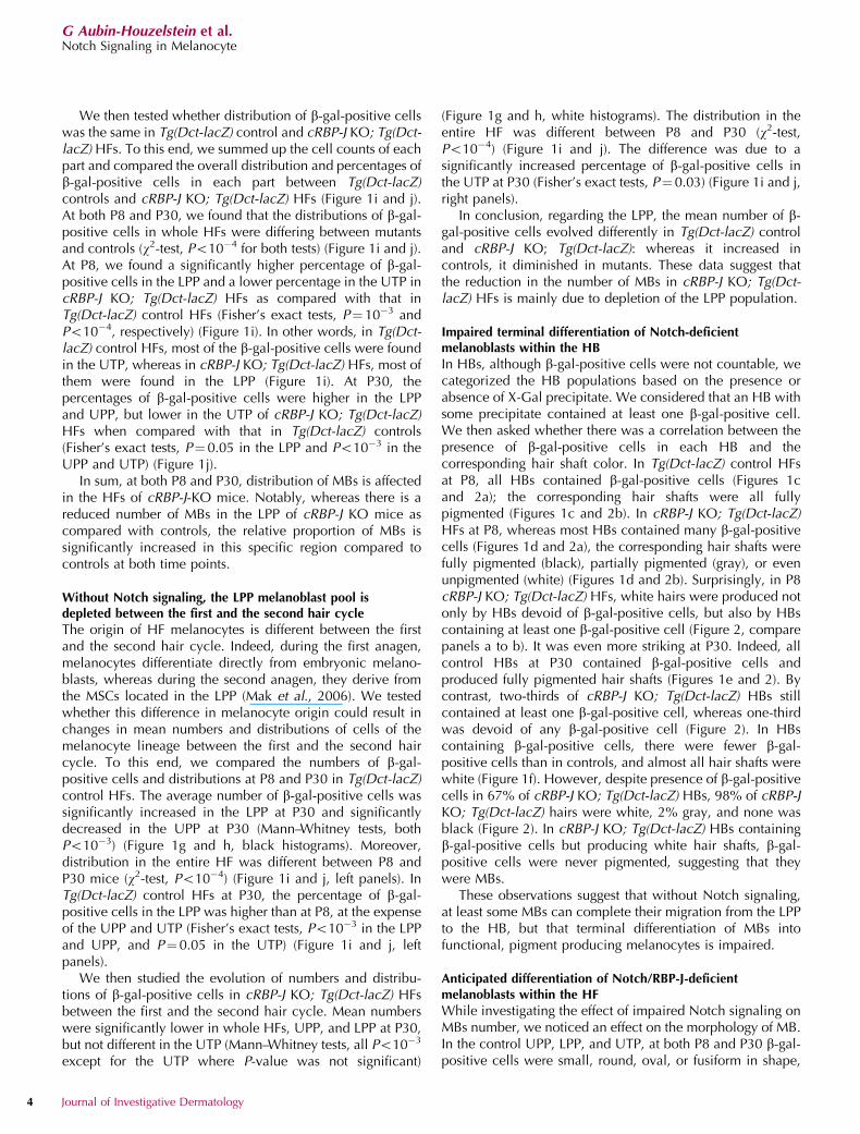
Impaired terminal differentiation of Notch-deficientmelanoblasts within the HB
In HBs, although b-gal-positive cells were not countable, wecategorized the HB populations based on the presence orabsence of X-Gal precipitate. We considered that an HB withsome precipitate contained at least one b-gal-positive cell.We then asked whether there was a correlation between thepresence of b-gal-positive cells in each HB and thecorresponding hair shaft color. In Tg(Dct-lacZ) control HFsat P8, all HBs contained b-gal-positive cells (Figures 1cand 2a); the corresponding hair shafts were all fullypigmented (Figures 1c and 2b). In cRBP-J KO; Tg(Dct-lacZ)HFs at P8, whereas most HBs contained many b-gal-positivecells (Figures 1d and 2a), the corresponding hair shafts werefully pigmented (black), partially pigmented (gray), or evenunpigmented (white) (Figures 1d and 2b). Surprisingly, in P8cRBP-J KO; Tg(Dct-lacZ) HFs, white hairs were produced notonly by HBs devoid of b-gal-positive cells, but also by HBscontaining at least one b-gal-positive cell (Figure 2, comparepanels a to b). It was even more striking at P30. Indeed, allcontrol HBs at P30 contained b-gal-positive cells andproduced fully pigmented hair shafts (Figures 1e and 2). Bycontrast, two-thirds of cRBP-J KO; Tg(Dct-lacZ) HBs stillcontained at least one b-gal-positive cell, whereas one-thirdwas devoid of any b-gal-positive cell (Figure 2). In HBscontaining b-gal-positive cells, there were fewer b-gal-positive cells than in controls, and almost all hair shafts werewhite (Figure 1f). However, despite presence of b-gal-positivecells in 67% of cRBP-J KO; Tg(Dct-lacZ) HBs, 98% of cRBP-JKO; Tg(Dct-lacZ) hairs were white, 2% gray, and none wasblack (Figure 2). In cRBP-J KO; Tg(Dct-lacZ) HBs containingb-gal-positive cells but producing white hair shafts, b-gal-positive cells were never pigmented, suggesting that theywere MBs.
These observations suggest that without Notch signaling,at least some MBs can complete their migration from the LPPto the HB, but that terminal differentiation of MBs intofunctional, pigment producing melanocytes is impaired.
Anticipated differentiation of Notch/RBP-J-deficientmelanoblasts within the HF
While investigating the effect of impaired Notch signaling onMBs number, we noticed an effect on the morphology of MB.In the control UPP, LPP, and UTP, at both P8 and P30 b-gal-positive cells were small, round, oval, or fusiform in shape,
4 Journal of Investigative Dermatology
G Aubin-Houzelstein et al.Notch Signaling in Melanocyte
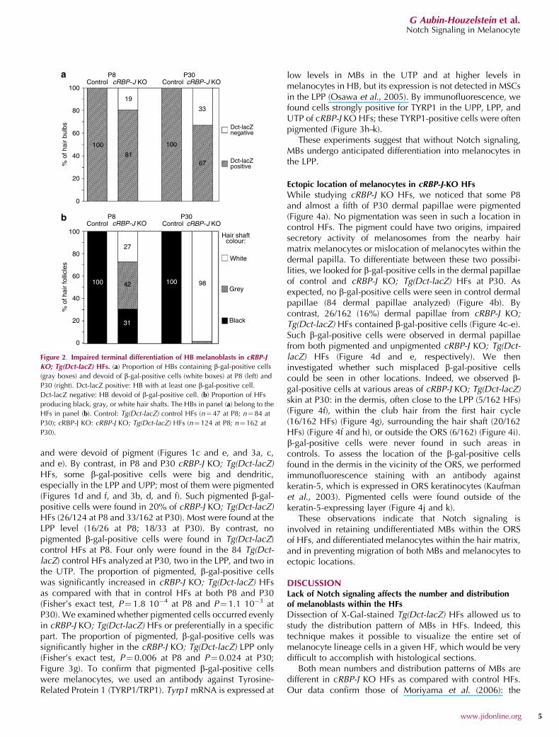
and were devoid of pigment (Figures 1c and e, and 3a, c,and e). By contrast, in P8 and P30 cRBP-J KO; Tg(Dct-lacZ)HFs, some b-gal-positive cells were big and dendritic,especially in the LPP and UPP; most of them were pigmented(Figures 1d and f, and 3b, d, and f). Such pigmented b-gal-positive cells were found in 20% of cRBP-J KO; Tg(Dct-lacZ)HFs (26/124 at P8 and 33/162 at P30). Most were found at theLPP level (16/26 at P8; 18/33 at P30). By contrast, nopigmented b-gal-positive cells were found in Tg(Dct-lacZ)control HFs at P8. Four only were found in the 84 Tg(Dct-lacZ) control HFs analyzed at P30, two in the LPP, and two inthe UTP. The proportion of pigmented, b-gal-positive cellswas significantly increased in cRBP-J KO; Tg(Dct-lacZ) HFsas compared with that in control HFs at both P8 and P30(Fisher’s exact test, P¼1.8 10�4 at P8 and P¼1.1 10�3 atP30). We examined whether pigmented cells occurred evenlyin cRBP-J KO; Tg(Dct-lacZ) HFs or preferentially in a specificpart. The proportion of pigmented, b-gal-positive cells wassignificantly higher in the cRBP-J KO; Tg(Dct-lacZ) LPP only(Fisher’s exact test, P¼0.006 at P8 and P¼0.024 at P30;Figure 3g). To confirm that pigmented b-gal-positive cellswere melanocytes, we used an antibody against Tyrosine-Related Protein 1 (TYRP1/TRP1). Tyrp1 mRNA is expressed at
low levels in MBs in the UTP and at higher levels inmelanocytes in HB, but its expression is not detected in MSCsin the LPP (Osawa et al., 2005). By immunofluorescence, wefound cells strongly positive for TYRP1 in the UPP, LPP, andUTP of cRBP-J KO HFs; these TYRP1-positive cells were oftenpigmented (Figure 3h–k).
These experiments suggest that without Notch signaling,MBs undergo anticipated differentiation into melanocytes inthe LPP.
Ectopic location of melanocytes in cRBP-J-KO HFs
While studying cRBP-J KO HFs, we noticed that some P8and almost a fifth of P30 dermal papillae were pigmented(Figure 4a). No pigmentation was seen in such a location incontrol HFs. The pigment could have two origins, impairedsecretory activity of melanosomes from the nearby hairmatrix melanocytes or mislocation of melanocytes within thedermal papilla. To differentiate between these two possibi-lities, we looked for b-gal-positive cells in the dermal papillaeof control and cRBP-J KO; Tg(Dct-lacZ) HFs at P30. Asexpected, no b-gal-positive cells were seen in control dermalpapillae (84 dermal papillae analyzed) (Figure 4b). Bycontrast, 26/162 (16%) dermal papillae from cRBP-J KO;Tg(Dct-lacZ) HFs contained b-gal-positive cells (Figure 4c–e).Such b-gal-positive cells were observed in dermal papillaefrom both pigmented and unpigmented cRBP-J KO; Tg(Dct-lacZ) HFs (Figure 4d and e, respectively). We theninvestigated whether such misplaced b-gal-positive cellscould be seen in other locations. Indeed, we observed b-gal-positive cells at various areas of cRBP-J KO; Tg(Dct-lacZ)skin at P30: in the dermis, often close to the LPP (5/162 HFs)(Figure 4f), within the club hair from the first hair cycle(16/162 HFs) (Figure 4g), surrounding the hair shaft (20/162HFs) (Figure 4f and h), or outside the ORS (6/162) (Figure 4i).b-gal-positive cells were never found in such areas incontrols. To assess the location of the b-gal-positive cellsfound in the dermis in the vicinity of the ORS, we performedimmunofluorescence staining with an antibody againstkeratin-5, which is expressed in ORS keratinocytes (Kaufmanet al., 2003). Pigmented cells were found outside of thekeratin-5-expressing layer (Figure 4j and k).
These observations indicate that Notch signaling isinvolved in retaining undifferentiated MBs within the ORSof HFs, and differentiated melanocytes within the hair matrix,and in preventing migration of both MBs and melanocytes toectopic locations.
DISCUSSIONLack of Notch signaling affects the number and distributionof melanoblasts within the HFs
Dissection of X-Gal-stained Tg(Dct-lacZ) HFs allowed us tostudy the distribution pattern of MBs in HFs. Indeed, thistechnique makes it possible to visualize the entire set ofmelanocyte lineage cells in a given HF, which would be verydifficult to accomplish with histological sections.
Both mean numbers and distribution patterns of MBs aredifferent in cRBP-J KO HFs as compared with control HFs.Our data confirm those of Moriyama et al. (2006): the
ControlP8
100
80
60
40
100 100
Dct-lacZ negative
Dct-lacZ positive
Hair shaft colour:
White
Grey
Black
33
6781
27
9842
31
100100
19
% o
f hai
r bu
lbs
20
0
100
80
60
40
% o
f hai
r fo
llicl
es
20
0
P30ControlcRBP-J KO cRBP-J KO
ControlP8 P30
ControlcRBP-J KO cRBP-J KO
Figure 2. Impaired terminal differentiation of HB melanoblasts in cRBP-J
KO; Tg(Dct-lacZ) HFs. (a) Proportion of HBs containing b-gal-positive cells
(gray boxes) and devoid of b-gal-positive cells (white boxes) at P8 (left) and
P30 (right). Dct-lacZ positive: HB with at least one b-gal-positive cell.
Dct-lacZ negative: HB devoid of b-gal-positive cell. (b) Proportion of HFs
producing black, gray, or white hair shafts. The HBs in panel (a) belong to the
HFs in panel (b). Control: Tg(Dct-lacZ) control HFs (n¼ 47 at P8; n¼ 84 at
P30); cRBP-J KO: cRBP-J KO; Tg(Dct-lacZ) HFs (n¼ 124 at P8; n¼162 at
P30).
www.jidonline.org 5
G Aubin-Houzelstein et al.Notch Signaling in Melanocyte

number of cRBP-J KO MBs is reduced in the whole HF, andMBs are especially depleted in the LPP. Moreover, they showthat the percentage of MBs in the LPP is increased in cRBP-J-KO HFs at both P8 and P30. This overrepresentation of MBswithin the LPP may reflect one or several of the followingdefects: (1) a reduced ability of MSCs to generate MBs, (2) animpaired ability of MBs to emigrate from the LPP, and (3) animpaired proliferation and/or survival of MBs within the UTP.Although these possibilities should be addressed in futurestudies, our observations show that the role of Notchsignaling on HF MBs is not limited to maintenance of theMSC pool.
Absence of Notch signaling impairs the timing of melanoblastdifferentiation within the HFs
Our data further indicate that inactivation of Notch signalingis associated with precocious expression of the melanogenic
enzyme TRP1 and with the anticipated synthesis of melaninin MBs located in the three upper regions of the HF, the LPP,UTP, and UPP. These observations indicate that Notchsignaling prevents differentiation of MBs into melanocytesin these regions. Such a function for Notch has recently beenreported in the embryonic development of limb muscles.Indeed, in the absence of RBP-J, myogenic progenitor cellsdifferentiate in an uncontrolled and premature manner atE11.5. Consequently, the progenitor pool is depleted and thesatellite cell population is missing at E18.5 (Vasyutina et al.,2007). Strikingly, during postnatal myogenesis, Notch signal-ing maintains PAX3-positive myogenic precursors in anundifferentiated state through upregulation of Pax3 anddownregulation of muscular differentiation genes (Conboyand Rando, 2002). A similar mechanism may well operate inMSCs that have been shown to express PAX3 as myogenicprecursors do (Osawa et al., 2005).
a
c
b
d
g
h i
kje f
P30 control Tg(Dct-lacZ)
UPP
LPP
UTP
P30 cRBP-J KO; Tg(Dct-lacZ)
14
12
10
8
6
4
2
0 0.0 0.0 0.0 0.03.2
12.9
ControlscRBP-J KO
4.8
1.8
7.4
2.4 2.4
11.1NS
NS
***
NS
NS
UPP UPPLPP
P8 P30
LPPUTP
P30 cRBP-J KO; Tg(Dct-lacZ) LPP
ep
UTP
% o
f HF
with
pig
men
ted
cells
TYRP1 DAPI
TYRP1 DAPI
P30 cRBP-J KO ; Tg(Dct-lacZ) UTP
Figure 3. Anticipated differentiation of melanoblasts in cRBP-J KO; Tg(Dct-lacZ) HFs. (a–f) Parts of Tg(Dct-lacZ) control (a, c, e) and cRBP-J KO; Tg(Dct-lacZ)
(b, d, f) HFs; control and mutant UPP (a, b), LPP (c, d) and UTP (e, f). HFs were dissected after X-Gal staining of the skin. Stars indicate the base of hair clubs
from the first hair cycle. Arrows point at pigment within b-gal-positive cells. Dotted lines delimit the HF. (g) Percentages of HFs with b-gal-positive, dendritic,
and/or pigmented cells in the UPP, LPP, or UTP at P8 (left panel) and P30 (right panel). Number of HFs analyzed are 47 and 124 at P8 and 84 and 162 at P30
for cRBP-J KO; Tg(Dct-lacZ) and Tg(Dct-lacZ) controls, respectively. Asterisks correspond to the P-value for Fisher’s exact test; *Po0.05; **Po0.01; NS,
non-significant. (h–k) Immunofluorescence staining of P30 cRBP-J KO; Tg(Dct-lacZ) skin sections using anti-TYRP1 antibody (green) and 4,6-diamidino-2-
phenylindole (blue); (h) optic section in LPP; ep, epidermis, with nonspecific TYRP1 labeling in keratinocytes; (j) optic section in UTP; panels (i) and (k) are the
Nomarski pictures of images in panels (h) and (j), respectively; arrowheads point at the same cells in fluorescent and Nomarski pictures. Sections thickness,
0.5mm. Bars¼ 20mm in panels (a)–(f) and 10 mm in panels (h)–(k).
6 Journal of Investigative Dermatology
G Aubin-Houzelstein et al.Notch Signaling in Melanocyte

Our results indicate that the proportion of pigmentedmelanocytes is significantly higher in the LPP but not in theUPP and UTP of cRBP-J KO HFs as compared with controls.As the LPP contains the MSCs as well as early MBs, oneexplanation for this observation could be that Notchactivation acts specifically on MSCs and early MBs tomaintain them in an undifferentiated state. Consequently,Notch-deficient melanocytes within the LPP would beinsensitive to the migrating cues that attract MBs out of theLPP toward the HB. The depletion of the MSC pool could be a
consequence of the anticipitated differentiation of MSCs intomelanocytes. However, the anticipated differentiation intomelanocytes occurs in a small percentage of the cRBP-J KOLPP (12.9 and 11.1% at P8 and P30, respectively), whereasdepletion of the MSC population within the cRBP-J KO LPP isa general event. Thus, if the anticipated differentiationcontributes to depletion of the MSC pool in the cRBP-J KOLPP, it cannot account for all of it. We conclude thatdepletion of the MSC pool in the cRBP-J KO LPP is aconsequence of at least two events that are not necessarily
P30 cRBP-J KO HBControl cRBP-J KO
P30 Tg(Dct-lacZ) HB
P30 cRBP-J KO; Tg(Dct-lacZ)
HB
UTP UTP UTP UTP
HB LPP LPP
K5 DAPI
a
d
h i kj
e f g
b c
Figure 4. Ectopic location of melanoblasts in cRBP-J KO; Tg(Dct-lacZ) HFs. (a) Histological section of P8 cRBP-J KO skin. The arrow points at pigment within
the dermal papilla. (b, c) Histological sections of P30 Tg(Dct-lacZ) control (b) and cRBP-J KO; Tg(Dct-lacZ) (c) HB; skins were stained with X-Gal before
sectioning. Dotted lines delimit the dermal papilla; white arrowhead points at a b-gal-positive cell within the dermal papilla. (d–i) Parts of P30 cRBP-J KO;
Tg(Dct-lacZ) HFs: HB (d, e), LPP (f, g), UTP (h, i); the corresponding skins were stained with X-Gal and HFs were dissected. Dotted lines delimit the dermal
papilla in panels (d) and (e) and the ORS in panels (f)–(i). Asterisks indicate the base of hair clubs from the first hair cycle. Arrowheads point at ectopic
b-gal-positive cells. (j) Immunofluorescence staining of a P30 cRBP-J KO; Tg(Dct-lacZ) UTP using anti-K5 antibody (green) and 4,6-diamidino-2-phenylindole
(blue). (k) Nomarski picture of the same section as in panel (j). Dotted lines delimit the ORS, arrowhead points at a pigmented cell outside the ORS. Sections
thickness: 10 mm in panels (a)–(c); 0.5mm in panels (j) and (k). Bars¼ 50 mm in panel (a); 20 mm in panels (b), (c), and (f)–(k); 40mm in panels (d) and (e).
www.jidonline.org 7
G Aubin-Houzelstein et al.Notch Signaling in Melanocyte
interrelated, MSC apoptosis as shown by Moriyama et al.(2006) and anticipated differentiation. Strikingly, in controlHFs, whereas no differentiated melanocytes are detected inthe UPP, LPP, and UTP at P8, some are found in the LPP andUTP at P30, although very rarely.
Notch/RBP-J-deficient melanoblasts are in reduced numberand lack terminal differentiation within the HBs
Many cRBP-J KO HFs cannot produce pigmented hair shaftsdespite presence of MBs within the hair matrix. These MBs,that are neither dendritic nor pigmented, stay undifferentiatedin a region where terminal differentiation occurs in wild-typeHFs. This is particularly true at P30 during the second haircycle when Notch/RBP-J-deficient MBs are in reducednumber in the HB. The absence of differentiation may bedirectly linked to the lack of Notch signaling in MBs withinthe HBs. Alternatively, absence of differentiation may be anindirect consequence of the lack of Notch signaling in MBs inthe LPP and UTP, leading to the reduced number of HB MBs.Indeed, in wild-type conditions, differentiation of MBs intopigment-producing melanocytes in the HB may require acritical number of MBs. Thus, presence of DCT-positive,unpigmented cells in cRBP-J KO HBs producing white hairshafts could be a consequence of the reduction in the numberof MBs reaching the HB, and not of the absence of Notchsignaling in HB MBs per se. Whatever the reason, Notchsignaling acts either directly or indirectly on terminaldifferentiation of MBs within the HB.
Notch/RBP-J-deficient melanoblasts and melanocytes arefound in ectopic locations in the skin
MBs and melanocytes are observed in various ectopiclocations in the skin of cRBP-J KO mice. We assume thatMBs and melanocytes migrated to inappropriate locationsdue to lack of Notch signaling. Indeed, Notch signaling couldparticipate in the communication between MBs and kerati-nocytes. In cRBP-J KO mice, such communication would bedisrupted eventually leading to altered MB migration.However, many HBs of cRBP-J KO mice contain MBs,indicating that most RBP-J-deficient MBs are still able toreach the hair matrix. Thus, altered migration is only a minorphenotype in Notch/RBP-J-deficient MBs.
Most of the DCT-positive cells seen in ectopic positionwere pigmented. It raises the possibility that impropermigration occurs whenever an RBP-J-deficient MB undergoesprecocious differentiation in the LPP, UPP, and UTP.Alternatively, within wild-type HFs, the environment mayinhibit differentiation of MBs into pigment-producing mela-nocytes; when they are out of their normal environment,RBP-J-deficient MBs would escape the inhibiting signals anddifferentiate into mature melanocytes.
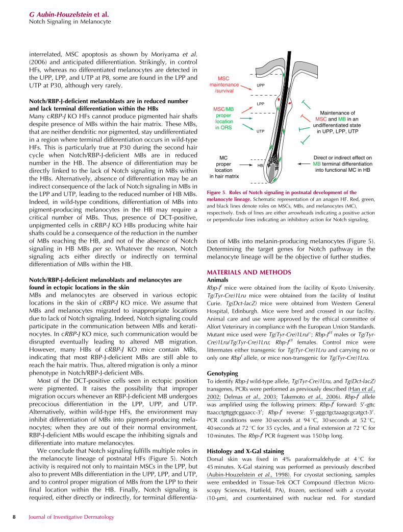
We conclude that Notch signaling fulfills multiple roles inthe melanocyte lineage of postnatal HFs (Figure 5). Notchactivity is required not only to maintain MSCs in the LPP, butalso to prevent MBs differentiation in the UPP, LPP, and UTP,and to control proper migration of MBs from the LPP to theirfinal location within the HB. Finally, Notch signaling isrequired, either directly or indirectly, for terminal differentia-
tion of MBs into melanin-producing melanocytes (Figure 5).Determining the target genes for Notch pathway in themelanocyte lineage will be the objective of further studies.
MATERIALS AND METHODSAnimals
Rbp-Jf mice were obtained from the facility of Kyoto University.
Tg(Tyr-Cre)1Lru mice were obtained from the facility of Institut
Curie. Tg(Dct-lacZ) mice were obtained from Western General
Hospital, Edinburgh. Mice were bred and crossed in our facility.
Animal care and use were approved by the ethical committee of
Alfort Veterinary in compliance with the European Union Standards.
Mutant mice used were Tg(Tyr-Cre)1Lru/1; Rbp-Jf/f males or Tg(Tyr-
Cre)1Lru/Tg(Tyr-Cre)1Lru; Rbp-Jf/f females. Control mice were
littermates either transgenic for Tg(Tyr-Cre)1Lru and carrying no or
only one Rbpf allele, or mice non-transgenic for Tg(Tyr-Cre)1Lru.
Genotyping
To identify Rbp-J wild-type allele, Tg(Tyr-Cre)1Lru, and Tg(Dct-lacZ)
transgenes, PCRs were performed as previously described (Han et al.,
2002; Delmas et al., 2003; Takemoto et al., 2006). Rbp-Jf allele
was amplified using the following primers: Rbp-Jf forward: 50-gttc
ttaacctgttggtcggaacc-30; Rbp-Jf reverse: 50-gggctgctaaagcgcatgct-30.
PCR conditions were 30 seconds at 94 1C, 30 seconds at 52 1C,
40 seconds at 72 1C for 35 cycles, and a final extension at 72 1C for
10 minutes. The Rbp-Jf PCR fragment was 150 bp long.
Histology and X-Gal staining
Dorsal skin was fixed in 4% paraformaldehyde at 4 1C for
45 minutes. X-Gal staining was performed as previously described
(Aubin-Houzelstein et al., 1998). For cryostat sectioning, samples
were embedded in Tissue-Tek OCT Compound (Electron Micro-
scopy Sciences, Hatfield, PA), frozen, sectioned with a cryostat
(10-mm), and counterstained with nuclear red. For standard
UPP
LPP
UTP
HB
MSC maintenance
/survival
MSC/MB proper locationin ORS
MC proper
location in hair matrix
Direct or indirect effect onMB terminal differentiationinto functional MC in HB
Maintenance ofMSC and MB in an
undifferentiated state in UPP, LPP, UTP
Figure 5. Roles of Notch signaling in postnatal development of the
melanocyte lineage. Schematic representation of an anagen HF. Red, green,
and black lines denote roles on MSCs, MBs, and melanocytes (MC),
respectively. Ends of lines are either arrowheads indicating a positive action
or perpendicular lines indicating an inhibitory action for Notch signaling.
8 Journal of Investigative Dermatology
G Aubin-Houzelstein et al.Notch Signaling in Melanocyte
histology, skin samples were fixed overnight in 4% formaldehyde/
phosphate-buffered saline (PBS) at 4 1C, embedded in paraffin,
sectioned (4-mm) following standard procedures, and counterstained
with hemalun–eosin.
Hair follicles dissection
Dorsal skin was dissected from two P8 and three P30 Tg(Dct-lacZ)
controls, and three P8 and three P30 cRBP-J KO; Tg(Dct-lacZ) mice.
The skin was fixed in 4% paraformaldehyde at 4 1C for 45 minutes,
rinsed in PBS, then cut in smaller pieces. For each mouse, three
groups of small skin pieces were kept: the first was obtained from
the anterior part of the dorsal skin sample, in the forelimb region;
the second from the middle part of the sample; and the third came
from the posterior part of the sample, from the hind-limb region. The
small pieces were stained with X-Gal for 48 hours at 32 1C.
Individual HFs were isolated from the X-Gal-stained small pieces
under a binocular microscope. The isolated HFs were mounted in
Mowiol (Polysciences Europe, Eppelheim, Germany).
b-Gal-positive-cell counting and statistical analyses
b-Gal-positive cells from individual HFs were counted under
microscope. The HFs were obtained from two cRBP-J KO; Tg(Dct-
lacZ) and three Tg(Dct-lacZ) control littermates at P8, and three
cRBP-J KO; Tg(Dct-lacZ) and three Tg(Dct-lacZ) control littermates at
P30. A comparable number of HFs was analyzed from each mouse.
The lower boundary for the LPP was anatomically located at the
junction of the dermis and the subcutis. In P30 HFs, it corresponded
to the base of the hair club from the first hair cycle. The upper
boundary for LPP was defined as the opening of the sebaceous gland
when visible, or placed 120mm above the lower boundary otherwise.
Statistical analyses were performed with StatViewF-4.51.3.PPC soft-
ware from Abacus Concepts (Berkeley, CA). Data are expressed as
means±SD. Distribution normality was tested by comparing the
observed distribution with a normal distribution with the same mean
and SD, and using a Kolmogorov–Smirnov test. Variances were
compared with an F-test. Means were compared with Student’s t-test
for normally distributed values with equal variances, and with a
Mann–Whitney U-test otherwise. Distributions were compared with a
w2-test. Percentages were compared with an Fisher’s exact test.
Immunofluorescence
Cryostat sections were rinsed in PBS. Blocking was performed in
10% normal goat serum in PBS for 1 hour at room temperature.
Sections were incubated overnight at 4 1C in primary antibody
solution (1:500 rabbit anti-mouse TYRP1/TRP1 antiserum; a kind gift
from Vincent Hearing) or 1:1,000 rabbit anti-mouse K5 (Covance,
Princeton, NJ) in 0.1% Tween-20 in PBS, and then for 1 hour at room
temperature in goat anti-rabbit IgG Alexa Fluor 488 (Invitrogen,
Cergy-pontoise, France) diluted 1:500 in 0.1% Tween-20 in PBS.
4,6-Diamidino-2-phenylindole was used for visualizing cell nuclei.
Between two incubation steps, the sections were washed in 0.1%
Tween-20 in PBS for 10 minutes. The sections were mounted in
Vectashield (Vector Laboratories, Peterborough, UK).
Image acquisition
Bright-field and fluorescence microscopy were performed with a
Zeiss Axioplan 2 Imaging microscope with ApoTome (upright stand),
using Plan-NEOFLUAR (� 10, � 25, and � 40 magnification) or
Plan-APOCHROMAT (� 63 and � 100 magnification) objective
lenses with 0.3, 0.8, 1.3, 1.4, and 1.4 numerical apertures,
respectively. Images were acquired with either an Axiocam HRc
(color imaging) or an Axiocam MRm (fluorescence imaging) camera
(Zeiss, Gottingen, Germany) coupled to Zeiss Axiovision 4.4
software. Minimal image processing was performed with Adobe
Photoshop.
CONFLICT OF INTERESTThe authors state no conflict of interest.
ACKNOWLEDGMENTSWe are grateful to T Honjo, I Jackson, and V Hearing for the kind gift of theRBPJf mouse, the Tg(Dct-lacZ) mouse, and the TRP1 antiserum, respectively;and Y Barrandon for his protocol for HF dissection. We thank A Casanova, MLDaupin, and C Koenen for dedicated animal husbandry; A Champeix andP Wattier for histological work; L Guillaud and P Salaun for technical help;Marie-Anne Nicola and Emmanuelle Perret from the Plateforme d’ImagerieDynamique at the Institut Pasteur for help with imaging; D Houzelstein forhelp with artwork; and M Chodkiewicz, M Cohen-Tannoudji, G Egidy-Maskos, X Montagutelli, and L Tiret for critical comments on the paper. GAH,FB, JDZ, and JJP were supported by the Association pour la Recherche contrele Cancer (ARC) (Grants 4332 and 99/7468) and by an agreement between theINRA and the Institut Pasteur; GAH and JJP were supported by the GrandProgramme Horizontal ‘‘Cellules Souches’’ of the Institut Pasteur; VD and LLwere supported by the Ligue Nationale contre le Cancer (Equipe Labellisee).JDZ was funded by the French Ministere de la Recherche et NouvellesTechnologies. This paper is dedicated to Charles Babinet.
SUPPLEMENTARY MATERIAL
Figure S1. X-Gal staining on isolated HFs from Tg(Dct-lacZ) and control mice.
REFERENCES
Artavanis-Tsakonas S, Matsuno K, Fortini ME (1995) Notch signaling. Science268:225–32
Artavanis-Tsakonas S, Rand MD, Lake RJ (1999) Notch signaling: cell fatecontrol and signal integration in development. Science 284:770–6
Aubin-Houzelstein G, Bernex F, Elbaz C, Panthier JJ (1998) Survival ofpatchwork melanoblasts is dependent upon their number in the hairfollicle at the end of embryogenesis. Dev Biol 198:266–76
Bolos V, Grego-Bessa J, de la Pompa JL (2007) Notch signaling indevelopment and cancer. Endocr Rev 28:339–63
Botchkareva NV, Khlgatian M, Longley BJ, Botchkarev VA, Gilchrest BA(2001) SCF/c-kit signaling is required for cyclic regeneration of the hairpigmentation unit. FASEB J 15:645–58
Chiba S (2006) Notch signaling in stem cell systems. Stem Cells 24:2437–47
Conboy IM, Rando TA (2002) The regulation of Notch signaling controlssatellite cell activation and cell fate determination in postnatalmyogenesis. Dev Cell 3:397–409
Delmas V, Martinozzi S, Bourgeois Y, Holzenberger M, Larue L (2003) Cre-mediated recombination in the skin melanocyte lineage. Genesis36:73–80
Han H, Tanigaki K, Yamamoto N, Kuroda K, Yoshimoto M, Nakahata T et al.(2002) Inducible gene knockout of transcription factor recombinationsignal binding protein-J reveals its essential role in T versus B lineagedecision. Int Immunol 14:637–45
Hurlbut GD, Kankel MW, Lake RJ, Artavanis-Tsakonas S (2007) Crossingpaths with Notch in the hyper-network. Curr Opin Cell Biol 19:166–75
Kato H, Sakai T, Tamura K, Minoguchi S, Shirayoshi Y, Hamada Y et al.(1996) Functional conservation of mouse Notch receptor familymembers. FEBS Lett 395:221–4
Kaufman CK, Zhou P, Pasolli HA, Rendl M, Bolotin D, Lim KC et al. (2003)GATA-3: an unexpected regulator of cell lineage determination in skin.Genes Dev 17:2108–22
www.jidonline.org 9
G Aubin-Houzelstein et al.Notch Signaling in Melanocyte
Kumano K, Masuda S, Sata M, Saito T, Lee SY, Sakata-Yanagimoto S et al.(2008) Both Notch1 and Notch2 contribute to the regulation ofmelanocyte homeostasis. Pigment Cell Melanoma Res 21:70–8
Lang D, Lu MM, Huang L, Engleka KA, Zhang M, Chu EY et al. (2005) Pax3functions at a nodal point in melanocyte stem cell differentiation. Nature433:884–7
MacKenzie MA, Jordan SA, Budd PS, Jackson IJ (1997) Activation of thereceptor tyrosine kinase Kit is required for the proliferation ofmelanoblasts in the mouse embryo. Dev Biol 192:99–107
Mak SS, Moriyama M, Nishioka E, Osawa M, Nishikawa S (2006) Indispen-sable role of Bcl2 in the development of the melanocyte stem cell.Dev Biol 291:144–53
Moriyama M, Osawa M, Mak SS, Ohtsuka T, Yamamoto N, Han H et al.(2006) Notch signaling via Hes1 transcription factor maintains survival ofmelanoblasts and melanocyte stem cells. J Cell Biol 173:333–9
Muller-Rover S, Handjiski B, van der Veen C, Eichmuller S, Foitzik K, McKayIA et al. (2001) A comprehensive guide for the accurate classification ofmurine hair follicles in distinct hair cycle stages. J Invest Dermatol117:3–15
Nishimura EK, Jordan SA, Oshima H, Yoshida H, Osawa M, Moriyama Met al. (2002) Dominant role of the niche in melanocyte stem-cell fatedetermination. Nature 416:854–60
Osawa M, Egawa G, Mak SS, Moriyama M, Freter R, Yonetani S et al. (2005)Molecular characterization of melanocyte stem cells in their niche.Development 132:5589–99
Paus R, Muller-Rover S, Van Der Veen C, Maurer M, Eichmuller S, Ling Get al. (1999) A comprehensive guide for the recognition and classifica-tion of distinct stages of hair follicle morphogenesis. J Invest Dermatol113:523–32
Pinnix CC, Herlyn M (2007) The many faces of Notch signaling in skin-derived cells. Pigment Cell Res 20:458–65
Roy M, Pear WS, Aster JC (2007) The multifaceted role of Notch in cancer.Curr Opin Genet Dev 17:52–9
Schouwey K, Delmas V, Larue L, Zimber-Strobl U, Strobl LJ, Radtke F et al.(2007) Notch1 and Notch2 receptors influence progressive hair grayingin a dose-dependent manner. Dev Dyn 236:282–9
Slominski A, Paus R (1993) Melanogenesis is coupled to murine anagen:toward new concepts for the role of melanocytes and the regulation ofmelanogenesis in hair growth. J Invest Dermatol 101:90S–7S
Takemoto Y, Keighren M, Jackson IJ, Yamamoto H (2006) Genomiclocalization of a Dct-LacZ transgene locus: a simple assay for transgenestatus. Pigment Cell Res 19:644–5
Tiede S, Kloepper JE, Bodo E, Tiwari S, Kruse C, Paus R (2007) Hair folliclestem cells: walking the maze. Eur J Cell Biol 86:355–76
Vasyutina E, Lenhard DC, Wende H, Erdmann B, Epstein JA, Birchmeier C(2007) RBP-J (Rbpsuh) is essential to maintain muscle progenitorcells and to generate satellite cells. Proc Natl Acad Sci USA 104:4443–8
Yoon K, Gaiano N (2005) Notch signaling in the mammalian central nervoussystem: insights from mouse mutants. Nat Neurosci 8:709–15
10 Journal of Investigative Dermatology
G Aubin-Houzelstein et al.Notch Signaling in Melanocyte
Related Documents


