MedPix Medical Image Database COW - Case of the Week Case Contributor: Steven J Goldstein

MedPix Medical Image Database COW - Case of the Week Case Contributor: Steven J Goldstein Affiliation: University of Kentucky.
Dec 15, 2015
Welcome message from author
This document is posted to help you gain knowledge. Please leave a comment to let me know what you think about it! Share it to your friends and learn new things together.
Transcript

MedPix Medical Image Database
COW - Case of the WeekCase Contributor: Steven J GoldsteinAffiliation: University of Kentucky

MedPix No: 13021 - HistoryPt Demographics: Age = 23 y.o. Gender = man23 year old Mexican man with 3 months of severe headache.
Downloaded by (-1)

MedPix No: 13021 - EXAM & LABSNon-contributory

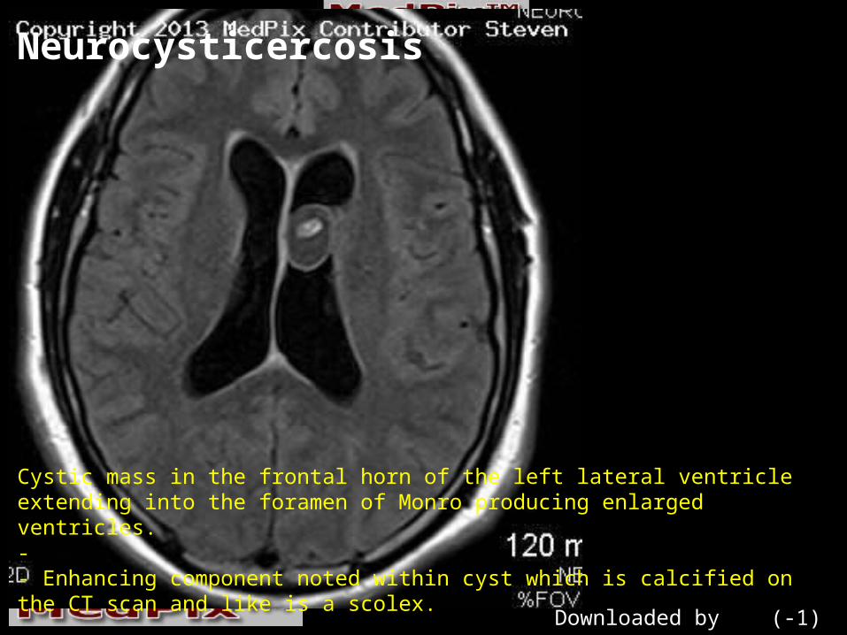
Neurocysticercosis
Cystic mass in the frontal horn of the left lateral ventricle extending into the foramen of Monro producing enlarged ventricles.- - Enhancing component noted within cyst which is calcified on the CT scan and like is a scolex.-
Downloaded by (-1)

Neurocysticercosis
Cystic mass in the frontal horn of the left lateral ventricle extending into the foramen of Monro producing enlarged ventricles.- - Enhancing component noted within cyst which is calcified on the CT scan and like is a scolex.-
Downloaded by (-1)

Neurocysticercosis
Cystic mass in the frontal horn of the left lateral ventricle extending into the foramen of Monro producing enlarged ventricles.- - Enhancing component noted within cyst which is calcified on the CT scan and like is a scolex.
Downloaded by (-1)

Cysticercosis Lifecycle
Eggs or proglottids in feces of humans infected with the adult tapeworm (cestode) in small intestines. Although often called the *pork tapeworm*, it is really a human tapeworm.- - By fecal contamination, either pigs or humans ingest the embyronated eggs or proglottids.- - The eggs mature in the pig*s intestines, and the oncospheres hatch, producing larvae. The larvae penetrate the intestinal wall, migrate through blood vessels, and finally encyst within pig*s muscle.- - Humans become infected by eating raw or undercooked pork.- - *Measly* pork is a phrase describing the cysticercosis larval cysts within the pig meat. In the war of 1812, *Uncle Sam* Wilson sold pork to the U.S. Troops. We hope it wasn*t measly.
Downloaded by (-1)

Neurocysticercosis
Cystic mass in the frontal horn of the left lateral ventricle extending into the foramen of Monro producing enlarged ventricles.- - Enhancing component noted within cyst which is calcified on the CT scan and like is a scolex.
Downloaded by (-1)
Neurocysticercosis
Cystic mass in the frontal horn of the left lateral ventricle extending into the foramen of Monro producing enlarged ventricles.- - Enhancing component noted within cyst which is calcified on the CT scan and like is a scolex.
Downloaded by (-1)

Neurocysticercosis
Cystic mass in the frontal horn of the left lateral ventricle extending into the foramen of Monro producing enlarged ventricles.- - Enhancing component noted within cyst which is calcified on the CT scan and like is a scolex.
Downloaded by (-1)

Neurocysticercosis
Cystic mass in the frontal horn of the left lateral ventricle extending into the foramen of Monro producing enlarged ventricles.- - Enhancing component noted within cyst which is calcified on the CT scan and like is a scolex.
Downloaded by (-1)

Neurocysticercosis
Cystic mass in the frontal horn of the left lateral ventricle extending into the foramen of Monro producing enlarged ventricles.- - Enhancing component noted within cyst which is calcified on the CT scan and like is a scolex.
Downloaded by (-1)

Neurocysticercosis
Cystic mass in the frontal horn of the left lateral ventricle extending into the foramen of Monro producing enlarged ventricles.- - Enhancing component noted within cyst which is calcified on the CT scan and like is a scolex.
Downloaded by (-1)

Neurocysticercosis
Cystic mass in the frontal horn of the left lateral ventricle extending into the foramen of Monro producing enlarged ventricles.- - Enhancing component noted within cyst which is calcified on the CT scan and like is a scolex.
Downloaded by (-1)

Neurocysticercosis
Cystic mass in the frontal horn of the left lateral ventricle extending into the foramen of Monro producing enlarged ventricles.- - Enhancing component noted within cyst which is calcified on the CT scan and like is a scolex.
Downloaded by (-1)

Neurocysticercosis
Cystic mass in the frontal horn of the left lateral ventricle extending into the foramen of Monro producing enlarged ventricles. The scolex is clearly shown (arrow). Additional labels:- 4 - fourth ventricle- Clivus- pons of brainstem- S - sphenoid sinus- t - cerebellar tonsil Downloaded by (-1)
Neurocysticercosis
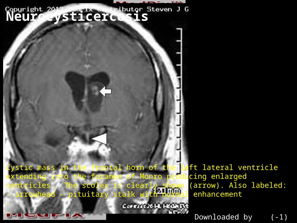
Cystic mass in the frontal horn of the left lateral ventricle extending into the foramen of Monro producing enlarged ventricles. The scolex is clearly shown (arrow). Also labeled:- arrowhead - pituitary stalk with normal enhancement- -
Downloaded by (-1)
Neurocysticercosis
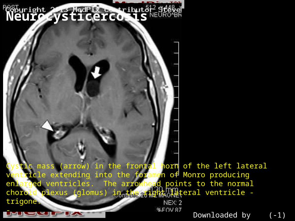
Cystic mass (arrow) in the frontal horn of the left lateral ventricle extending into the foramen of Monro producing enlarged ventricles. The arrowhead points to the normal choroid plexus (glomus) in the right lateral ventricle - trigone.-
Downloaded by (-1)

Neurocysticercosis
Cystic mass in the frontal horn of the left lateral ventricle extending into the foramen of Monro producing enlarged ventricles. The scolex is clearly shown (arrow). The arrowhead points to the normal choroid plexus in the body of right lateral ventricle.
Downloaded by (-1)

Neurocysticercosis
Cystic mass in the frontal horn of the left lateral ventricle extending into the foramen of Monro producing enlarged ventricles. The scolex is clearly shown (arrow).
Downloaded by (-1)
FINDINGS Hydrocephalus of both lateral ventricles Cystic mass in the left frontal horn of the lateral ventricle, extending into the foramen of Monro. The lesion has a central solid bit - a *target sign* Enhancing component noted within cyst which is calcified on the CT scan, may be a scolex.

DIFFERENTIAL DIAGNOSISWhat is your Differential Diagnosis? Arachnoid cyst- Subpendymal Giant Cell Astrocytoma- Central neurocytoma- Colloid Cyst- Cysticercosis larvae

Diagnosis: NeurocysticercosisDx Confirmed by: MRI, CT and social history

DISCUSSIONCysticercosis - taenia solium - as been a parasitic infection of man for over 10,000 years. Fecal contamination of pig feed leads to infection in swine - producing numerous cysts in the meat, or *measly* pork NOTE: While some sources suggest trichinae as the cause of *measly* pork - it is larval cysticercosis that produces cysts within the pork.

Related Documents


